Embed Size (px)
Citation preview

Lecture 12 Spring 2006 1
Materials with Biological Recognition (continued)
Last time: Biological recognition in vivo
Engineering biological recognition of biomaterials: adhesion/migration peptides
Today: Engineering biological recognition of biomaterials: enzymatic recognition and cytokine signaling
Reading: J.C. Schense et al., ‘Enzymatic incorporation of bioactive peptides into fibrin matrices
enhances neurite extension,’ Nat. Biotech. 18, 415-419 (2000) Supplementary Reading: -
ANNOUNCEMENTS:

Lecture 12 Spring 2006 2
Cell adhesion on biomaterials:Cell responses to non-biological, synthetic biomaterials
1. Protein adsorption2. Denaturation (unfolding)?3. Cell responses to expected
and unexpected epitopes4. Reorganization?
• Vroman effect: protein exchange
Lecture 12 Spring 2006 3
Control of cell attachment by mechanical properties of substrate
Polyelectrolyte multilayers (Rubner lab MIT):
Images removed due to copyright reasons.Please see:
Mendelsohn, Jonas D., Sung Yun Yang, Jeri'Ann Hiller, Allon I. Hochbaum, and Michael F. Rubner. "Rational Design of Cytophilic and Cytophobic Polyelectrolyte Multilayer Thin Films." Biomacromolecules 4 (2003): 96-106.

Lecture 12 Spring 2006 4
Control of cell attachment by mechanical properties of substrate
(Van Vliet and Rubner labs):
Graph removed due to copyright reasons.Please see:
Figure 3 in Thompson, M. T., et al. Biomaterials 26 (2005): 6836–6845.
Graph removed due to copyright reasons.Please see:
Figure 4 in Thompson, M. T., et al. Biomaterials 26 (2005): 6836–6845.

Lecture 12 Spring 2006 5
Controlling cell response to biomaterials by building in ECM cues on a ‘blank slate’ background

Lecture 12 Spring 2006 6
Design of protein adsorption-resistant surfaces

Lecture 12 Spring 2006 7
Design of protein adsorption-resistant surfacesSurface modification strategies:
Self-assembled monolayers (SAMs):
Surface grafting:
Graft copolymers or surface polymerization:

Lecture 12 Spring 2006 8
Limiting nonspecific cell adhesion
0
0.05
0.1
0.15
0.2
45 3020
Frac
tion
of c
ells
adh
ered
rela
tive
to T
CPS
weight fraction PEO units
9-unit side chains
O
O
OHO
O
OO
Ox y z
m n
Methyl methacrylate
Poly(ethylene glycol) methacrylates
PMMA
comb

Lecture 12 Spring 2006 9
Tailoring cell adhesion on biomaterials via immobilized ligands
PEO
peptide
PMMA
Peptide integrin-binding GRGDSP sequence
PEO short 6-9 unit side chains for protein resistance
PMMA backbone anchors hydrophilic side chains

Lecture 12 Spring 2006 10
Peptides used to modulate cell adhesion on biomaterials
Peptidesequence
Derived from Conjugatereceptor
Role
IKVAV Laminin α-chain LBP110 (110 KDalaminin bindingprotein)
Cell-ECMadhesion
RGD Laminin α-chain,fibronectin,collagen
Multiple integrins Cell-ECMadhesion
YIGSR Laminin β1-chain α1β1 and α3β1
integrinsCell-ECMadhesion
RNIAEIIKDI Laminin γ-chain unknown Cell-ECMadhesion
HAV N-cadherin N-cadherin Cell-celladhesion
DGEA Type I collagen α2β1 integrin Cell-ECMadhesion
VAPG Elastase Elastase receptor Cell-ECMadhesion
KQAGDV Fibrinogen γ-chain β3 integrins Cell-ECMadhesion
Lecture 12 Spring 2006 11
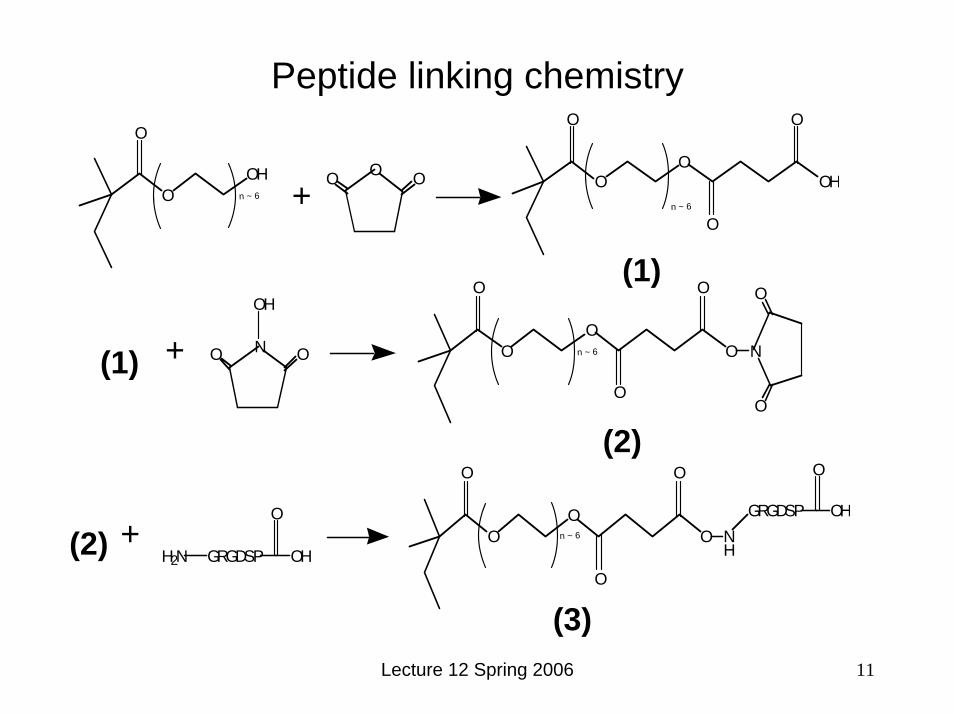
Peptide linking chemistry
O N O
OH
OO
O
O
O
O O
N
O
(1) + n ~ 6
(2)
OO
OH
O
O O OO
O
OH
O
O
n ~ 6 + n ~ 6
(1)
H2N GRGDSP
O
OHO
O
O
O
O
O
NH
GRGDSP
O
OH(2) + n ~ 6
(3)
(1)
(1)
(2)
(2)
(3)

Lecture 12 Spring 2006 12
0
0.1
0.2
0.3
0.4
0.5
GRGDSP GRGESPFrac
tion
Seed
ed C
ells
Adh
ered
+ solubleRGDTethered RGD
Cell responses to RGD

Lecture 12 Spring 2006 13
Cells respond to control of ligand density at the surface
0
0.1
0.2
0.3
0.4
0.5
0.6
0 1000 2000 3000 4000 5000
RGD Density (#/µm2)
% S
eede
d C
ells
A
dher
edTCPS
Frac
tion
seed
ed
cells
adh
ered

Lecture 12 Spring 2006 14
Cells respond to control of ligand density at the surface
Cell migration on fibronectin-coated substrates:
Graph removed due to copyright reasons. Please see:
Figure 1b in Palecek, S. et al. "Integrin-ligand Binding Properties Govern Cell Migration Speed Through Cell-substratum Adhesiveness." Nature 385 (6 February, 1997): 537 - 540.
Graphs removed due to copyright reasons. Please see:
Figure 2b in Palecek, S., et al. "Integrin-ligand Binding Properties Govern Cell Migration Speed Through Cell-substratum Adhesiveness." Nature 385 (6 February, 1997): 537 - 540.
Lauffenburger lab

Lecture 12 Spring 2006 15
Alternative functionalization approaches: avidin-biotin chemistry
Image removed due to copyright reasons. Please see:
Patel, et al. FASEB Journal 12 (1998): 1447-454.

Lecture 12 Spring 2006 16
Controlling gross physical distribution of cells
Images removed due to copyright reasons. Please see:
Patel, et al. FASEB Journal 12 (1998): 1447-454.

Lecture 12 Spring 2006 17
Cellular responses to physically patterned ligand- with nonadhesive background
Images removed due to copyright reasons.Please see:
Patel, et al. FASEB Journal 12 (1998): 1447-454.

Lecture 12 Spring 2006 18
Biomaterials recognized by cell-secreted enzymes:
synthetic ECMs

Lecture 12 Spring 2006 19
Enzymatic remodeling of synthetic ECMs

Lecture 12 Spring 2006 20
Enzymatic recognition of synthetic polymer backbones
Cleavage of synthetic polymers by enzymesCell source
Enzyme
Native
function
Acts on
Degradation Mechanism
Result
Various bacteria
lipases
protease
Polyesters, polyesteramides
III Monomers or dimers
Tritirachium album (mold) Proteinase K Protease Poly(lactide) III Monomers or dimers
Mammalian cells esterases protease Poly(alkyl cyanoacrylates)
II Water-soluble polymers
Mammalian cells Papain, pepsin proteases polyesteramides2 III Untested
Mammalian cells α-chymotrypsin Serine proteaseAromatic peptides in polyesteramides3 (e.g. Ala, Val, Leu)
III Untested
Mammalian cells elastase protease Polyesteramides III untested

Lecture 12 Spring 2006 21
Enzymatic degradation of polyesteramides
N. Paredes et al. J. Polym. Sci. A36, 1271 (1998)
Enzymatic breakdown by papain:Compare with hydrolysis:
(poly(ortho ester))
Graph removed due to copyright reasons.Please see:
Figure 12 in Paredes, N., et al. J. Polym. Sci. A 36, no. 1271 (1998).
Graph removed due to copyright reasons.Please see:
Figure 10 in Paredes, N., et al. J. Polym. Sci. A 36, no. 1271 (1998).

Lecture 12 Spring 2006 22
Esterase attack on poly(alkyl cyanoacrylates)Degradation of 250 nm-diam. porous particles:
Graph removed due to copyright reasons.Please see:
Figure 2 in Paredes, N., et al. J. Polym. Sci. A 36, no. 1271 (1998).
Graph removed due to copyright reasons.Please see:
Figure 11 in Paredes, N., et al. J. Polym. Sci. A 36, no. 1271 (1998).

Lecture 12 Spring 2006 23
Engineering enzymatic recognition of hydrogel biomaterials: recognition of peptide motifs
Enzymatic activity in vivo on peptide sequences:5,6 Cleavage Enzyme
Functions in vivo
Target amino acid sequences
Plasminogen activator (urokinase or tissue-type
plasminogen activator) / plasminogen → plasmin
Degradation of fibrin matrices, angiogenesis, tumor progression; urokinase can bind to cell surface receptor
on fibrinogen: Arg104-Asp105, Arg110-Val111, Lys206-Met207, Arg42-Ala43, Lys130-Glu131, Lys84-Ser85, Lys87-Met88
Matrix metalloproteinases (soluble and cell-surface): e.g. Fibroblast Collagenase (MMP I)
Facilitate cell migration Type I collagen: Gly775-Ile776 In smaller peptides: Gly-Leu or Gly Ile
bonds
Elastase Elastin remodeling Poly(Ala) sequences

Lecture 12 Spring 2006 24
Enzyme-sensitive crosslinks in hydrogelbiomaterials
PEG
photopolymerization
= =
Acrylate endgroups
peptides
-APGL-
-CH2CH2O-
collagenase sequence
collagenase
-APGL-collagenase
(West and Hubbell, 1999)

Lecture 12 Spring 2006 25
Effect of enzyme concentration
Gel containing collagenase sequence Gel containing elastase sequence
Graph removed due to copyright reasons.Please see:
Figure 1 in West, J.L. and J. A. Hubbell. “Polymeric Biomaterials with Degradation Sites for Proteases Involved in Cell Migration.”Macromolecules 32 (1999): 241-244.
Graph removed due to copyright reasons.Please see:
Figure 2 in West, J.L. and J. A. Hubbell. “Polymeric Biomaterials with Degradation Sites for Proteases Involved in Cell Migration.” Macromolecules 32 (1999): 241-244.

Lecture 12 Spring 2006 26
Cellular migration through enzymatically-recognized hydrogels
Biphasic migration response in 3D matrix:
Image removed due to copyright reasons.Please see:
Figure 4 in Gobin, A.S. and J. L. West. “Cell Migration Through Defined, Synthetic ECM Analogs.” Faseb J 16 (2002): 751-3.
Image removed due to copyright reasons.Please see:
Figure 6 in Gobin, A.S. and J. L. West. “Cell Migration Through Defined, Synthetic ECM Analogs.” Faseb J16 (2002): 751-3.

Lecture 12 Spring 2006 27
Enzymatic recognition of biomaterials II: Enzymatic cross-linking/modification of biomaterials
Example enzymes and their substrates:Enzyme Substrate in vivo Synthetic substrates Result
Transglutaminase Glutamines Glu-containing peptides Amide bond formation Factor XIII Fibrin γ-chain Peptides derived from γ-
chain FXIII binding site Amide bond formation
(Zhang et al. 2002)

Lecture 12 Spring 2006 28
Biomaterials that mimic signals from soluble factors or other cells

Lecture 12 Spring 2006 29
Cytokine receptor-based recognition of biomaterials
Diverse functions of cytokines:
•Induce cell migration/stop cell migration•Induce cell growth•Induce differentiation
•Upregulate tissue-specific functions
Characteristics:
•Typically potent, act at pmolconcentrations•Synergize with other receptor signals
•e.g. integrins
Receptor Monomer
GDP
Inactive Ras
EGF
Exterior
Cystosol
Dimeric Receptor
Step 1 Binding of hormone causesdimerization andautophosphorylation oftyrosine residues
P
P
P
P
P
P
Figure by MIT OCW.

Lecture 12 Spring 2006 30
Changes in signaling achieved by cytokine immobilization on surfaces
Image removed due to copyright reasons. Please see:
Figure 1 in Ito, Y., et al. "Tissue Engineering by Immobilized Growth Factors." Materials Science and Engineering C6 (1998): 267-274.

Lecture 12 Spring 2006 31
Immobilized insulin:
Image removed due to copyright reasons. Please see:
Figure 1 in Ito, Y., et al. "Tissue Engineering by Immobilized Growth Factors.“ Materials Science and Engineering C6 (1998): 267-274.

Lecture 12 Spring 2006 32
Local control of gene expression by non-diffusablecytokines:Patterned immobilization of EGF:
Images removed for copyright reasons.
Please see:
Figure 4 in Ito, Y. "Regulation of Cell Functions by Micropattern Immobilized Biosignal Molecules." Nanotechnology 9 (1998): 200-204.

Lecture 12 Spring 2006 33
Surface immobilization can induce new function in cytokines: case of tethered EGF-triggered neuronal cell
differentiation
PC12 cell line: • induced to differentiate and
extend axons under stimulation of NGF (nerve growth factor)
• induced to proliferate by EGF
Signal doesn’t trigger internalization of receptor; thus signal lasts longer and triggers differentiation
Signal triggers internalization of receptor; short signal triggers proliferation
Dimeric ReceptorDimeric ReceptorDimeric Receptor
P
P
P
P
P
P
Figure by MIT OCW.
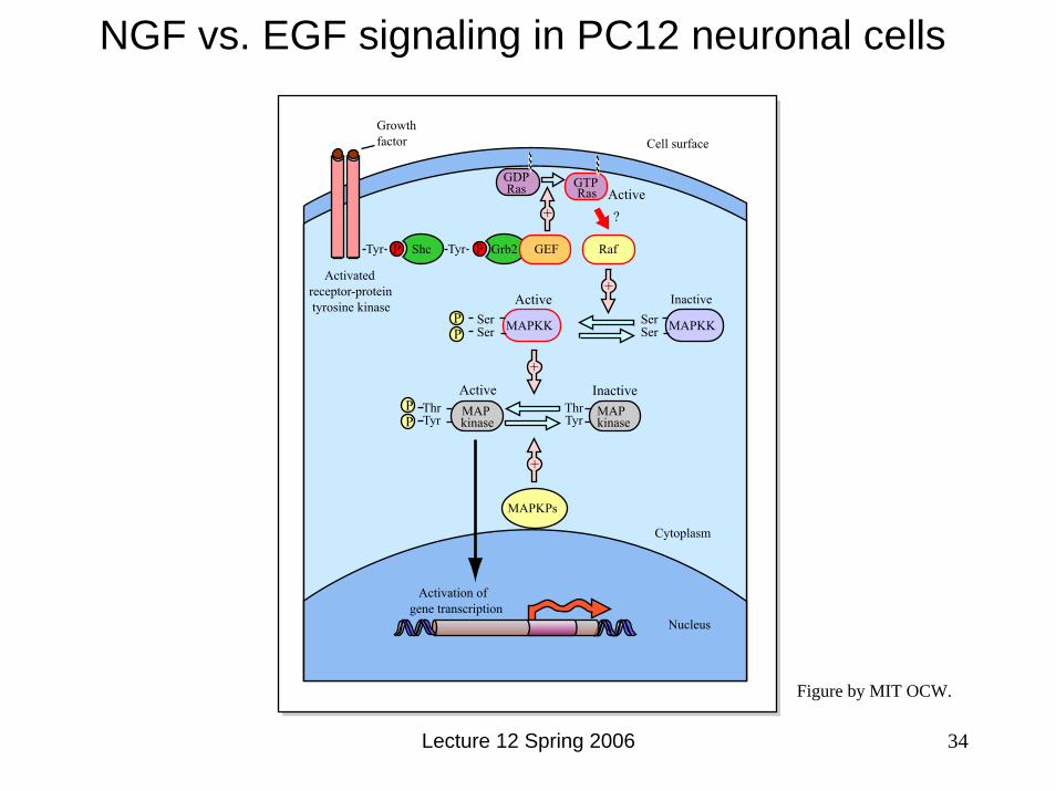
NGF vs. EGF signaling in PC12 neuronal cells
Lecture 12 Spring 2006
Shc GEF Raf
GTPRasRas
GDPActive
Active
Active+
MAPKK
MAP MAPkinase kinase
MAPKPs
ThrTyr
Tyr
ThrTyr
PP
PP
Inactive
Inactive
SerSerMAPKKSer
Ser
?
Activatedreceptor-protein tyrosine kinase
Cytoplasm
Nucleus
Activation of gene transcription
Growthfactor Cell surface
Tyr
+
+
+
++
Grb2PP GEF
34
Figure by MIT OCW.

Lecture 12 Spring 2006 36
Further Reading 1. Voet & Voet. in Biochemistry. 2. Paredes, N., Rodriguez, G. A. & Puiggali, J. Synthesis and characterization of a family of biodegradable
poly(ester amide)s derived from glycine. Journal of Polymer Science, Part A: Polymer Chemistry 36, 1271-1282 (1998).
3. Fan, Y., Kobayashi, M. & Kise, H. Synthesis and biodegradability of new polyesteramides containing peptide linkages. Polymer Journal 32, 817-822 (2000).
4. O, S. C. & Birkinshaw, C. Hydrolysis of poly (n-butylcyanoacrylate) nanoparticles using esterase. Polymer Degradation and Stability 78, 7-15 (2002).
5. Ekblom, P. & Timpl, R. Cell-to-cell contact and extracellular matrix. A multifaceted approach emerging. Curr Opin Cell Biol 8, 599-601 (1996).
6. Chapman, H. A. Plasminogen activators, integrins, and the coordinated regulation of cell adhesion and migration. Curr Opin Cell Biol 9, 714-24 (1997).
7. Mann, B. K., Gobin, A. S., Tsai, A. T., Schmedlen, R. H. & West, J. L. Smooth muscle cell growth in photopolymerized hydrogels with cell adhesive and proteolytically degradable domains: synthetic ECM analogs for tissue engineering. Biomaterials 22, 3045-51 (2001).
8. West, J. L. & Hubbell, J. A. Polymeric biomaterials with degradation sites for proteases involved in cell migration. Macromolecules 32, 241-244 (1999).
9. Gobin, A. S. & West, J. L. Cell migration through defined, synthetic ECM analogs. Faseb J 16, 751-3 (2002). 10. Sperinde, J. J. & Griffith, L. G. Control and prediction of gelation kinetics in enzymatically cross-linked
poly(ethylene glycol) hydrogels. Macromolecules 33, 5476-5480 (2000). 11. Sperinde, J. J. & Griffith, L. G. Synthesis and characterization of enzymatically-cross-linked poly(ethylene glycol)
hydrogels. Macromolecules 30, 5255-5264 (1997). 12. Zhang, Z. Y., Shum, P., Yates, M., Messersmith, P. B. & Thompson, D. H. Formation of fibrinogen-based
hydrogels using phototriggerable diplasmalogen liposomes. Bioconjug Chem 13, 640-6 (2002). 13. Sanborn, T. J., Messersmith, P. B. & Barron, A. E. In situ crosslinking of a biomimetic peptide-PEG hydrogel via
thermally triggered activation of factor XIII. Biomaterials 23, 2703-10 (2002). 14. Collier, J. H. et al. Thermally and photochemically triggered self-assembly of peptide hydrogels. J Am Chem Soc
123, 9463-4 (2001). 15. Collier, J. H. & Messersmith, P. B. Enzymatic modification of self-assembled peptide structures with tissue
transglutaminase. Bioconjug Chem 14, 748-55 (2003). 16. Schense, J. C., Bloch, J., Aebischer, P. & Hubbell, J. A. Enzymatic incorporation of bioactive peptides into fibrin
matrices enhances neurite extension. Nat Biotechnol 18, 415-9 (2000). 17. Ito, Y. Tissue engineering by immobilized growth factors. Materials Science and Engineering C 6, 267-274 (1998).18. Ito, Y. Regulation of cell functions by micropattern-immobilized biosignal molecules. Nanotechnology 9, 200-204
(1998). 19. Kuhl, P. R. & Griffith-Cima, L. G. Tethered epidermal growth factor as a paradigm for growth factor-induced
stimulation from the solid phase. Nat Med 2, 1022-7 (1996). 20. Chen, G. & Ito, Y. Gradient micropattern immobilization of EGF to investigate the effect of artificial juxtacrine
stimulation. Biomaterials 22, 2453-7 (2001). 21. Ito, Y. Surface micropatterning to regulate cell functions. Biomaterials 20, 2333-42 (1999).


![1 Protein Protein Interactions: An Overview · ADF/cofilin and profilin [10]. Biological (surface) recognition, like in the immune Biological (surface) recognition, like in the](https://img.dokumen.tips/doc/110x75/5b618dcc7f8b9a08478c7338/1-protein-protein-interactions-an-overview-adfcolin-and-prolin-10.jpg)