Embed Size (px)
Citation preview

Materials Science and Engineering C 64 (2016) 87–92
Contents lists available at ScienceDirect
Materials Science and Engineering C
j ourna l homepage: www.e lsev ie r .com/ locate /msec
Design and development of anisotropic inorganic/polystyrenenanocomposites by surface modification of zinc oxide nanoparticles
Xiao Han a,b, Shiming Huang c, Yilong Wang b,⁎, Donglu Shi b,d,⁎⁎a School of Materials Science and Engineering, Tongji University, Shanghai 200092, PR Chinab Research Center for Translational Medicine, East Hospital, the Institute for Biomedical Engineering & Nano Science, Tongji University School of Medicine, Shanghai 200092, PR Chinac Department of Physics, Tongji University, Shanghai 200092, PR Chinad The Materials Science and Engineering Program, College of Engineering and Applied Science, University of Cincinnati, Cincinnati, OH 45221, USA
⁎ Corresponding author at: Mailbox 155#, Siping Road 1200092, PR China.⁎⁎ Correspondence to: D. Shi, Research Center for Translthe Institute for Biomedical Engineering & Nano SciencMedicine, Shanghai 200092, PR China.
E-mail addresses: [email protected] (Y. Wang
http://dx.doi.org/10.1016/j.msec.2016.03.0580928-4931/© 2016 Elsevier B.V. All rights reserved.
a b s t r a c t
a r t i c l e i n f oArticle history:Received 16 December 2015Received in revised form 11 February 2016Accepted 19 March 2016Available online 22 March 2016
Anisotropic yolk/shell or Janus inorganic/polystyrene nanocomposites were prepared by combiningminiemulsion polymerization and sol–gel reaction. The morphologies of the anisotropic composites werefound to be greatly influenced by surface modification of zinc oxide (ZnO) nanoparticle seeds. Two differenttypes of the oleic acid modified ZnO nanoparticles (OA-ZnO) were prepared by post-treatment of commercialZnO powder and homemade OA-ZnO nanoparticles. The morphologies and properties of the nanocompositeswere investigated by transmission electronmicroscope (TEM), thermogravimetric analysis (TGA), Fourier trans-form infrared spectroscopy (FT-IR), dynamic light scattering (DLS), and energy dispersive X-ray spectroscopy(EDX). It was found that both post-treated OA-ZnO and in-situ prepared OA-ZnO nanoparticles resulted in theyolk–shell and Janus structure nanocomposites, but with varied size and morphology. These nanocompositesshowed stable and strong fluorescence by introducing quantum dots as the co-seeds. The fluorescent anisotropicnanocomposites were decorated separately with surface carboxyl and hydroxyl groups. These composites withunique anisotropic properties will have high potential in biomedical applications, particularly in bio-detection.
© 2016 Elsevier B.V. All rights reserved.
Keywords:Anisotropic nanocompositesSurface modified zinc oxideOleic acidMiniemulsion polymerization
1. Introduction
The concept of “Janus particles”, which named after the two-facedRoman God Janus, was first raised by P. G. de Gennes in his NobelPrize address in 1991 [1]. “Janus” particles are asymmetric and aniso-tropic in structure, shape, and surface characteristics with each surfacehaving distinctively different physical and chemical properties [2]. Dueto the unique geometrical asymmetry, Janus nanoparticles can be usedas ideal drug delivery carriers, catalysis, surfactants, and biosensor[3–5]. Several strategies have been developed to synthesize asymmetri-cal nanoparticles. These include the layer-by-layer self-assembly [6,7],evaporated metal particles [8–11], Pickering emulsion method [12],biphasic electrified jetting [13,14], photo-polymerization in amicrofluidicchannel [15,16], and polymer self-assembly [17,18]. Janus particles canassume different geometries: spherical, dumbbell, half raspberry-like,cylindrical, disk, and a variety of other shapes [2]. Besides Janus compos-ites, yolk–shell like nanocomposites was also a newly emerging systemfor biomedical applications [19,20]. The functional core and hollow shell
239, Yangpu District, Shanghai,
ational Medicine, East Hospital,e, Tongji University School of
), [email protected] (D. Shi).
of the yolk–shell particles can be tailored to the requirements ofthe nanoreactors [21] and biomedicine [22]. The common and typicalsynthesis of the yolk/shell particles includes the following, namely,selective etching or dissolution, bottom-up or soft-template, ship-in-bottle, Ostwald ripening or galvanic replacement process, andKirkendall diffusion methods [22]. The Janus or yolk–shell nanopar-ticles are usually composed of inorganic–inorganic dimmer orinorganic–polymer composites [23–26]. Among these, the silica/polystyrene composites is one of the most frequently studied systems[27,28] and the silica/magnetite/polymer composites have been devel-oped into the ternary system [29–31]. The straightforwardness andyield of the reaction have been important factors in the developmentof the syntheticmethods.Wehave previously reported a one-step facilemethod based on oleic acid modified iron oxide nanoparticles via com-bined miniemulsion polymerization and sol–gel reaction for preparingthe ternarymagnetic compositeswith Janus or yolk–shellmorphologies[32,33]. Suchmagnetic nanomaterials have demonstrated some advan-tages in bio-detection of rare target by magnetic collection utilizing theactive surface functionality. However, the role of the oleic acidmodifiedinorganic nanoparticles was not well understood in the formation ofthe ternary composites. Few literatures have been reported about thebilateral system, particularly on the influence of the surface chemistryof Titania hydrosols on the stability and morphology of the hybridnanoparticles [34].

88 X. Han et al. / Materials Science and Engineering C 64 (2016) 87–92
In this study,we investigated the effect of surfacemodification of theoleic acidmodified zinc oxide (OA-ZnO) nanoparticles on the compositesize and morphologies using a one-pot synthesis process. Two differentparticle systemswere investigated: the OA-ZnO nanoparticles were de-veloped in an in-situ synthesis with simultaneous surface modification,and the other by post-modification of commercialized zinc oxide powder.The oleic acid modified zinc oxide was used as the inorganic seeds.Polystyrene/ZnO@SiO2 nanocomposites were prepared via a com-bined process of miniemulsion polymerization and sol–gel reaction.For bio-detection applications, carboxyl group was introduced to thesurface of the polymer component for conjugation of antibody. Theternary systemwithout magnetic component was further developedinto a fluorescent probe by conjugating with the hydrophobic quantumdots for immune detection of biomarkers via a sandwich method.
2. Materials and experiments
2.1. Materials
All reagents used were analytical grade and available commercially.Zinc oxide (ZnO) was purchased from Emperor Nano Material Co.(Nanjing, China). Oleic acid (OA, 90%) was purchased from ShanghaiChemical Reagents Co. (Shanghai, China). Styrene (St), ammoniumhydroxide (NH4OH, 28 wt %), and Sodium hydroxide (NaOH) werepurchased from Sinopharm Chemical Regent Co. (Shanghai, China).Tetraethoxysilane (TEOS), sodium dodecyl sulfate (SDS) and 4, 4′-Azobis(4-cyanovaleric acid) (ACVA)were purchased from Sigma-Aldrich (USA).Hexadecane (HD)was purchased from Acros Organics (USA). Hydropho-bic quantum dots (CdSSe/ZnS core shell nanocrystals with particle size of9 nm and molecular weight of 7.5 × 10⁵) were purchased from Nanjingtechnology Co. (Hangzhou, China).
The aforementioned chemicals were used as received. Styrene waswashed first with 5 wt % sodium hydroxide solution and stored at−20 °C before using. High-purity nitrogen gas, provided by a localsupplier, was used for sparging solutions without further purification.Deionized (DI) water was used throughout the entire experiment.
2.2. Synthesis of oleic acid-modified zinc oxide nanoparticles
OA-ZnO-a nanoparticles were fabricated by surface modification ofthe commercial ZnO powder. With the index of particles' size byDynamic Laser Scattering (DLS) measurement, the orthogonal experi-ments were carried out to optimize liquid–solid ratio, mixing time,and the reaction temperature and time for surface modification of thecommercial ZnO powder. The optimum modification condition wasdetermined as follows. Briefly, oleic acid (1.94 mL) was dissolved inethanol (50 mL) with mechanical stirring. After adding ZnO nanoparti-cles (0.25 g), the dispersion was heated to 60 °C which under 200 Wsonication (KQ-400KDV, Kunshan Ultrasonic Instruments Co, Ltd.) for3 h. Finally, the dispersion was cooled down to room temperature.
Another type of OA-ZnO nanoparticles (OA-ZnO-b) was synthesizedvia a modified process according to Heyon's work [35]. Both typesof OA-ZnO nanoparticles were collected by a centrifugal machine(3000 rpm/min, 10 min), and the precipitate was dispersed in50 mL ethanol and DI water with ultrasonication for 10 min. Thewashing procedure was repeated for five times and the precipitatewas dried by freezing-drying for further use. The extra oleate andoleic acid was removed from both OA-ZnO-a and OA-ZnO-b nano-particles by washing.
2.3. Synthesis of polystyrene/ZnO@SiO2 nanocomposites
Anisotropic polystyrene/ZnO@SiO2 nanocomposites were preparedvia a combined process of miniemulsion polymerization and sol–gelreaction reported by our group [32]. The water phase was prepared bydissolving 0.092 g SDS in 39 g of DI water. The oil phase was prepared
by the following steps: HD (0.4 g) was first dissolved in St monomer(7 g) by 3 min sonication, then OA-ZnO nanoparticles powder (0.025 g)was then dissolved in the monomer phase. TEOS (2 g) was subsequentlyresolved into the mixture of the hydrophobic components. The waterphase and oil phase were mixed by dropping the oil dispersion dropwiseto the water phase under 200 W sonication (KQ-400KDV, Kun ShanUltrasonic Instruments Co., Ltd., China), followed by 250 rpmmechanicalstirring for 30 min. Miniemulsion droplets system was developed bysonicating the emulsions at 500 W in an ice bath with a duty cycle of50% for 20 min by a Scientz-IID sonifier (JY92-2D, Ningbo XinzhiBio-technology Co., Ltd., China). Upon neutralizing 0.12 g of ACVA by1 mL of NaOH solution (0.856 M) and added into the flask, the systemwas deoxygenized by nitrogen bubbling for 0.5 h with 250 rpmmechan-ical stirring. With a condenser under nitrogen protection, the flask wasmoved into 70 °C water bath to initiate St monomer polymerization.After reaction for 1.5 h, 50 μL of NH4OH was added into the reaction.After heating for another 5 h, the reaction system was stirred at roomtemperature overnight. The final polystyrene/ZnO@SiO2 nanocompositeswere obtained by centrifugal separation.
2.4. Synthesis of QDs@ polystyrene/ZnO@SiO2 nanocomposites
The fluorescent nanocomposites were developed by doping acontrolled amount of hydrophobic QDs (5 mg) with the OA-ZnO nano-particles into the oil phase. The steps were kept the same as synthesis ofpolystyrene/ZnO@SiO2 nanocomposites.
2.5. Characterization
Transmission electron microscopy (TEM). The washed compositeparticles were dispersed in DI water and dried onto carbon-coatedcopper grids before examination. TEM images and energy dispersiveX-ray (EDX) analysis were obtained with a Philips Tecnai 20 electronmicroscope.
Thermogravimetric analysis (TGA). A thermogravimetric analyzer(Netzsch TG209, Germany) was used to analyze the surface modifica-tion of the OA-ZnO. The measurement was performed under a nitrogenatmosphere from ambient temperature to 900 °C with a heating rate of10 °C/min.
Fourier transform infrared spectroscopy (FT-IR). FT-IR spectra ofunmodified ZnO and the OA-ZnO nanoparticles were obtained byusing the infrared spectrometer (Bruker Tensor 27, USA).
Dynamic light scattering (DLS). Hydrodynamic size of the nanocom-posites and purified OA-ZnO nanoparticles were measured by the DLSinstrument (Zetasizer Nano-ZS, Malvern, U.K.).
Fluorescence spectra. The fluorescence of QDs and compositenanoparticles were measured using a spectrophotometer (LS-55,Perkin–Elmer, USA) at room temperature. Fluorescent compositeswere dropped on glass slide.
Fluorescent imaging system. After dropping the fluorescent sampleonto a glass slide, the fluorescence imaging was performed by usingan inverted fluorescence microscope (Nikon ECLIPSE Ti, Japan).
3. Results and discussion
Fig. 1 shows the TEM images andDLS data of OA-ZnO-a andOA-ZnO-b. As shown in Fig. 1A, OA-ZnO-a is of irregular shape with an averagesize of 25 nm. In Fig. 1B, OA-ZnO-b mainly consists of mono-dispersedspherical particles of a size about 15 nm. Note that in the former(OA-ZnO-a), the oleic acid was functionalized by post-treatmentof the commercial ZnO powder and in the latter (OA-ZnO-b), thenanocrystals were surface-functionalized during the in-situ synthesis.With oleic acid coating, DLS data of the two kinds of OA-ZnO nanoparti-cles show good dispersion in the organic solvent. Upon surface modifica-tion, the hydrophobicity of the commercial ZnO particle was dramaticallyimproved. The hydrodynamic average diameter of OA-ZnO-a (Fig. 1C) is

Fig. 1. TEM images and DLS data of two kinds of oleic acid modified zinc oxide nanoparticles: OA-ZnO-a (A, C) and OA-ZnO-b (B, D).
89X. Han et al. / Materials Science and Engineering C 64 (2016) 87–92
close to 37 nm (Poly-dispersity Index, PDI: 0.315), indicating wellfunctionalized ZnO particles in this process. The DLS diameter ofOA-ZnO-b (Fig. 1D) is about 20 nm (PDI: 0.323), which is consistentwith TEM results.
The surface modification of the ZnO particles was further character-ized by FT-IR and TGA. Fig. 2A shows the FT-IR absorption spectra ofoleic acid (OA), OA-ZnO-a and OA-ZnO-b. As can be seen in Fig. 2A,there are strong vibrations bands of\\CH3 and\\CH2 groups fromboth OA-ZnO at 2850 and 2920 cm−1, indicating the nanoparticles'surface oleic acid molecules. However, peaks at 1725–1700 cm−1 dueto the stretching vibrations of C_O group in free oleic acid are notobserved in the spectra of OA-ZnO-a and OA-ZnO-b. Furthermore, thepeaks at 1544 cm−1 are attributed to the stretching vibrations ofCOO\\Zn, due to the interaction between the\\COOH group of theoleic acid and the\\OH group on the surface of ZnO nanoparticles[36–38]. These results show chemically well grafted surfaces of theZnO nanoparticles.
Fig. 2. FT-IR spectra and TGA analysis curv
Fig. 2B shows the thermogravimetric (TGA) curves of the originalcommercial ZnO powder, OA-ZnO-a, and OA-ZnO-b. The mass ofunmodified ZnO powders was found almost unchanged after theheating process. The weight loss of OA-ZnO-a in the temperaturerange of 150–700 °C was 47.0 wt %, due to thermal decomposition ofthe multi-layer oleate. Only 11 wt % weight loss of OA-ZnO-b nanopar-ticles, in the range of 150 to 700 °C, was detected resulting from thedecomposition of oleate. Due to excess oleic acid in ZnO-a, excessiveOA molecules are adsorbed on the first OA layer in the form of end-end hydrophobic interaction [39]. The primary layer is presumed tochemically adsorb on the surfaces of the nanoparticles. The irregularshape of the commercial ZnO nanoparticles may occupy more oleicacid molecules.
Fig. 3A and B shows the TEMmicrographs of the typical compositesobtained from the two types ZnO nanoparticles. The composites exhibitthe cell-like yolk–shell structures composed of the polystyrene core andinorganic shell with the OA-ZnO-a seeds. As shown in Fig. 3, only a small
es of the modified ZnO nanoparticles.

Fig. 3. TEM images and DLS data of ZnO/silica/polystyrene composites obtained when: (A, D) OA-ZnO-a as the seeds, (B, E) OA-ZnO-b as the seeds, and (C, F) without ZnO seeds.
90 X. Han et al. / Materials Science and Engineering C 64 (2016) 87–92
fraction displays the regular spherical shape, while the rest is ellipsoid.The average size of the composites and the polystyrene cores are 550and 370 nm, respectively. There is a clear space between the core andshell. Using OA-ZnO-b as the seeds, the product is Janus-like nanocom-posites. The structure is similar to the Fe3O4@silica/polystyrene ternarycomposites reported in our previous work [32]. Even under the samepolymerizing conditions, themorphologies of the two types of compos-ites (i.e. OA-ZnO-a and OA-ZnO-b) are entirely different. The differencein morphology is attributable to their surface properties resulting fromthe diverse oleic acid modification methods. Emulsification procedureinvolving a fusion/fission process during the mechanical stirring andsonication is necessary before polymerization in the miniemulsionsystem [40]. The monolayer or multilayer of OA on ZnO will have astrong effect on dispersion and morphology of the nanoparticles in theminiemulsion droplets [41].The Janus composites based on OA-ZnO-bhave the 100 nm PS cores as shown in Fig. 3B, and half-arc silica shellwith the thickness of around 30 nm. Fig. 3D shows, based on the DLSdata, an average size of 390 nm for the yolk–shell composites with apolydispersity index of 0.385, and 140 nm for Janus composites with apolydispersity index of 0.314. The differences in morphology and sizeindicate an important factor of using different inorganic seeds. Toinvestigate the role of the OA-ZnO nanoparticles in the reaction, weperformed the miniemulsion polymerization in the absence of anymodified ZnO nanoparticles under the same processing condition. As aresult, as shown in Fig. 3C, neither Janus nor yolk–shell structures isobtained. Only separated large polystyrene latexes and small silicaparticles are observed with hydrodynamic diameter about 600 nm.This result of control experiment shows the direct effect of the inorganicseeds on the final composite structures.
The nucleation of miniemulsion polymerization is distinctly differ-ent with conventional emulsion polymerization which takes placein the monomer droplets. When the solubility of the hydrophobe (i.e.co-stabilizer) in water increases, the rate of Ostwald ripening isincreased [42]. These oleic acid modified inorganic particles may act asthe co-stabilizer to engender the miniemulsification process [43,44].Based on the TGA and TEM data result, we found that the size of nano-composites and polystyrene corewas related to the amount of oleic acidgrafted on the surface of ZnO nanoparticles.
The composition of the inorganic layer was characterized byTEM-EDX. Fig. 4 shows the EDX spectra of a typical ternary compositeparticle. As can be seen in Fig. 4, there are elements of silicon and zinc
for each composites, indicating the hybrid composition of the inorganicshell.
Due to the unique structure and properties of yolk–shell and Januscomposites, the composites can be conjugated with plenty of QDs asfluorescent probes for cell labeling and tracking. Fig. 5 shows the fluo-rescence spectra and images of pure QDs and QDs/polystyrene/ZnO@SiO2 nanocomposites. The emission peak of pure QDs is at 605 nm.The fluorescence spectra of the QDs/yolk–shell composites-a (blackline) and QDs/Janus composite-b (blue line) show successful incorpora-tion of QDs into the ternary composite particles. Fig. 5B and C show thefluorescent images of the composites conjugatedwithQDs. By integratingthefluorescence and other functionalities via surface carboxyl groups, thenanocomposites will have potential applications in bio-detection such asthe immune-probes.
Fig. 6 is the schematic diagram showing the synthesis procedure ofthe composites particles via miniemulsion polymerization and sol–gelreaction. The one-pot miniemulsion process is similar to the methodreported in one of our earlier studies. The process can be described asfollows: (1) the typical miniemulsify process is developed from waterphase and oil phase. Styrene monomer, tetraethoxysilane, hexadecane,and oleic acid-functionalized zinc oxides are restricted in miniemulsiondroplets (stage b). (2) Polymerization of St monomers is initiated byinitiator 4,4′-Azobis(4-cyanovaleric acid) at 70 °C. As a result of Stmonomer polymerizing and phase separation, the intermediate productis produced (stage c). (3) After ammonium hydroxide is added, TEOSstarts to hydrolyze and poly-condense, simultaneously. The OA-ZnOfurther separates from the oil phase because the oleic acid moleculeswill transform to oleate in aqueous alkali. As a result of the lowestsystematic energy, the formed silica chains tend to hybridize with zincoxide particles. The St monomer diffuses inwardly to the PS particles,but TEOS outwardly to the inorganic shell. As the St monomer andTEOS precursors in the droplets are almost exhausted, Janus nanocom-posites with a PS core and an ZnO/silica hybrid shell are developed(stage d).
4. Conclusion
In summary, we have employed a one-pot synthesis for preparationof non-magnetic anisotropic nanocomposites. Typically yolk–shellor Janus composites are obtained with two different approaches:post-treatment of commercial ZnO and homemade OA-ZnO seeds.
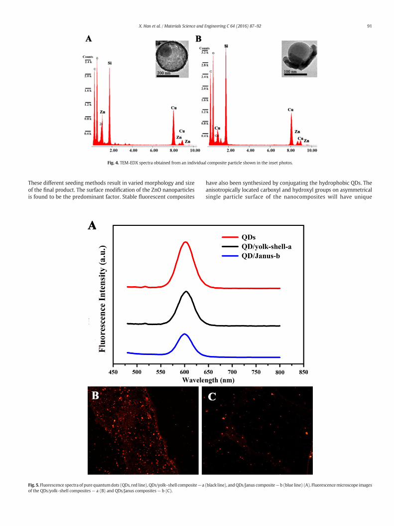
Fig. 4. TEM-EDX spectra obtained from an individual composite particle shown in the inset photos.
91X. Han et al. / Materials Science and Engineering C 64 (2016) 87–92
These different seeding methods result in varied morphology and sizeof the final product. The surface modification of the ZnO nanoparticlesis found to be the predominant factor. Stable fluorescent composites
Fig. 5. Fluorescence spectra of pure quantumdots (QDs, red line), QDs/yolk–shell composite— aof the QDs/yolk–shell composites— a (B) and QDs/Janus composites— b (C).
have also been synthesized by conjugating the hydrophobic QDs. Theanisotropically located carboxyl and hydroxyl groups on asymmetricalsingle particle surface of the nanocomposites will have unique
(black line), andQDs/Janus composite— b (blue line) (A). Fluorescencemicroscope images

Fig. 6. Schematic illustration shows the pathway for preparation of the nanocomposites via a combined process of miniemulsion polymerization and sol–gel reaction.
92 X. Han et al. / Materials Science and Engineering C 64 (2016) 87–92
applications in biodetection. The fluorescent experimental resultselementarily show that non-magnetic anisotropic nanocompositeswith good fluorescent performance are ideal carriers for bio-detectionapplications.
Acknowledgments
This work was supported by the National Natural Science Foundationof China (Nos. 51173135, 31571018), the Science and Technology Com-mission of Shanghai Municipality (Nos. 15ZR1443200, 15441905900).
References
[1] P.G. de Gennes, Rev. Mod. Phys. 64 (1992) 645–648.[2] J. Hu, S. Zhou, Y. Sun, X. Fang, L. Wu, Chem. Soc. Rev. 41 (2012) 4356–4378.[3] S. Berger, A. Synytska, L. Ionov, K.-J. Eichhorn, M. Stamm,Macromolecules 41 (2008)
9669–9676.[4] Y.K. Takahara, S. Ikeda, S. Ishino, K. Tachi, K. Ikeue, T. Sakata, T. Hasegawa, H. Mori,
M. Matsumura, B. Ohtani, J. Am. Chem. Soc. 127 (2005) 6271–6275.[5] P. Biji, A. Patnaik, Analyst 137 (2012) 4795–4801.[6] H.Y. Koo, D.K. Yi, S.J. Yoo, D.Y. Kim, Adv. Mater. 16 (2004) 274–277.[7] Z.F. Li, D.Y. Lee, M.F. Rubner, R.E. Cohen, Macromolecules 38 (2005) 7876–7879.[8] J.R. Howse, R.A.L. Jones, A.J. Ryan, T. Gough, R. Vafabakhsh, R. Golestanian, Physical
Review Letters, 99, 2007.[9] H. Takei, N. Shimizu, Langmuir 13 (1997) 1865–1868.
[10] O. Cayre, V.N. Paunov, O.D. Velev, J. Mater. Chem. 13 (2003) 2445–2450.[11] Y. Lu, H. Xiong, X.C. Jiang, Y.N. Xia, M. Prentiss, G.M. Whitesides, J. Am. Chem. Soc.
125 (2003) 12724–12725.[12] Y. Wang, B.-H. Guo, X. Wan, J. Xu, X. Wang, Y.-P. Zhang, Polymer 50 (2009)
3361–3369.[13] K.-H. Roh, M. Yoshida, J. Lahann, Langmuir 23 (2007) 5683–5688.[14] S. Hwang, K.-H. Roh, D.W. Lim, G.Wang, C. Uher, J. Lahann, Phys. Chem. Chem. Phys.
12 (2010) 11894–11899.[15] D. Dendukuri, P.S. Doyle, Adv. Mater. 21 (2009) 4071–4086.[16] R.F. Shepherd, J.C. Conrad, S.K. Rhodes, D.R. Link, M. Marquez, D.A. Weitz, J.A. Lewis,
Langmuir 22 (2006) 8618–8622.
[17] R. Erhardt, M.F. Zhang, A. Boker, H. Zettl, C. Abetz, P. Frederik, G. Krausch, V. Abetz,A.H.E. Muller, J. Am. Chem. Soc. 125 (2003) 3260–3267.
[18] I.K. Voets, R. Fokkink, T. Hellweg, S.M. King, P. de Waard, A. de Keizer, M.A.C. Stuart,Soft Matter 5 (2009) 999–1005.
[19] S. Li, J. Zheng, D. Chen, Y. Wu, W. Zhang, F. Zheng, J. Cao, H. Ma, Y. Liu, Nanoscale 5(2013) 11718–11724.
[20] J. Gao, G. Liang, J.S. Cheung, Y. Pan, Y. Kuang, F. Zhao, B. Zhang, X. Zhang, E.X. Wu, B.Xu, J. Am. Chem. Soc. 130 (2008) 11828–11833.
[21] J. Lee, J.C. Park, H. Song, Adv. Mater. 20 (2008) 1523–1528.[22] J. Liu, S.Z. Qiao, J.S. Chen, X.W. Lou, X. Xing, G.Q. Lu, Chem. Commun. 47 (2011)
12578–12591.[23] H.W. Gu, R.K. Zheng, X.X. Zhang, B. Xu, J. Am. Chem. Soc. 126 (2004) 5664–5665.[24] H. Yu, M. Chen, P.M. Rice, S.X. Wang, R.L. White, S.H. Sun, Nano Lett. 5 (2005)
379–382.[25] V. Nandwana, G.S. Chaubey, K. Yano, C.-b. Rong, J.P. Liu, Journal of Applied Physics,
105, 2009.[26] W. Qiang, Y. Wang, P. He, H. Xu, H. Gu, D. Shi, Langmuir 24 (2008) 606–608.[27] W. Mu, M. Fu, Microelectron. Eng. 96 (2012) 51–55.[28] Z. Han, L. Wang, J. Zhu, S. Zhang, W. Zhou, J. Appl. Polym. Sci. 122 (2011) 43–49.[29] M.D. Butterworth, L. Illum, S.S. Davis, Colloids Surf. A Physicochem. Eng. Asp. 179
(2001) 93–102.[30] M.D. Butterworth, S.A. Bell, S.P. Armes, A.W. Simpson, J. Colloid Interface Sci. 183
(1996) 91–99.[31] H. Xu, L. Cui, N. Tong, H. Gu, J. Am. Chem. Soc. 128 (2006) 15582–15583.[32] F. Wang, G.M. Pauletti, J. Wang, J. Zhang, R.C. Ewing, Y. Wang, D. Shi, Adv. Mater. 25
(2013) 3485–3489.[33] Y. Wang, F. Wang, B. Chen, H. Xu, D. Shi, Chem. Commun. 47 (2011) 10350–10352.[34] Y. Zhao, H. Wang, X. Song, Q. Du, Macromol. Chem. Phys. 211 (2010) 2517–2529.[35] S.H. Choi, E.G. Kim, J. Park, K. An, N. Lee, S.C. Kim, T. Hyeon, J. Phys. Chem. B 109
(2005) 14792–14794.[36] R.Y. Hong, T.T. Pan, J.Z. Qian, H.Z. Li, Chem. Eng. J. 119 (2006) 71–81.[37] M. Li, B. Hari, X. Lv, X. Ma, F. Sun, L. Tang, Z. Wang, Mater. Lett. 61 (2007) 690–693.[38] P. Liu, Z.X. Su, J. Macromol. Sci. Phys. B45 (2006) 131–138.[39] L.F. Shen, P.E. Laibinis, T.A. Hatton, Langmuir 15 (1999) 447–453.[40] M. Antonietti, K. Landfester, Prog. Polym. Sci. 27 (2002) 689–757.[41] F. Yan, J. Li, J. Zhang, F. Liu, W. Yang, J. Nanoparticle Res. 11 (2009) 289–296.[42] S. Lu, J. Forcada, J. Polym. Sci. A Polym. Chem. 44 (2006) 4187–4203.[43] C.S. Chern, T.J. Chen, Colloids Surf. A Physicochem. Eng. Asp. 138 (1998) 65–74.[44] C.S. Chern, H.T. Chang, Polym. Int. 51 (2002) 1428–1438.