Embed Size (px)
Citation preview

CASE REPORT Open Access
Massive odontoameloblastoma arising inthe maxilla: a case reportMasanori Kudoh1,2*, Hiroyuki Harada2, Yuriko Sato1,2, Ken Omura1,2,3 and Yoshimasa Ishii1,2
Abstract
Introduction: Odontoameloblastoma is an extremely rare mixed odontogenic tumor with both epithelial andmesenchymal components. The term odontoameloblastoma first appeared in the 1971 World Health Organizationclassification (Pindborg JJ., et al.) and is defined as “a neoplasm that includes odontogenic ectomesenchyme in additionto odontogenic epithelium that resembles an ameloblastoma in both structures and behavior.” Because of theaggressive nature and risk of recurrence of the tumor, complete resection is essential. In this report, we describe anextremely rare case of a patient with massive odontoameloblastoma arising in the maxilla and occupying maxillary sinus.
Case presentation: In 2013, an 11-year-old Japanese boy was referred to our department for a painless and large massof the right maxillary region. A panoramic X-ray showed a unilocular cystic lesion in the right maxilla containing acalcified mass in the lesion associated with an impacted tooth. Computed tomography showed a cystic lesion thatincluded calcified structures and measured 3.6×3.1×2.7 cm. In 2013, the patient underwent tumor extirpation combinedwith impacted tooth extraction. The histopathological diagnosis was an odontoameloblastoma. No recurrence wasnoted 27 months after the operation.
Conclusions: The patient has undergone postoperative occlusal guidance and functional orthodontic treatment, and hispostoperative condition is excellent. However, postoperative recurrence or malignant transformation can occur in casesof odontoameloblastoma, and close long-term follow-up will be continued for our patient.
Keywords: Maxillary sinus, Mixed odontogenic tumor, Odontoameloblastoma
IntroductionOdontoameloblastoma (OA) is an extremely rare mixedodontogenic tumor that is defined in the current WorldHealth Organization tumor classification system as atumor that includes odontogenic ectomesenchyme andodontogenic epithelium and resembles an ameloblastomain both structure and behavior [1]. Generally, the clinicalpresentation of OA resembles an odontoma; thus, a de-finitive diagnosis is based on histologic analysis followingexcision and curettage [2]. OA and complex odontoma be-long to a group of odontogenic tumors that consist ofodontogenic epithelium and odontogenic ectomesenchymewith or without dental hard tissue formation (so-calledmixed odontogenic tumors) [3, 4]. However, differential
diagnosis of OA is difficult compared with ameloblasticfibroodontoma or a developing complex odontoma [5].OA is usually found in young patients and has no sig-
nificant gender predilection [6, 7]. Clinically, the two maincomplaints are swelling and failure of tooth eruption.Radiological examination usually reveals a multilocularradiolucency with a well-defined boundary and oftenshows radiopaque areas resembling mature dental tissue[8]. If an unerupted tooth is present, the tumor is usuallysituated coronally to the crown of this tooth [6, 8]. We re-port an extremely rare case of a patient with massive OAarising in the maxilla and occupying the maxillary sinus.
Case presentationIn 2013, an 11-year-old Japanese boy was referred to ourdepartment for painless bone expansion in the rightmaxillary alveolus, delayed eruption of the permanentsecond molar teeth, and altered occlusion. He had nosignificant medical or family history. A panoramic X-rayshowed a unilocular cystic lesion in the right maxilla
* Correspondence: [email protected] of Oral and Maxillofacial Surgery, Ebina General Hospital, 1320,Kawaraguchi, Ebina, Kanagawa 243–0433, Japan2Oral and Maxillofacial Surgery, Department of Oral Restitution, Division ofOral Health Sciences, Graduate School, Tokyo Medical and Dental University,1–5–45 Yushima, Bunkyo-ku, Tokyo 113–8549, JapanFull list of author information is available at the end of the article
JOURNAL OF MEDICALCASE REPORTS
© 2015 Kudoh et al. Open Access This article is distributed under the terms of the Creative Commons Attribution 4.0International License (http://creativecommons.org/licenses/by/4.0/), which permits unrestricted use, distribution, andreproduction in any medium, provided you give appropriate credit to the original author(s) and the source, provide a link tothe Creative Commons license, and indicate if changes were made. The Creative Commons Public Domain Dedication waiver(http://creativecommons.org/publicdomain/zero/1.0/) applies to the data made available in this article, unless otherwise stated.
Kudoh et al. Journal of Medical Case Reports (2015) 9:278 DOI 10.1186/s13256-015-0743-0
containing a calcified large mass associated with an im-pacted tooth (Fig. 1). Computed tomography showed acystic lesion of size 3.6×3.1×2.7 cm that included calci-fied structures (Fig. 2a). A horizontal view showed rightmaxillary bone expansion (Fig. 2b).In 2013, the patient underwent tumor extirpation com-
bined with impacted tooth extraction. The incisional linewas started from the labial mucosa in the right maxillarycentral and lateral incisor area. It extended to the gingivain the right maxillary lateral incisor and canine area byarch-like incision, and to the gingiva of the right maxillarysecond molar by crestal incision with distal releasing inci-sion. The goal of this procedure was to make a mucoper-iosteal flap from the lower border of the pyriformaperture, vertically to the infraorbital foramen and sur-rounding areas, and horizontally to the lower border ofthe zygomatic bone. The infraorbital neurovascular bun-dles were preserved. Upon opening of the sinus from athin plate of bone in the canine fossa and surroundingareas in the anterior wall of the right maxilla (the samelevel as the lower border of the pyriform aperture), bone-like hard tissues that were strongly adhered to the maxillawere found. When these tissues were separated from thesurrounding areas and removed using a fissure bar, a solid,bone-like, hard odontogenic tumor (similar to a complexodontoma) was found to have occupied almost the wholesinus. The mass was too large to remove from the opening.For complete removal of the mass including the tumor
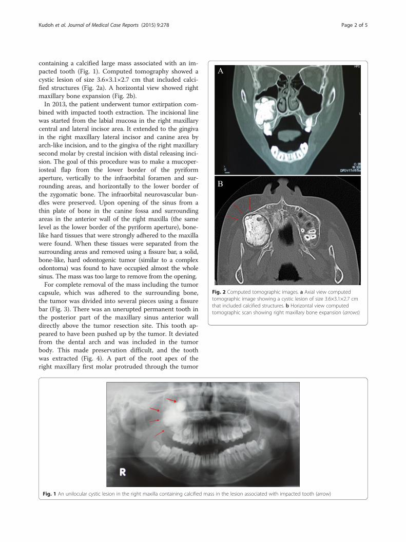
capsule, which was adhered to the surrounding bone,the tumor was divided into several pieces using a fissurebar (Fig. 3). There was an unerupted permanent tooth inthe posterior part of the maxillary sinus anterior walldirectly above the tumor resection site. This tooth ap-peared to have been pushed up by the tumor. It deviatedfrom the dental arch and was included in the tumorbody. This made preservation difficult, and the toothwas extracted (Fig. 4). A part of the root apex of theright maxillary first molar protruded through the tumor
Fig. 1 An unilocular cystic lesion in the right maxilla containing calcified mass in the lesion associated with impacted tooth (arrow)
Fig. 2 Computed tomographic images. a Axial view computedtomographic image showing a cystic lesion of size 3.6×3.1×2.7 cmthat included calcified structures. b Horizontal view computedtomographic scan showing right maxillary bone expansion (arrows)
Kudoh et al. Journal of Medical Case Reports (2015) 9:278 Page 2 of 5
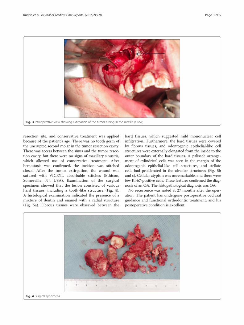
resection site, and conservative treatment was appliedbecause of the patient’s age. There was no tooth germ ofthe unerupted second molar in the tumor resection cavity.There was access between the sinus and the tumor resec-tion cavity, but there were no signs of maxillary sinusitis,which allowed use of conservative treatment. Afterhemostasis was confirmed, the incision was stitchedclosed. After the tumor extirpation, the wound wassutured with VICRYL absorbable stitches (Ethicon,Somerville, NJ, USA). Examination of the surgicalspecimen showed that the lesion consisted of varioushard tissues, including a tooth-like structure (Fig. 4).A histological examination indicated the presence of amixture of dentin and enamel with a radial structure(Fig. 5a). Fibrous tissues were observed between the
hard tissues, which suggested mild mononuclear cellinfiltration. Furthermore, the hard tissues were coveredby fibrous tissues, and odontogenic epithelial-like cellstructures were externally elongated from the inside to theouter boundary of the hard tissues. A palisade arrange-ment of cylindrical cells was seen in the margin of theodontogenic epithelial-like cell structures, and stellatecells had proliferated in the alveolar structures (Fig. 5band c). Cellular atypism was unremarkable, and there werefew Ki-67-positive cells. These features confirmed the diag-nosis of an OA. The histopathological diagnosis was OA.No recurrence was noted at 27 months after the oper-
ation. The patient has undergone postoperative occlusalguidance and functional orthodontic treatment, and hispostoperative condition is excellent.
Fig. 3 Intraoperative view showing extirpation of the tumor arising in the maxilla (arrow)
Fig. 4 Surgical specimens
Kudoh et al. Journal of Medical Case Reports (2015) 9:278 Page 3 of 5

DiscussionOA is an aggressive odontogenic tumor characterized bysimultaneous occurrence of an ameloblastoma and acompound or complex odontoma in the same tumor
mass [3, 9]. OA affects males and females equally andoccurs in the maxilla and mandible, with the molar-premolar area being the most common site. Clinicalsymptoms include slow, progressive swelling of the al-veolar plates, bone expansion, root resorption, dull pain,altered occlusion, tooth displacement, delayed eruption,and impacted teeth [6]. OA occurs between the ages of2 and 50 years and at a mean age of 20.2 years [6]. How-ever, as in our patient, most cases occur in childrenunder 16 years of age.The origin of an OA [10] has been proposed to be ma-
lignant transformation of the enamel epithelium afterodontogenesis [11] or malignant transformation of theepithelium and mesenchyme of supernumerary teethduring embryogenesis [12]. However, a definitive theoryhas yet to be established [13]. Furthermore, the diseaseis often accompanied by an impacted tooth, but thecausative link between the tumor and the impactedtooth is unclear. Thompson et al. [14] suggested that thetumor capsule may be derived from mesenchymal tissue,based on the property of this tissue to form dental hardtissue. The origin of our case was unclear, but betweentumor and tumor capsule that similar to the dental fol-licle around the impacted tooth was connected, with ir-regular structures and dentinal hyperplasia was seen, weguessed tumor transformation was caused by interactionbetween the odontogenic epithelium which degeneratedand mesenchymal tissue during the degenerated toothprocess. This suggests that tumor formation may occurduring the process of crown formation of the impactedtooth.Ameloblastoma, complex odontoma, compound odon-
toma, and dentigerous cysts may be differentiated by im-aging, but their diagnosis also requires histopathologicalexamination. A definitive diagnosis is difficult to make onthe basis of imaging and clinical findings alone; therefore,total excision is required. Masqueda-Taylor et al. [6] foundrecurrence in 3 (21.4 %) of 14 OA cases. On the basis ofthese data and findings that OA tends to occur at an earl-ier age than conventional ameloblastoma and because ithas a similar potential to produce bone expansion, rootresorption, and recurrence, it was suggested that OAshould be treated similarly to ameloblastoma, with wideexcision and close follow-up for at least 5 years [6]. There-fore, the first-choice treatment for OA is complete surgicalextirpation of the tumor, including wide excision. We alsosuggest that treatment of OA requires close cooperationof oral and maxillofacial surgeons with orthodontic andpedodontic specialists, including primary care dentists.This multidisciplinary treatment is required because mostcases of OA in pediatric patients are associated with dis-placed, unerupted permanent teeth.Clinically, OA starts as a slow-growing, painless mass
that expands the alveolus and vestibular cortex and
Fig. 5 a Histopathological image (hematoxylin and eosin stain,original magnification ×100). A histological examination indicatedthe presence of a mixture of dentin and enamel with a radialstructure. b, c Histopathological images (hematoxylin and eosinstain, original magnification ×200). A palisade arrangement ofcylindrical cells was seen in the margin of the odontogenicepithelial-like cell structures, and stellate cells had proliferated in thealveolar structures
Kudoh et al. Journal of Medical Case Reports (2015) 9:278 Page 4 of 5
prevents eruption of permanent teeth [9]. Our patienthad expansion in the right maxillary alveolus, preventionof eruption of permanent teeth, altered occlusion, toothdisplacement, delayed eruption, and impacted teeth.Most reported cases have had symptoms such as swell-ing or pain, but in our patient the tumor was asymptom-atic and was detected during a routine examination. OAalso tends to produce bone expansion in almost allcases, similarly to conventional ameloblastoma, whereasan odontoma seldom produces swelling of the affectedregion. Therefore, OA is often confused with compoundor complex odontomas, as in our patient. Our patienthas had no recurrence for 27 months postoperatively.However, the recurrence rate of OA is 21.4 % [6], andmalignant transformation may occur [15]. Therefore,long-term follow-up will be continued for our patient.
ConclusionsWe report an extremely rare case of a patient with massiveOA arising in the maxilla and occupying the maxillary sinus.
ConsentWritten informed consent was obtained from the patient’slegal guardian(s) for publication of this case report and anyaccompanying images. A copy of the written consent isavailable for review by the Editor-in-Chief of this journal.
AbbreviationOA: Odontoameloblastoma.
Competing interestsThe authors declare that they have no competing interests.
Authors’ contributionsMK contributed as main author and in surgical treatment and management,case report design, acquisition of data, analysis and interpretation of surgicaland pathological data, and drafting of the manuscript. HH contributed byobtaining academic support and in case report design, acquisition of data,analysis and interpretation of surgical and pathological data, and drafting ofthe manuscript. YS contributed through acquisition of data and providingtechnical support. KO contributed by obtaining academic support and insurgical treatment and management and supervision, as well as in thedrafting of the manuscript. YI contributed through medical support andsupervision. All authors read and approved the final manuscript.
Author details1Division of Oral and Maxillofacial Surgery, Ebina General Hospital, 1320,Kawaraguchi, Ebina, Kanagawa 243–0433, Japan. 2Oral and MaxillofacialSurgery, Department of Oral Restitution, Division of Oral Health Sciences,Graduate School, Tokyo Medical and Dental University, 1–5–45 Yushima,Bunkyo-ku, Tokyo 113–8549, Japan. 3Division of Oral and MaxillofacialSurgery, General Tokyo Hospital, 3–15–2 Ekota, Nakano-ku, Tokyo 165–0022,Japan.
Received: 3 July 2015 Accepted: 21 October 2015
References1. Kramer IRH, Pindborg JJ, Shear M. Histological typing of odontogenic
tumours. 2nd ed. Berlin: Springer; 1992.2. Martín-Granizo-López R, López-García-Asenjo J, De-Pedro-Marina M,
Domínguez-Cuadrado L. Odontoameloblastoma: a case report and a reviewof the literature. Med Oral. 2004;9:340–4.
3. Philipsen HP, Reichart PA, Slootweg PJ, Slater LJ, Sciubba JJ, Eversole LR, et al.Odontogenic tumours. In: Barnes L, Eveson JW, Reichart P, Sidransky D, editors.World Health Organization classification of tumours: pathology and genetics ofhead and neck tumours. Lyon, France: IARC Press; 2005. p. 283–327.
4. Philipsen HP, Reichart PA, Praetorius F. Mixed odontogenic tumours andodontomas. Considerations on interrelationship. Review of the literature andpresentation of 134 new cases of odontomas. Oral Oncol. 1997;33:86–99.
5. Reibel J, Grønbaek AB, Poulsen S. Peripheral ameloblastic fibroodontoma orperipheral developing complex odontoma: report of a case. Int J PaediatrDent. 2011;21:468–70.
6. Mosqueda-Taylor A, Carlos-Bregni R, Ramírez-Amador V, Palma-GuzmánJM, Esquivel-Bonilla D, Hernández-Rojase LA. Odontoameloblastoma:clinico-pathologic study of three cases and critical review of the literature.Oral Oncol. 2002;38:800–5.
7. Reichart PA, Philipsen HP, Gelderblom HR, Stratmann U. Ameloblastic fibro-odontoma—report of two cases with ultrastructural study of tumour dentalhard structures. Oral Oncol Extra. 2004;40:8–12.
8. LaBriola DJ, Steiner M, Bernstein ML, Verdi GD, Stannard PF.Odontoameloblastoma. J Oral Surg. 1980;38:139–43.
9. Gupta DS, Gupta MK. Odontoameloblastoma. J Oral Surg. 1986;44:146–8.10. Yoshikage N. A case of ameloblasto-odontoma [in Japanese]. Shikwa
Gakuho. 1959;59:1150–3.11. Goro I, Masatoyo A. Oral pathology II [in Japanese]. Revth ed. Kyoto, Japan:
Nagasue Bookstore; 1982. p. 506.12. Junji K, Masayuki I, Yoshio K, Etsuhide Y, Mayumi T, Kimiko Y, et al. Two
cases of odontoameloblastoma arising in the mandible [in Japanese]. Jpn JOral Maxillofac Surg. 1982;28:2002–8.
13. Takamasa A, Hiromitsu K, Kazunari S, Takamichi Y, Masahiro U, Wataru Y. Acase of odontoameloblastoma arising in the mandible [in Japanese]. Jpn JOral Maxillofac Surg. 1993;39:43–5.
14. Thompson IO, Philips VM, Ferreira R, Housego TG. Odontoameloblastoma:a case report. Br J Oral Maxillofac Surg. 1990;28:347–9.
15. Tetsuo I, Kinai T. A case of malignant transformation of theodontoameloblastoma arising in the mandible [in Japanese]. Nihon KōkūkaGakkai Zasshi. 1965;14:239–40.
Submit your next manuscript to BioMed Centraland take full advantage of:
• Convenient online submission
• Thorough peer review
• No space constraints or color figure charges
• Immediate publication on acceptance
• Inclusion in PubMed, CAS, Scopus and Google Scholar
• Research which is freely available for redistribution
Submit your manuscript at www.biomedcentral.com/submit
Kudoh et al. Journal of Medical Case Reports (2015) 9:278 Page 5 of 5