Embed Size (px)
Citation preview
Division of Oncology Stanford University School of Medicine Stanford Genome Technology Center Stanford Cancer Institute
Mass validation of variants identified by whole genome
sequencing
Georges Natsoulis, PhD Stanford University

Stanford University Division of Oncology Stanford Genome Technology Center
Stanford Cancer Institute
Cancer genome sequencing and personalized diagnostics
Complete Human Genomes
Exomes &
Transcriptomes
Genomic
Subsets
DNA diagnostic (e.g. CLIA)
“clinically actionable”
Clinical validation
studies
Discovery

Stanford University Division of Oncology Stanford Genome Technology Center
Stanford Cancer Institute
Two methods addressing multiple objectives
Objective Advances
Whole genome sequencing (WGS)
discovery Integration of targeting with
WGS
Validation of genome variants from cancer WGS Accelerating and improving
variant validation
Clinical implications from cancer populations
Facilitating analysis large clinical cohorts of archival
cancer samples
Clinical translation as diagnostic Rapid, accurate analysis for
prospective clinical review
Os-seq
Single–strand circularization

Stanford University Division of Oncology Stanford Genome Technology Center
Stanford Cancer Institute Method 1: OS-Seq Step1: synthesize capture probes on flow cell lawn
Primer probe
Myllykangas et al. 2011, Nat. Biotech.

Stanford University Division of Oncology Stanford Genome Technology Center
Stanford Cancer Institute
Step2: Capturing a target region from cancer genomes
Sequencing cancer
genes and other
regions
Stanford University Division of Oncology Stanford Genome Technology Center
Stanford Cancer Institute
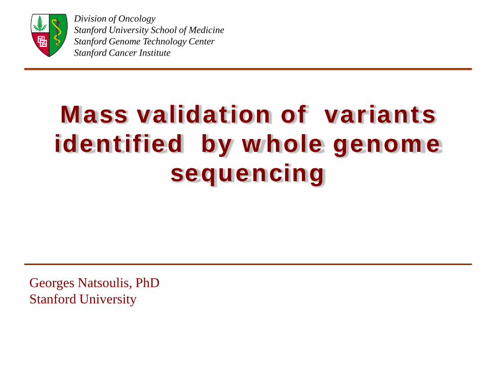
OS-Seq for targeting cancer genome regions
Strand-specific capture
P1 reads P2 reads
Stanford University Division of Oncology Stanford Genome Technology Center
Stanford Cancer Institute
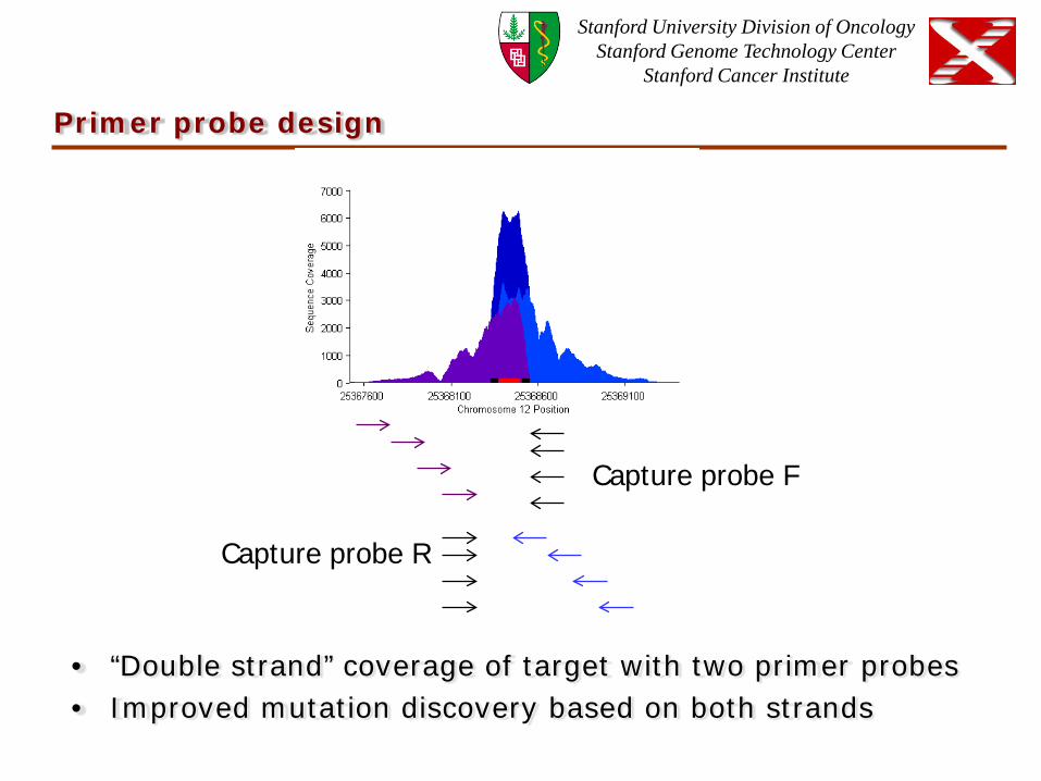
Primer probe design
• “Double strand” coverage of target with two primer probes • Improved mutation discovery based on both strands
Capture probe R
Capture probe F
Stanford University Division of Oncology Stanford Genome Technology Center
Stanford Cancer Institute
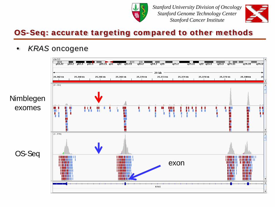
OS-Seq: accurate targeting compared to other methods
• KRAS oncogene
Nimblegen exomes
OS-Seq exon

Stanford University Division of Oncology Stanford Genome Technology Center
Stanford Cancer Institute
OS-Seq: Targeting loci like extended exons
• APC exon 15 (6.5 Kb)
OS-Seq
Primer probe placement
Nimblegen exomes

Stanford University Division of Oncology Stanford Genome Technology Center
Stanford Cancer Institute
OS-Seq: Even coverage of genomic region targets
• Primer probe yield
n = 344 cancer genes n = 10 cancer genes
n = 314 cancer genes
Stanford University Division of Oncology Stanford Genome Technology Center
Stanford Cancer Institute
OS-Seq advances and advantages
• Higher sensitivity and specificity mutation detection
with “deep” targeted resequencing
• Higher accuracy targeting of any nonrepetitive human
genome region
• Accurate variant discovery - overlapping primer probe
design improves variant detection
• Identification of rearrangement breakpoint sequences
• Efficient workflow of 1 day reduces experimental errors
• Low sample requirements (<1 ug DNA)

Stanford University Division of Oncology Stanford Genome Technology Center
Stanford Cancer Institute
5’ 3’
Single-stranded fragmented DNA
Capture probes
Mix in presence of Ampligase, TaqPol, exoI
Single stranded circle
Amplify with common primers
Population of double stranded linears
Key features: -Single-stranded substrate compatible with FFPE material. -Capture probes can be placed anywhere.
Illumina Nextgen Seqencing
Method 2: single strand genomic circularization

Stanford University Division of Oncology Stanford Genome Technology Center
Stanford Cancer Institute
Pilot demonstration of targeting and accuracy
• 628 genomic regions targeted (~200bp average size)
• 123 Kb of total size of genomic targets
• Samples
– Matched tissues from the same organ and individual
• High quality genomic DNA from flash frozen tissue
• Low quality DNA from matching FFPE tissue.
• Sequencing performed in triplicate

Stanford University Division of Oncology Stanford Genome Technology Center
Stanford Cancer Institute
Mutation discovery from clinical archival samples
1. Compare capture yields from high quality versus from
FFPE genomic DNA
2. Determine sensitivity of detection of heterozygote variants
in high quality genomic DNA compared to matched
archival genomic DNA (FFPE)
3. Evaluate FFPE-related DNA damage in variants in FFPE
genomic DNA but not in high quality genomic DNA
Stanford University Division of Oncology Stanford Genome Technology Center
Stanford Cancer Institute
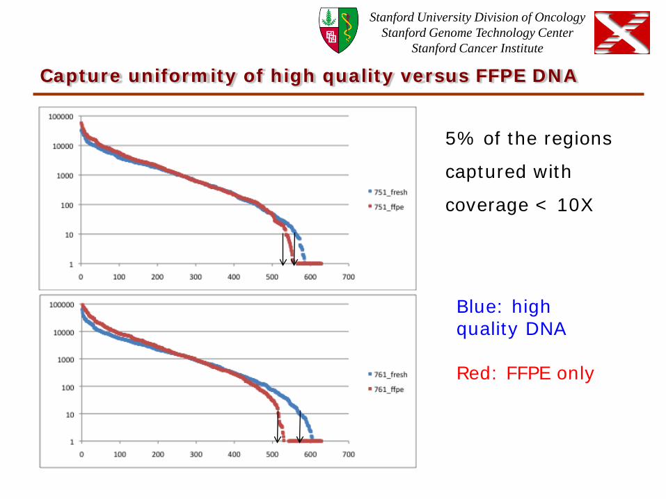
Capture uniformity of high quality versus FFPE DNA
5% of the regions
captured with
coverage < 10X
Blue: high quality DNA Red: FFPE only

Stanford University Division of Oncology Stanford Genome Technology Center
Stanford Cancer Institute
Artifacts introduced by FFPE processing
Percent variant FFPE DNA
Perc
ent
varian
t hig
h q
ula
ity
DN
A
Blue: high quality DNA and FFPE DNA Red: FFPE only
True Hets
WT
Noisy positions (variant single strand only)
FFPE induced errors
Stanford University Division of Oncology Stanford Genome Technology Center
Stanford Cancer Institute
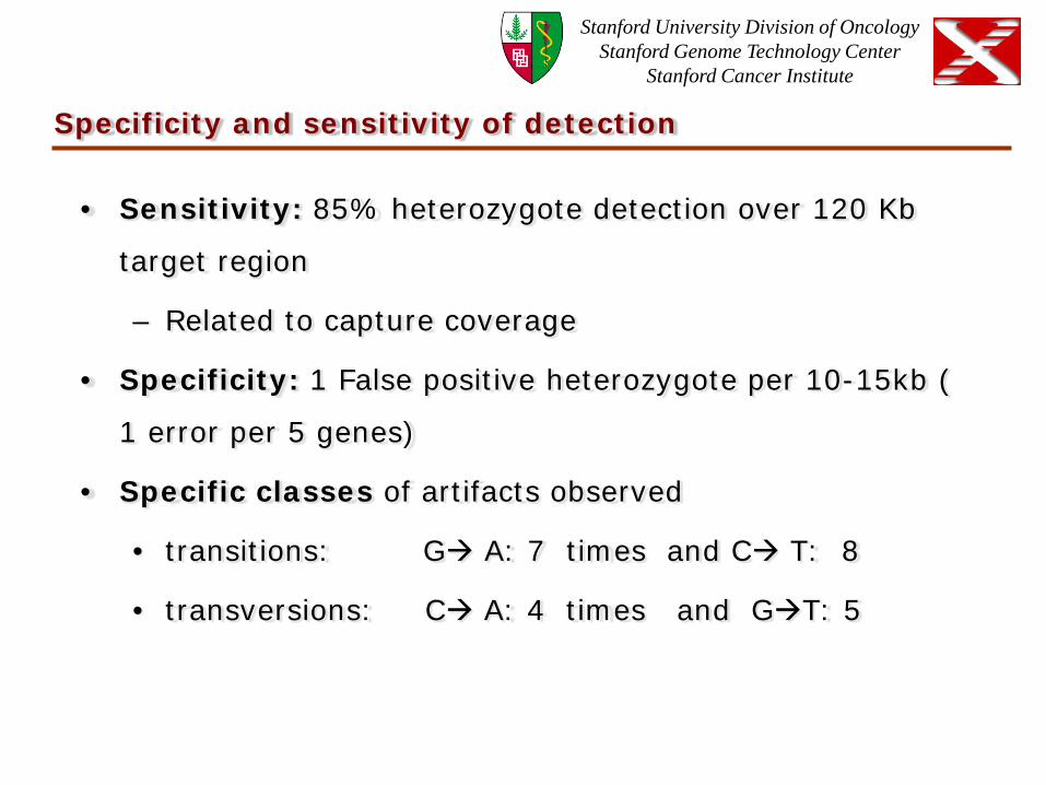
Specificity and sensitivity of detection
• Sensitivity: 85% heterozygote detection over 120 Kb
target region
– Related to capture coverage
• Specificity: 1 False positive heterozygote per 10-15kb (
1 error per 5 genes)
• Specific classes of artifacts observed
• transitions: G A: 7 times and C T: 8
• transversions: C A: 4 times and GT: 5

Stanford University Division of Oncology Stanford Genome Technology Center
Stanford Cancer Institute
Single lane mass-validation of whole genome sequencing
Whole Genome sequencing and exome sequencing of matched Normal blood/Primary gastric tumor/Ovarian metastasis
386 coding variants including SNVs, Indels and SVs
Validate all positions in parallel in a single lane of sequencing
From flash frozen tissue From FFPE
OS-seq capture
GAIIx or HiSeq
Single Strand Circularization
MiSeq

Stanford University Division of Oncology Stanford Genome Technology Center
Stanford Cancer Institute
Os-seq
Ovarian Metastasis
Gastric Tumor
Normal Blood

Stanford University Division of Oncology Stanford Genome Technology Center
Stanford Cancer Institute
Single strand genomic circularization
Ovarian Metastasis FFPE
Normal Flash frozen
Note : targeted amplicons are end-sequenced (150 by 150 bp) on MiSeq
Stanford University Division of Oncology Stanford Genome Technology Center
Stanford Cancer Institute
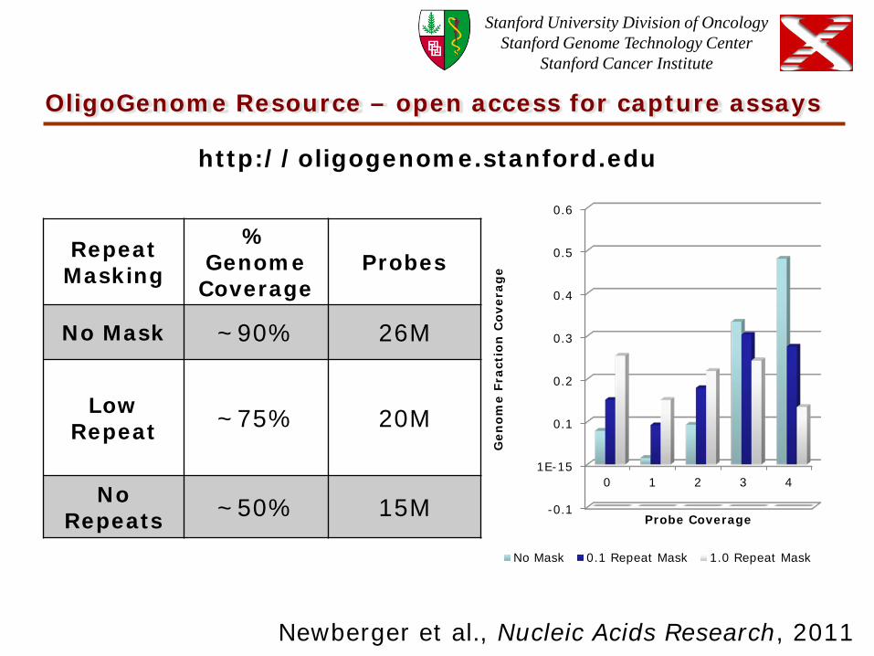
OligoGenome Resource – open access for capture assays
-0.1
1E-15
0.1
0.2
0.3
0.4
0.5
0.6
0 1 2 3 4 G
en
om
e F
ract
ion
Co
vera
ge
Probe Coverage
No Mask 0.1 Repeat Mask 1.0 Repeat Mask
Repeat Masking
% Genome Coverage
Probes
No Mask ~90% 26M
Low Repeat ~75% 20M
No Repeats ~50% 15M
http://oligogenome.stanford.edu
Newberger et al., Nucleic Acids Research, 2011
Stanford University Division of Oncology Stanford Genome Technology Center
Stanford Cancer Institute
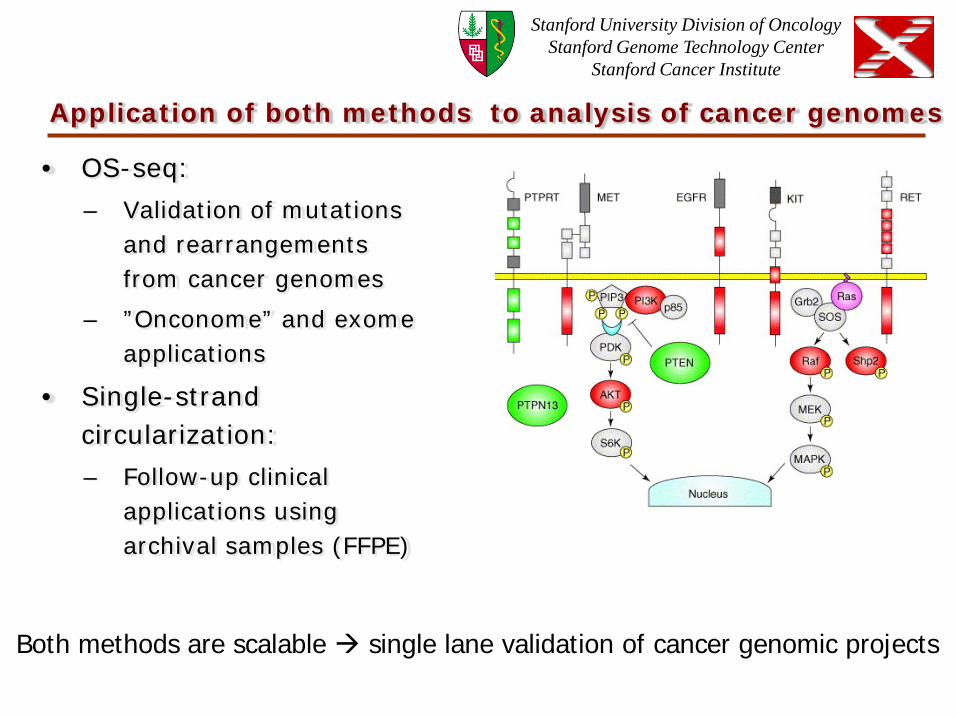
Application of both methods to analysis of cancer genomes
• OS-seq:
– Validation of mutations and rearrangements from cancer genomes
– ”Onconome” and exome applications
• Single-strand circularization:
– Follow-up clinical applications using archival samples (FFPE)
Both methods are scalable single lane validation of cancer genomic projects

Stanford University Division of Oncology Stanford Genome Technology Center
Stanford Cancer Institute
Acknowledgements
• Ji Research Group – Georges Natsoulis – Samuel Myllykangas – Jason Buenrostro – Erik Hopmans – Daniel Newburger – Laura Miotke – Hua Xu – Chris Xu – Sue Grimes
• Division of Oncology – Lincoln Nadauld
• Stanford Genome Technology Center – Michael Jensen
• Funding:
• NIH
– IMAT - National Cancer Institute (NCI)
– National Human Genome Research Institute (NHGRI)
• Doris Duke Foundation
• Howard Hughes Medical Foundation

![Small Variants Frequently Asked Questions (FAQ)Variants+FAQ.pdf · How do I identify somatic small variants? ... coverage-[ASM-ID].tsv: Reports number of bases in the reference genome](https://img.dokumen.tips/doc/110x75/5aa87f797f8b9a90188b9758/small-variants-frequently-asked-questions-faq-variantsfaqpdfhow-do-i-identify.jpg)


![Structural Variants in the Soybean Genome Localize to · PDF fileGenome Analysis Structural Variants in the Soybean Genome Localize to Clusters of Biotic Stress-Response Genes1[W][OA]](https://img.dokumen.tips/doc/110x75/5ab8426c7f8b9ad5338c9ea4/structural-variants-in-the-soybean-genome-localize-to-analysis-structural-variants.jpg)