Embed Size (px)
Citation preview

ISSSD 2015 September 26-30, 2015
Leon, Gto. Mexico
565
Mass attenuation coefficient (μ/ρ), effective atomic number (Zeff)
and measurement of x-ray energy spectra using based calcium
phosphate biomaterials: a comparative study
Madelon Aparecida Fernandes Zenobio1,2
, Elton Gonçalves Zenobio3
Teógenes Augusto da Silva 1,2
, Maria do Socorro Nogueira1,2
1 Development Center of Nuclear Technology- CDTN/CNEN
Avenida Presidente Antônio Carlos 6.627Campus UFMG
31270-901Belo Horizonte, Brazil. [email protected]
2Postgraduate Course on Nuclear Science and Techniques, UFMG
Av. Antônio Carlos 6627 Campus UFMG
31.270-90 Belo Horizonte, Brazil. [email protected]; [email protected].
3 PUCMINAS- Pontifíce Catholic University of Minas Gerais
Av. Dom José Gaspar 500
30535-901 Belo Horizonte, Brazil. [email protected]
Abstract In dentistry, alveolar bone regeneration procedures using based calcium phosphate
biomaterials have been shown effective. However,there are not reports in the literature of
studies the interaction of low energy radiation in these biomaterials used as attenuator and
not being then allowed a comparison between the theoretical values and experimental.The
objective of this study was to determine the interaction of radiation parameters of four
dental biomaterials - BioOss®, Cerasorb® M Dental, Straumann
® Boneceramic and
Osteogen®
for diagnostic radiology qualities. As a material and methods, the composition
of the biomaterials was determined by the analytical techniques. The samples with 0.181cm
to 0,297cm thickness were experimentally used as attenuators for the measurement of the
transmitted X-rays spectra in X-ray equipment with 50 to 90 kV range by spectrometric
system comprising the CdTe detector. After this procedure, the mass attenuation
coefficient, the effective atomic number were determined and compared between all the
specimens analyzed, using the program WinXCOM in the range of 10 to 200 keV. In all
strains examined observed that the energy spectrum of x-rays transmitted through the
BioOss®
has the mean energy slightly smaller than the others biomaterials for close
thickness. The μ/ρ and Zeff of the biomaterials showed its dependence on photon energy
and atomic number of the elements of the material analyzed. It is concluded according to
the methodology employed in this study that the measurements of x-ray spectrum, µ/ρ and
Zeff using biomaterials as attenuators confirmed that the thickness, density, composition of
the samples, the incident photon energy are factors that determine the characteristics of
radiation in a tissue or equivalent material.
Keywords: biomaterials, mass attenuation coefficient, X-rays spectra, effective atomic number
ISSSD 2015 September 26-30, 2015
Leon, Gto. Mexico
566
1.- INTRODUCTION
Studies on the interaction of low energy photons with biological samples are important in
the field of diagnostic radiology, medical radiation dosimetry, radiation shielding and
nuclear engineering materials showing an interdisciplinary science.
In the diagnostic radiology, the radiation interaction probability is a strong function of x-
ray energy, therefore the x-ray spectral distribution should be known in order to optimize
the radiographic imaging system; this would contribute to optimization and dose reduction
in patients [Maeda et al. 2005, Zenobio and Silva 2007; Zenobio et al. 2011].
For a comparison of the characteristics of radiation in a tissue or equivalent material, the
coefficient of mass attenuation (µ/ρ) of the photon and the coefficient of energy absorption
or the effective atomic number (Zeff) must be considered. Zeff was introduced to describe
the properties of composite materials in terms of equivalent elements [El Abd and Elkady
2014].
Advances in many specialty bioceramics have made significant contribution to the
development of modern health care industry and have improved the quality of human life.
In dentistry, alveolar bone regeneration procedures using based calcium phosphate
biomaterials have been shown effective and are the materials of choice in replacements of
teeth, repair for periodontal disease, maxillofacial reconstruction, augmentation and
stabilization of the jawbone. Theoretical and experimental investigations were carried out
in organic and inorganic materials in low energies presenting specific medical and
technological applications [Içelli and Erzeneoglu 2004; Morabad and Kerur 2010; Koç and
Ozyol 2000; Yang et al. 1987]. Nevertheless, there are no reports in literature concerning
the use of as based calcium phosphate biomaterials attenuation materials. This fact does not
allow for the establishment of comparisons between the theoretical and experimental
values. However, by means of investigation of the mass attenuation coefficient (µ/ρ),
effective atomic number (Zeff) and measurement of x-ray energy spectra using based

ISSSD 2015 September 26-30, 2015
Leon, Gto. Mexico
567
calcium phosphate biomaterials, invaluable information about the radiation interaction
process in diagnostic radiology x-ray qualities is expected to be provide.
2.- MATERIALS AND METHODS
This work was carried out in the Development Center of Nuclear Technology laboratories,
Brazil. Four bone substitute materials: BioOss®, Cerasorb® M Dental, Osteogen
®,
Straumann®
Boneceramic and Osteogen®
were obtained directly from the manufactures in
sealed vials in the powder form. Their chemical compositions were determined by the
technique of Neutron Activation Analysis (INAA), Elemental Analysis (EA), Mass
Spectrometry Inductively Coupled Plasma (ICP/AES) and X-ray Fluorescence (EDX)
following well established procedures.
The samples of the based calcium phosphate biomaterials were prepared in the form of
10mm diameter cylindrical pellets by pressing the weighed amount of the finely ground
powder in a Universal Instron 5882 - 100kN device at a pressure of 100 MPa. The
thickness of the Bio-Oss®, Cerasorb® M Dental, Osteogen
®, Straumann
® Boneceramic
range of 0.181cm to 0,297cm. The satisfactory geometrical arrangement has been employed
(Figure 1). Diagnostic radiology beam qualities RQR3, RQR5, RQR7 that are defined by
61267 International Electrotechnical Commission Standard (IEC, 2005), were established
in a 250 mm beryllium window constant potential Isovolt HS320 Pantak Seifert industrial
X-ray machine and 1 and 4mA tube current ranges [Kerur 2009; Zenobio et al. 2011].
Tungsten collimators with diameter aperture of 1 and 0.4 mm were placed inside the 35
mm-long spacer (Kit EXVC). X-ray energy spectra were measured with XR-100T model
CdTe detector; irradiation conditions were chosen to assure that pile-up effect was avoided.
In this research, the theoretical values of the mass attenuation coefficient (µ/ρ) of the
biomaterials samples were calculated by the WinXCOM software. For materials composed
of various elements, one may assume that the contribution of each element to the total
interaction of the photon is additive “Mixture Rule” [Morabad and Kerur 2010]. In
ISSSD 2015 September 26-30, 2015
Leon, Gto. Mexico
568
accordance with this rule, the total mass attenuation coefficient of a composite is the sum of
the weight proportion of each individual atom present in it [Morabad and Kerur 2010].
Therefore:
(
)
∑( ) (
)
(1)
In which: (µ/ρ)comp is the mass attenuation coefficient for the composite, (µ/ρ)i is the mass
attenuation coefficient of each individual element and wi is the fractionated weight of the
elements in the composite. The weight fractions of each element present in the biomaterials
samples were determined by analytical means mentioned above.
The Zeff values were determined directly for total interactions within a radiological low
photon energy range, from 10 to 200 keV [Özdemir and Kurudirek 2009; Rao et al. 1985;
Yang et al. 1987]. The atomic cross-section in barn/atom was computed from the µ/ρ using
the following relation:
∑ (2)
where Ai represents atomic number of the constituent elements, while N represents the
Avogadro constant [White, 1977]. Plots were drawn between the atomic cross-section in
the individual element and the atomic numbers of the elements for each energy level. From
these plots, the Zeff of each material was obtained by interpolation.

ISSSD 2015 September 26-30, 2015
Leon, Gto. Mexico
569
Figure 1.- Experimental set-up
3.- RESULTS AND DISCUSSIONS
The chemical composition of based calcium phosphate biomaterials that were
obtained by analytical techniques are given in the Table 1. The samples presented
different chemical compositions and high and different concentrations of Ca, P and
organic material.
Table 1- Chemical composition of based calcium phosphate biomaterials.
Elements Bio-Oss® Cerasorb
M Dental
Straumann
Boneceramic®
Osteogen®
H 0,38 0,03 0,39
C 1,12 0,12 0,23
O 37,99 38,85 41,42 39,83
Na 1,96 2,00 0,50 1,60
Mg 0,30 0,08 0,28
Al 0,40 0,32 0,36 0,38
Si 1,20 1,37 1,21 1,26
P 16,71 17,69 17,58 17,88
S 0,21 0,17 0,33 0,20
Cl 0,15 0,10 0,05 0,06
K 0,03 0,02

ISSSD 2015 September 26-30, 2015
Leon, Gto. Mexico
570
Ca 39,59 39,39 38,10 38,17
Figures 2 to 4 show the x-ray spectra of transmitted beams through different thicknesses of
biomaterials samples for the 50, 70 and 90 kV. In this experiment the average energy varied
from 33.96 to 46.43 keV. Results show that the transmitted spectra have slightly
differences in the penetration power and in the spectral distribution. It was clear in the
Figure 4, for the 90kV. The transmitted spectrum for the Straumann® Boneceramicand and
Cerasorb® M Dental show that the their mean energy is higher than the Bio-Oss
® and
Osteogen® even for close thicknesses.
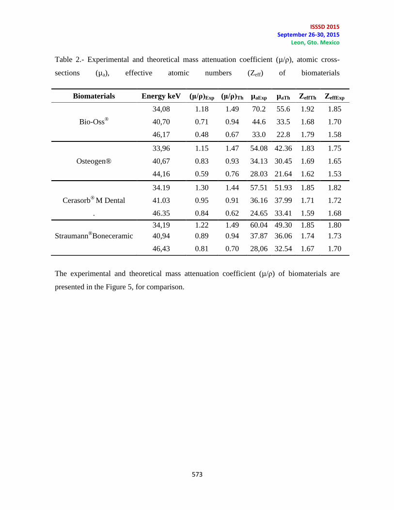
The table 2 shows variation of the experimental and theoretical mass attenuation coefficient
(µ/ρ), atomic cross-sections (µa) and effective atomic numbers (Zeff) of the biomaterials
samples with the energy. The value of the μ/ρ of the samples of biomaterials were inversely
proportional to the energy in agreement studies of Morabad and Kerur [2010] and Koç and
Ozyol [2000]. In this study there were significant differences between experimental and
theoretical values (5.1 to 35.6%) for all biomaterials samples when evaluatted the µ/ρ, µa.
However, in relation to the Zeff the experimental results are, in general, consistent with
theoretical data (0.9 to 6.2%) that were calculated using the independent atomic model, or
in other word, mixture rule which theoretical value has done by considering the cross-
section for isolated atom. Besides, Straumann ® Boneceramic and Cerasorb® M Dental
show µ/ρ experimental and theoretical values were higher than those of Bio-Oss® and
Osteogen®. These indicate that Straumann ® Boneceramic and Cerasorb® M Dental are
better absorber than of Bio-Oss® and Osteogen®.
Experimental data were not found in the literature to compare with, nevertheless Zenobio
[2011], Içelli and Erzeneoglu [2004] found that the uncertainties in the sample thicknesses
and elemental composition of materials affected experimental results during transmitted
spectrum measurements of material compounds for determining effective atomic numbers.
According to Han and Demir [2009] at low photon energies the process of photoelectrical
interaction and the dependence of atomic number is dominant when there is interaction of
photon with matter. The discrepancies in the effective atomic number may be reduced by
considering the molecular, chemical, or crystalline environment of the atom [Içelli, 2008;
Singh and Badiger, 2014]. The density is additional influence factors and in biomaterial the

ISSSD 2015 September 26-30, 2015
Leon, Gto. Mexico
571
calcination and sintering processes need to be considered when the density is evaluated
[Kim et al. 2003]. These biomaterials, as well as enamel, dentin, bone, present in their
composition high and different concentration of Ca and P; consequently, their interactions
with low energy photons are expected to be different from other biological samples in
agrement the literature [Lakamaa and Rytomaa 1977; Koç and Ozyol 2000; Zenobio et al.
2011]. Thus, deep knowledge of the purity of the elemental materials, the composition of
alloys or mixtures is required in order to obtain accurate experimental and accurate
calculated values [Zenobio et al. 2011; Kucuk et al. 2013].
Figure 2.- Photons spectra of 50 kV x-ray beam transmitted through different thicknesses
of biomaterials.
0
5E+09
1E+10
1.5E+10
2E+10
2.5E+10
3E+10
10 20 30 40 50
Ph
oto
ns
spec
tra
Energy
"BioOss" "Osteogen" "Cerasorb"
"Boneceramic" "Without Attenuator"

ISSSD 2015 September 26-30, 2015
Leon, Gto. Mexico
572
Figure 3.- Photons spectra of 70 kV x-ray beam transmitted through different thicknesses of
biomaterials.
Figure 4.- Photons spectra of 90 kV x-ray beam transmitted through different thicknesses of
biomaterials
0
5E+09
1E+10
1.5E+10
2E+10
10 20 30 40 50 60 70 80
Ph
oto
ns
spec
tra
Energy
BioOss Osteogen Cerasorb
Boneceramic "Without attenuator"
0
2E+09
4E+09
6E+09
8E+09
1E+10
1.2E+10
1.4E+10
1.6E+10
10 20 30 40 50 60 70 80 90 100
Ph
oto
ns
spec
tra
Energy
Biooss Osteogen Cerasorb Boneceramic
ISSSD 2015 September 26-30, 2015
Leon, Gto. Mexico
573
Table 2.- Experimental and theoretical mass attenuation coefficient (µ/ρ), atomic cross-
sections (µa), effective atomic numbers (Zeff) of biomaterials
The experimental and theoretical mass attenuation coefficient (µ/ρ) of biomaterials are
presented in the Figure 5, for comparison.
Biomaterials Energy keV (μ/ρ)Exp (μ/ρ)Th µaExp µaTh ZeffTh ZeffExp
34,08 1.18 1.49 70.2 55.6 1.92 1.85
Bio-Oss®
40,70 0.71 0.94 44.6 33.5 1.68 1.70
46,17 0.48
0.67 33.0 22.8 1.79 1.58
33,96 1.15
1.47 54.08 42.36 1.83 1.75
Osteogen® 40,67 0.83 0.93 34.13 30.45 1.69 1.65
44,16 0.59
0.76 28.03 21.64 1.62 1.53
34.19 1.30
1.44 57.51 51.93 1.85 1.82
Cerasorb®
M Dental 41.03 0.95 0.91 36.16 37.99 1.71 1.72
. 46.35 0.84 0.62 24.65 33.41 1.59 1.68
34,19 1.22
1.49 60.04 49.30 1.85 1.80
Straumann®
Boneceramic 40,94 0.89
0.94
37.87 36.06 1.74 1.73
46,43 0.81 0.70 28,06 32.54 1.67 1.70

ISSSD 2015 September 26-30, 2015
Leon, Gto. Mexico
574
Figure 6.- µ/ρ in biomaterials samples in the present study
The mass attenuation coefficient (µ/ρ) of the biomaterials samples were calculated by the
WinXCOM software. The behavior of Zeff values with respect to applied energy levels for
total interaction in biomaterials samples were plotted in the Figure 6, for comparison with
the literature, in the energy range from 10 to 200 keV.
In the low photon energy region, the Zeff decrease with the increasing of the incident photon
energy. However, several jumps in values have been observed in the energy range from 30
keV to 50 keV for all materials except for the Osteogen®. Second El-Kateb et al. [2000]
and Elmahroug et al. [2015] these jumps are due to the absorption K-edge of the high Z-
element (Na, Al, S, Si, Ca, Mn, Fe, Zn and Sr) and this explains the differences between the
values of in this region is very large compared to other regions.
.
0.4
0.8
1.2
33 38 43 48
µ/ρ
Energy (keV)
BioOss exp. BioOss ThOsteogen® Exp. Osteogen® Th.Cerasorb®M Dental Exp. Cerasorb®M Dental Th.Straumann ® Boneceramic Exp. Straumann ® Boneceramic Th.

ISSSD 2015 September 26-30, 2015
Leon, Gto. Mexico
575
Figure 6.- Zeff in biomaterials samples in the present study and the literature.
The maximum errors in mass attenuation coefficients were calculated from errors in
incident ( I0 ) and transmitted ( I) intensities and areal density (t) and statistical counting
[Ozdemir and Kurudirek 2009]; given that the values estimated in this study were from
0.08% to 0.03% in the biomaterials.
5.- CONCLUSIONS
The mass attenuation coefficient (µ/ρ), effective atomic number (Zeff) and measurement of
x-ray energy spectra using based calcium phosphate biomaterials- Bio-Oss®, Cerasorb
® M
5
6
7
8
9
10
11
12
13
14
15
16
0 50 100 150 200
Zef
f
Energy (keV)
Bio-Oss® Osteogen®Cerasorb® Boneceramic®Bone- Shivaramu [2002] Dentin- Zenóbio [2011]Enamel- Zenóbio [2011]

ISSSD 2015 September 26-30, 2015
Leon, Gto. Mexico
576
Dental, Osteogen®
, Straumann® Boneceramic- were investigated for diagnostic radiology
qualities.
The measurements of x-ray spectrum, µ/ρ and Zeff using biomaterials as attenuators
confirmed that the thickness, density, composition of the samples, the incident photon
energy are factors that determine of the characteristics of radiation in a tissue or equivalent
material.
The data presented on photon interaction parameters are expected to be helpful in
dosimetry; radiation shielding and other applications of radiation physics. Further
investigation on the photon interaction parameters in different compounds and/or
composites still remain to be done in order to confirm the validity of both.
Acknowledgments
The authors are thankful to CDTN/CNEN, PUCMinas, CAPES, FAPEMIG and Ministry of
Science and Technology - MCT/Brazil, through the Brazilian Institute of Science and
Technology (INCT) for Radiation Metrology in Medicine
REFERENCES
El-Kateb A.H.; Rizk R.A.M.; Abdul-Kader A.M. (2000). Determination of atomic cross-
sections and effective atomic numbers for some alloys. Annals of Nuclear Energy 27:
1333-43.
Elmahroug Y.; Tellili B.; Souga C (2015). Determination of total mass attenuation
coefficients, effective atomic numbers and electron densities for different shielding
materials. Annals of Nuclear Energy 75: 268–74.
El Abd A.A.; Elkady A.S (2014). A Method for Simultaneous Determination of Effective
Removal Cross-section for Fast Neutrons and Mass Absorption Coefficient for
Gamma Rays. SOJ Mater Science Engineering 2: 1-6.

ISSSD 2015 September 26-30, 2015
Leon, Gto. Mexico
577
Han I.; Demir L. (2009). Mass attenuation coefficients, effective atomic and electron
numbers of Ti and Ni alloys. Radiation Measurements 44: 289-94.
IçellI O.; Erzeneoglu S. (2004). Effective atomic numbers of some vanadium and nickel
compounds for total photon interactions using transmission experiments. Journal of
Quantitative Spectroscopy and Radiative Transfer 85: 115-24
Icelli O. and Erzeneoglub S. Determination of molecular, atomic, electronic cross-sections
and effective atomic number of some boron compounds and TSW, Nuclear
Instruments and Methods in Physics Research 266 (2008) 3226–30
IEC-International Organization for Standardization, 2005. Medical Diagnostic X-ray
Equipment- Radiation Conditions for Use in the Determination of Characteristics.
ISO/IEC 61267, second ed. Geneva.
Kerur B.R.; Manjula V.T.; Lagare M.T. Anil Kumar S. (2009). Mass attenuation coefficient
of saccharides for X-rays in the energy range from 8 keV to 32 keV. Radiation
Measurements 44: 63-7.
Kim S.R; Lee H.; Kim Y.T.; Riu D.H.; Jung S.J; Lee Y.J; Chung S.C; Kim Y.H. (2003).
Synthesis of Si, Mg substituted hydroxyapatites and their sintering behaviors.
Biomaterials,. 24: 1389-98.
Koç N.; Ozyol H. (2000) Z-dependence of partial and total photon interactions in some
biological samples. Radiation Physics and Chemistry 59: 339-45.
Kucuk N.; Cakir M.; Isitman N. A. (2013). Mass attenuation coefficients, effective atomic
numbers and effective electron densities for some polymers. Radiation Protection and
Dosimetry 153: 127-34.
Maeda K.; Matsumoto M.; Taniguchi A. (2005). Compton-scattering measurement of
diagnostic x-ray spectrum using high-resolution Schottky CdTe detector. Medical
Physics 32: 1542-7.
Morabad R.B.; Kerur B.R. (2010). Mass attenuation coefficients of X-rays in different
medicinal plants. Applied Radiation and Isotopes 68: 271-4,
Ozdemir Y.; Kurudirek M.A (2009). Study of total mass attenuation coefficients, effective
atomic numbers and electron densities for various organic and inorganic compounds
at 59.54 keV. Annals of Nuclear Energy 36: 1769-73,
Rao B.V.T.; Raju M.L.N.; K.L. Narasimham; Parthasaradhi K.and Rao B.M.(1985).
Interaction of low-energy photons with biological materials and the efecctive atomic
number. Medical Physics 12: 745-8.

ISSSD 2015 September 26-30, 2015
Leon, Gto. Mexico
578
Shivaramu (2002). Effective atomic numbers for photon energy absorption and photon
attenuation of tissues from human organs. Medical Dosimetry 27:1-9.
Singh V.P.; Badiger N.M. (2014). Effective atomic numbers of some tissue substitutes by
different methods: A comparative study. Journal of Medical Physics 39: 24-31.
White D.R. (1977). An analysis of the Z-dependence of photon and electron interactions.
Physics in Medicine and Biology 22: 219-28.
Yang N.C.; Leichner P.K.; Hawkins W.G. (1987). Effective atomic numbers for low-
energy total interactions in human tissues. Medical Physics 14: 59-66.
Zenóbio M.A.F.; Silva T.A. (2007). Absorbed dose on patients undergoing tomographics
exams for pre-surgery planning of dental implants. Applied Radiation and Isotopes
65: 708-11.
Zenóbio M.A.F.; Tavares M.S.N.; Zenóbio E.G.; Squair P.L; Santos M.A.P.; Silva T.A.
(2011). Measurement of spectra for intra-oral x-ray beams using biological materials
as attenuator. Radiation Measurements 46: 2100-2.
Zenóbio M.A.F.; Tavares M.S.N.; Zenóbio E.G.; Silva T.A. (2011). Elemental composition
of dental biologic tissues: study by means of different analytical techniques. Journal
of Radioanaytical Nuclear Chemistry 289: 161-6.