Embed Size (px)
Citation preview

Chapter 26Chapter 26

Figure 26.1 An Introduction to the Urinary System
Suprarenalgland
Renal arteryand vein
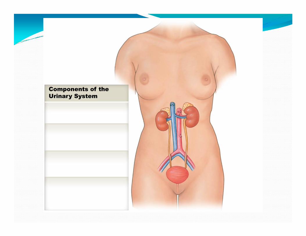
Components of the
Urinary System
Kidney
Produces urine
© 2015 Pearson Education, Inc.
Aorta
and vein
Inferiorvena cava
Produces urine
Ureter
Transports urine towardthe urinary bladder
Urinary Bladder
Temporarily storesurine prior to elimination
Urethra
Conducts urine to exterior;in males, transports semenas well
Components of the
Urinary System

Diaphragm
Celiac trunk
Left ureter
Superiormesenteric artery
Left renal vein
Left suprarenal gland
Left kidney
Left renal artery
Figure 26.3a The Urinary System in Gross Dissection© 2015 Pearson Education, Inc.
Iliacus muscle
Psoas majormuscle
Rectum (cut)

Function

AortaInferior
vena cava
Connective Tissue
Parietal peritoneum
Stomach
Pancreas
Vertebra
Spleen
Figure 26.2b© 2015 Pearson Education, Inc.
Pararenalfat
Renal fascia
Perinephric fat
Fibrous capsule
Connective TissueLayers Protectingthe Kidneys
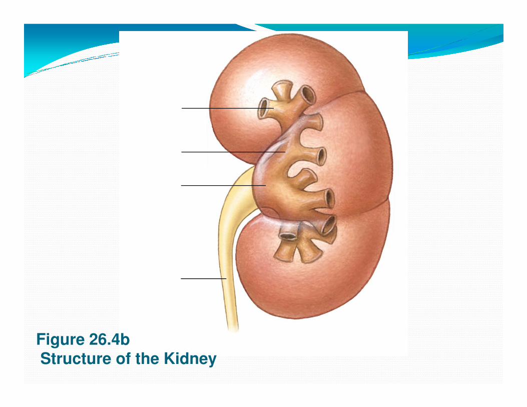
Figure 26.4bStructure of the Kidney

Renal sinus
Adipose tissuein renal sinus
Renal pelvis
Cortex
Medulla
Renal pyramid
Connection tominor calyx
Minor calyx
Major calyxHilum
Medulla
Outer layer offibrous capsule
Renalpyramids
Renal sinus
Renal pelvis
Major calyx
Figure 26.4a Structure of the Kidney© 2015 Pearson Education, Inc.
Hilum
Renal papilla
Ureter
Renal lobe
Renal columns
Outer layer offibrous capsule
Ureter
Major calyx
Minor calyx
Renal papilla
Renal lobe
Fibrous capsule

Typical kidney� Size: 10 cm long, 5.5 cm wide, 3 cm thick, 150g
� Sectional view:
� Medial indentation is the __________
� Renal _________ enter at the hilum
� Renal _________ and ___________ exit at the hilum

Interlobar
arteries
Segmental
artery
Suprarenal
artery
Renal
artery
Figure 26.5a Blood Supply to the Kidneys © 2015 Pearson Education, Inc.
Arcuate
arteries
Cortical
radiate
arteries
Blood supply to kidneys1. Beginning with blood in the _________, blood flows
to:
2. ___________ arteries
3. ____________ arteries3. ____________ arteries
4. _____________ arteries
5. _________ ____________arteries
6. ______________ arterioles
7. ________________ capillaries
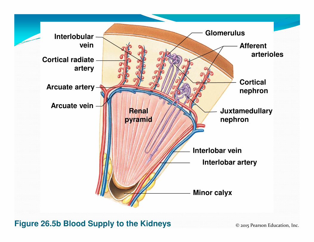
Interlobularvein
Cortical radiate
artery
Arcuate veinRenal
pyramid
Glomerulus
Afferent
arterioles
Juxtamedullarynephron
Cortical
nephronArcuate artery
Figure 26.5b Blood Supply to the Kidneys © 2015 Pearson Education, Inc.
pyramid nephron
Interlobar vein
Interlobar artery
Minor calyx
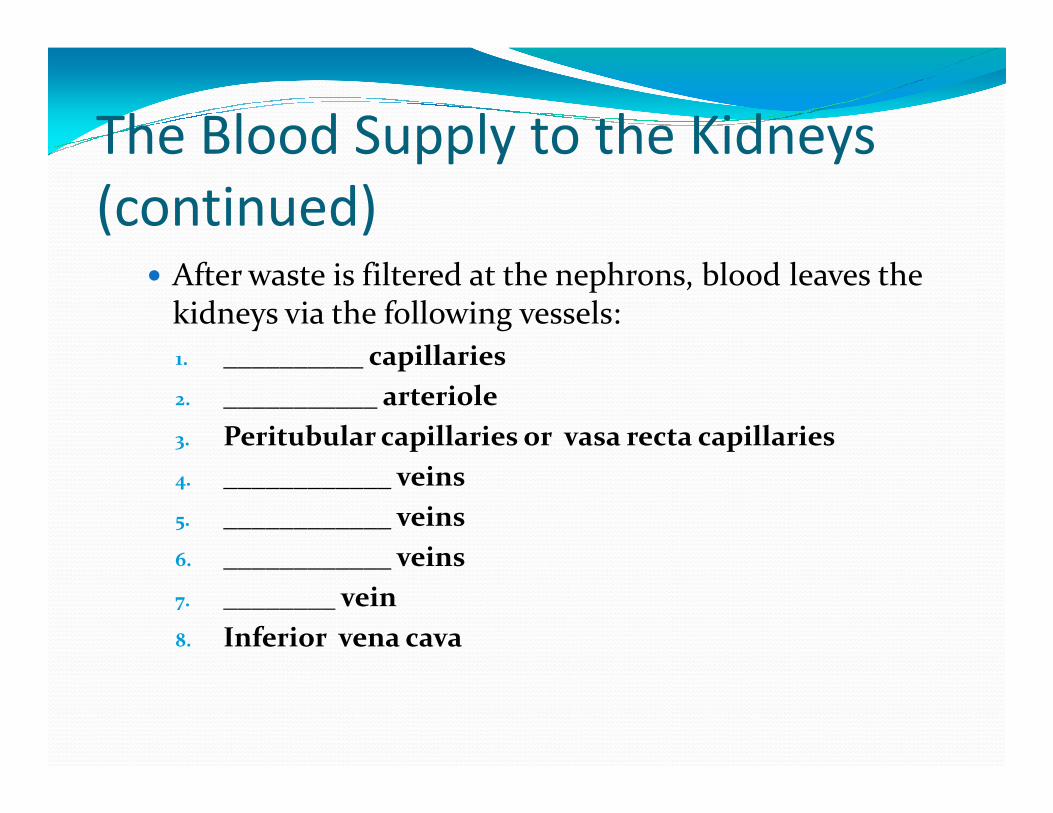
The Blood Supply to the Kidneys
(continued) � After waste is filtered at the nephrons, blood leaves the
kidneys via the following vessels:
1. __________ capillaries
2. ___________ arteriole2. ___________ arteriole
3. Peritubular capillaries or vasa recta capillaries
4. ____________ veins
5. ____________ veins
6. ____________ veins
7. ________ vein
8. Inferior vena cava

7. Renal
vein
6. Interlobar
veins5. Arcuate
veins
Arcuate
arteries

The Kidneys� Innervation of the Kidneys
� Urine production is regulated by autoregulation
� Involves reflexive changes in the diameter of nephron arteriolesarterioles
� Receives sympathetic nerve fibers from the celiac and inferior mesenteric ganglia
� Nerve innervation serves to:
� Regulate renal blood flow and pressure
� Stimulate renin release
� Stimulate water and sodium ion reabsorption

Structure and Function of the
Nephron
� Waste (glomerular filtrate) material leaves theglomerular capillaries and enters:
1. Glomerular capsule
2. _____________________________(PCT)
3. Nephron loop
4. _____________________________(DCT)
5. Collecting Duct

Figure 26.8a Histology ofthe Nephron
Cortex
Cortical
nephron
Juxtamedullary
nephron
2. Proximalconvoluted
tubule
1. Renal corpuscle
4. Distal convolutedtubule
5. Connecting tubules
© 2015 Pearson Education, Inc.
a
3. Nephronloop
Thin descendinglimb
Thick ascendinglimb
Medulla
5. Collecting duct
Papillary duct
Renal papilla
Minor calyx
Orientation of cortical and juxtamedullary nephrons.
Structure and Function of the
Nephron
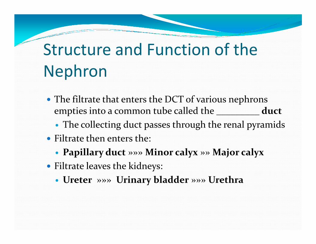
� The filtrate that enters the DCT of various nephrons empties into a common tube called the _________ duct
� The collecting duct passes through the renal pyramids� The collecting duct passes through the renal pyramids
� Filtrate then enters the:
� Papillary duct »»» Minor calyx »» Major calyx
� Filtrate leaves the kidneys:
� Ureter »»» Urinary bladder »»» Urethra
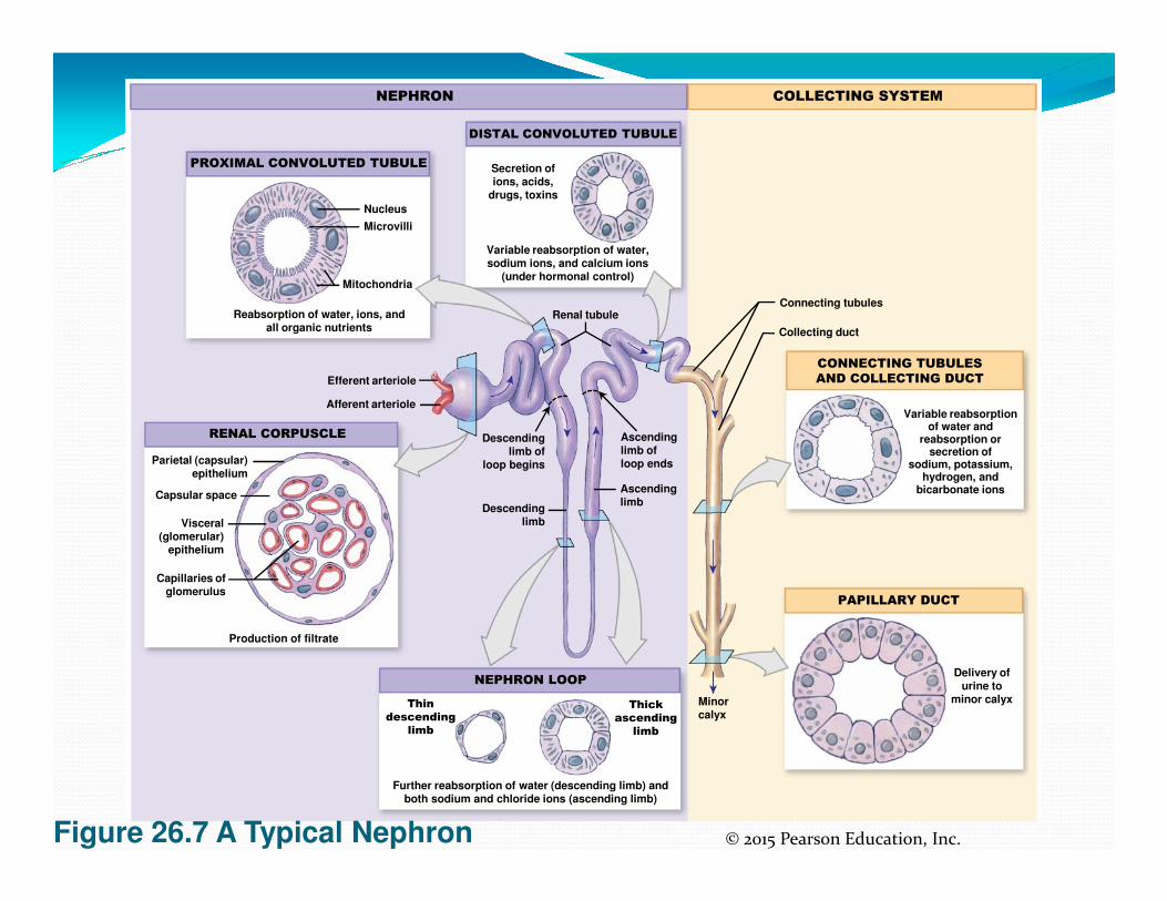
Nucleus
Microvilli
Mitochondria
Efferent arteriole
Afferent arteriole
Reabsorption of water, ions, andall organic nutrients
Renal tubule
Variable reabsorption of water,sodium ions, and calcium ions
(under hormonal control)
Secretion ofions, acids,
drugs, toxins
Descendinglimb of
Ascendinglimb of
PROXIMAL CONVOLUTED TUBULE
DISTAL CONVOLUTED TUBULE
RENAL CORPUSCLE
NEPHRON
CONNECTING TUBULES
AND COLLECTING DUCT
COLLECTING SYSTEM
Connecting tubules
Collecting duct
Variable reabsorptionof water and
reabsorption orsecretion of
Figure 26.7 A Typical Nephron © 2015 Pearson Education, Inc.
Minorcalyx
limb ofloop begins
limb ofloop ends
Descendinglimb
Ascendinglimb
Parietal (capsular)epithelium
Capsular space
Visceral(glomerular)
epithelium
Capillaries ofglomerulus
Production of filtrate
Thin
descending
limb
Thick
ascending
limb
Further reabsorption of water (descending limb) andboth sodium and chloride ions (ascending limb)
NEPHRON LOOP
PAPILLARY DUCT
secretion ofsodium, potassium,
hydrogen, andbicarbonate ions
Delivery ofurine to
minor calyx

Structure and Function of the
Nephron� Two main types of nephrons
� ___________ nephrons
� 85 percent of the nephrons are cortical
� Most of the nephron is located in the cortex� Most of the nephron is located in the cortex
� Have a relatively short nephron loop
� _____________nephrons
� 15 percent of the nephrons are juxtamedullary
� Capsule is located near the border of the cortex and the medulla
� Have a long nephron loop

Structure and Function of the
Nephron
� Main functions of the nephron
� _____________________________________________� _____________________________________________
� Urine processing
� ____________________________________________
� Prevents dehydration
� ____________________________________________
� Urine processing
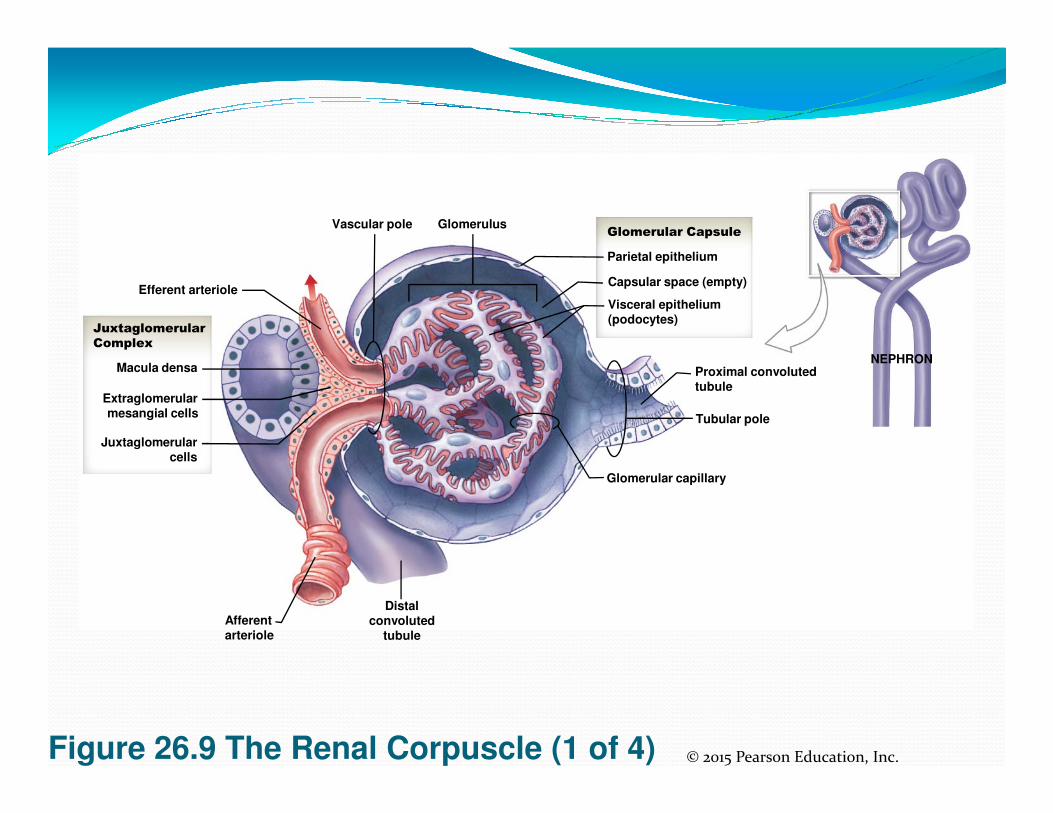
The Kidneys� The Renal Corpuscle
� Consists of:
� Glomerular capsule
� Glomerular capillaries (glomerulus)� Glomerular capillaries (glomerulus)
� Glomerular capsule consists of:
� Parietal layer
� Made of squamous cells
� Visceral layer
� Makes up the epithelial lining of the capillaries

GlomerulusVascular pole
Efferent arteriole
Juxtaglomerular
Complex
Macula densa
Extraglomerularmesangial cells
Proximal convolutedtubule
NEPHRON
Visceral epithelium(podocytes)
Capsular space (empty)
Parietal epithelium
Glomerular Capsule
Figure 26.9 The Renal Corpuscle (1 of 4) © 2015 Pearson Education, Inc.
mesangial cells
Juxtaglomerularcells
Afferentarteriole
Distalconvoluted
tubule
Glomerular capillary
Tubular pole
The Kidneys� The Renal Corpuscle
� Filtration within the renal corpuscle involves three layers
� ___________________________� ___________________________
� Fenestration (.06-.1 µ)
� ___________________________
� Surrounds capillary endothelium (large proteins can’t pass)
� ___________________________
� Consists of specialized cells called podocytes
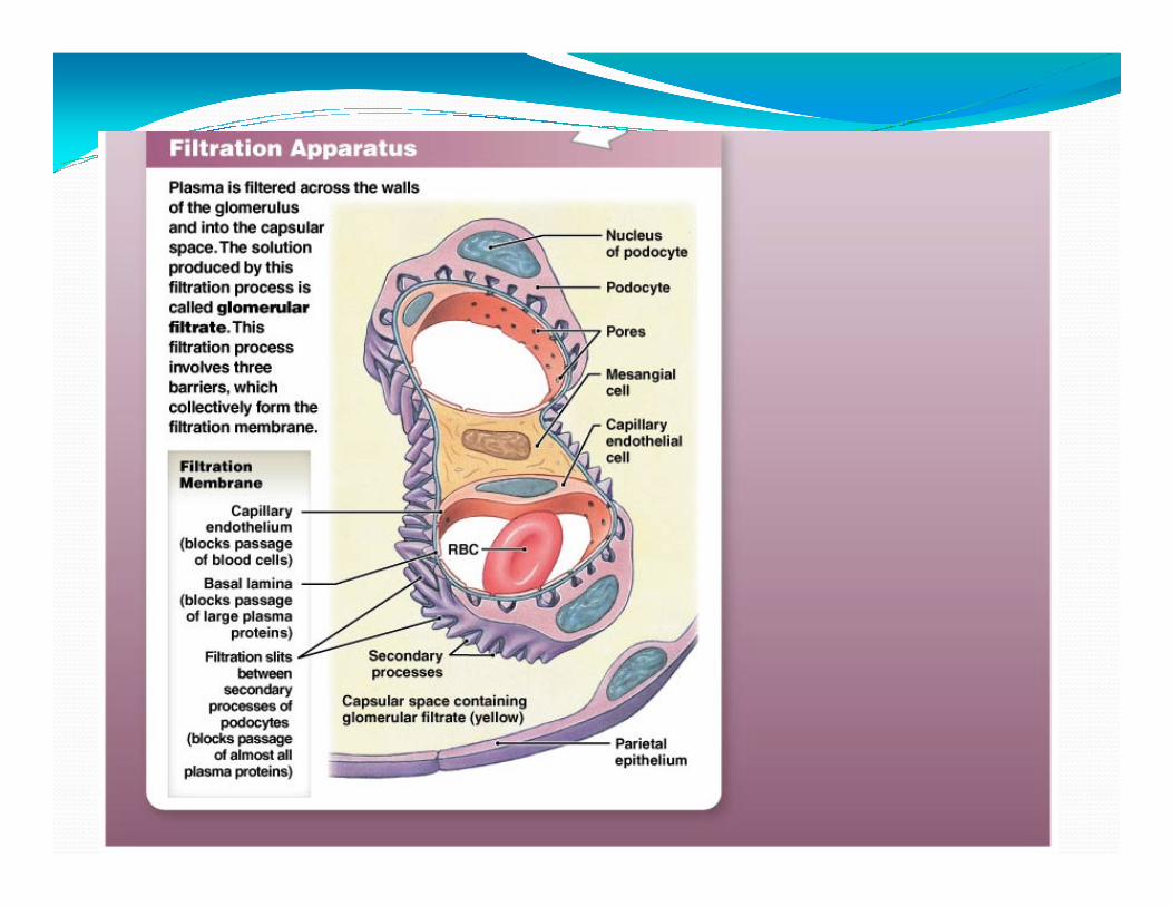

Glomerular epithelium� ____________ have long extensions that wrap around
basal lamina
� Extensions have filtration slits
� Filtrate passing through consists of water, ions, small � Filtrate passing through consists of water, ions, small organic molecules (glucose, fatty acids, amino acids, vitamins)
� Any potential useful product will be reabsorbed in proximal convoluted tubule (PCT)
___________� Lined with cuboid epithelium
� Reabsorbs all:
� Organic nutrients
Plasma protein� Plasma protein
� 60% sodium and chloride ions and water
� Calcium, potassium, magnesium, bicarbonate, phosphate, sulfate ions

Nucleus
Microvilli
Mitochondria
Efferent arteriole
Afferent arteriole
Reabsorption of water, ions, andall organic nutrients
Renal tubule
Variable reabsorption of water,sodium ions, and calcium ions
(under hormonal control)
Secretion ofions, acids,
drugs, toxins
Descendinglimb of
Ascendinglimb of
PROXIMAL CONVOLUTED TUBULE
DISTAL CONVOLUTED TUBULE
RENAL CORPUSCLE
NEPHRON
CONNECTING TUBULES
AND COLLECTING DUCT
COLLECTING SYSTEM
Connecting tubules
Collecting duct
Variable reabsorptionof water and
reabsorption orsecretion of
Figure 26.7 A Typical Nephron © 2015 Pearson Education, Inc.
Minorcalyx
limb ofloop begins
limb ofloop ends
Descendinglimb
Ascendinglimb
Parietal (capsular)epithelium
Capsular space
Visceral(glomerular)
epithelium
Capillaries ofglomerulus
Production of filtrate
Thin
descending
limb
Thick
ascending
limb
Further reabsorption of water (descending limb) andboth sodium and chloride ions (ascending limb)
NEPHRON LOOP
PAPILLARY DUCT
secretion ofsodium, potassium,
hydrogen, andbicarbonate ions
Delivery ofurine to
minor calyx

_______________� Descending portion
� Reabsorption of water into blood stream »» vasa recta
� Ascending portion
Pumps ions (Na and Cl ions) out of ascending loop to � Pumps ions (Na and Cl ions) out of ascending loop to prevent their loss
� Impermeable to water

____________________(DCT)
� Active secretion of ions and acids
� Selective reabsorption of Na and Ca ions
� Very little reabsorption of water

Juxtaglomerular complex� Located region of vascular pole. Consists of:
� Macula densa cells, juxtaglomerular cells, mesangialcells
� Produce two hormones:� Produce two hormones:
� Renin
� Erythropoietin
GlomerulusVascular pole
Efferent arteriole
Juxtaglomerular
Complex
Macula densa
Extraglomerularmesangial cells
Proximal convolutedtubule
NEPHRON
Visceral epithelium(podocytes)
Capsular space (empty)
Parietal epithelium
Glomerular Capsule
Figure 26.9 The Renal Corpuscle (1 of 4) © 2015 Pearson Education, Inc.
mesangial cells
Juxtaglomerularcells
Afferentarteriole
Distalconvoluted
tubule
Glomerular capillary
Tubular pole

____________� Ureters exit at the hilum area
� Transport urine from kidneys to urinary bladder
� Enter urinary bladder on the posterior/inferior side
The ureteral openings enter at the ____________� The ureteral openings enter at the ____________
� Consists of three tunic layers (mucosa, muscular, adventitia)
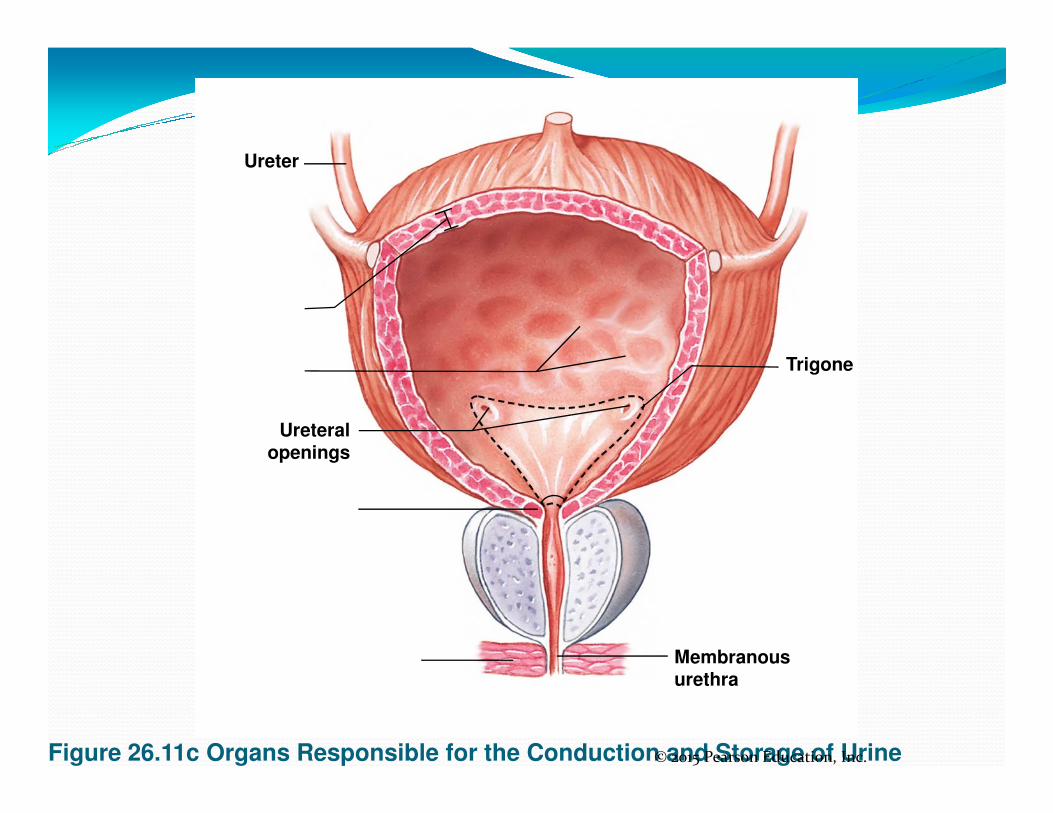
Trigone
Ureter
Figure 26.11c Organs Responsible for the Conduction and Storage of Urine© 2015 Pearson Education, Inc.
Ureteralopenings
Membranousurethra
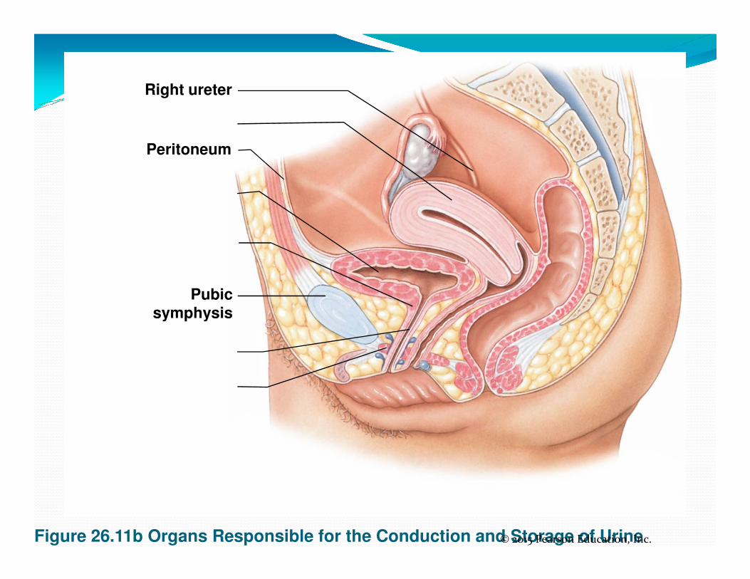
Right ureter
Peritoneum
Pubic
Figure 26.11b Organs Responsible for the Conduction and Storage of Urine© 2015 Pearson Education, Inc.
Pubicsymphysis

Figure 26.11a Organs Responsible for the Conduction and Storage of Urine
Peritoneum
Pubic
symphysis
Left
ureter
Prostate
gland
© 2015 Pearson Education, Inc.
Urogenital
diaphragm
Spongy
urethra
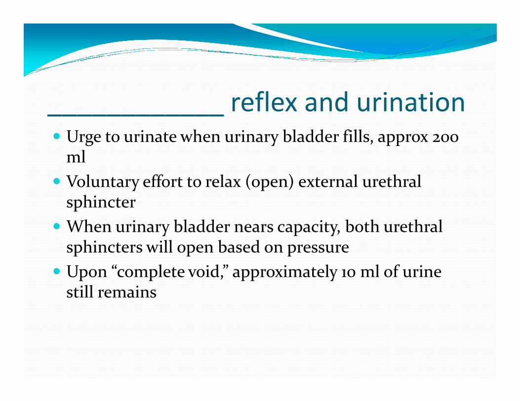
____________ reflex and urination� Urge to urinate when urinary bladder fills, approx 200
ml
� Voluntary effort to relax (open) external urethral sphinctersphincter
� When urinary bladder nears capacity, both urethral sphincters will open based on pressure
� Upon “complete void,” approximately 10 ml of urine still remains


![IBS DelCarpio 2013 [Read-Only] · Dr. Delcarpio 11-18-2013Delcarpio 2013 ... The Renal Corpuscle 6. ... Mature Renal Corpuscle, components: 1. The glomerular](https://img.dokumen.tips/doc/110x75/5b85807c7f8b9a4a488e67cf/ibs-delcarpio-2013-read-only-dr-delcarpio-11-18-2013delcarpio-2013-the.jpg)