Embed Size (px)
Citation preview
Corneal transplant outcome – a Swedish register
Margareta Claesson MD
Department of Ophthalmology Institute of Neuroscience and Physiology
University of Gothenburg Sweden
December 2008

The patient goes about his business on his own, managed the fairly lengthy journey here alone, has been able to find his way without difficulty in this unfamiliar town and is able at home to take care of lighter agricultural duties such as raking up and turning the hay, cleaning and feeding the cattle, mucking out the cowshed, etc. Edward Zirm, 1905, about the first successful penetrating keratoplasty.
To the patients

Abstract
Aim. The aim of this study was to present different aspects of the outcome after corneal transplantation based on data from the Swedish Cornea Register.
Papers. The first paper describes the register and gives descriptive statistics and analysis of data from a two-year follow up, while the last paper presents data from a ten-year follow up. Papers two and three deal with two specific problems in corneal transplantation, astigmatism and corneal oedema after cataract surgery (bullous keratopathy, BK). Paper four and five compare the Swedish patients with a cohort from the Middle East.
Results. The major indications were keratoconus (29%), BK (21%) and a mixed group of other diagnoses (32%), including regraft. The overall incidence of rejection at two years was 15%, and regrafting, which occurred in 10% of cases was related to rejection and other complications.
Visual acuity (VA) after two years improved most in keratoconus and this was still the case after ten years. Most changes in visual outcome after PK in all indications occurred during the first two postoperative years. Graft survival and VA at ten years depended mainly on complications occurring before two years postoperative.
The mean value of astigmatism at two years was 4.6 D (95% CI 4.4-4.7), independent of indication and preoperative astigmatism. In a group with high astigmatism (mean value 8.4 D) relaxing incisions reduced the astigmatism by 50%. At ten years there was a small increase in astigmatism in all indications.
Bullous keratopathy was one of the indications with poorest outcome. The risk of developing BK at the time of cataract surgery was influenced by pre-existing endothelial disease and cataract surgery done by phaco-emulsification.
In the Palestinian Territories the preponderance of keratoconus was higher than in Sweden. The patients came to surgery with a more advanced disease and more risk factors. They also developed more postoperative complication and the outcome was poorer, even though most gained some visual acuity.
Conclusion. Through the data analysed from the register our knowledge of the outcome after corneal transplantation has increased. The register will also allow evaluation of new techniques of corneal transplantation. Key words: Corneal transplantation, quality register, long term follow up, astigmatism, bullous keratopathy, graft survival, visual outcome. ISBN-978-91-628-7552-7

Contents
Abstract Original papers Abbreviations Glossary Introduction 1 Prevalence of corneal disease 1 Indications for corneal transplantation 1 History 2 Penetrating keratoplasty 4 Postoperative treatment 5 Anatomy and physiology of the cornea 6 Tear film 7 Epithelium 7 Stroma 8 Endothelium 9 Avascularity 10 Innervation 10 Immunology and rejection 11 Risk factors 12 Rejection 12 Prevention and treatment of rejection 12 Gene therapy 13 Wound healing 13 Quality register 15 Aims 15 Methods 16 Data acquisition 16 Data storage and processing 16 Data analysis 17 Patients 17
Results 18 Paper 1 - Visual outcome 18 Paper 2 – Astigmatism 20 Paper 3 – Bullous keratopathy 20 Paper 4 – Keratoplasty in the Palestinian territories 21 Paper 5 – Outcome of keratoplasty in the Palestinian territories 22 Paper 6 – Long term follow up 22 Discussion General 23 Paper 1 25 Paper 2 26 Paper 3 27 Paper 4 29 Paper 5 30 Paper 6 30 Conclusion 32 Future development 33 References 36 Acknowledgements Addendum 1 – Swedish Cornea Register forms Addendum 2 – Published articles
Original papers M Claesson, WJ Armitage, P Fagerholm, U Stenevi. Visual outcome in corneal grafts: a preliminary analysis of the Swedish Corneal Transplant Register. Br J Ophthalmol 2002;86:174-180 M Claesson, WJ Armitage. Astigmatism and the Impact of Relaxing Incisions After Penetrating Keratoplasty. J Refract Surg. 2007;23:284-289 M Claesson, WJ Armitage. Corneal oedema after cataract surgery: predisposing factors and corneal graft outcome. Acta Ophthalmologica. Published on line June 2008. doi: 10.1111/j.1755-3768.2008.01180.x. M Claesson, WJ Armitage. Corneal grafts at St John Eye Hospital, Jerusalem, January 2001- November 2002. Br J Ophthalmol 2004;88:858-860 M Claesson, WJ Armitage, K Olsson-Abdellatif, N Sargent. Corneal transplant outcome in the Palestinian Territories: a two year follow-up study. Eye, published on line 15/8/08. doi:10.1038/eye.2008.263 M Claesson, WJ Armitage. Ten-year follow up of graft survival and visual outcome after penetrating keratoplasty in Sweden. Submitted. CORNEA-D-08-00310R.

Abbreviations
ABK Aphakic bullous keratopathy
ACAID Anterior chamber associated immune deviation
BK Bullous keratopathy
CCTS Collaborative Corneal Transplantation Studies
CI Confidence interval (statistics)
D Dioptre
EBAA Eye Bank Association of America
ECCE Extracapsular cataract extraction
EEBA European Eye Bank Association
EU European Union
HLA Human leukocyte antigen
HSV Herpes simplex virus
ICCE Intracapsular cataract extraction
IL Interleukin
IOL Intraocular lens
LASIK Laser-assisted in situ keratomileusis
MHC Major histocompatibility complex
M-K McCarey-Kaufman (medium for preservation of corneal grafts)
OR Odds ratio (statistics)
PBK Pseudophakic bullous keratopathy
PK Penetrating keratoplasty
SD Standard deviation (statistics)
TBI Tissue Banks International
TNF Tumour necrosis factor
VA Visual acuity
VEGF Vascular endothelial growth factor
WHO World Health Organization

Glossary
Allograft Transplant between individuals of the same species Amniotic membrane Membrane holding a developing fetus and used in ocular
surface surgery Astigmatism Refractive error due to an irregularly shaped cornea or
lens Autograft Graft taken from one part of the body and placed in
another site in the same individual Cataract Opacification of the crystalline lens Dioptre Unit for measuring optical power of a lens or a curved
mirror Extracapsular cataract extraction Method of cataract extraction in which the content of the
lens is removed manually, but the lens capsule is left in situ
Intracapsular cataract extraction Method of cataract extraction in which the whole lens,
including the capsule is removed from the eye Lamellar keratoplasty Partial thickness transplant in the cornea Myopic Short-sighted Penetrating keratoplasty Transplantation of the full thickness of the cornea Phacoemulsification Method of cataract extraction in which the lens is
fragmented by ultrasonic vibrations and simultaneously aspirated
Pseudophakic Having an artificial lens in the eye Snellen chart Table with letters in decreasing size for subjective
measurement of visual acuity Xenograft Transplant between individuals of different species

1
Corneal transplant outcome – a Swedish register Introduction Prevalence of corneal disease Corneal disease is a major cause of blindness worldwide, second only to cataract in overall importance. Using the World Health Organization (WHO) definition of blindness as a visual acuity of 6/60 or less, it was estimated that in 1995 45 million individuals worldwide were bilaterally blind and hundreds of millions disabled by monocular visual loss. However, nearly 75% of the blindness is avoidable and due to the work of a global initiative, The Right to Sight, with the campaign Vision 2020, the number of blind individuals had decreased to 37 million in 2002. Ninety percent of the blind are living in developing countries (Pizzarello et al. 2004). The major cause of corneal blindness is trachoma, with an estimated number of 4.9 million people. Trachoma leads to blindness mainly through vascularisation and scarring of the cornea. Other major causes of corneal blindness are onchocerciasis and leprosy as well as trauma and corneal ulceration. About 1.5 million children around the world suffer from blindness, and 5 million are visually handicapped. The diagnoses resulting in childhood blindness include xerophthalmia (vitamin A deficiency), ophthalmia neonatorum (infection caused by Neisseria gonorrhoeae) and, less frequently, Herpes simplex infection and vernal keratoconjunctivitis, all affecting the cornea (Whitcher et al. 2001). Interestingly refractive errors and the lack of correcting spectacles are notably high on WHO’s list of causes of blindness.
There is a large variation in prevalence of the different diseases between countries, and in the western world the panorama of corneal diseases is completely different again. Indications for corneal transplantation The leading indications for corneal transplantation in Europe are keratoconus, pseudophakic corneal oedema and regraft (Vail et al. 1997; Legeais et al. 2001). The number of patients undergoing corneal transplantation for keratoconus has been fairly stable through the years, in spite of better contact lenses, whereas pseudophakic corneal oedema and regraft have increased substantially. This is also the case in the USA where more than 30 000 corneal transplants are performed each year (Niederkorn 1999; Cosar et al. 2002). In the early 1980s pseudophakic (PBK) and initially aphakic bullous keratopathy (ABK) became an indication for corneal transplantation as a consequence of the increasing number of cataract operations (Haamann et al. 1994). Initially it was noted, especially in the USA where large numbers of artificial intraocular lenses were implanted in the anterior chamber, that these lenses frequently caused corneal oedema (Brady et al. 1989). However, even now with more developed techniques for cataract surgery and more physiological intraocular lenses implanted in the posterior chamber in the lens capsule, PBK remains one of the leading indications for corneal transplantation in the USA and Europe, exceeded only by retransplantation in some areas. Looking into the original indication for transplantation in cases that eventually need to be regrafted, here PBK is also the dominant cause. As very few patients are nowadays left aphakic, ABK is becoming less frequent (Cosar et al. 2002).
2
In Australia keratoconus is the leading indication, accounting for 31% of transplants (Williams et al. 2004; Williams et al. 2007), and in New Zealand the percentage is even higher at 45%, followed by PBK and regraft (Edwards et al. 2002).
There is also a variation over time in sight hindering corneal diseases, as better prevention and treatment of infectious diseases have been possible through antibiotics and antiviral drugs; for example, onchocerciasis and leprosy in the developing world, and HSV and gonorrhoeae in the western world.
In spite of improved medical treatment of some corneal diseases, the overall dominant treatment is corneal transplantation. It is difficult to estimate the true prevalence of the different indications, as patients with only mild symptoms would never be seen by an ophthalmologist, and are therefore not registered. For example the incidence of keratoconus has been reported to be 1-3/100 000 per year in the Western world (Georgiou et al. 2004; Nielsen et al. 2007), with considerably higher numbers from Asian populations, but many of these individuals never undergo keratoplasty. The time point when to operate, for all indications, must essentially vary between patients. In keratoconus this is usually when the patient can no longer achieve satisfactory visual acuity with tolerable correction, in most cases stable contact lenses. Many patients with Fuchs’ dystrophy are not discovered before they need cataract surgery, sometimes not until they have developed corneal oedema after the cataract operation. Patients with bullous keratopathy may seek help because the expected improvement in visual acuity after cataract surgery was never achieved, or they may wait till they have developed a painful corneal oedema. In every case there is a question of balancing expected benefit against the risks. As will be seen in this study some indications, such as bullous keratopathy and regraft, have rather poor prognosis for graft survival and visual outcome. It may still be the best option to perform keratoplasty, but it is important for both patient and surgeon to be aware of expected outcome. History Up till the 20th century all attempts to transplant corneas failed, whether allografts or xenografts, and grafts would not remain clear beyond the initial one to two weeks. The two most important developments for future corneal transplantation that were made during the 19th century were probably ether and chloroform anaesthesia and Lister’s principles of antiseptic surgery. In 1886 Arthur von Hippel (1841-1916) reported the first partly successful lamellar graft. He believed that leaving the recipient’s endothelium and Descemet’s membrane in place was crucial for graft survival (Moffatt et al. 2005).
In 1905, however, the Austrian ophthalmologist Eduard Zirm (1863-1944), working in the Moravian town of Olmütz, performed the first successful full-thickness corneal transplant in a 45 year old man who suffered lime burns, making him bilaterally blind due to opaque corneas (Zirm 1906). In 1905 there were no microsurgical instruments or operating microscopes, there were no antibiotics or steroids, tools that we nowadays would consider essential for a corneal graft to succeed. The knowledge of the physiology and immunology of the cornea was also very minor in those days. Moreover, lime burn is even today a very high-risk case for corneal transplantation, with a poor prognosis (Wagoner 1997). So, as is stated in a comment on Zirm’s paper published in 1906, serendipity must have played some part in this remarkable achievement that paved way for the successful treatment of many thousands of patients around the world with corneal disease (Armitage et al. 2006).

3
During the first four decades of the 20th century only a few penetrating keratoplasties were performed. The main cause of failure was believed to be poor adhesion of the graft, and lamellar grafts were somewhat more successful. Anton Elschnig (1863-1939) in Prague, at that time the world centre of corneal transplantation, performed 180 corneal transplants, 22% of which showed optical improvement (Elschnig 1930). New trephines and the slit-lamp biomicroscope were developed during the 1930s which helped improve the operating technique, and made proper examination of the eyes possible.
Vladimir Filatov (1875-1956), a Russian Ophthalmologist from Odessa, performed more than 3500 corneal transplants with increasing success. He overcame many technical problems and complications and improved the instruments used. Filatov started to use corneas from cadaver eyes stored in moist chambers in ice, and used an egg membrane to secure the graft (Filatov 1935).
In the 1940s corneal transplant surgery evolved dramatically with the availability of antibiotics and would benefit further from the introduction of steroids in the subsequent decade. However, corneal tissue for transplantation was always in short supply. Enucleated eyes from living persons remained the main source until Filatov’s pioneering work. In1944 Richard Townley Patton (1901-1984) founded the world’s first eye bank in New York, initially using corneas from executed prisoners (Paton 1991). This quickly grew into a network of eye banks in the USA and a donation programme was started.
In the 1950s instruments and techniques continued to develop, such as atraumatic needles, which enabled direct suturing of the corneal button to the host. Previously the corneal graft had to be fixated with various forms of splints and straddling sutures. A landmark was reached in 1953 when Frederick Stocker (1893-1974) for the first time described the structure and function of the corneal endothelium (Stocker FW 1953). These developments led to a great increase in the prognosis for clear grafts and corneal transplantation became much more widespread in many countries.
Corneal allograft rejection was, and remains, the most important threat to graft survival. Following the classic work by Sir Peter Medawar and colleagues, immunologically mediated graft rejection was recognized in the 1950s (Medawar 1948). Edward Maumanee (1913-1998) was the first to report corneal graft rejection as a clinical entity (Maumenee 1941; Maumenee 1948). This led to intensified research in the area of transplant immunology, and immunosuppressive agents such as corticosteroids and Cyclosporine A were developed.
The major indications for corneal transplantation vary not only between countries and different parts of the world, but with time. In the beginning of the corneal transplantation era the patients often suffered from acute corneal inflammatory and infectious disease, often in the presence of compromised ocular surface. These conditions can now often be treated non-surgically, or even be prevented. Today, chronic non-inflammatory conditions dominate as indications for corneal transplantation. This has no doubt contributed to the improved results.
A factor that nowadays is crucial for cornea transplantation is eye banking. Although Townley-Patton created the first eye banks in the 1940s the tissue was then ‘fresh’ (i.e., whole eyes in moist chambers) and had to be used within 2-3 days, which made surgery difficult to plan. It was not until 1974 that McCarey and Kaufman introduced M-K medium, which made longer storage possible, initially for 5 days but after further

4
improvement of the medium, for up to 10 days (McCarey B 1974). However, eye banks typically stored corneas in M-K medium for less than 5 days. The corneoscleral discs were stored under hypothermic conditions and the quality turned out to be as good as corneas from moist-chamber eyes. This increased the availability of corneal grafts immensely and made it possible to schedule the surgery, to the benefit for patients and hospitals. Eye banks in Europe, with Niels Ehlers in Aarhus, Denmark, as a leading pioneer, further developed the organ culture storage method, originally developed by Doughman (Doughman DJ 1976), (Anderson J 1986). The tissue is stored in organ culture medium at physiological temperature for up to 30 days, allowing for microbiological tests and tissue typing.
Two parallel organisations, the Eye Bank Association of America (EBAA) and the European Eye Bank Association (EEBA, www.europeaneyebanks.org) have been very important for standardisation of eye banking and helped in creating new eye banks. Medical standards for eye donation defined by these Associations have undoubtedly improved the safety and quality of corneas. New EU-regulations will furthermore insure safety and traceability in the European eye banks (http://ec.europa.eu/health/). Penetrating keratoplasty Although the technique for penetrating keratoplasty may vary somewhat between centres around the world, the principles are in large part the same. The operation can be performed under general or local anaesthesia, the importance being to have a comfortable patient with an immobilized eye. The surgical conditions should be such that there is no pressure from the surrounding tissues affecting the eye. The size of the graft varies, depending on size of the patient’s cornea and the diseased area of the cornea, usually between 6 and 8 mm in diameter.
Figure 1. Corneal transplant showing running suture (Author’s picture) The transplant is usually delivered to the operating theatre as a corneoscleral disc in storage medium. To cut both recipient and donor corneas circular trephines are used, either manual or motor driven. In the late 1990s Naumann and his group developed a technique to cut the corneas with an excimer laser, a technique that so far is used only in very specialized centres (Seitz et al. 1999).The preferred size of the transplant can be cut either with the corneoscleral disc in a support, endothelium up, or in an artificial anterior chamber, endothelium down. If the cornea is cut from the endothelial side, the donor trephine is usually oversized by 0.25–0.5 mm compared with the trephine used to excise the patient’s cornea, because the donor cornea is flattened when cut in this way, producing a smaller graft than expected when it returns to its natural curved shape.
5
The awareness of the importance to protect the endothelium led to the introduction of viscoelastic substances, mainly based on hyaluronic acid, in the 1970s (Pape and Balazs 1980). A thin layer is applied on the corneal graft, and usually also in the anterior chamber of the patient. The transplant is then sutured in place on the recipient bed. The technique of suturing was greatly helped with introduction of monofilament sutures, sizes10/0 and 11/0, on atraumatic needles in the 1970s. A variation of only interrupted sutures, only running suture (single or double), or a combination is used. The different techniques aim to get a well positioned graft with a watertight wound interface, and to minimize postoperative astigmatism, which, although generally not a serious problem, is the most frequent complication after corneal transplantation (Karabatsas CH 1998; Dolorico et al. 2003). However, the suturing technique does not seem to play an important role in the final astigmatism of the transplant, even if adjusting the sutures during the long healing process (12-18 months) can temporarily decrease the amount of astigmatism (Vail et al. 1997).
As mentioned earlier, lamellar corneal transplantation was actually successful before penetrating keratoplasty became the method of choice. However, due to less satisfying optical results the lamellar grafts have for the past 40–50 years been little used. Since the late 1990s, lamellar keratoplasty has undergone a renaissance, due to pioneers such as Gerrit Melles and Mark Terry. The idea of removing only the diseased part of the cornea is a very interesting concept and offers many advantages, depending on the type of lamellar transplantation, such as a more stable eye, fewer suture complications, quicker rehabilitation, less astigmatism and less risk of rejection (Melles et al. 1998; Melles et al. 1999; Terry and Ousley 2003; Terry and Ousley 2005). However, the technique is more difficult and therefore has more of a learning curve, and there have been data showing a slightly inferior optical result compared with penetrating keratoplasty. The lamellar procedure can be divided into deep or superficial anterior, and posterior. It can be performed completely manually, but mainly now with the aid of automated keratomes or laser. It is only recently that the Swedish Corneal Transplant Register started to collect data on lamellar grafts and so they do not feature in the studies presented here, but these are techniques that may be important in the future. Postoperative treatment With the introduction of antibiotics in the 1940s and antiviral drugs in the 1980s the need for emergency keratoplasty for severely infected, sometimes melting corneas has decreased substantially (Kaufman 1980). Antibiotics, however, also play an important role in the postoperative care of patients having undergone corneal transplantation. In routine cases the epithelium of the graft is intact by the first week, also covering the sutures. Until this is achieved there are epithelial defects on the cornea. In patients with corneal disease that affects the limbal area, these defects can remain much longer. The epithelium is an important barrier for microorganisms, but as long as there are defects in the epithelial layers the integrity of this barrier is compromised. Antibiotics are therefore given locally during the first 1-2 weeks after transplantation to prevent corneal infection. As long as the sutures are in place there is also an increased risk of infection, especially if the sutures become loose or superficial. In cases of previous Herpes simplex virus infections, the trauma of the operation and the steroid treatment after the operation can both trigger recurrence of the virus. Prolonged systemic antiviral treatment given after the transplant can reduce the recurrence of HSV (van Rooij J 2003).

6
The introduction of steroids in the 1950s revolutionised organ transplantation. It has also been beneficial in corneal transplantation to reduce immunological rejection, although this is still the most common cause of corneal graft failure. The reasons for this are not clear, but under-treatment may possibly be one. The cornea has traditionally been looked upon as an immunologically privileged tissue, mainly because a healthy cornea lacks blood vessels. It is also true that a pathologically vascularised cornea has a much higher risk of rejection. Other risk factors are eyes inflamed at the time of surgery, such as uveitis and Herpes simplex keratitis. Previously failed grafts, young recipient age and multiple surgical procedures at the time of transplantation also increase the risk of immune reactions. Large grafts, being closer to the limbus, are also considered being more at risk (Dua 1999). To prevent graft rejection patients are nowadays always treated with topical steroids after the transplantation, the amount and length of time varying somewhat between centres (Coster 2003). Furthermore, some studies have shown the benefit of tissue matching, while others doubt the effect (CCTS 1992; Vail et al. 1997; Sundmacher 2003).
Most rejection episodes can be reversed with the help of high dose topical steroids. Systemic immunosuppression, such as steroids, Cyclosporine A and Tacrolimus have also been shown to have a beneficial effect, both for treatment and prevention of rejection in high-risk grafts (Sloper CM 2001). Using these powerful systemic drugs, however, it is important to monitor side effects. The immunology of the eye especially that of the cornea, the mechanism of rejection and immunosuppression, will be discussed in more detail later. Anatomy and physiology of the cornea To understand the background to different corneal diseases, the mechanism of wound healing after corneal transplantation and immunological rejection of grafts, it is important to know about corneal structure and physiology, as well as basic immunology.
Figure 2. Transverse section of human cornea: a, epithelium; b, Bowman’s layer; c, stroma; d, Descemet’s membrane; e, endothelium (Hogan et al. 1971 reproduced with permission).

7
The cornea is the major refractive component of the eye, contributing approximately 70 percent of the total dioptric power; yet it also serves as a strong barrier protecting the inner structures of the eye against infection and trauma. These unique optical and mechanical properties are consequences of the structure and shape of the cornea, the intraocular pressure, and maintenance of transparency through active control of hydration. The greater part of the cornea consists of a collagenous stroma, which is bounded on its outer surface by a multilayered epithelium with its associated basement membrane, and on its inner surface by Descemet’s membrane and a monolayer of endothelial cells (Fig. 2). The anterior stroma beneath the epithelial basement membrane is modified to form Bowman’s layer. The epithelial cells are derived from the ectoderm, whereas the endothelium and stromal keratocytes are of mesenchymal origin.
The human cornea is 0.52 mm (SD 0.04 mm) thick at its centre, increasing to 0.66 mm (SD 0.08 mm) at the periphery, and the anterior surface has a radius of curvature of 7.68 mm (SD 0.26 mm). Viewed from the outside the cornea is slightly elliptical (horizontal axis 11.7 mm, vertical axis 10.6 mm), while the inner aspect is circular with a diameter of 11.7 mm. There is no difference in corneal thickness between males and females, but the other corneal dimensions are slightly less in females (Armitage 1999). Tear film
The surface of the cornea must be kept moist to prevent damage to the epithelium, and this moisture must be evenly spread across the anterior membranes of the epithelial cells to prevent local drying. The moisture is provided by the preocular tear film. The tear film, composed of the lipid, aqueous and mucin layers, has many functions. It presents a mechanical and antimicrobial barrier and ensures an optical refractive surface. The lipid component originates from the Meibomian glands of the tarsus and forms the superficial layer of the tear film. The aqueous component contains electrolytes, water and a large variety of proteins, and is primarily secreted by the lacrimal gland. Mucins are glycoproteins expressed by goblet cells in the conjunctiva. They protect tissues by functioning as antioxidants, providing lubrication, and inhibiting bacterial adherence. Smooth lid margins and good blinking ability are necessary for the spreading of the tear film (Klyce 1998; Ohashi et al. 2006). Epithelium
The corneal epithelium consists of a non-keratinized, stratified squamous epithelium, which is five to seven cells thick and accounts for approximately 10 per cent of the thickness of the cornea. The basal cells, adjacent to Bowman’s layer are columnar in shape, whereas the two outermost cell layers consist of highly flattened squamous cells. Between the basal and superficial cells are three layers of polygonal, wing-shaped cells (Fig. 3). In the superficial layers there are increasing numbers of desmosomal attachments between cells and tight junctions. There is a continual loss of the superficial epithelial cells through desquamation, and an equal replacement of cells through mitotic division. Only the basal cells of the corneal epithelium are capable of division, they then migrate towards the surface and also towards the centre of the cornea. The primary source of corneal epithelium is believed to be a population of epithelial stem cells in the basal layer of the limbal epithelium (Kruse 1994). All stem cells are characterized by their undifferentiated state, longevity, high potential proliferative capacity, and slow cell cycle. The basal limbal epithelial cells are the least differentiated
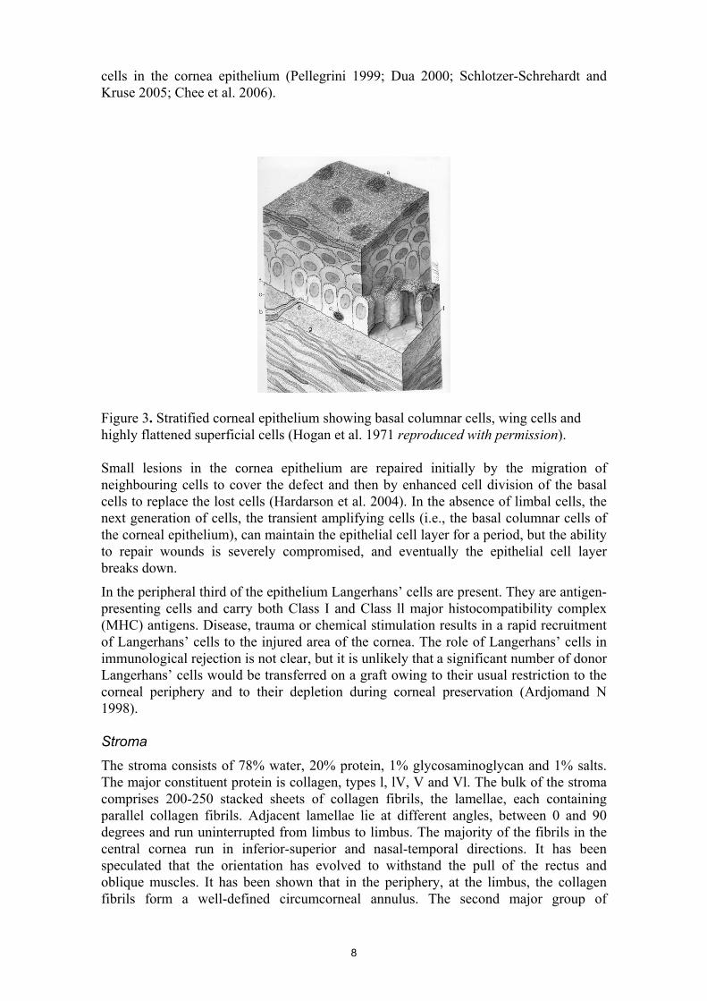
8
cells in the cornea epithelium (Pellegrini 1999; Dua 2000; Schlotzer-Schrehardt and Kruse 2005; Chee et al. 2006).
Figure 3. Stratified corneal epithelium showing basal columnar cells, wing cells and highly flattened superficial cells (Hogan et al. 1971 reproduced with permission). Small lesions in the cornea epithelium are repaired initially by the migration of neighbouring cells to cover the defect and then by enhanced cell division of the basal cells to replace the lost cells (Hardarson et al. 2004). In the absence of limbal cells, the next generation of cells, the transient amplifying cells (i.e., the basal columnar cells of the corneal epithelium), can maintain the epithelial cell layer for a period, but the ability to repair wounds is severely compromised, and eventually the epithelial cell layer breaks down.
In the peripheral third of the epithelium Langerhans’ cells are present. They are antigen-presenting cells and carry both Class I and Class ll major histocompatibility complex (MHC) antigens. Disease, trauma or chemical stimulation results in a rapid recruitment of Langerhans’ cells to the injured area of the cornea. The role of Langerhans’ cells in immunological rejection is not clear, but it is unlikely that a significant number of donor Langerhans’ cells would be transferred on a graft owing to their usual restriction to the corneal periphery and to their depletion during corneal preservation (Ardjomand N 1998). Stroma
The stroma consists of 78% water, 20% protein, 1% glycosaminoglycan and 1% salts. The major constituent protein is collagen, types l, lV, V and Vl. The bulk of the stroma comprises 200-250 stacked sheets of collagen fibrils, the lamellae, each containing parallel collagen fibrils. Adjacent lamellae lie at different angles, between 0 and 90 degrees and run uninterrupted from limbus to limbus. The majority of the fibrils in the central cornea run in inferior-superior and nasal-temporal directions. It has been speculated that the orientation has evolved to withstand the pull of the rectus and oblique muscles. It has been shown that in the periphery, at the limbus, the collagen fibrils form a well-defined circumcorneal annulus. The second major group of

9
extracellular proteins in the stroma comprises the proteoglycans. They bind at specific sites along the collagen fibrils and are believed to control the regular arrangement of the collagen fibrils.
Figure 4. Corneal stroma showing arrangement of collagen fibrils and a keratocyte (Komai and Ushiki 1991 reproduced with permission). This regular arrangement is believed to be responsible for the shape and strength of the cornea (Meek 2003) as well as for corneal transparency. The cornea transmits 86-94 %, depending on wavelength, of visible light. The precise explanation for this remarkable degree of transparency is still not completely known. Although the short range ordering of the collagen fibrils does not seem to be sufficiently regular to explain the minimal light scattering by the cornea, the degree of long-range ordering is probably sufficient (Maurice 1957).
Scattered through the stroma and lying between the lamellae are the fibroblast-like keratocytes. These highly flattened cells secret components of the stroma and are thus important both for maintenance and in wound repair (Fig. 4). Endothelium
The endothelium comprises a monolayer of mostly hexagonal cells that forms a continuous mosaic completely covering the posterior surface of the cornea (Fig. 5). In humans, corneal endothelial cells only rarely undergo mitotic division and the loss of cells is compensated by the migration and spreading of neighbouring cells. There is a continuous decline in cell density with increasing age from approximately 3500 cells/mm2 in 10-19 year olds to 2300 cells/mm2 in 80-89 year olds. Descemet’s membrane is the endothelial basement membrane. It is a thick and strong membrane composed of collagens, mainly type Vlll. The endothelium is a leaky cell layer, with three-fold higher water permeability than the epithelium, and because the stroma has a tendency to imbibe water and solutes, there is a continuous influx of ions, small molecules and water across the endothelium from the aqueous humour into the stroma. This is necessary, in the absence of blood supply, for the nutrition of the stromal
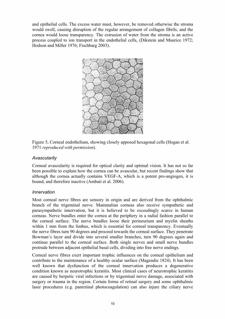
10
and epithelial cells. The excess water must, however, be removed otherwise the stroma would swell, causing disruption of the regular arrangement of collagen fibrils, and the cornea would loose transparency. The extrusion of water from the stroma is an active process coupled to ion transport in the endothelial cells, (Dikstein and Maurice 1972; Hodson and Miller 1976; Fischbarg 2003).
Figure 5. Corneal endothelium, showing closely apposed hexagonal cells (Hogan et al. 1971 reproduced with permission). Avascularity
Corneal avascularity is required for optical clarity and optimal vision. It has not so far been possible to explain how the cornea can be avascular, but recent findings show that although the cornea actually contains VEGF-A, which is a potent pro-angiogen, it is bound, and therefore inactive (Ambati et al. 2006). Innervation
Most corneal nerve fibres are sensory in origin and are derived from the ophthalmic branch of the trigeminal nerve. Mammalian corneas also receive sympathetic and parasympathetic innervation, but it is believed to be exceedingly scarce in human corneas. Nerve bundles enter the cornea at the periphery in a radial fashion parallel to the corneal surface. The nerve bundles loose their perineurium and myelin sheaths within 1 mm from the limbus, which is essential for corneal transparency. Eventually the nerve fibres turn 90 degrees and proceed towards the corneal surface. They penetrate Bowman’s layer and divide into several smaller branches, turn 90 degrees again and continue parallel to the corneal surface. Both single nerves and small nerve bundles protrude between adjacent epithelial basal cells, dividing into free nerve endings.
Corneal nerve fibres exert important trophic influences on the corneal epithelium and contribute to the maintenance of a healthy ocular surface (Magendie 1824). It has been well known that dysfunction of the corneal innervation produces a degenerative condition known as neurotrophic keratitis. Most clinical cases of neurotrophic keratitis are caused by herpetic viral infections or by trigeminal nerve damage, associated with surgery or trauma in the region. Certain forms of retinal surgery and some ophthalmic laser procedures (e.g. panretinal photocoagulation) can also injure the ciliary nerve

11
fibres as they sweep anteriorly between the sclera and the choroid. Keratorefractive procedures such as LASIK, and indeed keratoplasty, also damage the stromal and epithelial nerves. The cause of neurotrophic keratitis is probably a combination of desiccation of the corneal surface due to diminished lacrimal secretions, diminished protective blink reflexes, abnormal epithelial cell metabolism with subsequent failure to resist effects of trauma, drying and infection, and the loss of trophic influences supplied by the corneal nerves, the latter probably being the most important (Muller et al. 2003). Immunology and rejection Graft rejection is one of the leading causes of failure in corneal transplantation. Rejection occurs in 30–40% of keratoplasty cases (Williams et al. 1995), but not all rejections result in corneal opacity, most can be treated and the cornea remains transparent, especially in low-risk patients such as keratoconus. In high risk corneas, with previous rejection, active inflammation or vascularisation of the cornea, the failure rate caused by rejection can be anything between 50 and 100%. Nonetheless the cornea was the first solid tissue to be transplanted successfully in animals and humans. In spite of no routinely performed tissue matching, and usually no systemic immunosuppressive treatment, corneal grafting at least initially exceeds the survival rate of transplanted organs. This apparent exemption from the laws of transplantation immunology has led to the presumption that the cornea is an immunologically privileged tissue.
Several features of the cornea appear to contribute to this privilege. Unlike other epithelial surfaces such as skin, the central cornea is devoid of Langerhans’ cells. These are bone-marrow-derived dendritic antigen-presenting cells and are potent inducers of delayed-type hypersensitivity. In addition the cornea lacks blood and lymphatic vessels, which limits both the afferent and efferent limbs of immune responsiveness. The cornea and iris secret immunosuppressive factors that create local microenvironments that suppress certain immune and inflammatory responses within the anterior segment of the eye. Moreover the corneal epithelium and endothelium express Fas ligand that induces apoptosis of activated T-cells, which attack the corneal graft. Furthermore a deviant immune response is elicited when antigens are inoculated in the anterior chamber of the eye (anterior chamber associated immune deviation, ACAID) (Yoichiro 1997; Niederkorn 1999).
The discovery of the mechanisms behind this relative immune privilege gives rise to the possibility of new treatments, including therapeutic antibodies, such as anti-TNFα (Sugar 2008), to help further improve the success rate in corneal transplantation (Atsushi 1995).
As previously stated the cornea is, in spite of its relative immune privilege, not as protected from rejection as previously thought. Several studies have therefore been performed in order to find the possible benefits of tissue matching. Experimental transfer of tumours from one mouse strain to the other led to the discovery of the major histocompatibility complex (MHC) (Gorer 1937). Klein et al. (1983) divided the MHC antigens into different classes according to their expression patterns. Class I antigens comprise HLA - A, -B and -C. They are expressed on epithelial, stromal and endothelial cells of the cornea and are recognized by cytotoxic T-cells leading to destruction of cells presenting allo-antigens. Class II molecules are expressed on specific antigen-presenting cells, such as dendritic cells, B-cells and Langerhans’ cells as well as activated T-cells. Histocompatibility matching has been used for more than four decades for solid organ transplantation, but has not become generally accepted for cornea transplantation due to
12
inconclusive or contradictory results (CCTS 1992; Baggesen et al. 1996; Volker-Dieben et al. 2000; Sundmacher 2003; Volker-Dieben et al. 2003). The reason for such varying results can probably be explained by variations in methods for HLA-typing, heterogenic groups of patients and different postoperative immune suppression therapy. It has been suggested that blood group (ABO) matching may be of value (CCTS 1992) as ABO blood-group antigens are expressed on the epithelium and endothelium (Salisbury and Gebhardt 1981). The uncertainty of the benefit of tissue typing in combination with the cost of performing the test and the consequence of longer waiting times for the patients to receive a matched graft has limited the use of this approach to reducing the risk of rejection. Risk factors
Vascularisation of the cornea is by most surgeons accepted as a risk factor for graft rejection. It is, however, possible that it is not the vascularisation as such that increases the risk for immune response, but rather the causes that evoke neovascularisation, such as infection, trauma, phagocytic stimuli and the transplantation process itself, including sutures, that directly induces an immune reaction (Niederkorn 1999). Other known factors increasing the risk of rejection are young recipients (< 10 years of age), large grafts and possibly combined suturing (running and interrupted)(Vail et al. 1997). Young recipients are believed to have a more alert immune response, large grafts involve the periphery of the cornea where Langerhans’ cells can be found. The reason why combined suturing increases the risk for rejection is not known. Previous rejections and an inflamed eye at the time of keratoplasty, or even earlier, erode the mechanisms that contribute to the immune privilege and predispose for rejection. Previous grafts in the same eye causes sensitisation and is also a major risk factor. Rejection
Rejection can occur in any of the three layers of the cornea, epithelium, stroma and endothelium. The clinically most important rejection is that of the endothelium. The clinical symptoms include moderate pain or irritation, increased tearing and dim vision. Ciliary injection can be noted. Inspection in the slit lamp reveals varying degrees of corneal oedema, typically in one sector, following a line of leucocytes progressing across the endothelium (endothelial rejection). Multifocal deposits, keratic precipitates can also occur. The endothelial cells will be seriously injured unless immunosuppressive treatment is started promptly (Claerhout et al. 2003). Prevention and treatment of rejection
Patients are routinely treated with local steroids after the transplantation. With this treatment the overall rejection rate is about 15% (Claesson et al. 2002).The glucocorticosteroids prevent or reverse rejection through multiple mechanisms(Barshes et al. 2004), one of the most important probably the inhibition of leukocyte migration into the cornea. In spite of steroid treatment rejection does still occur, especially in high risk grafts where the rejection rate can be up to 100%. In these cases other immunosuppression would be beneficial, and is sometimes needed also in low risk patients that respond to steroids with high intraocular pressure. The most used such medication is Cyclosporine (Sandimmun), which has only proved effective in systemic treatment. Studies using Cylclosporine as topical drops have not been able to show any reduction in rejection rate (Price and Price 2006). Cyclosporine suppresses the proliferation of activated T- and B-cells by blocking IL-2 receptor. Used systemically it

13
has by some been shown to reduce rejection (Sundmacher et al. 1992; Hill 1995), whereas others failed to find it beneficial (Inoue et al. 2001; Poon et al. 2001). With the systemic treatment there are also risks of significant side effects. Another T-cell specific drug, Tacrolimus (FK506)(Sloper CM 2001), also suppresses T-cell and dendritic cell activities, as does Rapamycin (Birnbaum et al. 2006), both of which are derived from the fungus Streptomyces spp. These drugs have slightly different blocking mechanisms and can therefore be combined, which in the future may be successful in preventing rejection. Gene therapy
Gene therapy is an interesting concept for modifying corneal endothelial cells. The process of gene delivery is facilitated by the relatively simple anatomy and the ability to maintain corneas in culture for long periods. Ex vivo transduction of the cornea with gene transfer vectors has been shown to be feasible.
With today’s knowledge there are three areas where gene transfer could become useful in cornea:
A) to stimulate mitosis of endothelial cells in allografts or in a patient’s own cornea in vivo, to treat endothelial cell deficiency(Mimura and Joyce 2006),
B) to prevent / reduce neovascularisation of the cornea(Ng et al. 2006), C) to block mechanisms of rejection of corneal transplants.
In contrast to the use of gene therapy for the treatment of single gene disorders, for example the recent report concerning Leber’s congenital amaurosis (Bainbridge et al. 2008), the mechanism of rejection is multifactorial and probably several different transgenes are needed to down-regulate the rejection process (Fu et al. 2008). One way would be to induce production of Interleukin 10 (IL-10) to down-regulate MHC class ll and dendritic cell activation of T-cells. This has been investigated in a sheep model of corneal transplantation (Klebe et al. 2001). Although rejection was delayed it was not entirely prevented, which underlines the redundancy in the rejection process.
To transfer genes a vector is needed that can penetrate the target cell. Viral vectors have been used experimentally, but these can be immunogenic and, as a side effect, cause infection. Currently available non-viral vectors are less efficient, but as only a transient exposure is needed, they are possibly a better and safer option. Wound healing Corneal wound healing is the end result of a sequence of events that are controlled by many factors. It takes place over a long period of time, and the strength of corneal scars never reaches that of uninjured corneal tissue, which has to be considered also in the context of surgically induced injury, such as penetrating keratoplasty.
The epithelium heals firstly by migration of cells to cover the wounded area, while polymorphonuclear leucocytes, derived from the tear fluid, deal with the removal of necrotic cells. Only once migration of the epithelial monolayer is complete does it become more firmly anchored to the basement membrane and to Bowman’s layer by newly synthesised hemidesmosomes and anchoring filaments. The adhesion structures are restored approximately six to eight weeks after injury. Factors affecting adhesion complex formation include age, depth of the corneal wound and the nature of any underlying condition. The next phase of epithelial healing is proliferation of epithelial cells until normal epithelial thickness is restored. The sources for these new cells are

14
believed to be located in stem cells in the basal layer of the limbal epithelium. Stem cells first produce rapidly dividing cells termed transient amplifying cells. These further divide into more differentiated, post mitotic cells, moving more centrally and superficially in the cornea, to end up, finally, as terminally differentiated cells (Fig. 6). In cases of deficient or absent limbal stem cells the corneal epithelium will not heal permanently, which has devastating consequences for the cornea.
Figure 6. Migration of corneal epithelial cells from putative stem cell niche at limbus (Kruse 1994) (reproduced with permission). Healing of stromal wounds is slower than in other connective tissues, presumably due to its avascularity. The acellular Bowman’s layer and Descemet’s membrane do not heal; the cut ends of the membranes remain retracted. Stromal regeneration depends on co-ordinated interaction between epithelial cells and keratocytes, and does not take place until the epithelium is covering the wound. Polypeptide growth factors play an important role. Following stromal wounding, keratocytes undergo proliferation, migration, and fibroblastic transformation. The fibroblasts produce collagen, glycoproteins and proteoglycans, which form the new stromal matrix. Initially the collagen fibrils in the new matrix vary in diameter and are quite disorganised. Stromal remodelling continues for several years after the injury and after two years the lamellar collagen pattern is almost back to normal, but with shorter and narrower lamellae.
As was mentioned earlier human endothelial cells have minimal or no capacity to replicate by mitosis. Therefore, endothelial wound healing is largely dependant on enlargement and movement of the surrounding cells to cover the wound site. The endothelium is responsible for the deposition of a new basement membrane throughout the wound area. There is a substantial overcapacity of the endothelial cells to keep the cornea dehydrated, so the loss of endothelial cells at the time of penetrating keratoplasty does usually not affect the transparency of the cornea. (Steele 1999; Connon and Meek 2003; Connon and Meek 2004; Dupps and Wilson 2006).
15
Quality register Clearly, the cornea is functionally a complex tissue. Loss of normal function can cause decreased vision, sometimes blindness, and also severe pain. The only treatment for many corneal diseases is keratoplasty, where the impaired cornea is replaced with healthy tissue from a human donor. The result of this operation is affected by a great number of factors. In order to find out more about graft outcome and the factors that influence it, our colleagues in Australia showed great foresight by starting a corneal transplant register in 1986 (Williams 1987; Williams 1989; Williams et al. 2007). A register for following up corneal transplants is running in the UK (Vail et al. 1997).
Sweden is a small country and routine corneal grafting is concentrated to just seven centres, with 15-20 surgeons performing the operation. It therefore seemed an ideal situation to start a national register, as the collecting of data would be easier within such a small group. With this in mind the Swedish Corneal Transplant Register was started in 1996 (Claesson et al. 2002). The register has since 2002 been recognized by the Swedish Health Authority as a Quality Register, one of now 64 in Sweden, and is therefore financially supported by the government. At the start of 2007 the register became web-based, which has great advantages for the efficient collection of data. The aim of the register is not only, like in some international registers, to document the survival of the graft, but also to record the visual outcome, the actual benefit for the patients. The final goal for the register is to learn more about the optimal treatment for each patient. This means analyzing risk factors and postoperative complications and how they affect the outcome and then draw the consequences of this in choosing method of treatment, such as type of operation, postoperative treatment and also to be able to inform the patients about expected outcome in individual cases.
Aims The overall aim of this study is to present the register and, through findings described in six papers, demonstrate its value.
Paper 1 To assess visual outcome and the incidence of complications at 2 years postoperatively in corneal grafts reported to the Swedish Corneal Transplant Register.
Paper 2 To determine the impact of relaxing incisions for correcting post-operative astigmatism following penetrating keratoplasty.
Paper 3 To find risk factors for developing corneal oedema after cataract surgery and factors that influence subsequent survival of the graft and the visual outcome.
Paper 4 To compare a cohort of corneal graft patients in the Palestinian Territories with one in Sweden.
Paper 5 To review the two-year corneal transplant outcome in the cohort of patients from the Palestinian Territories described in Paper 4.
Paper 6 To determine factors influencing graft survival and visual outcome ten years after penetrating keratoplasty.

16
Methods
The core methodology covering data acquisition, storage and analysis, was similar for all the papers. The register permits observational longitudinal studies on corneal transplant outcome to be undertaken. The only inclusion criterion was that patients had a corneal transplant and there were no additional exclusion criteria. Patients were recruited at the time of transplant and followed up at two years. A cohort of patients was also followed up at ten years (Paper 6). Data acquisition Forms for data collection at the time of keratoplasty and at the two year follow up were developed in cooperation with all the users, i.e. the corneal surgeons in Sweden. (Addendum 1). Where applicable the form for two year follow-up was used also for the ten year follow-up.
At the time of keratoplasty patient details such as age, sex, diagnosis, type of procedure, visual acuity and lens status were registered. The diagnosis, or indication for surgery, was divided into five groups, keratoconus, Fuchs’ endothelial dystrophy, bullous keratopathy, stromal dystrophy and “other diagnosis”. Preoperative visual acuity was measured in both eyes. The type of operation was defined as penetrating keratoplasty, penetrating keratoplasty combined with cataract surgery and IOL implantation ( triple procedure) or “other procedure”.
All the corneas for the Swedish patients were provided by the five eye banks in Sweden and Denmark, all using organ culture storage. For the patients from the Palestinian Territories corneas were supplied by TBI (Tissue Banks International) in Baltimore, USA, where hypothermic storage was used.
At the 2-year follow up visual acuity, any postoperative complications, including rejection, were registered, and whether regrafting had been necessary. Astigmatism was measured, and if refractive surgery had been performed the astigmatism before and after this procedure was noted. The refractive surgery consisted in all cases of relaxing incisions down to Descemet’s membrane in the graft/host interface, 45 degrees around the steepest meridian. The visual acuity with best preferred correction was also recorded in the grafted eye, and whether other sight hindering disease was detected. For the ten year follow up (Paper 6) the same data set as at two years was collected. However, it was uncertain in many cases whether there had been any additional complications, including rejection episodes, between the two and ten year follow-ups. It was therefore decided to use data concerning postoperative complications only the first two postoperative years.
In Paper 3, additional data were collected from the patients’ notes describing circumstances around the cataract surgery, time and type of operation, including complications, time between operation and development of corneal oedema and duration of corneal oedema before penetrating keratoplasty. It was also noted whether the patients had known endothelial disease prior to cataract surgery or any other sight-hindering disease. Data storage and processing Initially the data were stored in an Excel spreadsheet (Microsoft Corporation). As the data set grew and the need for more sophisticated data processing became necessary, the

17
data were transferred into a custom designed Access database (Microsoft Corporation). Data were entered through screen-based forms, designed to be similar in structure to the paper forms completed by the surgeons. The data were stored in tables and interrogated through a series of queries. These allowed both descriptive statistics to be compiled and data sets to be generated for further statistical analysis. Based on data from the queries, clinic specific reports were sent to participating surgeons to identify missing or incomplete follow up information.
Since 2007 the data collection has been web based and maintained through EyeNet Sweden, an organisation for Swedish quality registers. However, the data in these six papers were collected from the register before it was web-based. Data analysis The data in all six papers were analysed using SPSS (initially v 10.0.5, subsequently later versions) statistical software. Multifactorial statistical methods were applied to determine the influence of recipient factors on graft outcome. For astigmatism, multiple linear regression was used following square root transformation of the outcome indicator (dioptres of astigmatism). To describe the difference in astigmatism before and after refractive surgery and between the two and ten year follow-ups a subtraction method was used (Paper 1 and 6). For more fundamental analysis of the actual change in astigmatism vector analysis was applied (Paper 2)(Olsen and Dam-Johansen 1994). Back transformed means and 95% confidence intervals (95% CI) are reported. Logistic regression was used for analysing the influence of factors on visual acuity (≥0.5; ≤0.2; VA operated eye≥ VA contra lateral eye), and on the incidence of rejection, regrafting, and other complications. Odds ratios (OR) and 95% CI are reported from the logistic regression analysis. The final statistical models were derived by backwards stepwise regression. The level of significance was set at 5%.
Patients All of the analyses were undertaken using anonymized data that masked individual patient identification. In view of this, the Sahlgrenska University Hospital Ethics Committee advised that specific ethical approval was not required to undertake these studies. Moreover these retrospective observational studies involved no specific patient interventions beyond the normal course of their treatment.
In paper 1 all the patients that were registered between 1996 and 2001 were included in the study. We had data from the time of operation for 1957 eyes, which, when compared with the numbers of corneal transplants from operating theatre records, consisted of 87% of the grafted patients in Sweden. From the 2-year follow up visit data for 520 grafts were available, representing approximately 50% of those eligible for follow up.
In paper 2 the astigmatism was analysed in all the eyes that had a complete 2-year follow up. At the time of the study the number of eyes was 1161. Of these 131 eyes had intolerable astigmatism and underwent relaxing incisions.
In the third paper 273 patients were included. These were all the eyes that had undergone corneal transplantation for corneal oedema after cataract surgery, had complete 2- year follow up data, and where the patients’ notes from the cataract operation were available.

18
During 2001–2002 the author worked as a cornea consultant at St John Eye Hospital in Jerusalem. The hospital is the main provider of ophthalmic health care for the Palestinian population. During that time 161 corneal transplants were performed. It was decided to add this cohort to the Swedish Cornea Transplant Register in order to allow comparison of two different populations concerning demographic data, indication for corneal transplantation, severity of disease and the outcome of transplantation expressed as graft survival and visual outcome. All 161 patients undergoing corneal transplantation were entered in the Register and analysed concerning preoperative data (Paper 4). Ninety-nine of these patients were available for follow up after two years (Paper 5).
For the ten year follow up, described in Paper 6, the 242 patients that underwent corneal transplantation at Sahlgrenska University Hospital 1996-1998, which accounted for about 20% of the patients being grafted in Sweden at that time, were chosen. 140 of these patients were available for follow up. Results Paper 1 Keratoconus, bullous keratopathy and “other diagnosis” were the most frequent indications for grafting in this group of 1957 patients reported to the Swedish Cornea Register. As expected, the mean age of keratoconus patients was substantially lower than for the other diagnostic groups. In keratoconus the most common type of operation was PK, whereas in Fuchs’ dystrophy the triple procedure dominated. In bullous keratopathy PK was combined with other procedures, such as vitrectomy and exchange of IOL, in 15% of cases. Table 1. Post-operative astigmatism and complications at 2 years. Diagnosis Astigmatism (D)1 Complications (%) Co-morb2 (%) Rejection Regraft Other Keratoconus 4.0 (3.5, 4.5, n=105) 11.7 6.3 13.4 9.7 Fuchs’ dystrophy 4.2 (3.4, 5.1, n=48) 9.7 9.5 21.6 38.5 Bullous keratopathy 4.7 (4.0, 5.3, n=54) 17.9 14.2 29.2 67.3 Stromal dystrophy 4.4 (2.5, 6.6, n=9) - - - - Other 4.3 (3.7, 5.0, n=64) 18.8 11.8 37.9 64.7 Total 4.3 (4.0, 4.6, n=280) 14.9 10.4 26.2 45.7 Notes: 1Back-transformed means (95% CI) 2Other conditions in the operated eye that may adversely affect sight The overall incidence of rejection and other postoperative complications was highest in the “other diagnosis” group, followed by the bullous keratopathy group. The risk of rejection was similar in the Fuchs’ dystrophy and keratoconus groups. The likelihood of regrafting was higher for grafts that had suffered rejection, and when other complications or other pathology were reported in the grafted eye (Table 1).

19
In the whole group of patients, 86% had visual acuity ≤0.2 before surgery, whereas at the 2 year follow up the figure was only 39%. After grafting 48% had a visual acuity ≥0.5. In the keratoconus group the improvement of visual acuity was even more marked: the percentage of patients with visual acuity ≤0.2 fell from 75% preoperatively to only 8% postoperatively while those with visual acuity ≥0.5 correspondingly increased from 12 to 86%. In Fuchs’ endothelial dystrophy there was also a significant improvement, whereas the bullous keratopathy group did not do so well. “Other diagnosis” showed the poorest results concerning visual acuity, with only 24% achieving ≥ 0.5 after grafting (Fig. 7). A higher postoperative visual acuity was associated with uncomplicated PK, high preoperative visual acuity, low astigmatism and the absence of other sight hindering pathology.
Lines of change on Snellen Chart - All(n = 2216)
0
5
10
15
20
25
-7 -6 -5 -4 -3 -2 -1 0 1 2 3 4 5 6 7 8 9 10 11 12
%
Lines of change on Snellen Chart - Keratoconus(n = 632)
0
5
10
15
20
25
-7 -6 -5 -4 -3 -2 -1 0 1 2 3 4 5 6 7 8 9 10 11 12
%
Lines of change on Snellen Chart - Fuchs'
(n = 434)
0
5
10
15
20
25
-7 -6 -5 -4 -3 -2 -1 0 1 2 3 4 5 6 7 8 9 10 11 12
%
Lines of change on Snellen Chart - BKP(n = 404)
0
5
10
15
20
25
-7 -6 -5 -4 -3 -2 -1 0 1 2 3 4 5 6 7 8 9 10 11 12
%
Lines of change on Snellen Chart - Re-graft
(n = 229)
0
5
10
15
20
25
-7 -6 -5 -4 -3 -2 -1 0 1 2 3 4 5 6 7 8 9 10 11 12
%
Lines of change on Snellen Chart - Other (n = 414)
0
5
10
15
20
25
-7 -6 -5 -4 -3 -2 -1 0 1 2 3 4 5 6 7 8 9 10 11 12
%
Figure 7. Lines of change on Snellen chart after PK for different indications. Comparing the visual acuity in the grafted eye with the other eye, 20% of eyes to be grafted had visual acuity equal to or better than the contralateral eye before surgery. After surgery this more than doubled to 48%. Also here the keratoconus group showed the best improvement: in only 11% was the eye to be grafted the better eye, but after surgery the grafted eye was the better in 59%. The data for Fuchs’ dystrophy were similar, whereas in the bullous keratopathy and “other diagnosis” groups there was less

20
improvement in the percentage of grafted eyes having as good as or better visual acuity than the other eye.
The only factors that influenced the level of astigmatism at 2 years were the diagnosis and the clinic. Refractive surgery, in the form of relaxing incisions, was performed after suture removal in 32 patients, reducing the astigmatism by about 50%. Paper 2 The mean post-operative astigmatism for all grafts with complete follow-up data was 4.56 D, with 46% of grafts having 4 D or less astigmatism. There was a small increase in post-operative astigmatism with increasing age. The only other factor that influenced astigmatism was clinic, two of the participating clinics having significantly lower values. However, the final regression model explained less than 5% of the variability of the data.
Of the 131 grafts that underwent relaxing incisions to reduce postoperative astigmatism 96% had more than 4 D of astigmatism before refractive surgery, compared with only 40% after surgery. The mean reduction was 4.5 D (95% CI: 4.0-5.0, p<0.001) using subtraction (Fig. 8). Vector analysis was used to show the actual reduction of astigmatism, which came to a mean of 7.9 D (95% CI: 7.2-8.7 p<0.001).
Figure 8. Change in astigmatism before and after refractive surgery (back-transformed mean dioptres with 95% confidence interval). Compared with grafts with no refractive surgery there was a trend that suggested that corrected visual acuity was improved following relaxing incision, the prevalence of sight-hindering co-morbidity being similar in the two groups. Paper 3 In this study, 273 eyes with aphakic or pseudophakic bullous keratopathy were analysed. Of these 43% developed persistent corneal oedema at the time of cataract
21
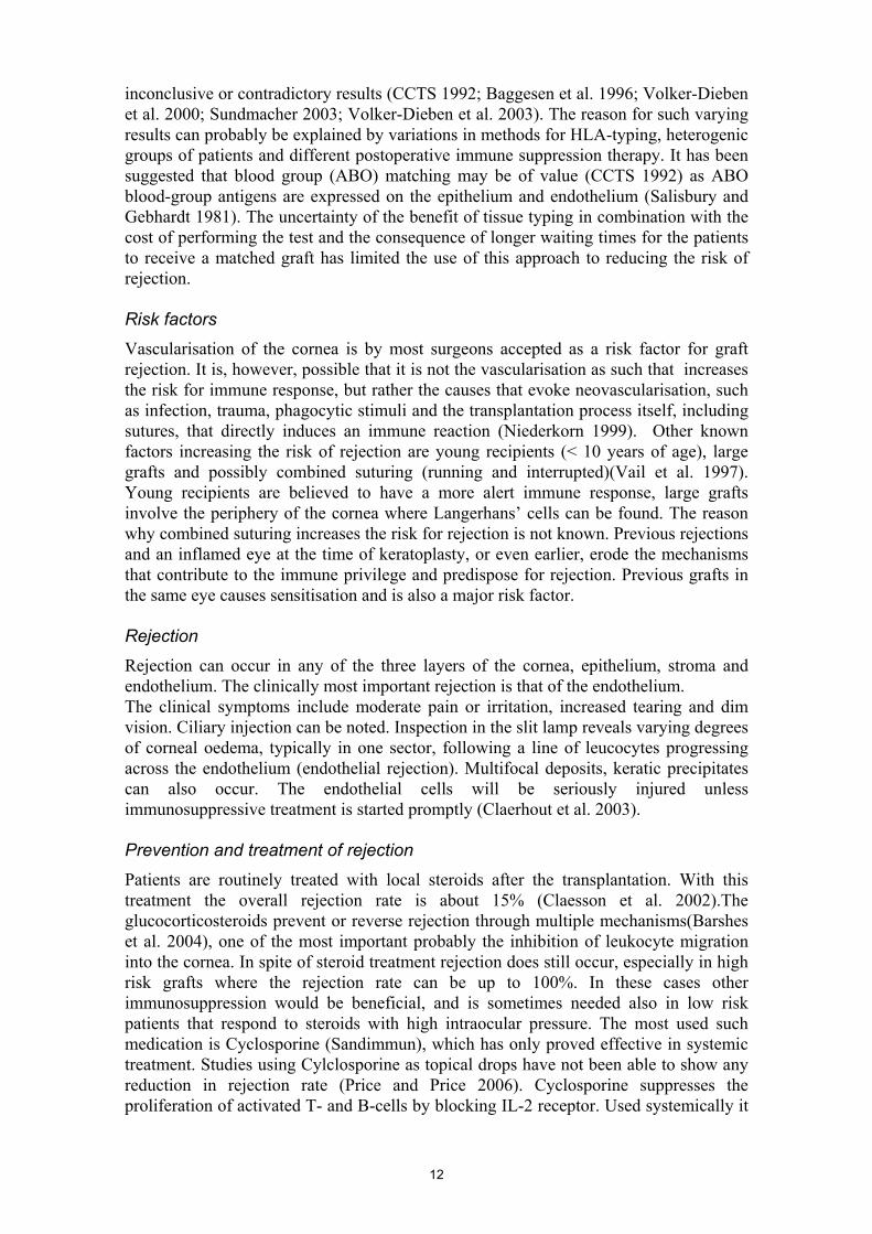
surgery. The median onset time for the other patients was 5 years. Factors that increased the risk of developing oedema at the time of cataract surgery included pre-existing endothelial disease and cataract operations done using the phaco-emulsification technique rather than extracapsular cataract extraction or, in a few cases, intracapsular techniques.
The risk of graft failure was mainly increased by rejection episodes and complications following PK. Patients that had had rejection episodes suffered from failed grafts in 76% of cases, whereas with no rejection only 24% failed. Graft failure was less likely in eyes that had developed persistent oedema at the time of cataract surgery than in those that developed oedema later.
0
10
20
30
40
50
60
70
Freq
uenc
y (%
)
Post-cataract Pre-PK Post-PK
Figure 9. Visual acuity after cataract surgery, before PK and 2-years after PK: percentage of patients with VA >0.1 (n=272). Two years after PK, 50% of the grafted eyes had achieved visual acuity of >0.1, an improvement from 10% before the operation (Fig. 9). Sight-hindering co-morbidity and grafts performed for pain relief affected the visual outcome negatively, as did rejection episodes. A shorter duration of the oedema and PK combined with intraocular lens exchange both had a positive influence on visual outcome. Paper 4 One hundred and sixty-one patients underwent corneal transplantation at St John Eye Hospital in Jerusalem 2001-2002. These patients were compared with the Swedish patients entered into the Cornea Register, which at the time of analysis amounted to 3431 transplants. Keratoconus was the most frequent indication for transplantation in both cohorts, but more so in the Palestinian group, with 51% of the patients compared with 27% in Sweden. In Sweden the majority of keratoconus patients were male (male:female 75:25), whereas in the Palestinian Territories this was reversed with a preponderance of females (male:female 39:61).Only 3 of the 161 Palestinian patients suffered from Fuchs’ endothelial dystrophy, whereas in Sweden 17% of grafts were for Fuchs’ dystrophy. Most of the patients coming for surgery to St John had more advanced disease than the Swedish patients, with all having visual acuity ≤ 0.1. In Sweden 14% still had vision >0.2 before the operation.
22
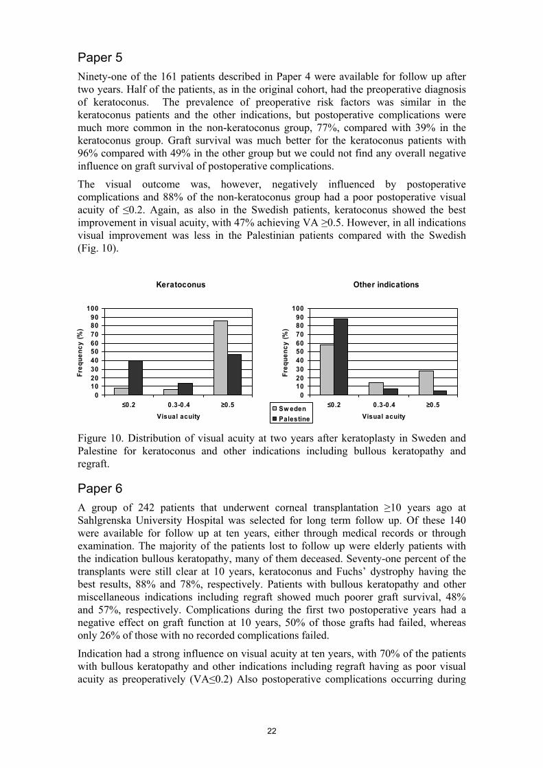
Paper 5 Ninety-one of the 161 patients described in Paper 4 were available for follow up after two years. Half of the patients, as in the original cohort, had the preoperative diagnosis of keratoconus. The prevalence of preoperative risk factors was similar in the keratoconus patients and the other indications, but postoperative complications were much more common in the non-keratoconus group, 77%, compared with 39% in the keratoconus group. Graft survival was much better for the keratoconus patients with 96% compared with 49% in the other group but we could not find any overall negative influence on graft survival of postoperative complications.
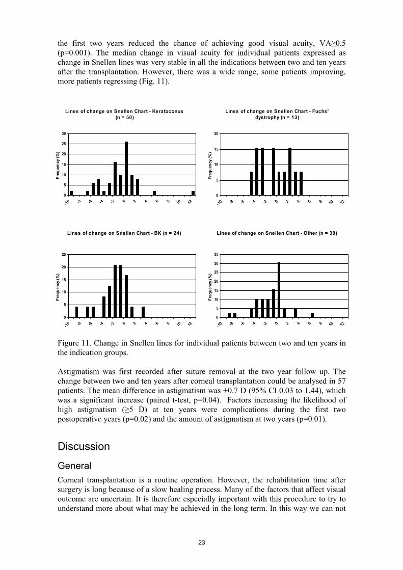
The visual outcome was, however, negatively influenced by postoperative complications and 88% of the non-keratoconus group had a poor postoperative visual acuity of ≤0.2. Again, as also in the Swedish patients, keratoconus showed the best improvement in visual acuity, with 47% achieving VA ≥0.5. However, in all indications visual improvement was less in the Palestinian patients compared with the Swedish (Fig. 10). Figure 10. Distribution of visual acuity at two years after keratoplasty in Sweden and Palestine for keratoconus and other indications including bullous keratopathy and regraft. Paper 6 A group of 242 patients that underwent corneal transplantation ≥10 years ago at Sahlgrenska University Hospital was selected for long term follow up. Of these 140 were available for follow up at ten years, either through medical records or through examination. The majority of the patients lost to follow up were elderly patients with the indication bullous keratopathy, many of them deceased. Seventy-one percent of the transplants were still clear at 10 years, keratoconus and Fuchs’ dystrophy having the best results, 88% and 78%, respectively. Patients with bullous keratopathy and other miscellaneous indications including regraft showed much poorer graft survival, 48% and 57%, respectively. Complications during the first two postoperative years had a negative effect on graft function at 10 years, 50% of those grafts had failed, whereas only 26% of those with no recorded complications failed.
Indication had a strong influence on visual acuity at ten years, with 70% of the patients with bullous keratopathy and other indications including regraft having as poor visual acuity as preoperatively (VA≤0.2) Also postoperative complications occurring during
Other indications
0102030405060708090
100
≤0.2 0.3-0.4 ≥0.5Visual acuity
Freq
uenc
y (%
)
Sw edenPalestine
Keratoconus
0102030405060708090
100
≤0.2 0.3-0.4 ≥0.5Visual acuity
Freq
uenc
y (%
)
23
the first two years reduced the chance of achieving good visual acuity, VA≥0.5 (p=0.001). The median change in visual acuity for individual patients expressed as change in Snellen lines was very stable in all the indications between two and ten years after the transplantation. However, there was a wide range, some patients improving, more patients regressing (Fig. 11). Figure 11. Change in Snellen lines for individual patients between two and ten years in the indication groups. Astigmatism was first recorded after suture removal at the two year follow up. The change between two and ten years after corneal transplantation could be analysed in 57 patients. The mean difference in astigmatism was +0.7 D (95% CI 0.03 to 1.44), which was a significant increase (paired t-test, p=0.04). Factors increasing the likelihood of high astigmatism (≥5 D) at ten years were complications during the first two postoperative years (p=0.02) and the amount of astigmatism at two years (p=0.01). Discussion General Corneal transplantation is a routine operation. However, the rehabilitation time after surgery is long because of a slow healing process. Many of the factors that affect visual outcome are uncertain. It is therefore especially important with this procedure to try to understand more about what may be achieved in the long term. In this way we can not
Lines of change on Snellen Chart - Keratoconus (n = 50)
0
5
10
15
20
25
30
-10 -8 -6 -4 -2 0 2 4 6 8 10 12
Freq
uenc
y (%
)
Lines of change on Snellen Chart - Fuchs' dystrophy (n = 13)
0
5
10
15
20
-10 -8 -6 -4 -2 0 2 4 6 8 10 12
Freq
uenc
y (%
)
Lines of change on Snellen Chart - BK (n = 24)
0
5
10
15
20
25
-10 -8 -6 -4 -2 0 2 4 6 8 10 12
Freq
uenc
y (%
)
Lines of change on Snellen Chart - Other (n = 39)
0
5
10
15
20
25
30
35
-10 -8 -6 -4 -2 0 2 4 6 8 10 12
Freq
uenc
y (%
)

24
only improve patient selection for corneal transplantation, but also better inform our patients, giving them more realistic expectations. In previous studies much of the interest has often been focused on graft survival. A major aim of other studies (Vail A 1997; Williams 2000) and the Swedish Corneal Transplant Register is to understand more about visual outcome in our patients as the majority of grafts are done to improve vision.
The register may also be used to monitor changes in the indications for corneal transplantation and provide clinical outcome data when new surgical procedures are introduced. This is very relevant now as so many new surgical techniques, such as different kinds of lamellar keratoplasty, are developing, but also less invasive techniques including intracorneal inlays (Intacs) and crosslinking. The data from the Swedish Cornea Register provide a firm baseline for comparing results from these new techniques. It can also help make surgeons aware of important risk factors and postoperative complications. Data can teach and give guidance concerning optimal postoperative treatment. The register thus works as a quality control, providing feedback to the surgeons, the clinics, medical authorities and, most importantly, to patients. One advantage of the Swedish Corneal Transplant Register is that a small group of surgeons perform all the corneal grafting in the country. Meetings are held once a year to discuss the design of the register and, as a result, there is a good agreement in surgical and clinical interpretation, and improvements can be made to the data collection forms with increasing experience. The response rate is good, with 89% of the grafts in Sweden being reported to the register.
There are of course always pitfalls, and there are limitations to what a register can do and to the presumed reliability of the data. There are three main levels of potential bias:
• Reporting bias through operations not being entered in the register and missing follow up data
• Recording bias resulting from inaccuracies in the data included in the register • Recording bias through varying clinical interpretation by the surgeons; for
example, extent of vascularisation, reporting of rejection, reason for failure.
Addressing the first point, the numbers of operations entered are compared annually with the actual number of corneal transplants performed at each clinic. These figures show that approximately 90% of grafts in Sweden are reported to the register. So far as data validation is concerned, the web-based register has built in filters, so that unrealistic data cannot be accepted. For many of the data, such as visual acuity, the values are chosen from a drop down list on the data entry screen. Since the register became web-based 2006 the surgeons feed in data on line into the database directly, not via a paper form, which means there is one less step in the handling of data, which in turn should reduce the risk of faulty entries. However, wrong values can still be entered and further validation is necessary and planned. When it comes to the third point subjective evaluation always carries a risk of individual differences. To come as close as possible to a true and consistent result frequent discussions between the participants is very important. The 15 surgeons running the Cornea Register are in close contact and have the opportunity to discuss such issues at least once a year.
25
Paper 1 The distribution of diagnoses and patient age were consistent with other studies from outside North America. In North America data show a higher proportion of transplants for bullous keratopathy and a lower proportion for keratoconus. The reason for this is believed to be a consequence of the early frequent use of anterior chamber intraocular lenses after cataract surgery in the US, a procedure known to have an increased risk of corneal oedema due to endothelial damage (Lindquist et al. 1991; Haamann et al. 1994).
Whether the lower proportion of keratoconus in North America is a consequence of the higher proportion of the bullous keratopathy or a true absolute figure is not known, but it has been discussed that development of better contact lenses should make the need for corneal grafting in keratoconus patients less (Garcia-Lledo et al. 2006).
The “other diagnosis” group turned out to be substantial, accounting for 32% of the grafts. Since these grafts suffered a high proportion of complications and had poor visual outcome, further investigations have been made since this report was published as to the different diagnoses hiding in this group. The data collection form has been accordingly modified to allow other diagnoses to be specified, and a special code for “regraft” (previously included in “other diagnosis”) has been added.
Visual outcome at 2 years was determined by overall improvement in visual acuity and by the amount of astigmatism. Although not necessarily accepted (Brahma et al. 2000) Williams et al (1991) have suggested that for patients to perceive a visual benefit from a corneal graft, the operated eye has to achieve a visual acuity at least as good as the other eye. In terms of visual acuity, the keratoconus patients did substantially better than the other diagnostic groups: 86% achieved a visual acuity ≥0.5 at 2 years and 59% had a visual acuity in the grafted eye at least as good as the other eye. The success was due in part to the low incidence of other pathology in these eyes. That 41% of grafted eyes in keratoconic patients had poorer visual acuity than the other eye needs to be further investigated; one reason for this could be that the other eye had already been grafted, and, as a consequence, the data collection form has been modified accordingly to collect this information.
The Fuchs’ dystrophy and even more so the bullous keratopathy patients had a higher incidence of postoperative complications and of accompanying pathology that could adversely affect vision than the keratoconus group. These groups comprised elderly patients accounting for the high incidence of other sight hindering disease, such as age related macular degeneration, and the bullous keratopathy patients would in part suffer from consequences of previous cataract surgery, such as macular oedema. The latter was reflected in a 30% greater incidence of accompanying pathology compared to the Fuchs’ dystrophy group. Also, some of the grafts in the bullous keratopathy group were done for pain relief in patients with poor visual potential.
The worst visual outcome overall was in the “other diagnosis” group. Sixty-five percent of the eyes in this group had other conditions, such as trauma to other parts of the eye, affecting sight. As mentioned earlier, this group is going to be investigated further and the data collection form has now been altered for this purpose. The postoperative visual acuities in the different diagnostic groups are broadly similar to those reported in the Australian Corneal Graft Registry (Williams 2000; Williams et al. 2007) and to UK data (Vail et al. 1997), albeit the latter at only 1 year follow up.

26
Postoperative astigmatism clearly contributes to an unsatisfactory visual result after grafting. The number of grafts with ≤ 3 D of astigmatism at two years and after suture removal was only 27%, which is similar to the 34% reported in the Australian registry (Williams 2000). The degree of astigmatism before transplantation (i.e., high in keratoconus and low in bullous keratopathy) did not affect the postoperative astigmatism, which suggests that it is the transplantation itself, and the healing process, that causes the astigmatism. A centre effect was noted in that two clinics showed less astigmatism in their post graft patients than the others. In trying to explain this we studied the different operating techniques, and it turned out that there were some differences between surgeons (see paper 2).
In this study we could show in a small group of patients that relaxing incisions were effective in reducing the degree of astigmatism. As astigmatism is the major practical problem to deal with after PK we wished to investigate it further and the findings are reported in the second paper of this study. Paper 2 Astigmatism may not be the most serious complication after penetrating keratoplasty, but it is indeed the most common reason for unsatisfactory vision after grafting (Williams 2004). It is a complicated phenomenon to describe owing to the subtle variations in the corneal topography. It has many different patterns (Bogan 1990; Karabatsas 1999), which can best be illustrated by videokeratography and wave front analysis (Pantanelli et al. 2007) but such data are difficult to reduce to a form that can be readily analysed. In this paper we therefore used keratometry readings, a method also easily available to clinics.
Some factors that influence astigmatism are still not known. It has been discussed whether preoperative diagnosis influences the postoperative astigmatism. Keratoconus, for example, has a very high pre-operative astigmatism, whereas bullous keratopathy hardly has any at all. As was shown in this study all indications have very much the same amount of astigmatism after PK, which suggests that astigmatism is more related to the actual grafting procedure and the wound healing, rather than the nature of the underlying disease or recipient factors. Nevertheless, different ways of trephining the cornea and different techniques for suturing have been tried in order to reduce astigmatism, all with little effect on the end result, although some suture adjustment may decrease the astigmatism during the early postoperative period.
However, the two factors we found that influenced the postoperative astigmatism were recipient age and the clinic where the operation was performed. The effect of age was small, but significant, so that the astigmatism increased slightly with increasing age. The reason for this is not clear, but clinical observations suggest that delayed healing and changes in the tissue’s rigidity with age may be contributory factors. The fact that two of the seven clinics had significantly lower astigmatism, even after adjusting for all other factors, probably meant that slight variations in the operating technique may after all influence the outcome. We know for example that in the clinic achieving the lowest astigmatism, the cornea was cut with a diamond knife, rather than scissors, after trephination.
The influence of graft size and over/under sizing of the graft has also been investigated. Larger graft diameter can have a positive effect on visual acuity and central corneal astigmatism, but the risk of rejection increases (Vail et al. 1997). Under sizing of the graft (i.e., using same trephine size for recipient and donor) may have some effect on

27
the curvature of the cornea and the spherical equivalent, but has not been shown to reduce astigmatism (Jaycock et al. 2008). The slow healing process of the corneal stroma is therefore thought to have a large impact on astigmatism. As was mentioned earlier, remodelling of the stromal lamellae continues for many years after the injury (Connon and Meek 2003), which could also explain the fact that the astigmatism after grafting continues to change for many years.
Some astigmatism is always expected after penetrating keratoplasty. This can often be corrected to a comfortable and satisfactory degree with glasses or hard contact lenses. However, astigmatism that cannot be corrected by these means causes great discomfort and blurred vision for the patient. The amount of astigmatism that the patients can tolerate varies between individuals. In our study the patients who underwent refractive surgery had astigmatism between 4 and 14 dioptres, in varying angles.
There are several surgical methods to deal with post-keratoplasty astigmatism, re-grafting being the ultimate step. Excimer laser can reduce myopic astigmatism by up to 6 dioptres using a two step LASIK procedure (Riddle et al. 1998; Rashad 2000) and a very irregular corneal surface can be dealt with by excimer laser assisted by sodium hyaluronidate. (Alio et al. 2001). For astigmatism of more than 8 dioptres, and in cases of cataract, implantation of toric posterior chamber intraocular lenses can be helpful (Nuijts et al. 2004).
Excimer laser is, however, still too expensive for many clinics around the world and it has its limitations when it comes to reducing high astigmatism. There is also the risk of flap complications and epithelial ingrowth (Kwito S 2002; Chan and Rootman 2004; Solomon et al. 2004). Relaxing incisions may therefore be considered a preferred initial intervention that may preclude the need for subsequent laser refractive surgery. It is therefore important to evaluate what can be achieved with this less resource-demanding technique. With this technique the astigmatism measured in dioptres decreased by approximately 50%. The mean reduction of 4.5 D was similar to previous reports (Kirkness et al. 1991; Claesson et al. 2002). Before refractive surgery only 4% of the patients had 4 or less dioptres of astigmatism, whereas after the procedure 60% were within this low range. To get a measure of the overall reduction of astigmatism it is necessary to take the angle into consideration (Kaye et al. 1992). As expected, vector analysis of astigmatic changes after relaxing incisions showed a greater reduction than the simple subtraction method.
Comparison of the distribution of visual acuities after relaxing incisions with that of grafts with no refractive surgery showed a trend towards fewer grafts with poor visual acuity and more grafts with good visual acuity. Whether this trend reflected a true improvement in visual acuity consequent to the relaxing incisions remains to be confirmed, but the data are suggestive.
Overall, our study confirmed that use of relaxing incisions is an effective; low-risk method to reduce the astigmatism induced by penetrating keratoplasty and may also have a beneficial influence on visual acuity. Paper 3 Patients undergoing corneal transplantation for bullous keratopathy following cataract surgery form only a small subset of all cataract patients, but this serious condition remains one of the main indications for PK (Vail et al. 1997; Aiken-O'Neill P 2002; Claesson et al. 2002; Williams et al. 2004). Since graft survival and visual outcome

28
after PK can be poor in this group (Yoichiro 1997; Claesson et al. 2002), and much worse than for similarly aged patients with Fuchs’ dystrophy, this indication was selected for further investigation. The aim was to identify factors that influenced the development of corneal oedema after cataract surgery and that had an impact on subsequent PK outcome.
The patients included in this study had all undergone cataract surgery and developed corneal oedema at varying intervals after the cataract operation. This separates them from the Fuchs’ dystrophy group in the register, who were transplanted as a result of primary endothelial disease. However, in the bullous keratopathy group we found that 65% had known endothelial disease before cataract surgery, and this increased the risk of developing persistent oedema at the time of cataract surgery by almost four-fold. This may raise the question for these patients whether triple procedure (cataract extraction, IOL implantation and PK) would be beneficial rather than doing the cataract surgery followed by PK some time later. Triple procedure has its disadvantages, being more complicated than just cataract operation or PK on its own and, as the refractive power of the cornea can change substantially after grafting, there are difficulties in calculating the correct power of the intraocular lens. Even so, it has been shown to have a better survival rate than staged procedures (Williams et al. 2004).
In Sweden, as in most countries in Europe, America and Australasia, phacoemulsification has been the dominating procedure for cataract surgery for more than 10 years. Presently more than 70 000 cataract operations are performed each year in Sweden and 98% of those are done by phacoemulsification (Lundström 2006). In this study only 54% of the patients that developed bullous keratopathy had undergone phacoemulsification, which implies that other techniques, such as extracapsular or intracapsular cataract extraction are more likely to cause corneal oedema. However, in a prospective randomized trial comparing phacoemulsification with ECCE, endothelial cell loss at one year was similar for these two techniques of cataract extraction (Bourne et al. 2004).
Looking at the time point when oedema developed, we found that a large group of patients, 43%, developed persisting oedema at the time of cataract surgery and that the risk for this increased 3.6 times if the cataract operation was done by phacoemulsification. The other type of operation, ECCE, was much more associated with a later onset of oedema, and a mean time from cataract surgery to oedema being 5 years. The reason for this is not clear, but it could be speculated that the phaco energy may have a direct effect on the corneal endothelium (Bourne et al. 2004; Lundberg et al. 2005).
We could not obtain information about the density of the cataract in the individual cases, which could have been useful, as dense cataracts with hard nuclei demand more ultrasonic power during the phaco procedure. When phacoemulsification was introduced as a new technique for cataract surgery it became clear that there was a learning curve (Seward et al. 1993; Thomas et al. 1994); but the technique has now been the predominant method for many years and so surgeon inexperience alone cannot explain the increased risk for immediate corneal oedema.
The patients that developed immediate persistent corneal oedema underwent PK sooner than the ones that developed oedema later, i.e. they suffered from corneal oedema for a shorter time. These patients had a higher chance of graft survival and also better visual acuity. It is well known from the clinic that patients with long term corneal oedema, especially with epithelial involvement with bullae and erosions, develop inflammation,

29
often with vascularisation of the cornea. Inflammation and vascularisation of the cornea are risk factors for graft survival; hence this may be a reason for the better prognosis in patients with shorter duration of corneal oedema and could be an incentive for earlier grafting.
As epithelial bullae and inflammation cause severe pain, patients with bullous keratopathy sometimes undergo corneal transplantation solely for pain relief, even if the visual potential is low due to co-morbidity, such as macular degeneration or persistent macular oedema after a complicated cataract operation. This of course has to be taken into consideration when interpreting overall visual outcome.
In cases where IOLs were displaced or damaged, the data showed that IOL exchange at the time of PK improved the chances for good visual rehabilitation. Importantly, we also found that this more complicated operation did not jeopardize survival of the graft. The group as a whole had, as previously stated, a high proportion of failed grafts at two years. There was a strong association with rejection and other complications, of which the most common was glaucoma, followed by suture related problems, such as loose or broken suture, with vascular response and occasionally keratitis. This should be a message to surgeons to optimize the postoperative care, probably including extended use of steroids, better glaucoma control and more frequent postoperative monitoring. Paper 4 Corneal transplantation is a resource-demanding procedure with long rehabilitation and many factors affecting the outcome. Through the Swedish Cornea Transplant Register we have started to learn more about these factors in order to optimise the patient selection, the method of surgery and the postoperative treatment. However, so far all our data are from Sweden, a country of good socio-economic status, considered to be providing good health care. There is only a limited number of reports from other parts of the world (Williams et al. 2007; Tan et al. 2008), and even fewer from developing countries (Dandona et al. 1997; Tabin et al. 2004; Tilahun et al. 2005). It was therefore decided that when the author had the opportunity to work as a cornea consultant at the St John Eye Hospital in Jerusalem for just under two years, the patients undergoing corneal transplantation at that time should be added on to the Swedish register. This enabled comparison with the Swedish cornea patients. In the first of two papers pre- and peri-operative data are compared between the two groups.
St John Eye Hospital in East Jerusalem provides the majority of the eye care for the three million Palestinians living in the West bank, Gaza and East Jerusalem. The political situation during the time when data were collected was difficult, with the latest Intifada having just started. People had severe difficulties travelling to and from the hospital. This not only affected which patients could come for surgery, but it also meant that the post operative follow up suffered. The travelling difficulties may partly explain why the preponderance of keratoconic patients was so marked as these younger patients were generally healthier and more able to cope. Also, as was stated in the paper, the Palestinian Territories have a far higher proportion of young people than Sweden. The fact that more women than men with keratoconus were seen at the hospital, which is the contrary to most other reports from the western world, can possibly be partly explained by social reasons. The young women were approaching marriageable age and did not want to be seen as having a visual handicap. However, variations in both incidence and severity of keratoconus between men and women in different populations have been reported (Fink et al. 2005).
30
Most of the patients grafted at St John had far more advanced disease than in Sweden across all indications. The reason for this could be that the patients, because of difficulties travelling and poor economy, would be seeking help much later than would be the case in Sweden. Another explanation could be the high prevalence of vernal catarrh, which is a known predisposing factor for keratoconus (Khan et al. 1988; Totan et al. 2001) and which adds inflammation and corneal scarring and vascularisation to the keratoconic eye. Patients also often suffered from severe ocular surface disease, aggravated by the dry and dusty conditions in the country.
Interestingly there were very few patients with Fuchs’ dystrophy seen at St John Eye Hospital. The reason for this is unknown; again speculations can be made whether this is genetic or just an expression of the difficulties getting medical help. An inventory of eye diseases in the country would be helpful. This has been planned, but is difficult to perform during present circumstances. Paper 5 The situation, politically and socio-economically, has not changed since paper 4 was written. This can well explain why only 99 of the 161 patients originally transplanted at St John Eye Hospital in Jerusalem were available for a two year follow up. Many of the patients had also not been able to come to the hospital for regular examinations during the postoperative period. This sometimes resulted in complications that otherwise could have been avoided, such as loosening sutures leading to ulceration, infection, dehiscence of the graft and vascularisation. The inflammatory reactions following these complications also led to increased risk for rejection (Sundmacher et al. 1992; Hargrave et al. 2003). These conditions were often not treated in time and were probably also under-reported - the patient could come to the hospital with an already opaque, failed graft, and the reason would be unknown.
The patients who underwent corneal transplantation in Jerusalem had more preoperative risk factors than in Sweden. This again can be explained by the fact that the patients came to the hospital with more advanced disease and often with vernal catarrh or other ocular surface problems leading to inflammation and vascularisation. The indication and the preoperative risk factors mainly influenced graft survival, whereas postoperative complications affected visual acuity at two years. The keratoconus patients suffered much less from postoperative complications than the other patients and also had a much better visual outcome, but the preoperative risk factors were surprisingly equal between the two groups. Despite this, grafts for keratoconus had a higher survival rate. This is not easy to explain from our data, but mirrors the higher survival rate seen in Sweden and other countries. On the other hand, the visual outcome of grafts for keratoconus, while far better than for other indications, was poorer than in Sweden. Paper 6 As can be seen in other reports (Muraine et al. 2003; Thompson et al. 2003; Williams et al. 2007), there is no stable end-stage after penetrating keratoplasty. A two year follow up was chosen for the cornea register because most grafts would have had sutures removed, a process which induces refractive changes, and is therefore the soonest time point to make a reasonable assessment of visual outcome. However, the healing process continues (Wilson et al. 2001) and complications can still occur. A group of patients was therefore chosen for 10-year follow up. As expected many of the older patients had died since the operation, most of them having the indication bullous keratopathy (BK).

31
The graft outcome in the patients with BK that were lost to follow up is unknown, but their mean age was higher than that in the group available for follow up. It would therefore be plausible to suppose that the visual outcome in the group lost to follow up would be worse, i.e. the poor visual results in the BK group may in fact be even poorer than our analysis shows. Patients with BK but also these in the Other indications group both approach the preoperative visual acuity by ten years. Looking at the change in Snellen lines there is very little change in any of the indication groups between two and ten years, but in the BK and Other group also very little change between preoperative and two year values. The range is wide, however, and for an individual patient there was often some gain in VA. However, ten year graft survival was also poor in both groups, 48% and 57% respectively. Such poor results both concerning graft survival and visual outcome raise the question how far corneal transplantation is beneficial in these two indication groups. Newer lamellar techniques or a more conservative approach may be a better solution, which continued analysis of data from the register should help to clarify.
It is also important to register actual patient satisfaction. For patients with poor visual acuity in both eyes a small improvement in one eye can be of great importance. In elderly patients the ten year result may not be of such significance as the life expectancy may not be that long. Other patients again may undergo keratoplasty to relieve pain from an oedematous cornea, and be very happy with the result even with little or no improvement in visual acuity. To be able to assess the real benefit from corneal transplantation the data from the register must be combined with a quality of life assessment. A questionnaire for this purpose is under development.
The patients with keratoconus and to some extent Fuchs’ dystrophy showed much better results. Both indications had good ten year survival rates, of 88 and 78% respectively, of these available for follow up. A high proportion of the keratoconus patients also still had good visual acuity after ten years, with >70% having VA ≥0.5. Patients with Fuchs’ dystrophy, being of an older age group with more co-morbidity had four-fold less chance of VA≥0.5, than the keratoconus patients. The patients with Fuchs’ dystrophy had, however, in general better visual acuity than the patients with BK, although being of the same age group. The reason for this is believed to be that the BK patients had more compromised eyes, with retinal complications and possibly chronic inflammation after complicated cataract surgery and longstanding corneal oedema.
Complications, including rejection, during the first two postoperative years, had a negative effect both on graft survival, with 50% failure rate at ten years, and on visual outcome, with a four-fold decreased chance of visual acuity ≥0.5, looking at all the indications. This emphasises the importance of adequate postoperative treatment and follow up, but also information to patients concerning symptoms and easy access to ophthalmic care, in order to prevent complications or to treat them at an early stage.
Astigmatism was not influenced by indication at either two or ten years. It has been discussed whether keratoconus can recur in the graft, and also progress in the peripheral host cornea after keratoplasty, which is a clinical impression of the author (MC). Although the number of patients across all indications with refractive measurements at both two and ten years was relatively small (n=57), an increase in astigmatism of 0.7 D (95% CI 0.03, 1.44) was found (p=0.04) The factors influencing astigmatism at ten years included the amount of astigmatism at two years, which seems unsurprising, but also complications occurring during the first two postoperative years. Though not reported in the papers, we have seen that many of the postoperative complications were

32
suture related. A broken or loose suture, regardless of whether it leads to resuturing or not would, as well as a melt or a keratitis adjacent to the suture invariably cause a topographic change of the cornea. The irregular astigmatism resulting from this is often difficult to correct with refractive surgery. It may also well be the case that in patients that have had complications after keratoplasty, including rejection, the surgeon is reluctant to perform more surgery, such as relaxing incisions. Conclusion A register that enables clinical follow up of the outcome of medical and surgical interventions is an important instrument for evaluating quality and improving standards. The Swedish Corneal Transplant Register has proven to be such a tool for corneal transplantation. There are always, inevitably, shortcomings in registers. Not all factors that influence the procedure may be captured, and there is always a limit to the amount of data that can be collected simply because of the time required for surgeons to complete the follow-up forms. The strength of the Swedish register is that it covers a whole nation, and it has developed by consensus amongst the participating corneal surgeons. It includes a very high proportion of patients undergoing corneal transplantation in Sweden, which makes the results reliable.
The first paper describes the purpose of the register. Descriptive statistics on this patient population, such as distributions of age, gender, diagnosis and preoperative visual acuity, as well as statistical analyses of the factors that influence graft survival, graft rejection, and visual outcome are presented. Working with the register in this way guided more specific analyses, focusing on particular problems in corneal transplantation, such as astigmatism in the second paper, and bullous keratopathy in the third paper. Comparing the Swedish patients with those from a different socio-economic environment in Papers 4 and 5 highlights the differences in indications, risk factors and outcome. There have been few studies on the factors influencing long term outcome after corneal transplantation. This topic is introduced in Paper 6.
The ultimate reason for the register is to help improve the treatment of patients through evidence-based advances. It is therefore important to get information back to the surgeons in order to improve patient selection, operating techniques, and postoperative management, and also to be able to inform patients about expected outcome. As the Register continues to grow with about 500 new patients recorded each year, there are still many questions to be answered and investigations to be done.
Rejection is still a significant challenge and very little has changed in its management since the discovery of corticosteroids; however, research into the prevention and treatment of rejection continues to be carried out. More direct advice to the clinicians could emanate from the register; for example should topical steroids be used continuously postoperatively in some of the indications, such as bullous keratopathy, at least until better ways of preventing rejection are available? Astigmatism is a major postoperative complication. It does not jeopardise graft survival and we do indeed have ways to decrease it, but we understand frustratingly little about its causes and prevention. Can the register help to elucidate these problems?
One indication for corneal transplantation, bullous keratopathy, is actually caused by surgeons as an unintended consequence of cataract surgery. This is embarrassing and should be avoided as much as possible, especially as the outcome after corneal

33
transplantation for this indication is indeed very poor (Paper 3). As cataract surgery has become the most common surgical operation with more than 70 000 every year in Sweden the procedure has been increasingly standardized in the drive towards greater efficiency. This is in many ways inevitable, but sometimes can go too far, for example cataract surgeons do not always see their patients before the operation. This examination is important in order to be able to tailor the operation for each individual patient. Those cases where the endothelium is not healthy would be revealed, allowing means to be taken to protect it during the operation. If the cataract is very dense this may also affect the surgical approach. In cases of severe guttata and even early decompensation of the cornea a triple procedure may well be the surgery of choice as shown in Paper 3.
Regrafting is internationally, as in Sweden, becoming one of the most common indications for corneal transplantation (Patel et al. 2000; Cosar et al. 2002). The outcome after regrafting is with few exceptions poor. Data from the register show that indications such as bullous keratopathy and postoperative complications including rejection increase the risk for graft failure and thus the need for retransplantation. Unfortunately, many of the factors influencing graft survival are beyond the control of the surgeon; however, some are amenable to intervention. As new techniques for corneal transplantation develop, such as different methods for lamellar keratoplasty, and our knowledge about rejection and methods of preventing or treating this and other complications improves, a register that enables long term follow up becomes an even more essential tool to measure and evaluate outcomes. Future developments In Sweden, as in many other countries, it is acknowledged that the medical profession must be able to show the quality of its work. To this end, a quality registry can be a useful tool. In the field of corneal transplantation, the development of new treatments and surgical techniques can be very fast. The assessment of new procedures based on evidence is essential for their effective application and subsequent improvement.
Our understanding of the immunological processes that decide if a graft will be accepted or rejected is still not complete. There is dispute whether HLA matching is of value or not (CCTS 1992; Baggesen et al. 1996; Vail et al. 1997; Volker-Dieben et al. 2003). Work is in progress in developing different immunomodulation and immunosuppressive agents to be used locally or systemically, in different combinations (Atsushi 1995). The hope is to be able to decrease steroid treatment to avoid the negative side effects of this drug. In the future gene modification may help to ameliorate the problems of rejection and neovascularisation, and even promote endothelial cell proliferation.
In the field of corneal surgery many interesting new techniques have been developed. For many years since the 1950’s, when penetrating keratoplasty became the method of choice, the development was fairly slow. As has been mentioned in the Introduction, lamellar keratoplasty went out of fashion some 50 years ago because of its inferior optical results. However, within the last decade it has had a renaissance. To put it simply, there are two different approaches for lamellar techniques, anterior and posterior. Anterior lamellar keratoplasty aims to replace diseased stroma while leaving the recipient’s Descemet’s membrane and endothelium intact. It can be divided into superficial or deep depending on the extent of replacement of the stroma. In posterior
34
lamellar keratoplasty, non-functioning endothelium is replaced leaving the rest of the cornea intact.
There are many potential advantages to these techniques compared to penetrating keratoplasty, such as a safer operation, not working with an open eye, a more stable eye post-operatively, shorter rehabilitation, less suture related problems and with the posterior approach hardly no induced astigmatism. In anterior lamellar keratoplasty, where the recipient endothelium is left intact, it is thought that the risk for rejection may be lower as most rejection episodes are endothelial (Shimmura and Tsubota 2006). It is also believed that the risk for rejection is lower in posterior lamellar keratoplasty, but the reason is not yet fully understood (Allan et al. 2007). The steroid treatment can therefore be reduced, avoiding complications such as cataract formation and glaucoma.
The disadvantages with the lamellar technique are that it is technically more difficult and therefore has a longer learning curve. As a consequence there are more immediate postoperative complications, such as dehiscence of the graft, requiring a new intervention. The handling of the delicate endothelial graft is obviously, at least in many surgeons’ hands, harmful for the endothelium and the loss of endothelial cells in the early postoperative period is greater than with conventional PK. Lamellar keratoplasty has also been considered to give a slightly poorer optical result compared to penetrating keratoplasty in some cases (Saini et al. 2003).
An even less invasive method of surgically improving the optics of the cornea is Intacs (Siganos et al. 2003; Colin 2006). Intrastromal corneal ring segments (Intacs; Addition Technology Inc., Fremont, California, USA) were designed to achieve a refractive adjustment by flattening the central corneal curvature while maintaining clarity in the optical zone. This requires a clear cornea and is suitable only for keratoconus patients without central corneal scarring. It can be useful in early stages of keratoconus, to improve uncorrected or contact lens corrected visual acuity, but little is known about predictability and the long-term effect.
A new non-invasive method has been introduced for the treatment of progressive keratoconus using collagen crosslinking by the photosensitizer riboflavin and ultraviolet A light. Three and five year results indicate that the progression of the ectasia can at least be stopped and, in up to 50% of cases, slightly reversed with a flattening of the cornea by up to 2.8 dioptres (Wollensak 2006). It may in the future become a standard treatment for progressive keratoconus; however, laboratory studies suggest that there can be damage to the corneal endothelium if the thickness of the cornea is less than 400 micrometers.
As mentioned in Paper 3, bullous keratopathy patients are sometimes grafted for pain relief only, with no prospect of visual rehabilitation. As corneal transplantation is, especially in this group of patients, associated with many problems, there are good reasons to try to help patients with a less invasive method. Amniotic membrane transplantation can heal epithelial defects and provide pain relief in up to 90% of cases (Pires et al. 1999), for at least two years (Espana et al. 2003).
Corneal surface problems can sometimes be related to epithelial stem cell deficiency (Kruse 1994). Damage to the limbal area of the eye, such as chemical injuries, Stevens-Johnson syndrome, multiple surgery, serious inflammation or even long-term contact lens wear can seriously affect the replacement of epithelial cells, which in turn causes breakdown of the corneal epithelial layer (Dua et al. 2000). Without epithelium the stroma is also severely compromised. Penetrating keratoplasty is not the cure for these

35
patients because the allograft does not heal - the epithelium is not replaced by the host, causing the graft to melt. Different methods are being developed for replacing the epithelial stem cells (Dogru and Tsubota 2005). In cases with unilateral limbal deficiency, a limbal graft from the contralateral eye can be used. Where there is bilateral disease an allograft is needed, requiring substantial immunosuppression to prevent rejection. Penetrating keratoplasty with a decentred graft including part of the donor’s limbal area can also be used (Egarth et al. 2005). During recent years techniques for cultivating stem cells have been developed, so that only a very small sample of limbal tissue from the patient can produce a sufficient amount of stem cells for transplantation (Rama et al. 2001).
These are a few examples of new techniques developing to cure corneal disease. The register is there to help the surgeon choose the right strategy for each patient.

36
References Aiken-O'Neill P and Mannis MJ. Summary of corneal transplant activity, Eye Bank
Associations of America. Cornea 2002; 21: 1-3. Alio JL, Belda JI and Shalaby AM. Correction of irregular astigmatism with excimer
laser assisted by sodium hyaluronate. Ophthalmology 2001; 108: 1246-1260. Allan BDS, Terry MA, Price FW, Jr., Price MO, Griffin NB and Claesson M. Corneal
transplant rejection rate and severity after endothelial keratoplasty. Cornea 2007; 26: 1039-1042.
Ambati BK, Nozaki M, Singh N, Takeda A, Jani PD, Suthar T, Albuquerque RJC, Richter E, Sakurai E, Newcomb MT, Kleinman ME, Caldwell RB, Lin Q, Ogura Y, Orecchia A, Samuelson DA, Agnew DW, St Leger J, Green WR, Mahasreshti PJ, Curiel DT, Kwan D, Marsh H, Ikeda S, Leiper LJ, Collinson JM, Bogdanovich S, Khurana TS, Shibuya M, Baldwin ME, Ferrara N, Gerber H-P, De Falco S, Witta J, Baffi JZ, Raisler BJ and Ambati J. Corneal avascularity is due to soluble VEGF receptor-1. Nature 2006; 443: 993-997.
Anderson J and Ehlers N. Corneal transplantation using long-term cultured donor material. Acta Ophthalmologica 1986; 64: 93-96.
Ardjomand N, Berghold A and Reich ME. Loss of corneal Langerhans cells during storage in organ culture medium, Optisol and McCarey-Kaufman medium. Eye 1998; 12: 134-138.
Armitage WJ. Anatomy and physiology of the cornea. In: Oxford Textbook of Ophthalmology, Easty DL and Sparrow JM, Eds. Oxford: Oxford University Press, 1999; Vol. 1: 373-380.
Armitage WJ, Tullo AB and Larkin DFP. The first successful full-thickness corneal transplant: a commentary on Eduard Zirm's landmark paper of 1906. British Journal of Ophthalmology 2006; 90: 1222-1223.
Atsushi K. Corneal transplantation and immunosuppressive therapy. 1st Annual Meeting of the Kyoto Cornea Club, Kyoto, Japan. Amsterdam: Kugler Publications, 1995.
Baggesen K, Lamm LU and Ehlers N. Significant effect of high-resolution HLA-DRB1 matching in high-risk corneal transplantation. Transplantation 1996; 62: 1273-1277.
Bainbridge JWB, Smith AJ, Barker SS, Robbie S, Henderson R, Balaggan K, Viswanathan A, Holder GE, Stockman A, Tyler N, Petersen-Jones S, Bhattacharya SS, Thrasher AJ, Fitzke FW, Carter BJ, Rubin GS, Moore AT and Ali RR. Effect of gene therapy on visual function in Leber's congenital amaurosis. New England Journal of Medicine 2008; 358: 2231-2239.
Barshes NR, Goodpastor SE and Goss JA. Pharmacologic immunosuppression. Frontiers in Bioscience 2004; 9: 411-420.
Birnbaum F, Reis A, Bohringer D, Sokolowska Y, Mayer K, Voiculescu A, Oellerich M, Sundmacher R and Reinhard T. An open prospective pilot study on the use of rapamycin after penetrating high-risk keratoplasty. Transplantation 2006; 81: 767-772.
Bogan SJ, Waring lll GO, Ibrahim O, Drews C and Curtis L. Classification of normal corneal topography based on computor-assisted videokeratography. Archives of Ophthalmology 1990; 108: 945-949.
Bourne RR, Minassian DC, Dart JK, Rosen P, Kaushal S and Wingate N. Effect of cataract surgery on the corneal endothelium: modern phacoemulsification

37
compared with extracapsular cataract surgery. Ophthalmology 2004; 111: 679-685.
Brady SE, Rapuano CJ, Arentsen JJ, Cohen EJ and Laibson PR. Clinical indications for and procedures associated with penetrating keratoplasty, 1983-1988. American Journal of Ophthalmology 1989; 108: 118-122.
Brahma A, Ennis F, Harper R, Ridgway A and Tullo A. Visual function after penetrating keratoplasty for keratoconus: a prospective longitudinal evaluation. British Journal of Ophthalmology 2000; 84: 60-66.
The Collaborative Corneal Transplantation Studies Research Group. The collaborative corneal transplantation studies (CCTS). Effectiveness of histocompatibility matching in high-risk corneal transplantation. Archives of Ophthalmology 1992; 110: 1392-1403.
Chan CC and Rootman DS. Corneal lamellar flap retraction after LASIK following penetrating keratoplasty. Cornea 2004; 23: 643-646.
Chee KYH, Kicic A and Wiffen SJ. Limbal stem cells: the search for a marker. Clinical & Experimental Ophthalmology 2006; 34: 64-73.
Claerhout I, Beele H, De Bacquer D and Kestelyn P. Factors influencing the decline in endothelial cell density after corneal allograft rejection. Investigative Ophthalmology & Visual Science 2003; 44: 4747-4752.
Claesson M, Armitage WJ, Fagerholm P and Stenevi U. Visual outcome in corneal grafts: a preliminary analysis of the Swedish Corneal Transplant Register. British Journal of Ophthalmology 2002; 86: 174-180.
Colin J. European clinical evaluation: use of Intacs for the treatment of keratoconus. Journal of Cataract & Refractive Surgery 2006; 32: 747-755.
Connon CJ and Meek KM. Organization of corneal collagen fibrils during the healing of trephined wounds in rabbits. Wound Repair & Regeneration 2003; 11: 71-78.
Connon CJ and Meek KM. The structure and swelling of corneal scar tissue in penetrating full-thickness wounds. Cornea 2004; 23: 165-171.
Cosar CB, Sridhar MS, Cohen EJ, Held EL, Alvim Pde T, Rapuano CJ, Raber IM and Laibson PR. Indications for penetrating keratoplasty and associated procedures, 1996-2000. Cornea 2002; 21: 148-151.
Coster DJ and Williams KA. Management of high-risk corneal grafts. Eye 2003; 17: 996-1002.
Dandona L, Naduvilath TJ, Janarthanan M, Ragu K and Rao GN. Survival analysis and visual outcome in a large series of corneal transplants in India. British Journal of Ophthalmology 1997; 81: 726-731.
Dikstein S and Maurice DM. The metabolic basis to the fluid pump in the cornea. Journal of Physiology 1972; 221: 29-41.
Dogru M and Tsubota K. Current concepts in ocular surface reconstruction. Seminars in Ophthalmology 2005; 20: 75-93.
Dolorico AM, Tayyani R, Ong HV and Gaster RN. Shortterm and longterm visual and astigmatic results of an opposing 10-0 nylon double running suture technique for penetrating keratoplasty. Journal of the American College of Surgeons 2003; 197: 991-999.
Doughman DJ, Harris JE and Schmidt MK. Penetrating keratoplasty using 37 C organ cultured cornea. Transactions of the American Academy of Ophthalmology and Otolaryngology 1976; 81: 778-793.
Dua HS and Azuara-Blanco A. Limbal stemcells of the corneal epithelium. Survey of Ophthalmology 2000; 44: 415-425.

38
Dua HS and Azuara-Blanco A. Corneal allograft rejection: risk factors, diagnosis, prevention and treatment. Indian Journal of Ophthalmology 1999; 47: 3-9.
Dua HS, Saini JS, Azuara-Blanco A and Gupta P. Limbal stem cell deficiency: concept, aetiology, clinical presentation, diagnosis and management. Indian Journal of Ophthalmology 2000; 48: 83-92.
Dupps WJ, Jr. and Wilson SE. Biomechanics and wound healing in the cornea. Experimental Eye Research 2006; 83: 709-720.
Edwards M, Clover GM, Brookes N, Pendergrast D, Chaulk J and McGhee CN. Indications for corneal transplantation in New Zealand: 1991-1999. Cornea 2002; 21: 152-155.
Egarth M, Hellkvist J, Claesson M, Hanson C and Stenevi U. Longterm survival of transplanted human corneal epithelial cells and corneal stem cells. Acta Ophthalmologica 2005; 83: 456-461.
Elschnig A. Keratoplasty. Archives of Ophthalmology 1930; 4: 165-173. Espana EM, Grueterich M, Sandoval H, Solomon A, Alfonso E, Karp CL, Fantes F and
Tseng SCG. Amniotic membrane transplantation for bullous keratopathy in eyes with poor visual potential. Journal of Cataract & Refractive Surgery 2003; 29: 279-284.
Filatov V. Transplantation of the cornea. Archives of Ophthalmology 1935; 13: 321-347.
Fink BA, Wagner H, Steger-May K, Rosenstiel C, Roediger T, McMahon TT, Gordon MO and Zadnik K. Differences in keratoconus as a function of gender. American Journal of Ophthalmology 2005; 140: 459-468.
Fischbarg J. On the Mechanism of Fluid Transport Across Corneal Endothelium and Epithelia in General. Journal of Experimental Zoology 2003; 300A: 30-40.
Fu H, Larkin DFP and George AJT. Immune modulation in corneal transplantation. Transplantation Reviews 2008; 22: 105-115.
Garcia-Lledo M, Feinbaum C and Alio JL. Contact lens fitting in keratoconus. Comprehensive Ophthalmology Update 2006; 7: 47-52.
Georgiou T, Funnell CL, Cassels-Brown A and O'Conor R. Influence of ethnic origin on the incidence of keratoconus and associated atopic disease in Asians and white patients. Eye 2004; 18: 379-383.
Gorer PA. The genetic and antigenetic basis of tumor transplantation. Journal of Pathology 1937; 44: 691.
Haamann P, Jensen OM and Schmidt P. Changing indications for penetrating keratoplasty. Acta Ophthalmologica 1994; 72: 443-446.
Hardarson T, Hanson C, Claesson M and Stenevi U. Time-lapse recordings of human corneal epithelial healing. Acta Ophthalmologica 2004; 82: 184-188.
Hargrave S, Chu Y, Mendelblatt D, Mayhew E and Niederkorn J. Preliminary findings in corneal allograft rejection in patients with keratoconus. American Journal of Ophthalmology 2003; 135: 452-460.
Hill JC. Systemic cyclosporine in high-risk keratoplasty: long-term results. Eye 1995; 9: 422-428.
Hodson S and Miller F. The bicarbonate ion pump in the endothelium which regulates the hydration of rabbit cornea. Journal of Physiology. 1976; 263: 563-577.
Hogan MJ, Alvaradeo JA and Weddell JA. Histology of the human eye. Philadelphia: Saunders (1971).
Inoue K, Kimura C, Amano S, Sato T, Fujita N, Kagaya F, Kaji Y, Tsuru T and Araie M. Long-term outcome of systemic cyclosporine treatment following penetrating keratoplasty. Japanese Journal of Ophthalmology 2001; 45: 378-382.

39
Jaycock PD, Jones MNA, Males J, Armitage WJ, Cook SD, Tole DM and Kaye SB. Outcomes of Same-Sizing versus Oversizing Donor Trephines in Keratoconic Patients Undergoing First Penetrating Keratoplasty. Ophthalmology 2008; 115: 268-275.
Williams KA, Hornsby NB, Bartlett CM, Holland HK, Esterman A and Coster DJ. Report from the Australian Corneal Graft Registry. Adelaide: Snap Printing, 2004.
Karabatsas CH, Cook SD, Figueiredo FC, Diamond JP and Easty DL. Combined interrupted and continuous versus single continuous adjustable suturing in penetrating keratoplasty: a prospective, randomized study of induced astigmatism during the first postoperative year. Ophthalmology 1998; 105: 1991-1998.
Karabatsas CH, Cook SD and Sparrow JM. Proposed classification for topographic patterns seen after penetrating keratoplasty. British Journal of Ophthalmology 1999; 83: 403-409.
Kaufman H. Antimetabolite drug therapy in herpes simplex. Ophthalmology 1980; 87: 135-139.
Kaye SB, Campbell SH, Davey K and Patterson A. A method for assessing the accuracy of surgical technique in the correction of astigmatism. British Journal of Ophthalmology 1992; 76: 738-740.
Khan MD, Kundi N, Saeed N, Gulab A and Nazeer AF. Incidence of keratoconus in spring catarrh. British Journal of Ophthalmology 1988; 72: 41-43.
Kirkness CM, Ficker LA, Steele AD and Rice NS. Refractive surgery for graft-induced astigmatism after penetrating keratoplasty for keratoconus. Ophthalmology 1991; 98: 1786-1792.
Klebe S, Sykes PJ, Coster DJ, Krishnan R and Williams KA. Prolongation of sheep corneal allograft survival by ex vivo transfer of the gene encoding interleukin-10. Transplantation 2001; 71: 1214-1220.
Klein J, Figueroa F and Nagy ZA. Genetics of the major histocompatibility complex: The final act. Annual Review of Immunology 1983; 1: 119.
Klyce S and Beuerman RW. Structure and Function of the Cornea. In: The Cornea. Kaufman H, Barron BA and McDonald MB, Eds. Boston: Butterworth-Heinemann, 1998: 3-50.
Komai Y and Ushiki T. The three-dimensional organization of collagen fibrils in the human cornea and sclera. Investigative Ophthalmology & Visual Science 1991; 32: 2244-2258.
Kruse FE. Stem cells and corneal epithelial regeneration. Eye 1994; 8: 170-183. Kwito S Marinho DR, Rymer S and Ramos Filho S. Laser in situ keratomileusis after
penetrating keratoplasty. Journal of Cataract and Refractive Surgery 2002; 28: 7-8.
Legeais JM, Parc C, d'Hermies F, Pouliquen Y and Renard G. Nineteen years of penetrating keratoplasty in the Hotel-Dieu Hospital in Paris. Cornea 2001; 20: 603-606.
Lindquist TD, McGlothan JS, Rotkis WM and Chandler JW. Indications for penetrating keratoplasty: 1980-1988. Cornea 1991; 10: 210-216.
Lundberg B, Jonsson M and Behndig A. Postoperative corneal swelling correlates strongly to corneal endothelial cell loss after phacoemulsification cataract surgery. American Journal of Ophthalmology 2005; 139: 1035-1041.
Lundström M. The Swedish National Cataract Register. Annual Report. (2006)

40
Magendie J. De la influence de la cinquime paire nerfts sur la nutrition et les functions de l'oeil. Journal of Physiology. 1824; 4: 176-182.
Maumenee A. The influence of donor-recipient sensitisation on corneal grafts. American Journal of Ophthalmology 1941; 34: 142.
Maumenee A and Kornbluet W. Symposium: corneal transplantation IV, pathophysiology. Transactions of the American Academy of Ophthalmology and Otolaryngology 1948; 52: 331-340.
Maurice DM. The structure and transparency of the cornea. Journal of Physiology 1957; 136: 263-286.
McCarey B and Kaufman H. Improved corneal storage. Investigative Ophthalmology & Visual Science 1974; 13: 165-173.
Medawar P. Immunity to homologous grafted skin. lll. The fate of skin homografts transplanted to the brain, to subcutanous tissue, and to the anterior chamber of the eye. British Journal of Experimental Pathology 1948; 29: 58-69.
Meek KM and Boote C. The organization of collagen in the corneal stroma. Experimental Eye Research 2003; 78: 503-512.
Melles GR, Eggink FA, Lander F, Pels E, Rietveld FJ, Beekhuis WH and Binder PS. A surgical technique for posterior lamellar keratoplasty. Cornea 1998; 17: 618-626.
Melles GR, Lander F, Rietveld FJ, Remeijer L, Beekhuis WH and Binder PS. A new surgical technique for deep stromal, anterior lamellar keratoplasty. British Journal of Ophthalmology 1999; 83: 327-333.
Mimura T and Joyce NC. Replication competence and senescence in central and peripheral human corneal endothelium. Investigative Ophthalmology & Visual Science 2006; 47: 1387-1396.
Moffatt SL, Cartwright VA and Stumpf TH. Centennial review of corneal transplantation. Clinical & Experimental Ophthalmology 2005; 33: 642-657.
Müller LJ, Marfurt CF, Kruse F and Tervo TMT. Corneal nerves: structure, contents and function. Experimental Eye Research 2003; 76: 521-42.
Muraine M, Sanchez C, Watt L, Retout A and Brasseur G. Long-term results of penetrating keratoplasty. A 10-year-plus retrospective study. Graefe’s Archives of Clinical and Experimental Ophthalmology 2003; 241: 571-576.
Ng Y-S, Krilleke D and Shima DT. VEGF function in vascular pathogenesis. Experimental Cell Research 2006; 312: 527-537.
Niederkorn JY. The immunology of corneal transplantation. Developments in Ophthalmology 1999; 30: 129-140.
Nielsen K, Hjortdal J, Aagaard Nohr E and Ehlers N. Incidence and prevalence of keratoconus in Denmark. Acta Ophthalmologica 2007; 85: 890-892.
Nuijts RM, Abhilakh Missier KA, Nabar VA and Japing WJ. Artisan toric lens implantation for correction of postkeratoplasty astigmatism. Ophthalmology 2004; 111: 1086-1094.
Ohashi Y, Dogru M and Tsubota K. Laboratory findings in tear fluid analysis. Clinica Chimica Acta 2006; 369: 17-28.
Olsen T and Dam-Johansen M. Evaluating surgically induced astigmatism. Journal of Cataract & Refractive Surgery 1994; 20: 517-522.
Pantanelli S, MacRae S, Jeong TM and Yoon G. Characterizing the Wave Aberration in Eyes with Keratoconus or Penetrating Keratoplasty Using a High-Dynamic Range Wavefront Sensor. Ophthalmology 2007; 114: 2013-2021.
Pape LG and Balazs EA. The use of sodium hyaluronate (Healon) in human anterior segment surgery. Ophthalmology 1980; 87: 699-705.
41
Patel NP, Kim T, Rapuano CJ, Cohen EJ and Laibson PR. Indications for and outcomes of repeat penetrating keratoplasty, 1989-1995. Ophthalmology 2000; 107: 719-724.
Paton D. The founder of the first eyebank: R.Townley Paton. Refractive & Corneal Surgery 1991; 7: 190-5.
Pellegrini G, Golisano, O, Paterna P, Lambiase A, Bonini S, Rama P and de Luca M. Location and clonal analysis of stem cells and their differentiated progeny in the human ocular surface. Journal of.Cell Biology 1999; 145: 769-782.
Pires RT, Tseng SC, Prabhasawat P, Puangsricharern V, Maskin SL, Kim JC and Tan DT. Amniotic membrane transplantation for symptomatic bullous keratopathy. Archives of Ophthalmology 1999; 117: 1291-1297.
Pizzarello L, Abiose A, Ffytche T, Duerksen R, Thulasiraj R, Taylor H, Faal H, Rao G, Kocur I and Resnikoff S. VISION 2020: The Right to Sight: a global initiative to eliminate avoidable blindness. Archives of Ophthalmology 2004; 122: 615-620.
Poon AC, Forbes JE, Dart JK, Subramaniam S, Bunce C, Madison P, Ficker LA, Tuft SJ, Gartry DS and Buckley RJ. Systemic cyclosporin A in high risk penetrating keratoplasties: a case-control study. British Journal of Ophthalmology 2001; 85: 1464-1469.
Price MO and Price FW Jr. Efficacy of topical cyclosporine 0.05% for prevention of cornea transplant rejection episodes. Ophthalmology 2006; 113: 1785-1790.
Rama P, Bonini S, Lambiase A, Golisano O, Paterna P, De Luca M and Pellegrini G. Autologous fibrin-cultured limbal stem cells permanently restore the corneal surface of patients with total limbal stem cell deficiency. Transplantation 2001; 72: 1478-1485.
Rashad KM. Laser in situ keratomileusis for correction of high astigmatism after penetrating keratoplasty. Journal of Refractive Surgery 2000; 16: 701-710.
Riddle HK Jr., Parker DA and Price FW Jr. Management of postkeratoplasty astigmatism. Current Opinion in Ophthalmology 1998; 9: 15-28.
Saini JS, Jain AK, Sukhija J and Saroha V. Indications and outcome of optical partial thickness lamellar keratoplasty. Cornea 2003; 22: 111-113.
Salisbury JD and Gebhardt BM. Blood group antigens on human corneal cells demonstrated by immunoperoxidase staining. American Journal of Ophthalmology 1981; 91: 46-50.
Schlötzer-Schrehardt U and Kruse FE. Identification and characterization of limbal stem cells. Experimental Eye Research 2005; 81: 247-264.
Seitz B, Langenbucher A, Kus MM, Kuchle M and Naumann GO. Nonmechanical corneal trephanation with the excimer laser improves outcome after penetrating keratoplasty. Ophthalmology 1999; 106: 1156-1164.
Seward HC, Dalton R and Davis A. Phacoemulsification during the learning curve: risk/benefit analysis. Eye 1993; 7: 164-8.
Shimmura S and Tsubota K. Deep anterior lamellar keratoplasty. Current Opinion in Ophthalmology 2006; 17: 349-355.
Siganos CS, Kymionis GD, Kartakis N, Theodorakis MA, Astyrakakis N and Pallikaris IG. Management of keratoconus with Intacs. American Journal of Ophthalmology 2003; 135: 64-70.
Sloper CM, Powell RJ and Dua HS. Tacrolimus in the management of high-risk corneal and limbal grafts. Ophthalmology 2001; 108: 1838-1344.
Solomon R, Donnenfeld ED and Perry HD. Photorefractive keratectomy with mitomycin C for the management of a LASIK flap complication following a penetrating keratoplasty. Cornea 2004; 23: 403-405.

42
Steele C. Corneal wound healing: a review. Optometry today 1999: 28-32. Stocker FW. The endothelium of the cornea and its clinical implications. Transactions
of the American Academy of Ophthalmology and Otolaryngology 1953; 51: 669-786.
Sugar A and LUCIDA Study Group. Design of Phase 2/3 clinical Trials of a Cyclosporine A Eluting Implant, LX201, for the Prevention and Treatment of Corneal Allograft Rejection or Graft Failure in Patients at High Risk. Investigative Ophthalmology & Visual Science 2008; 49: E-abstract 1146.
Sundmacher R, Ed. Adequate HLA Matching in Keratoplasty. Dusseldorf: Karger, 2003. Sundmacher R, Reinhard T and Heering P. Six years' experience with systemic
cyclosporin A prophylaxis in high-risk perforating keratoplasty patients. A retrospective study. German Journal of Ophthalmology 1992; 1: 432-436.
Tabin GC, Gurung R, Paudyal G, Reddy HS, Hobbs CL, Wiedman MS and Ruit S. Penetrating keratoplasty in Nepal. Cornea 2004; 23: 589-596.
Tan DTH, Janardhanan P, Zhou H, Chan Y-H, Htoon HM, Ang LPK and Lim LS. Penetrating keratoplasty in Asian eyes: the Singapore Corneal Transplant Study. Ophthalmology 2008; 115: 975-982.
Terry MA and Ousley PJ. Replacing the endothelium without corneal surface incisions or sutures: the first United States clinical series using the deep lamellar endothelial keratoplasty procedure. Ophthalmology 2003; 110: 755-764.
Terry MA and Ousley PJ. Small-incision deep lamellar endothelial keratoplasty (DLEK): six-month results in the first prospective clinical study. Cornea 2005; 24: 59-65.
Thomas R, Braganza A, Raju R, Lawrence and Spitzer KH. Phacoemulsification-a senior surgeon's learning curve. Ophthalmic Surgery 1994; 25: 504-9.
Thompson RW Jr., Price MO, Bowers PJ and Price FW Jr. Long-term graft survival after penetrating keratoplasty. Ophthalmology 2003; 110: 1396-1402.
Tilahun Y, Bejiga A and Alemayehu W. Outcome of corneal transplantation. Ethiopian Medical Journal 2005; 43: 27-30.
Totan Y, Hepsen IF, Cekic O, Gunduz A and Aydin E. Incidence of keratoconus in subjects with vernal keratoconjunctivitis: a videokeratographic study. Ophthalmology 2001; 108: 824-827.
Vail A, Gore SM, Bradley BA, Easty DL, Rogers CA and Armitage WJ. Conclusions of the corneal transplant follow up study. Collaborating Surgeons. British Journal of Ophthalmology 1997; 81: 631-636.
van Rooij J Rijneveld WJ, Remeijer L, Völker-Dieben HJ, Eggink CA, Geerards AJ, Mulder PG, Doornenbal P and Beekhius WH. Effect of oral acyclovir after penetrating keratoplasty for herpetic keratitis: a placebo-controlled multicenter trial. Ophthalmology 2003; 110: 1916-1919.
Völker-Dieben HJ, Claas FH, Schreuder GM, Schipper RF, Pels E, Persijn GG, Smits J and D'Amaro J. Beneficial effect of HLA-DR matching on the survival of corneal allografts. Transplantation 2000; 70: 640-648.
Völker-Dieben HJ, Schreuder GM, Claas FH, Doxiadis, II, Schipper RF, Pels E, Persijn GG, Smits J and D'Amaro J. Histocompatibility and corneal transplantation. Developments in Ophthalmology. 2003; 36: 22-41.
Wagoner MD. Chemical injuries of the eye:current consepts in pathophysiology and therapy. Survey of Ophthalmology 1997; 41: 275-313.
Whitcher JJP, Srinivasan MM and Upadhyay MMP. Corneal blindness: a global perspective. Bulletin of the World Health Organization 2001; 79: 214-221.

43
Williams KA, Ash JK, Pararajasegaram P, Harris S and Coster DJ. Long-term outcome after corneal transplantation. Visual result and patient perception of success. Ophthalmology 1991; 98: 651-657.
Williams KA, Hornsby NB, Bartlett CM, Holland HK, Esterman A and Coster DJ. The Australian Corneal Graft Registry 2004 report. Adelaide: Snap Printing, 2004.
Williams KA, Lowe MT, Bartlett CM, Kelly L and Coster DJ. The Australian Corneal Graft Registry 2007 report. Adelaide: Flinders University Press, 2007.
Williams KA, Muehlberg SM, Lewis RF and Coster DJ. How successful is corneal transplantation? A report from the Australian Corneal Graft Register. Eye 1995; 9: 219-227.
Williams KA, Muehlberg SM, Bartlett CM, Esterman A, Coster DJ. Australian Corneal Graft Registery 1999 Report. Adelaide: University of Adelaide, 2000.
Williams KA, Sawyer M, Alfrich SJ, Joseph L, White MA, Coster DJ, Mahmood M. First report of the Australian Corneal Graft Registry. Australia and New Zealand Journal of Ophthalmology 1987; 15: 291-302.
Williams KA, Sawyer M, White MA, Mahmood M, Coster DJ. Report from the Australian Corneal Graft Registry. Transplantation Proceedings 1989; 21: 3142-3144.
Wilson SE, Mohan RR, Ambrosio R Jr., Hong J and Lee J. The corneal wound healing response: cytokine-mediated interaction of the epithelium, stroma, and inflammatory cells. Progress in Retinal and Eye Research 2001; 20: 625-637.
Wollensak G. Crosslinking treatment of progressive keratoconus: new hope. Current Opinion in Ophthalmology 2006; 17: 356-360.
Yoichiro S. Immune response in corneal transplantation. 3rd Annual Meeting of the Kyoto Cornea Club. Kyoto, Japan. The Hague: Kugler Publications, 1997.
Zirm E. Eine erfolgreiche totale Keratoplastik. Graefe’s Archive of Ophthalmology 1906; 64: 580-593.

Acknowledgements
I would like to express my sincere gratitude to all those who made this work possible and have helped me during these years. In particular, I would like to thank:
• Bengt Hedbys, docent, who all those years ago awakened my interest in cornea.
• Johan Sjöstrand, professor, who enthusiastically introduced me to research.
• Ulf Stenevi, professor, my supervisor, for stimulating discussions, thoughts and guidance, but also for humour and encouragement.
• John Armitage, professor, collaborator and co-supervisor, for help and advice on registers, data analysis and the Queen’s English, but also on ‘Life the Universe and Everything’. Thank you for always being there for me, even if ‘overseas’.
• Per Fagerholm, professor, thoughtful and insightful collaborator. I appreciate the many discussions in the warmth of your kitchen.
• The members of the Swedish Cornea Surgeon Society, past and present, who provided data, advice and opinions: Anders Behndig, Berit Byström, Anna Cardiakidis-Myers, Per Fagerholm, Ingrid Florén, Virpi Kalifeh-Barke, Stefan Latkovic, Carl-Gustaf Laurell, Eva Lydahl, Per Montan, Jes Mortensen, Margareta Neumann, Alf Nyström, Anders Petrelius, Bo Philipson, Alexander Podskochy, Hossein Shams, Ulf Stenevi, Anne-Sophie Strömbeck, Helena Sönne, Ulf Wihlmark, Anna Wikberg-Matsson, Charlotta Zetterström, Arne Öhrström.
• All my collegues and staff at the eye clinic, Sahlgrenska University Hospital, especially Hossein Shams and Karin Sundelin who gave me the time away from clinical commitments, often under difficult circumstances, to undertake this research and to Karin Österberg, who managed to find missing forms and patients. Thank you all for making me feel appreciated!
• Eva Corneliusson and Stefan Ek, transplant coordinators, for their dedication to the eye bank and for all the high quality corneas they provided for the patients .
• My Mother for her support and for always believing in me.
• Anders, Sara and Ellen, for growing up to become such true and kind supporting friends to their hard working mother.

Addendum 1 – Swedish Cornea Register forms