Embed Size (px)
Citation preview

MARCH 4, 2013
STANDARD: SAP3
WARM-UP:
Complete ARG 50.2-Write the answers only in your composition notebook.

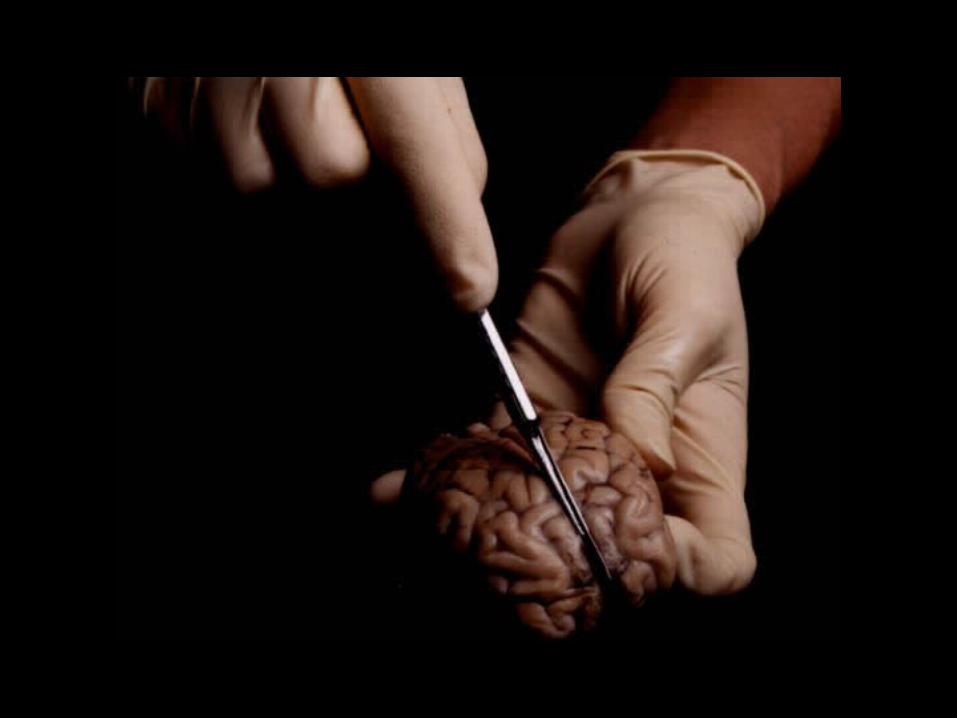
Nervous System
Part II
The Central Nervous System


The Brain
• During embryonic development the CNS first appears as a neural tube – the anterior end starts to expand at four weeks and brain formation begins.


The Four Parts of the Brain:
• Cerebrum
• Diencephalon
• Brain Stem
• Cerebellum


Cerebrum
• Left and right paired hemispheres
• Hemispheres connected by bridge of nerve fibers that allows them to communicate with each other – corpus callosum
• The cerebrum is the largest area – it covers all other regions of the brain


• Gyri (elevated ridges) and sulci (shallow grooves) increase surface area of brain to hold more neurons
• Fissures – deeper grooves– Longitudinal fissure – divides cerebrum into
hemispheres (left & right)– Other fissures divide hemispheres into lobes (frontal,
parietal, temporal, occipital)
• Lobes – are named for cranial bones that life over them


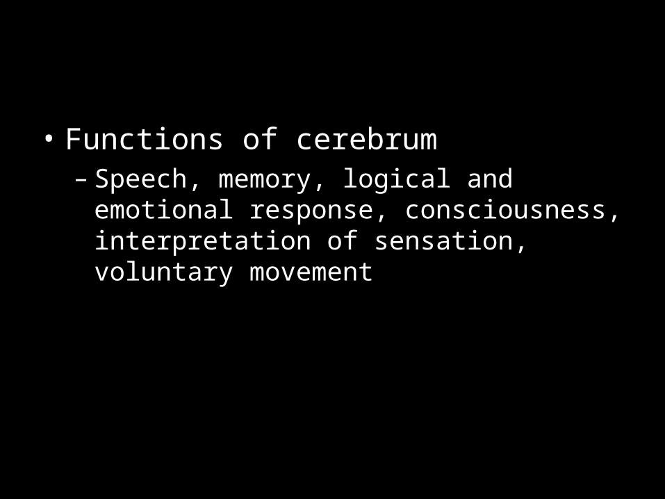
• Functions of cerebrum– Speech, memory, logical and emotional
response, consciousness, interpretation of sensation, voluntary movement

Functional areas of cerebrum:
• Frontal lobe – primary motor area– This area controls voluntary movement of
skeletal muscle
• Prefrontal cortex – judgement– This area is not completely wired until
adulthood
• Broca’s area – speech– Only in one hemisphere
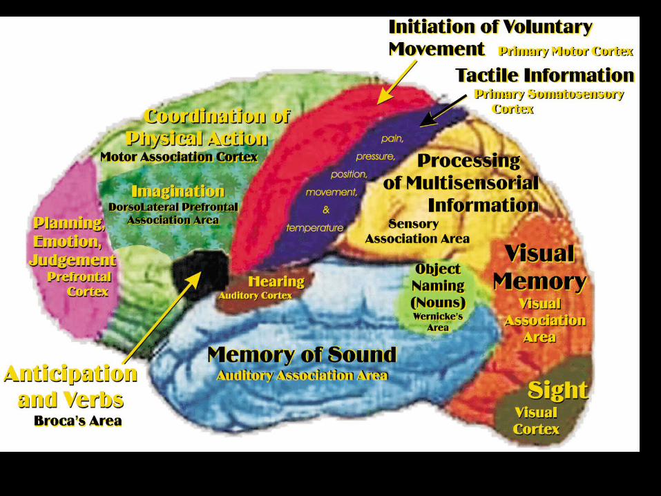

• Sensory and motor pathways are crossed – left side of brain receives impulses from right side of the body

• Cerebral cortex – thin outer layer of brain composed of gray matter (cell bodies)– Only 3-4 mm thick.– Where the conscious mind is located
• White matter – located beneath cerebral cortex; tracts that connect cell bodies of cerebral cortex with other parts of nervous system


Basal Nuclei of Cerebrum
• Islands of gray matter in white matter.– Located in deepest parts of the brain.
• Two diseases associated with abnormal function of basal nuclei:– Huntington’s chorea
• Too much dopamine produced by neurons.• Continuous movement
– Parkinson’s disease• Not enough dopamine produced by neurons.• Slowed movement, tremors


Diencephalon
• Located at the top of the brain stem
• Covered by cerebrum
• Hypothalamus– Thermoregulation, water balance, metabolism– Controls thirst, appetite, sex, pain– Contains pleasure center – Regulates pituitary gland

Brain Stem
• Midbrain, pons, medulla oblongata
• Medulla oblongata– Merges with spinal cord at foramen magnum– Contains centers that control heart rate, blood
pressure, breathing, swallowing and vomiting
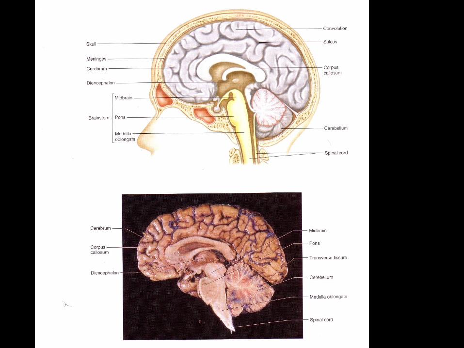


Cerebellum
• Looks like cauliflower
• Balance and equilibrium
• Ataxia – clumsy and disorganized movements caused by damage to cerebellum – cannot touch finger to nose with eyes closed; may be confused with drunkenness

The effect of alcoholism on the cerebellum

Hemisphere Dominance Inventory
1. ____ If you had to give someone directions to your house, which of the following methods would you most likely use?
a. Write a paragraph that explains where and when to turnb. Draw a road map
2. ____Which of the following are you better at solving?a. Jigsaw puzzle b. Crossword puzzle
3. ____Do you remember faces easily? a. Yes b. No4. ____Do you think you’d earn higher grades in a geometry class or in an algebra class?
a. geometry b. algebra5. ____Imagine that you’re vacationing at a resort. Which of the following would you most likely do?
a. Obtain a brochure of local attractions and plan what you’d like to do for the dayb. Drive around without a plan and decide what you’d like to do as you drive along
6. ____Was it usually easy or difficult to learn grammar in school? a. difficult b. easy7. ____Imagine enrolling in a music coarse. You and a partner in the course must write a song. Which of the following would you prefer to do?
a. Write the lyrics b. Compose the melody8. ____When you read a new chapter in a textbook, which of the following are you most likely to do?
a. Skim through the entire chapter first to get a general idea of what the chapter is aboutb. Read the chapter from beginning to end without doing much skimming
9. ____ In which of the following English classes would you most likely enroll?a. Journalismb. Creative writing
10. ____ Imagine that you volunteered to work for the school newspaper. Which of the following would you rather do?

11. ____ After reading a new chapter in a textbook, which of the following would you rather do?a. summarize the chapter b. outline the chapter
12. ____ if you had an important project due in a class, would you prefer to work? a. in a group b. alone
13. ____Which of the following classroom situations do you prefer?a. A teacher announces assignments on a weekly basis and sets specific weekly
due datesb. A teacher announces all the assignments at the beginning of the course and
allows you to complete them at any time before the end of the course14. ____ Which of the following statements best applies to you?
a. I’m good at guessing a person’s mood by his or her body languageb. I’m not good at guessing a person’s mood by his or her body language
15. Which of the following would you rather play? a. Scrabble b. Checkers16. With which of the following statements do you most agree?
a. We should continue exploring outer space since one day this exploration may benefit us
b. We should continue exploring outer space only if we can be sure ahead of time of certain benefits we would receiveScoringHow many “a” answers did you have for odd-numbered questions? ___________How many “b” answers did you have for even-numbered questions? ___________
LEFT HEMISPHERE TOTAL _______________How many “a” answers did you have for even-numbered questions? ___________How many “b” answers did you have for odd-numbered questions? ___________
RIGHT HEMISPHERE TOTAL _______________

CRANIAL NERVES(12 PAIRS)
Oh - Olfactory (smell)Oh - Optic (vision)Oh – Oculomotor (eye muscles)To – Trochlear (external eye muscle)Touch – Trigeminal (skin of face, chewing muscles)And – Abducens (lateral muscle of eye)Feel – Facial (facial expression)Very – Vestibulocochlear (balance, hearing)Good – Glossopharyngeal (throat, swallowing, saliva)Velvet – Vagus (pharynx, larynx, heart)Ah – Accessory (sternocleidomastoid and trapezius) Hypoglossal (tongue movements)

Protection of the CNS
• Four elements of protection:– Bones of dorsal cavity– Meninges– Cerebrospinal fluid– Blood-Brain Barrier

Bones of Dorsal Cavity
• Skull– Helmet-like protection of brain
• Vertebral Column– Cervical, thoracic, and lumbar vertebrae

Meninges
• Protective connective tissue membranes
• Three layers wrap brain and spinal cord
Dura mater• Leathery outer layer• Ends in sac below the layer of the spinal cord

– Arachnoid mater• Web-like (spider) middle layer• Thread-like extensions extend into sub-arachnoid
space which contains cerebrospinal fluid (CSF)
– Pia mater• Most delicate layer which clings to the surface of
the brain

• Pathologies:– Meningitis – inflammation of meninges
caused by bacteria or viruses; can spread to CNS.
– Encephalitis – inflammation of brain (West Nile Virus).


Figure 28-21 Pyogenic meningitis. A thick layer of suppurative exudate covers the brain stem and cerebellum and thickens the leptomeninges. (From Golden JA, Louis DN: Images in clinical medicine: Acute bacterial meningitis. N Engl J Med 333:364, 1994.)
Downloaded from: Robbins & Cotran Pathologic Basis of Disease (on 7 April 2005 12:46 AM)
© 2005 Elsevier

Cerebrospinal Fluid (CSF)
• Watery fluid – similar to blood plasma.
• Cushions brain and cord, protecting fragile nervous tissue from trauma.
• Fills subarachnoid space, ventricles of brain, and central canal of spinal cord.
• Volume – about ½ cup.

• Lumbar puncture (spinal tap) – diagnostic tool to check for bacteria or viruses in CSF (meningitis).
• Needle is inserted between L3-L4 or
L4-L5.
• Spinal cord ends around level of L1-L2.


CSF Pathologies
• Subdural hematoma – Blood from brain injury collects in space
between dura and arachnoid mater.– Pressure can kill brain tissue.
• Hydrocephalus– CSF drainage is obstructed.– CSF accumulates and puts pressure on the
brain.


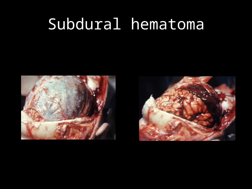
Subdural hematoma

Figure 28-12 A, Large organizing subdural hematoma attached to the dura. B, Coronal section of the brain showing compression of the hemisphere underlying the hematoma.
Downloaded from: Robbins & Cotran Pathologic Basis of Disease (on 7 April 2005 12:46 AM)
© 2005 Elsevier

Downloaded from: Robbins & Cotran Pathologic Basis of Disease (on 7 April 2005 12:46 AM)
© 2005 Elsevier
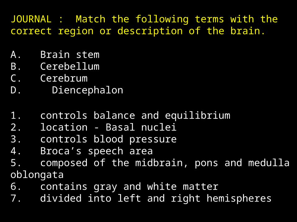
JOURNAL : Match the following terms with the correct region or description of the brain.
A. Brain stemB. CerebellumC. CerebrumD. Diencephalon
1. controls balance and equilibrium2. location - Basal nuclei3. controls blood pressure4. Broca’s speech area5. composed of the midbrain, pons and medulla oblongata6. contains gray and white matter7. divided into left and right hemispheres

Blood-Brain Barrier
• The brain is absolutely dependent on a constant internal environment.– Fluctuations in sodium and potassium levels
can result in uncontrollable firing of neurons.
• Blood Brain Barrier:– The cells of the brain are separated from
substances in the blood by the least permeable capillaries in the body.
• These capillaries are the thickest capillaries in the body.

What substances can cross the blood-brain barrier?
• Water soluble substances that can cross the barrier:– Water, glucose, essential amino acids.
• BBB is useless against fat-soluble substances:– This is why alcohol, nicotine, and anesthetics can
affect the brain.
• This barrier often prevents antibiotics from reaching the brain or CSF in sufficient concentrations to battle infections, such as meningitis.

Pathology of the CNS
• Concussion – caused by a blow to the head.– Patient may lose consciousness briefly.– May be accompanied by vomiting and blurred
vision.


Headaches
• Although brain tissue lacks pain receptors, the meninges and blood vessels are well supplied with them.
• Tension headaches– Muscular headaches– Prolonged contraction of skeletal muscle.– Related to stress, anxiety, frustration.

Migraine Headaches
• Vascular headaches (blood vessels)• Initial vasoconstriction of blood vessel causes
aura of light and/or numbness of limbs.– When the vessel becomes smaller, it restricts blood
flow to an area.
• Reflex vasodilation of affected vessel results in intense pain, usually unilateral.– When the vessel expands, it presses on nerve fibers.


Brain Tumors - Gliomas
• Brain tumors are not composed of neurons because neurons can’t divide.– Brain tumors are almost always composed of
neuroglial cells.
• Grade I Tumor– Usually only symptom is seizure.– Good prognosis – Usually found in children and young adults.


• Grade IV – Glioblastoma– Most malignant tumor– Infiltrate brain tissue with fingers that cannot
be removed surgically.– Prognosis: 51 weeks with most aggressive
treatment.


Cerebrovascular Accident (CVA)
• May be a stroke or an aneurysm.
• Blood circulation to an area of the brain is blocked and brain tissue dies.
• Ischemia – loss of blood supply; oxygen and glucose are blocked.
• Most common cause: blood clot.
• Aneurysm – ballooning of blood vessel.– Vessel is weakened and prone to rupture.


Alzheimer’s Disease
• Primarily affects individuals over 65 years of age.
• Gradual loss of short term and long term memory.
• Beta-amyloid plaques form between neurons in the brain.
• Neurofibrillary tangles form inside neurons, causing their destruction.

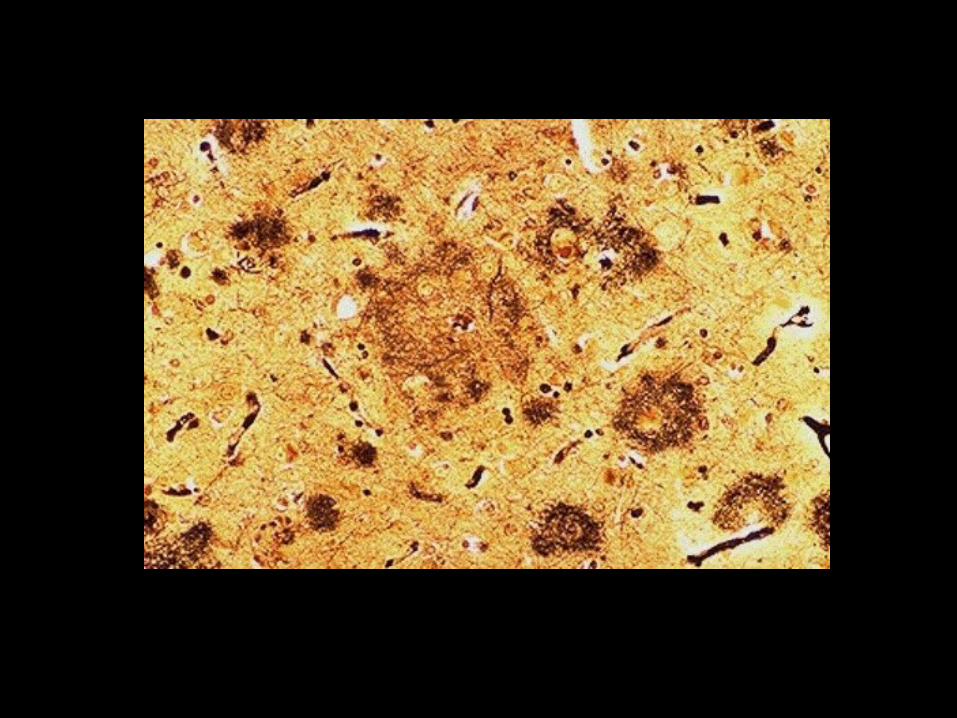


Normal brain vs. Alzheimer’s


Effects of Drugs on the Brain
• The Pleasure Center is a small bundle of nervous tissue located inside the hypothalamus.– This area reinforces our drives to eat, drink and
reproduce.– Our ability to feel good involves neurotransmitters
such as dopamine, norepinephrine, endorphins and serotonin.
– Recreational drugs work by artificially stimulates the reward pathways in the pleasure center.

CNS Stimulants
• Result in increased vasoconstriction, leading to increased heart rate and blood pressure.
• Amphetamines– Cause increased secretion of norepinephrine
and/or dopamine which stimulates the sympathetic nervous system (similar to adrenalin).
– The more you take artificially, the less your body makes; can quickly lead to dependence or addiction.


Meth Mouth

• Cocaine– Blocks reabsorption of dopamine– It stays in the synapse and continues to stimulate
receptor cells so that the drug’s effects are felt for an extended period.
– Dopamine is washed away from the synapse instead of being reabsorbed into axonal terminals – neuron runs out of dopamine.
– User becomes anxious and unable to experience pleasure without the drug.
– Highly addictive.

Hallucinogens - LSD
• Acts as artificial neurotransmitter that stimulates the brain.
• Synesthesia – hearing colors or seeing sound.
• Hallucinations – false sensory perceptions.
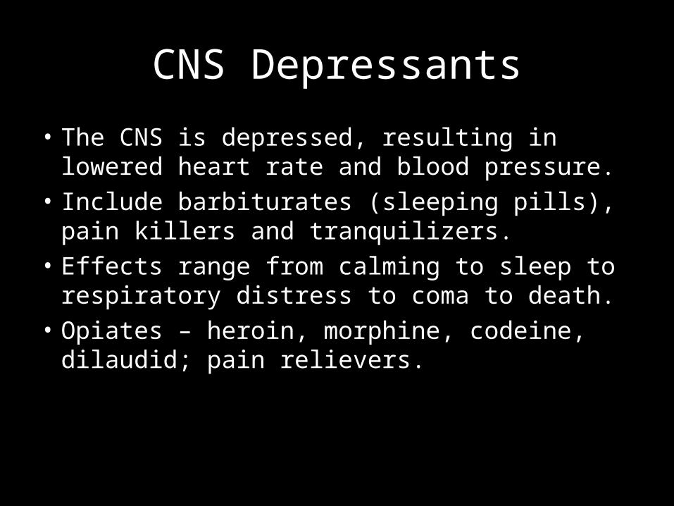
CNS Depressants
• The CNS is depressed, resulting in lowered heart rate and blood pressure.
• Include barbiturates (sleeping pills), pain killers and tranquilizers.
• Effects range from calming to sleep to respiratory distress to coma to death.
• Opiates – heroin, morphine, codeine, dilaudid; pain relievers.
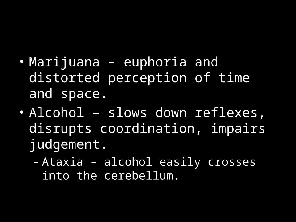
• Marijuana – euphoria and distorted perception of time and space.
• Alcohol – slows down reflexes, disrupts coordination, impairs judgement.– Ataxia – alcohol easily crosses into the
cerebellum.

Spinal Cord
• Begins where the nervous tissue leaves the cranial cavity at the foreman magnum.
• Terminates between the 1st and 2nd lumbar vertebrae.
• Structure:– 31 segments – each gives rise to a pair of
spinal nerves.– Spinal nerves branch out to body parts,
connecting them to CNS.


Functions of the Spinal Cord
1. Conduction of nerve impulses– The spinal cord is a two-way communication
system between the brain and parts of the body.
2. Serve as a center for spinal reflexes.

Spinal Cord Injuries
• 10,000 per year; mostly in males 18-30.
• If the spinal cord is damaged, loss of function may be only temporary. If function does not return within 48 hours, paralysis is permanent in most cases.
• If nerve fibers are severed, function is likely to be completely lost.


• Transection (cutting) of the spinal cord at any level results in motor and sensory loss in body regions inferior to the site of damage.
• The loss is bilateral (both sides).– Paraplegia – T1-L1, both lower limbs are
paralyzed.– Quadriplegia – cervical region; all 4 limbs
affected.