Embed Size (px)
Citation preview
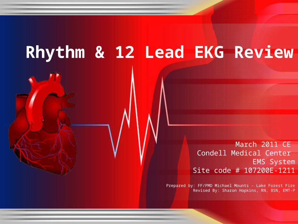
March 2011 CE Condell Medical Center
EMS SystemSite code # 107200E-1211
Prepared by: FF/PMD Michael Mounts – Lake Forest FireRevised By: Sharon Hopkins, RN, BSN, EMT-P
Rhythm & 12 Lead EKG Review
Objectives
Upon successful completion of this module, the EMS provider will be able to:
• Identify the components of a rhythm strip
• Identify what the components represent on the rhythm strip
• Identify criteria for sinus rhythms
• Identify criteria for atrial rhythms
• Identify AV/junctional rhythms
Objectives cont.• Identify ventricular rhythms• Identify rhythms with AV blocks• Identify treatments for different rhythms• Identify criteria for identification of ST elevation
on 12 lead EKG’s• Identify EMS treatment for patients with acute
coronary syndrome (ST elevation)• Demonstrate standard & alternate placement
of ECG electrodes for monitoring
Objectives cont.• Demonstrate placement of electrodes for
obtaining a 12 lead EKG• Demonstrate the ability to identify a variety
of static or dynamic EKG rhythm strips• Demonstrate the ability to identify the
presence or absence of ST elevation when presented with a 12 lead EKG
• Review department’s process to transmit 12 lead EKG to hospital, if capable
• Successfully complete the post quiz with a score of 80% or better.
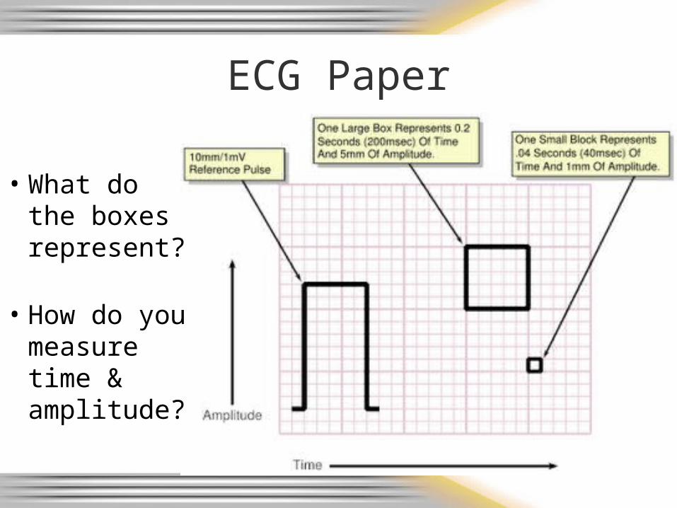
ECG Paper
• What do the boxes represent?
• How do you measure time & amplitude?
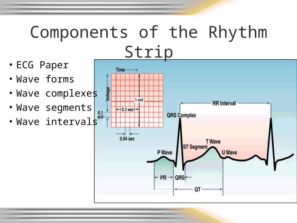
Components of the Rhythm Strip
• ECG Paper• Wave forms• Wave complexes• Wave segments• Wave intervals
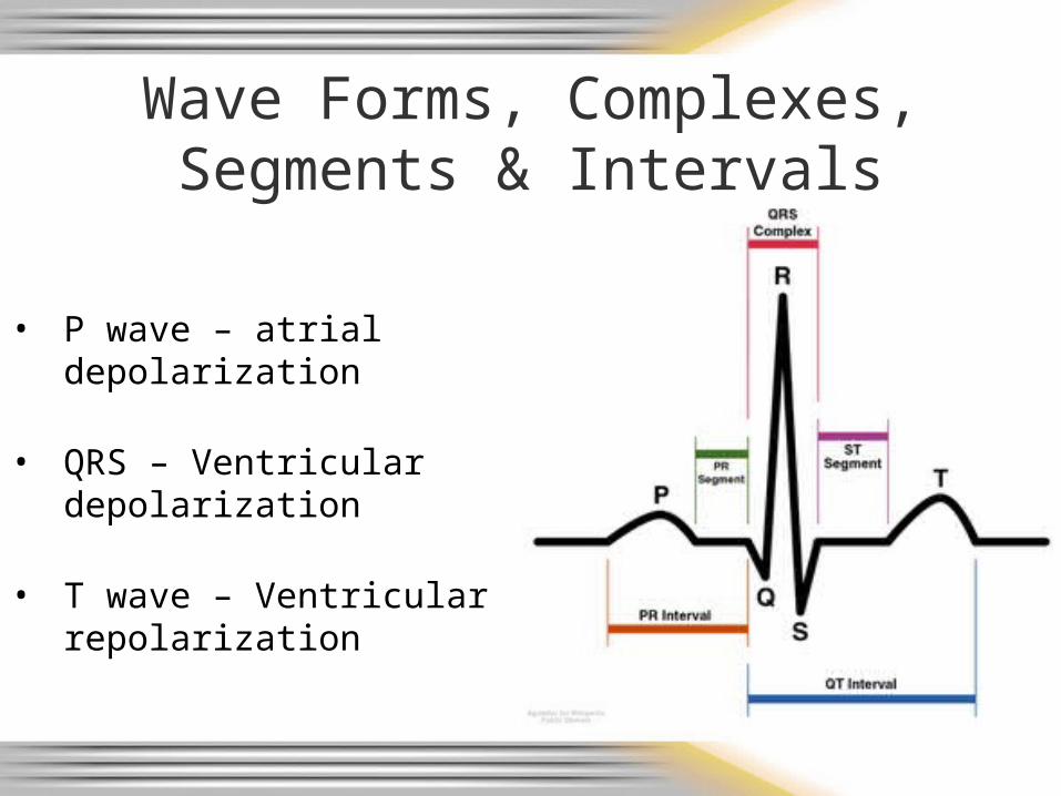
Wave Forms, Complexes, Segments & Intervals
• P wave – atrial depolarization
• QRS – Ventricular depolarization
• T wave – Ventricular repolarization
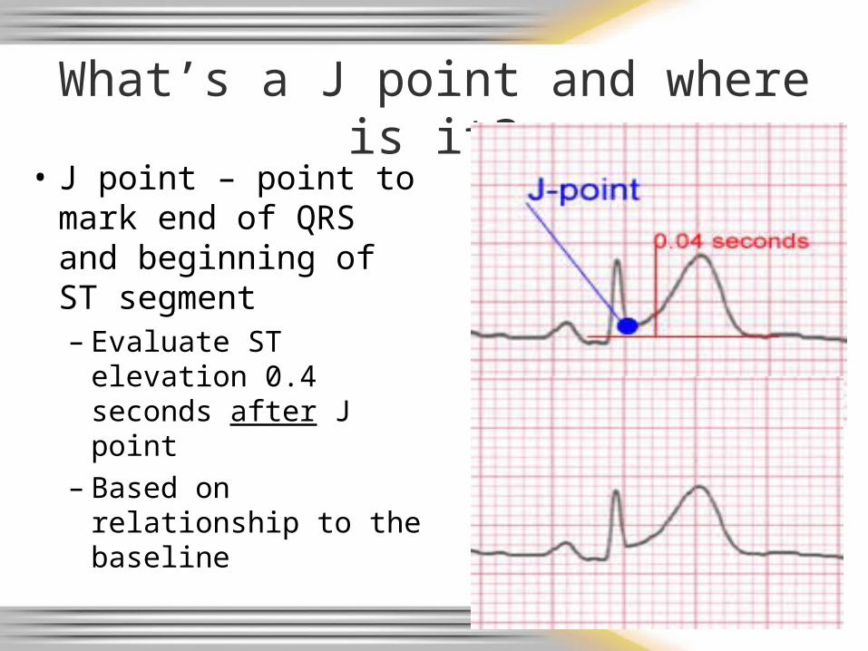
What’s a J point and where is it?
• J point – point to mark end of QRS and beginning of ST segment– Evaluate ST elevation
0.4 seconds after J point
– Based on relationship to the baseline
Intervals and Complexes
• PR interval – atrial and nodal activity– Includes atrial depolarization & delay in the AV
node (PR segment)
• QRS complex– Corresponds to the patient’s palpated pulse– Large in size due to reflection of ventricular
activity
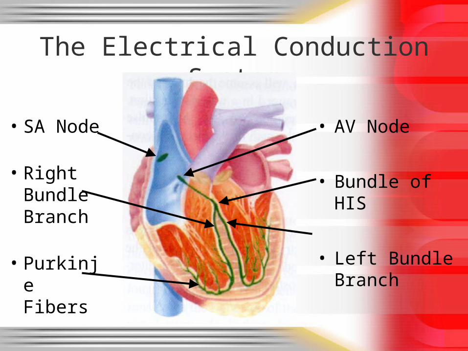
The Electrical Conduction System
• AV Node
• Bundle of HIS
• Left Bundle Branch
• SA Node
• Right Bundle Branch
• Purkinje Fibers
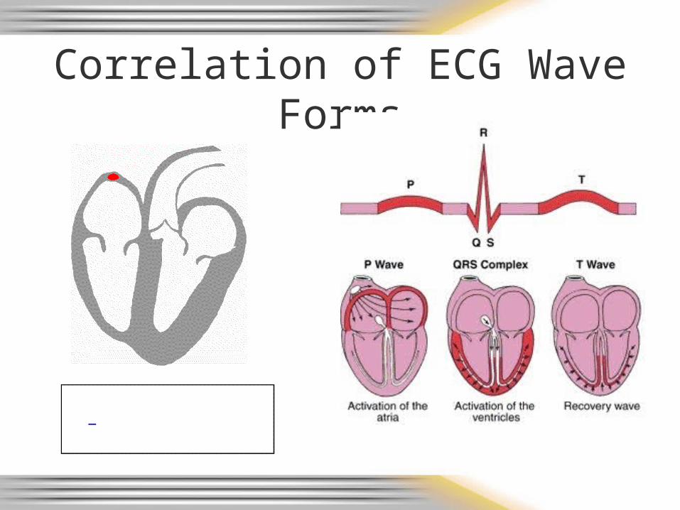
Correlation of ECG Wave Forms
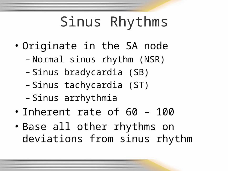
Sinus Rhythms
• Originate in the SA node– Normal sinus rhythm (NSR)– Sinus bradycardia (SB)– Sinus tachycardia (ST)– Sinus arrhythmia
• Inherent rate of 60 – 100
• Base all other rhythms on deviations from sinus rhythm
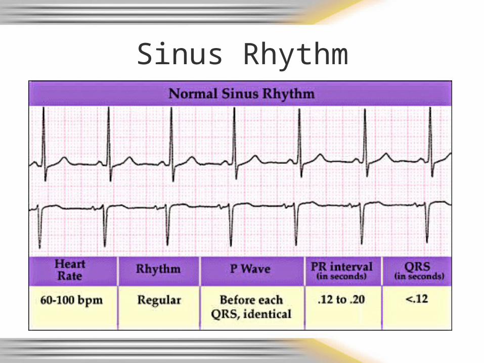
Sinus Rhythm
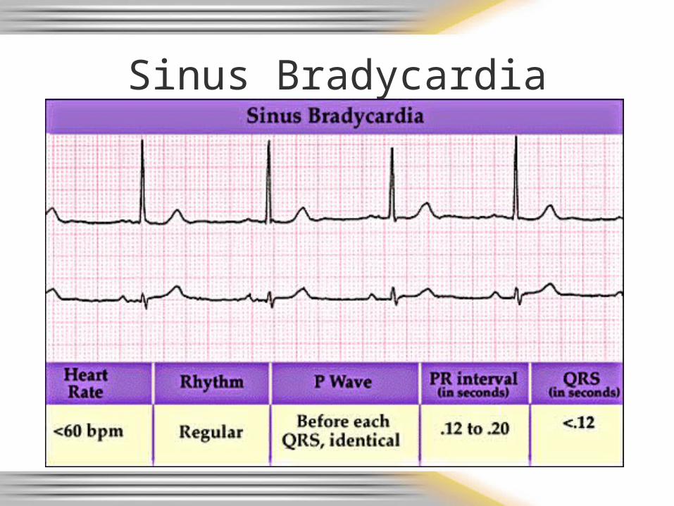
Sinus Bradycardia
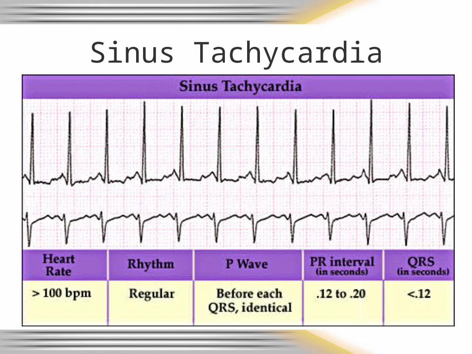
Sinus Tachycardia
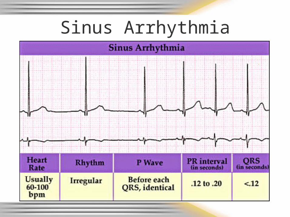
Sinus Arrhythmia
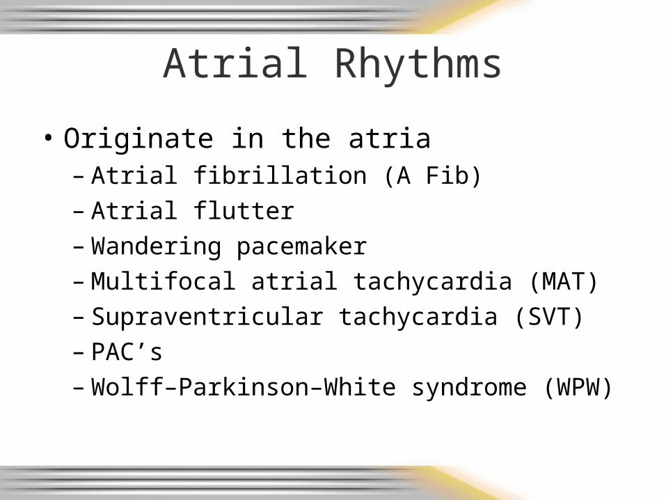
Atrial Rhythms
• Originate in the atria
– Atrial fibrillation (A Fib)– Atrial flutter– Wandering pacemaker– Multifocal atrial tachycardia (MAT)– Supraventricular tachycardia (SVT)– PAC’s– Wolff–Parkinson–White syndrome (WPW)

A - Fib
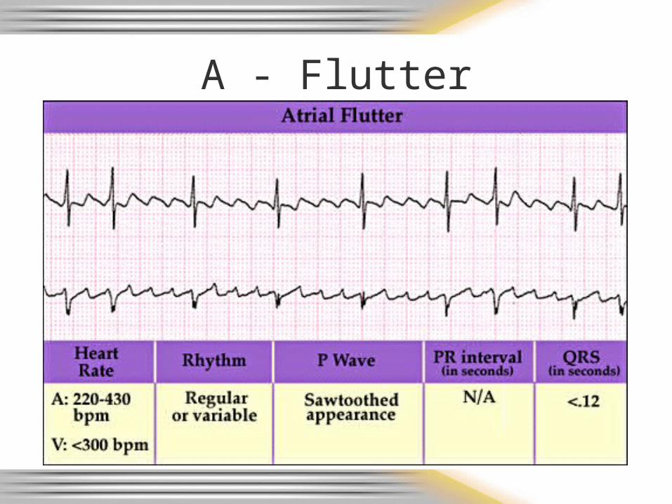
A - Flutter
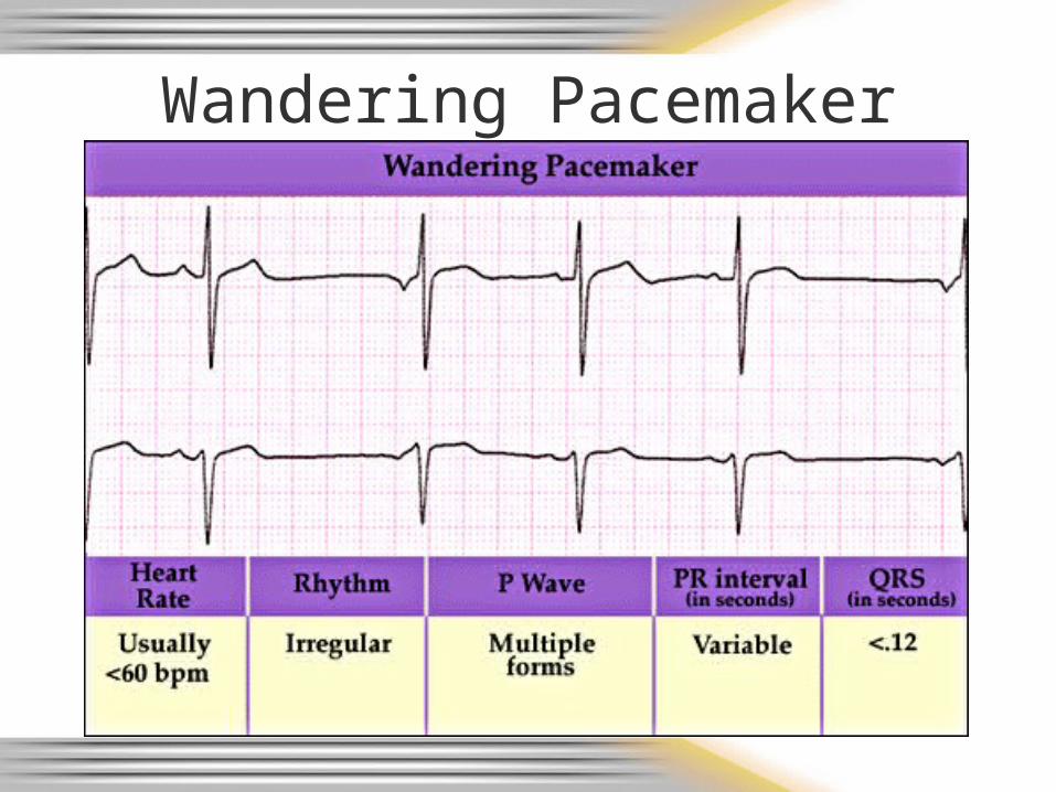
Wandering Pacemaker
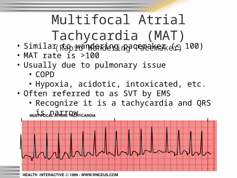
Multifocal Atrial Tachycardia (MAT)(Rapid Wandering Pacemaker)
• Similar to wandering pacemaker (< 100)• MAT rate is >100• Usually due to pulmonary issue
• COPD• Hypoxia, acidotic, intoxicated, etc.
• Often referred to as SVT by EMS• Recognize it is a tachycardia and QRS is narrow
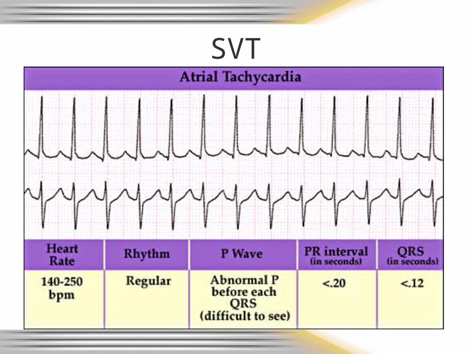
SVT

PAC’s
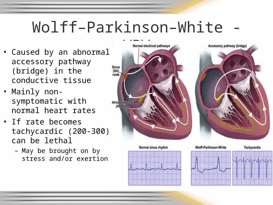
Wolff–Parkinson–White - WPW
• Caused by an abnormal accessory pathway (bridge) in the conductive tissue
• Mainly non-symptomatic with normal heart rates
• If rate becomes tachycardic (200-300) can be lethal– May be brought on by
stress and/or exertion
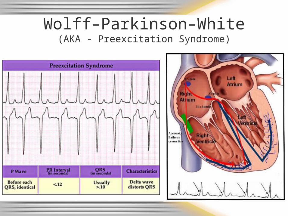
Wolff–Parkinson–White(AKA - Preexcitation Syndrome)
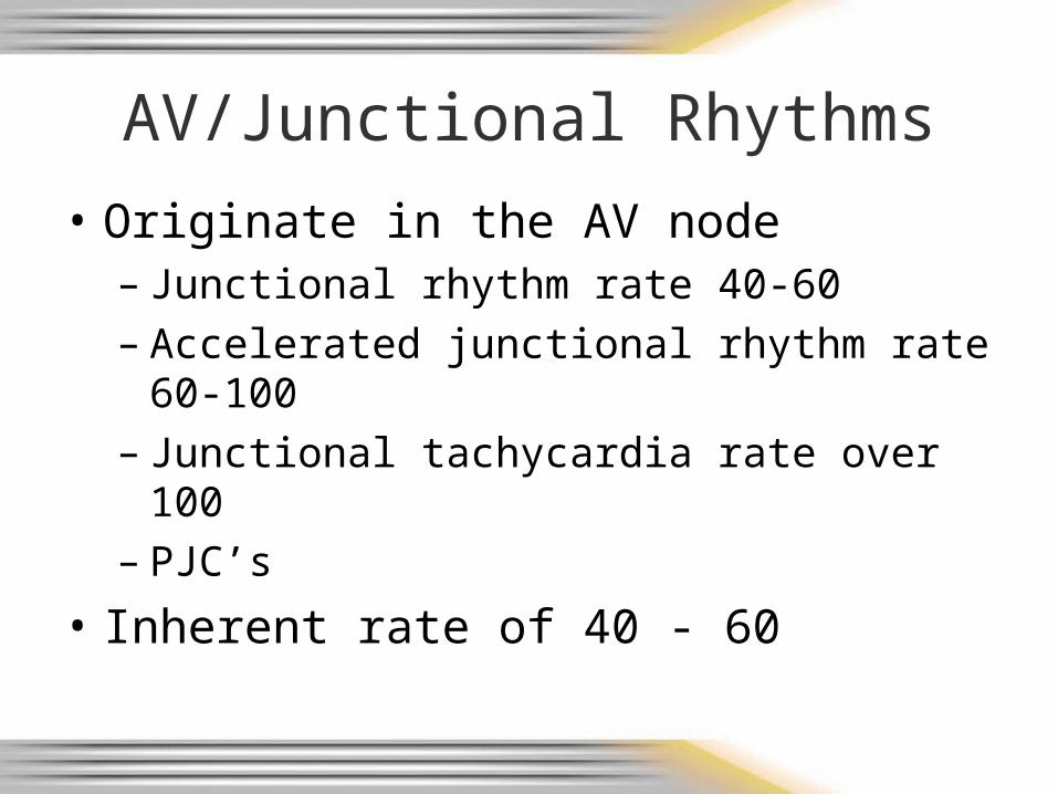
AV/Junctional Rhythms
• Originate in the AV node– Junctional rhythm rate 40-60– Accelerated junctional rhythm rate 60-100– Junctional tachycardia rate over 100– PJC’s
• Inherent rate of 40 - 60
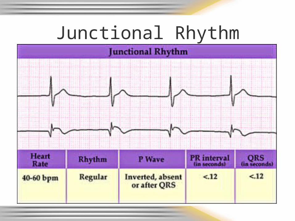
Junctional Rhythm
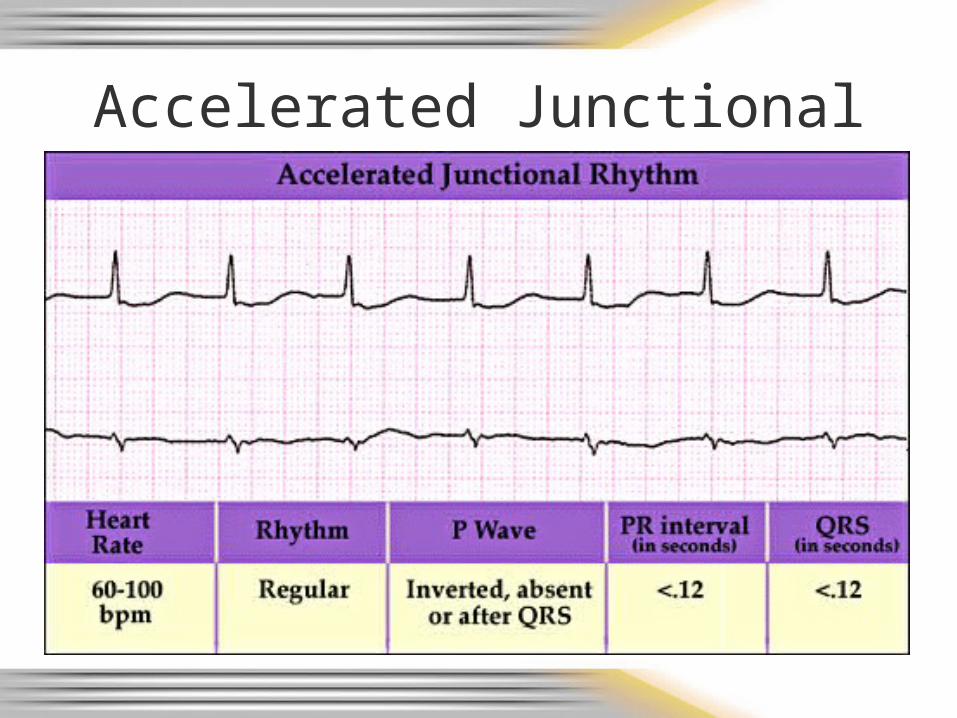
Accelerated Junctional
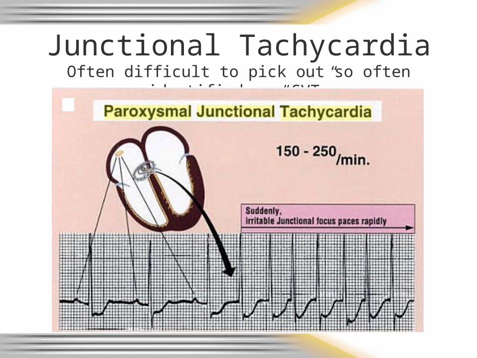
Junctional TachycardiaOften difficult to pick out so often identified as “SVT”
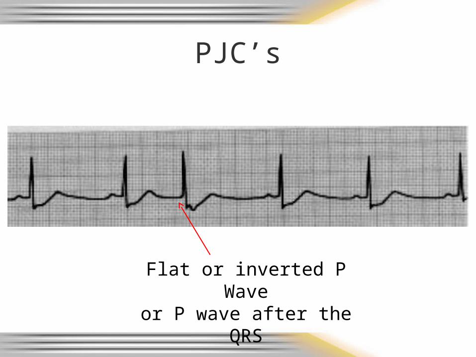
PJC’s
Flat or inverted P Waveor P wave after the QRS
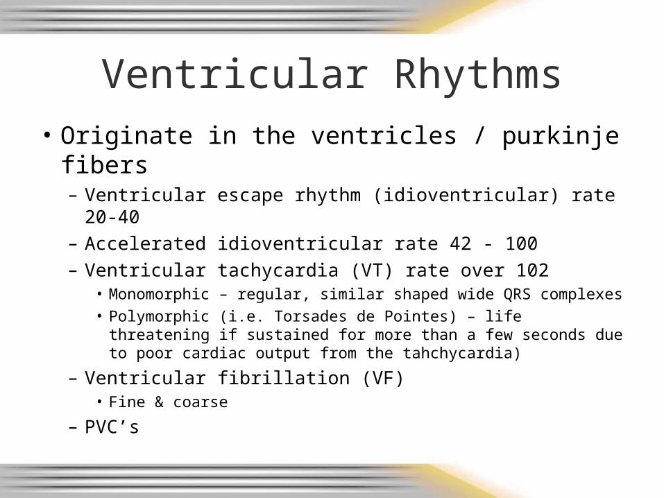
Ventricular Rhythms
• Originate in the ventricles / purkinje fibers– Ventricular escape rhythm (idioventricular) rate 20-40– Accelerated idioventricular rate 42 - 100– Ventricular tachycardia (VT) rate over 102
• Monomorphic – regular, similar shaped wide QRS complexes• Polymorphic (i.e. Torsades de Pointes) – life threatening if
sustained for more than a few seconds due to poor cardiac output from the tahchycardia)
– Ventricular fibrillation (VF)• Fine & coarse
– PVC’s
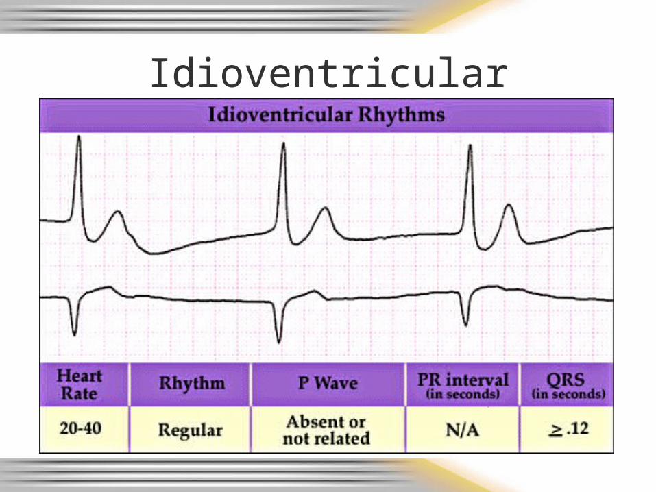
Idioventricular
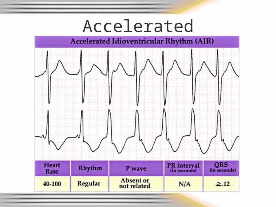
Accelerated Idioventricular
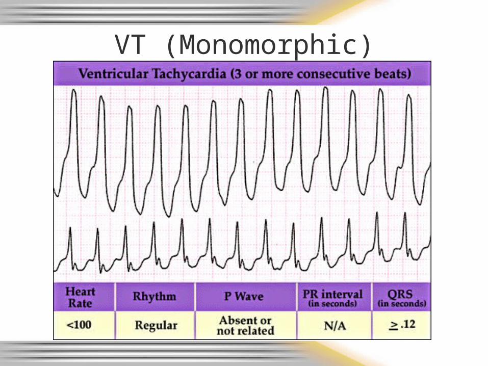
VT (Monomorphic)
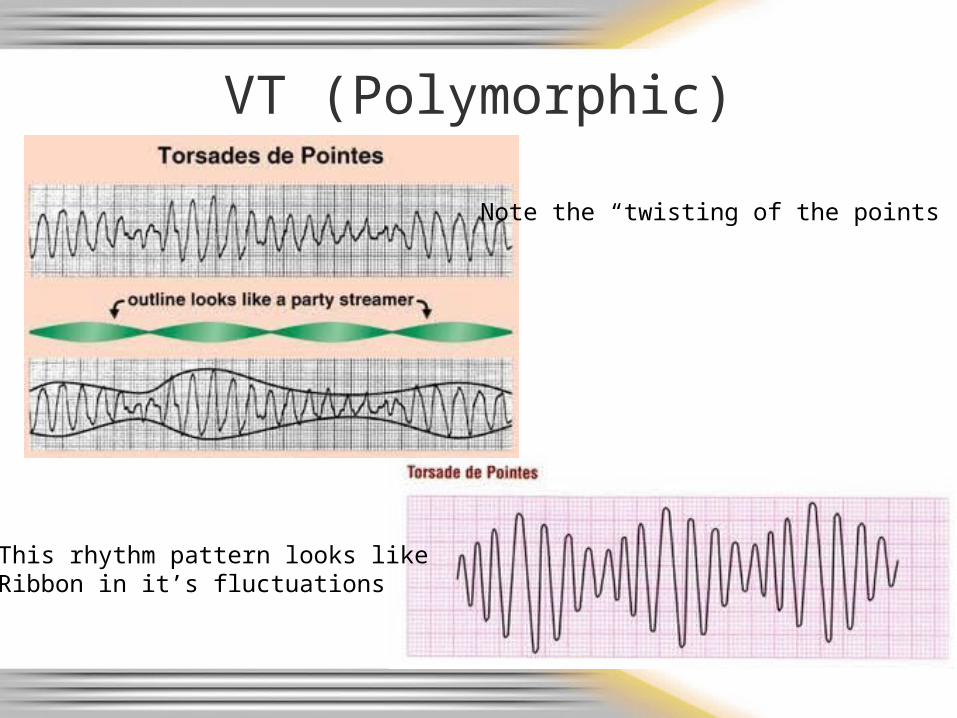
VT (Polymorphic)
Note the “twisting of the points”
This rhythm pattern looks likeRibbon in it’s fluctuations
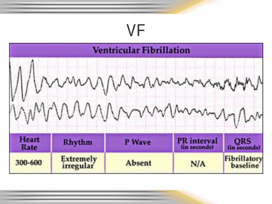
VF
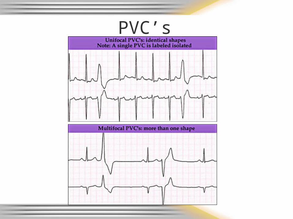
PVC’s
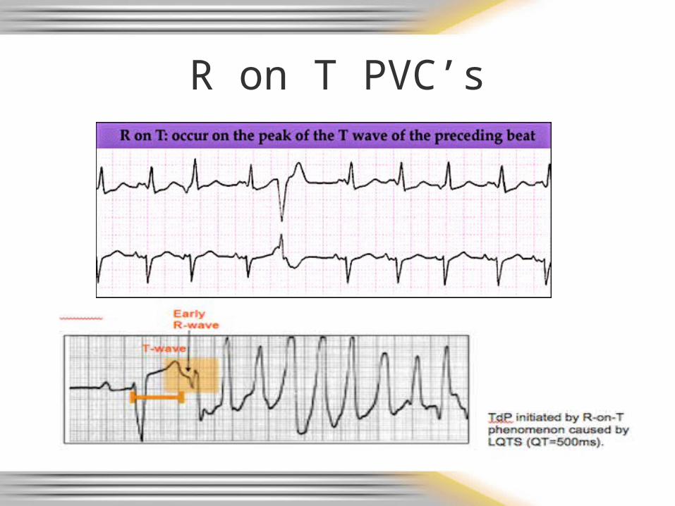
R on T PVC’s
R on T PVC’s cont.• Why is R on T so bad?
– Downslope of T wave is the relative refractory period• Some cells have repolarized and can be stimulated
again to depolarize/discharge– Relatively strong impulse can stimulate cells to
conduct electrical impulses but usually in a slower, abnormal manner
» Can result in ventricular fibrillation• Absolute refractory period is from the beginning of
the QRS complex through approximately the first half of the T wave
– Cells not repolarized and therefore cannot be stimulated
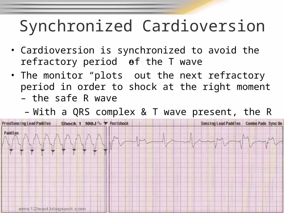
Synchronized Cardioversion• Cardioversion is synchronized to avoid the refractory period
of the T wave• The monitor “plots” out the next refractory period in order to
shock at the right moment – the safe R wave– With a QRS complex & T wave present, the R wave can
be predicted (cannot work in VF – no wave forms present)
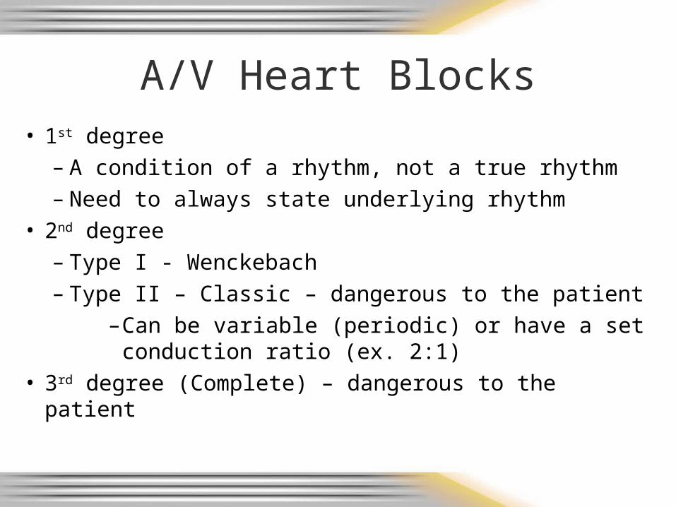
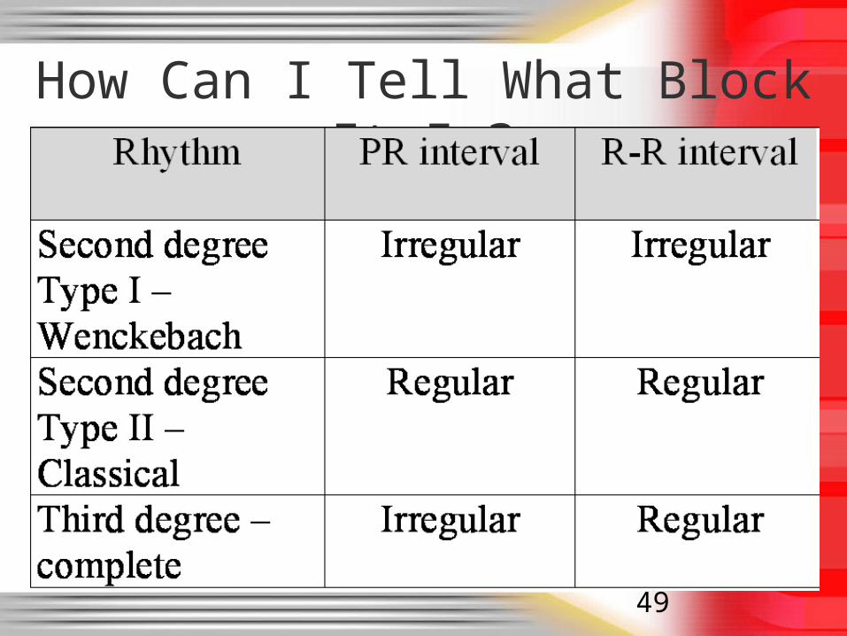
A/V Heart Blocks• 1st degree
– A condition of a rhythm, not a true rhythm– Need to always state underlying rhythm
• 2nd degree– Type I - Wenckebach– Type II – Classic – dangerous to the patient
–Can be variable (periodic) or have a set conduction ratio (ex. 2:1)
• 3rd degree (Complete) – dangerous to the patient
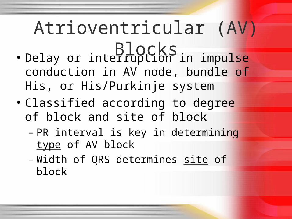
Atrioventricular (AV) Blocks
• Delay or interruption in impulse conduction in AV node, bundle of His, or His/Purkinje system
• Classified according to degree of block and site of block– PR interval is key in determining type of
AV block– Width of QRS determines site of block
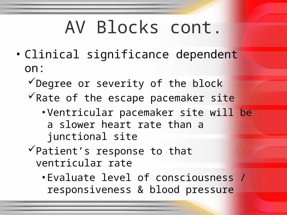
AV Blocks cont.
• Clinical significance dependent on:Degree or severity of the blockRate of the escape pacemaker site
• Ventricular pacemaker site will be a slower heart rate than a junctional site
Patient’s response to that ventricular rate• Evaluate level of consciousness /
responsiveness & blood pressure
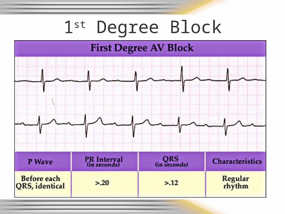
1st Degree Block
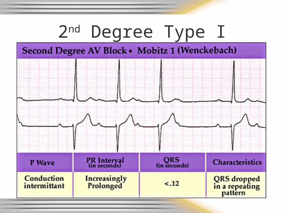
2nd Degree Type I
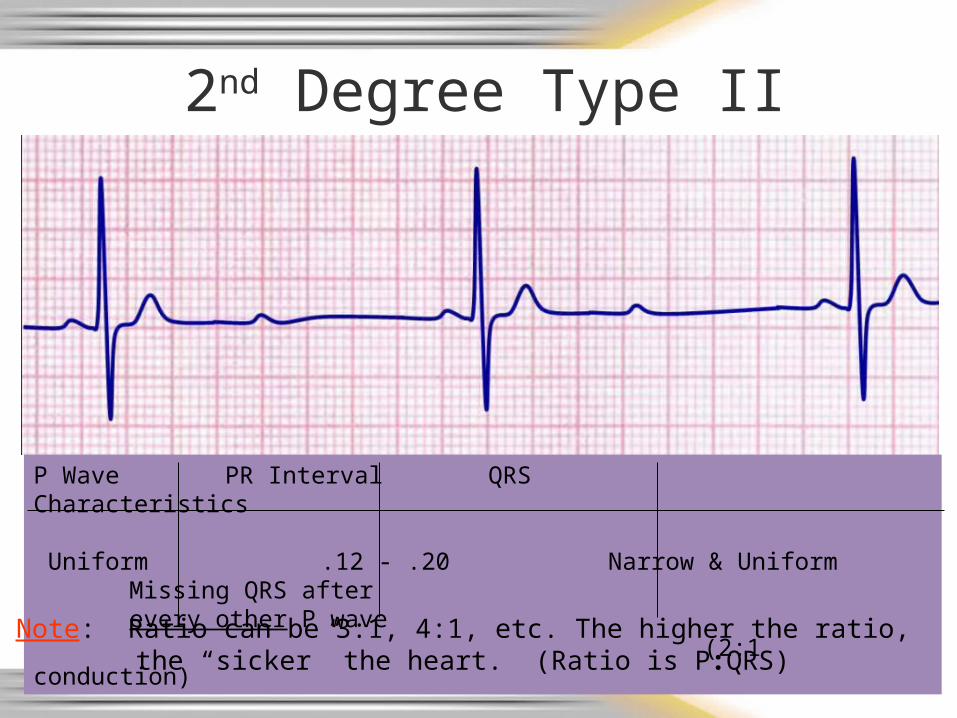
2nd Degree Type II (constant)
P Wave PR Interval QRS Characteristics
Uniform .12 - .20 Narrow & Uniform Missing QRS after every other P wave(2:1 conduction)
Note: Ratio can be 3:1, 4:1, etc. The higher the ratio, the “sicker” the heart. (Ratio is P:QRS)

2nd Degree Type II (periodic)
P Wave PR Interval QRS Characteristics
Uniform .12 - .20 Narrow & Uniform Missing QRS after some P waves
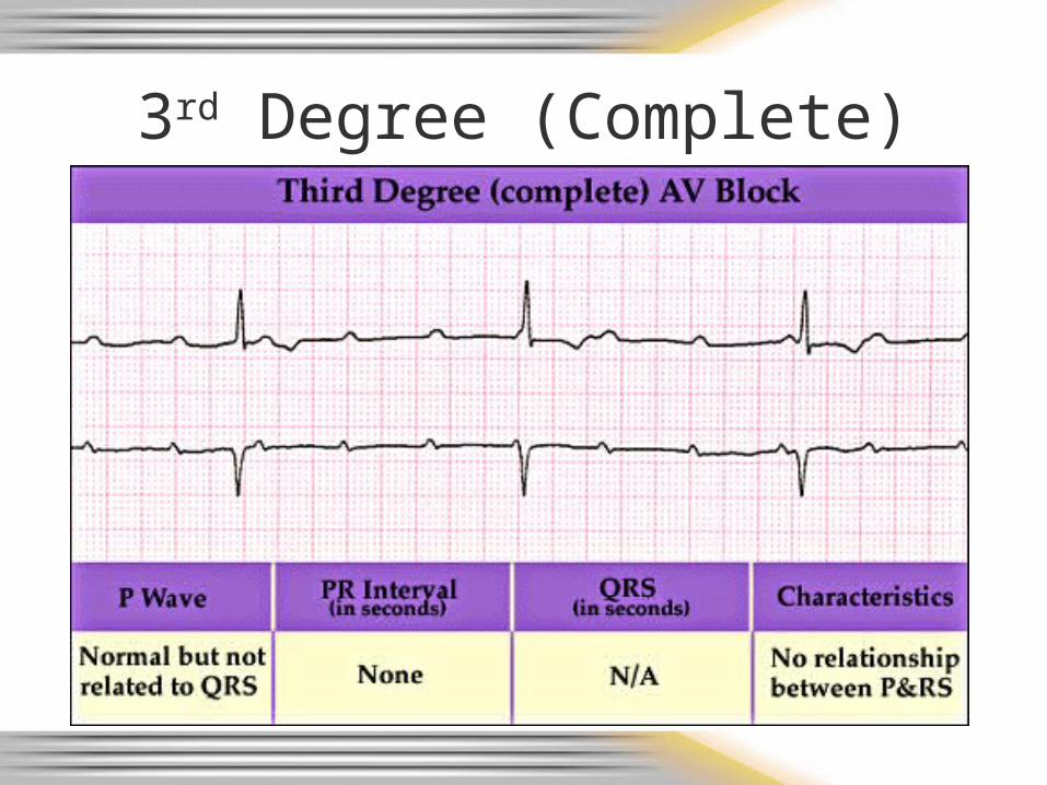
3rd Degree (Complete)
49
How Can I Tell What Block It Is?
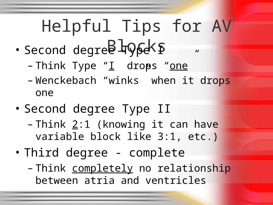
Helpful Tips for AV Blocks• Second degree Type I
– Think Type “I” drops “one”– Wenckebach “winks” when it drops one
• Second degree Type II– Think 2:1 (knowing it can have variable
block like 3:1, etc.)
• Third degree - complete– Think completely no relationship between
atria and ventricles
5151
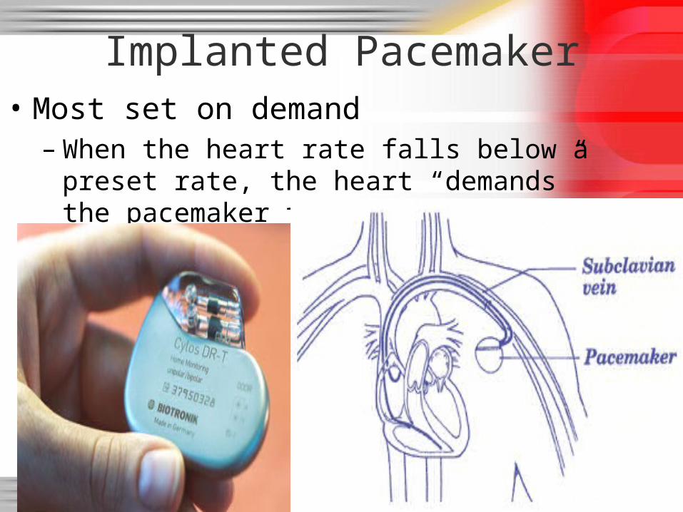
Implanted Pacemaker• Most set on demand
– When the heart rate falls below a preset rate, the heart “demands” the pacemaker to take over
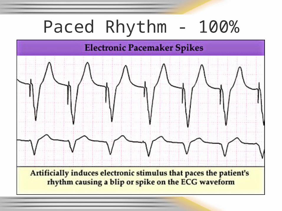
Paced Rhythm - 100% Capture
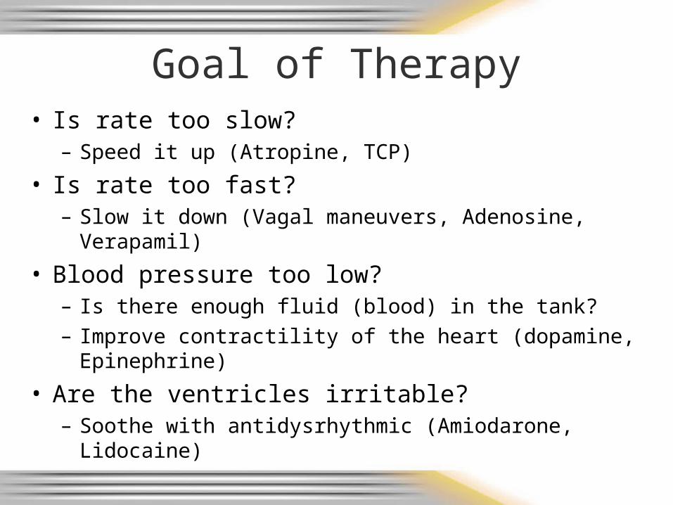
Goal of Therapy• Is rate too slow?
– Speed it up (Atropine, TCP)
• Is rate too fast? – Slow it down (Vagal maneuvers, Adenosine, Verapamil)
• Blood pressure too low? – Is there enough fluid (blood) in the tank?– Improve contractility of the heart (dopamine,
Epinephrine)
• Are the ventricles irritable?– Soothe with antidysrhythmic (Amiodarone, Lidocaine)
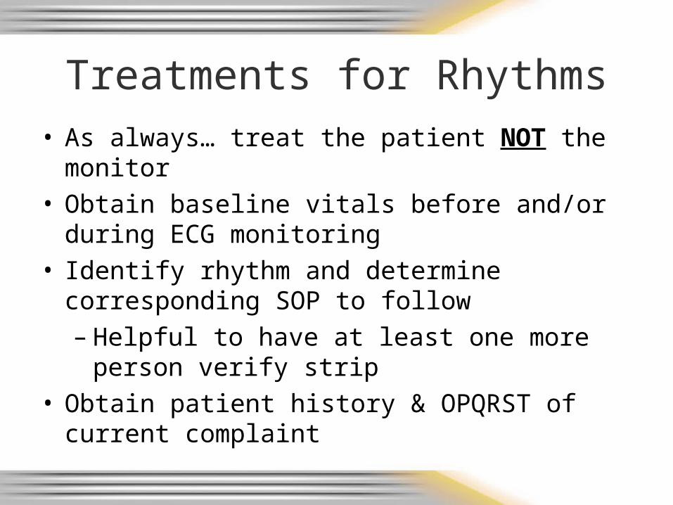
Treatments for Rhythms• As always… treat the patient NOT the monitor• Obtain baseline vitals before and/or during ECG
monitoring• Identify rhythm and determine corresponding
SOP to follow– Helpful to have at least one more person
verify strip• Obtain patient history & OPQRST of current
complaint
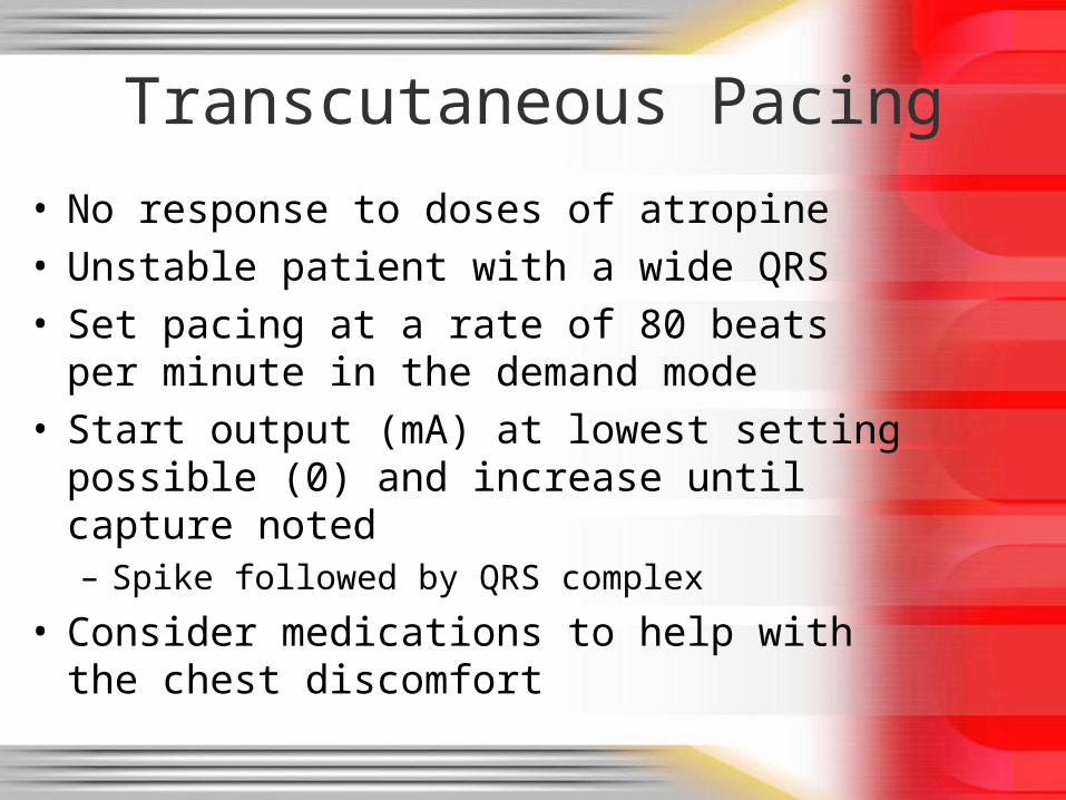
Transcutaneous Pacing
• No response to doses of atropine• Unstable patient with a wide QRS • Set pacing at a rate of 80 beats per minute in
the demand mode• Start output (mA) at lowest setting possible
(0) and increase until capture noted– Spike followed by QRS complex
• Consider medications to help with the chest discomfort

Tachycardias
• Can be generally well tolerated rhythmsOR
• Can become lethal usually related to the heart rate and influence on cardiac output
• Ask 2 questions:– Is the patient stable or unstable?
• If unstable, needs cardioversion– If stable, determine if the QRS is narrow or
wide• QRS width drives decisions for therapy in
stable patient

12-lead ECG Review
• Lead placement– Lead II monitoring– Obtaining 12 lead EKG
• Lead / location correlation of ST elevation
• ST elevation criteria
• 12 – lead practice EKG’s
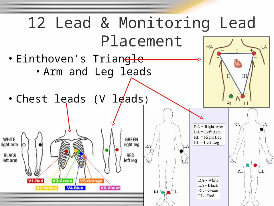
12 Lead & Monitoring Lead Placement
• Einthoven’s Triangle• Arm and Leg leads
• Chest leads (V leads)
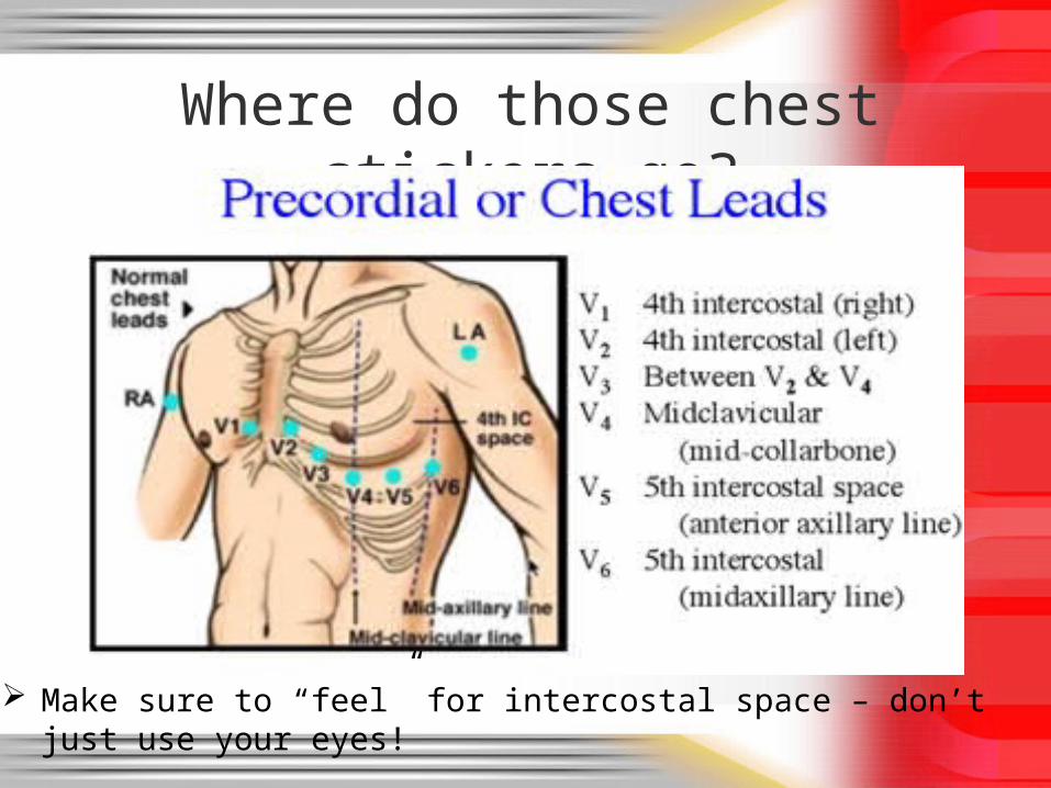
Where do those chest stickers go?
Make sure to “feel” for intercostal space – don’t just use your eyes!

……and the FEMALES
• Bras loosened, people, if in your way
• Not all nipple lines are created equal
• Cover the chest with a towel/sheet after leads are placed to preserve modesty
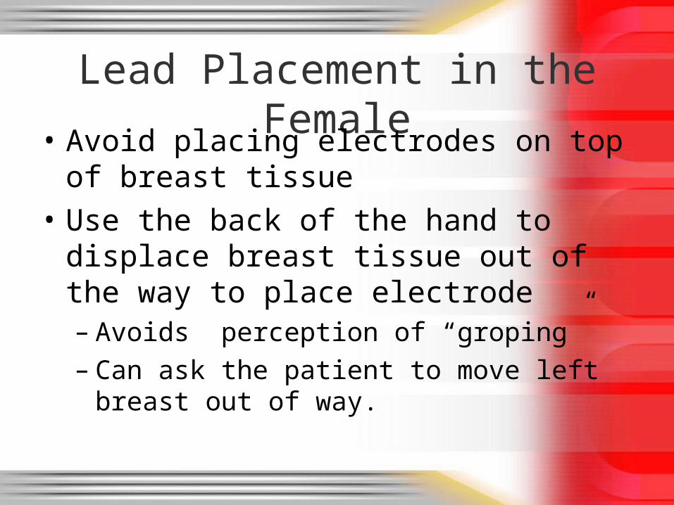
Lead Placement in the Female
• Avoid placing electrodes on top of breast tissue
• Use the back of the hand to displace breast tissue out of the way to place electrode– Avoids perception of “groping”– Can ask the patient to move left breast out of
way.
Lead Placement• The more accurate the lead placement, the
more accurate the 12-lead interpretation• 12-leads are often evaluated on a sequential
basis, each interpretation considering the previous one
• V4 - V6 should be in a gentle upward curve following the same 5th intercostal rib space
• CMC has done many trainings– We should be doing this right by now– Placement was standardized in 1938; this is
proven science and placement must be accurate!
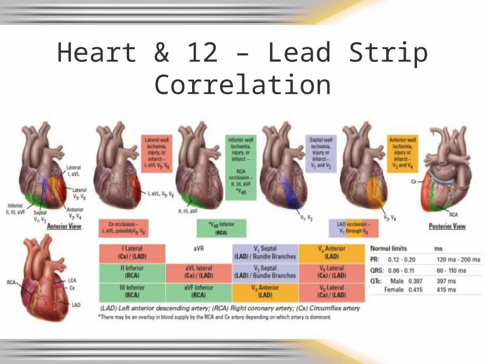
Heart & 12 – Lead StripCorrelation
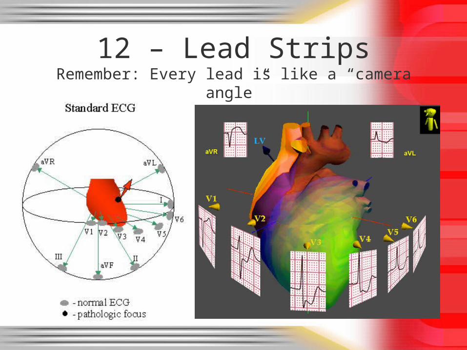
12 – Lead StripsRemember: Every lead is like a “camera angle”
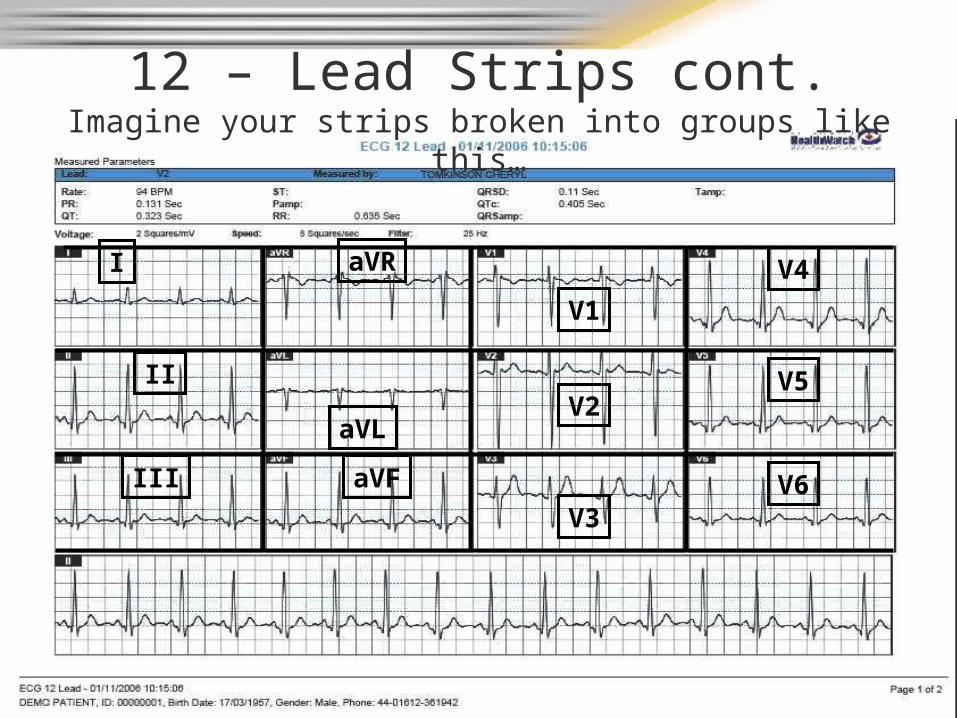
12 – Lead Strips cont.Imagine your strips broken into groups like this…
I
II
III
aVL
aVF
V1
V2
V3
V4
V5
V6
aVR
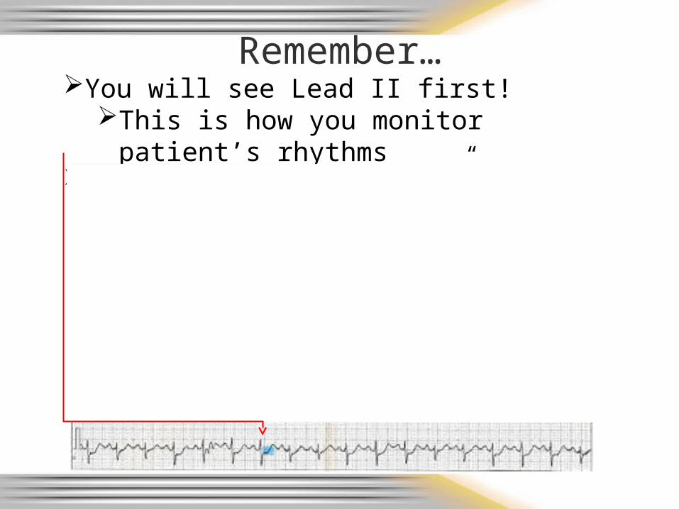
Remember…You will see Lead II first!
This is how you monitor patient’s rhythmsMay see “reciprocal changes” as ST depression
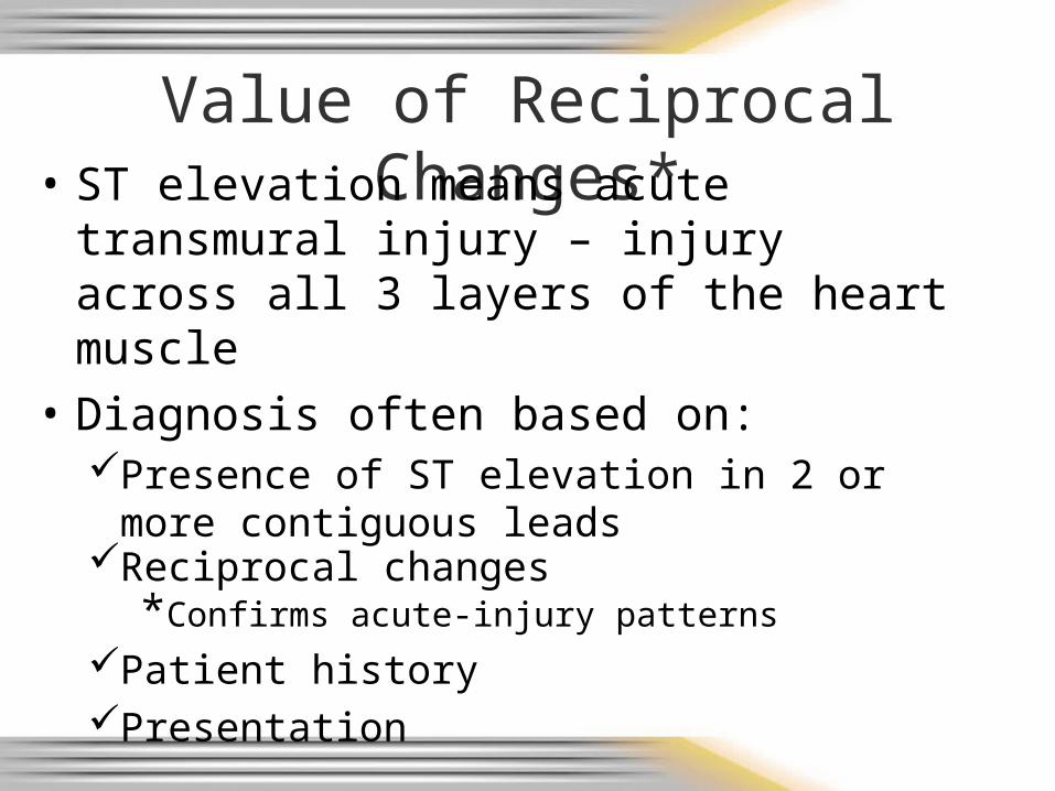
Value of Reciprocal Changes*• ST elevation means acute transmural
injury – injury across all 3 layers of the heart muscle
• Diagnosis often based on:Presence of ST elevation in 2 or more
contiguous leadsReciprocal changes
*Confirms acute-injury patterns
Patient historyPresentation
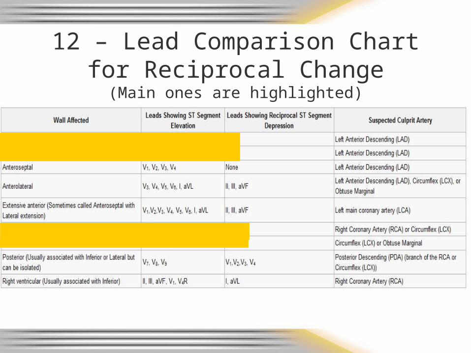
12 – Lead Comparison Chart for Reciprocal Change(Main ones are highlighted)
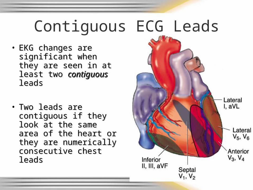
Contiguous ECG Leads• EKG changes are EKG changes are
significant when they significant when they are seen in at least two are seen in at least two contiguouscontiguous leads leads
• Two leads are Two leads are contiguous if they look contiguous if they look at the same area of the at the same area of the heart or they are heart or they are numerically numerically consecutive chest consecutive chest leadsleads
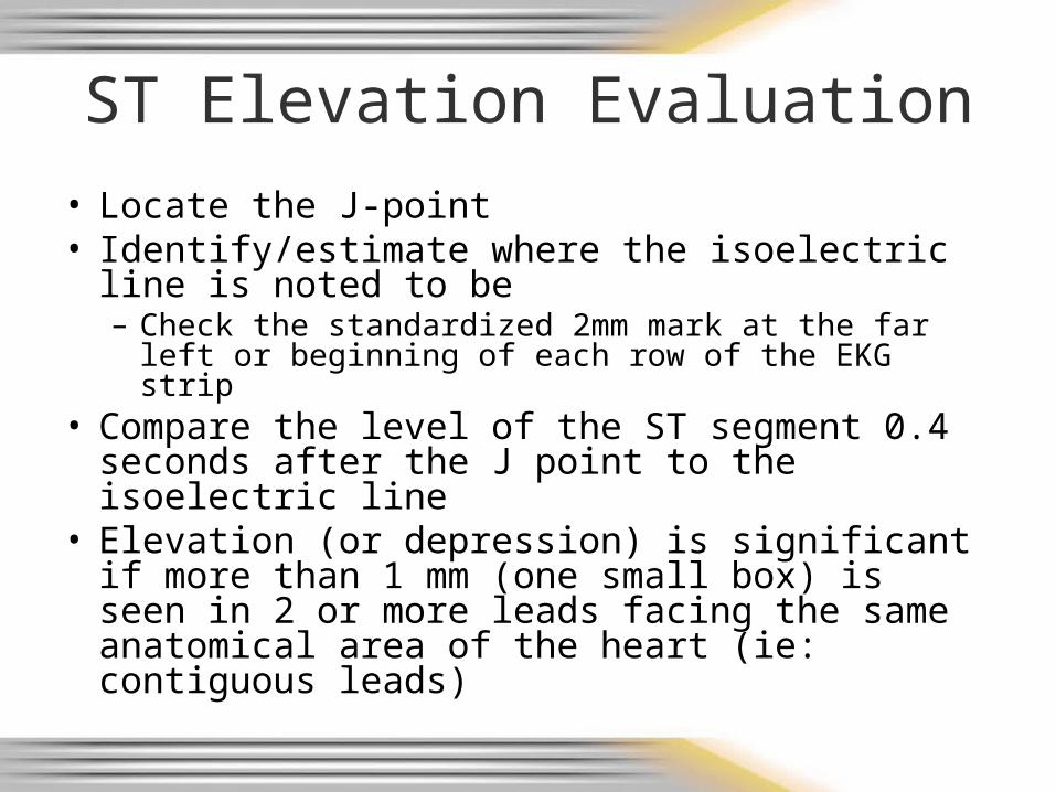
ST Elevation Evaluation
• Locate the J-point• Identify/estimate where the isoelectric line is
noted to be– Check the standardized 2mm mark at the far left or
beginning of each row of the EKG strip• Compare the level of the ST segment 0.4
seconds after the J point to the isoelectric line• Elevation (or depression) is significant if more
than 1 mm (one small box) is seen in 2 or more leads facing the same anatomical area of the heart (ie: contiguous leads)
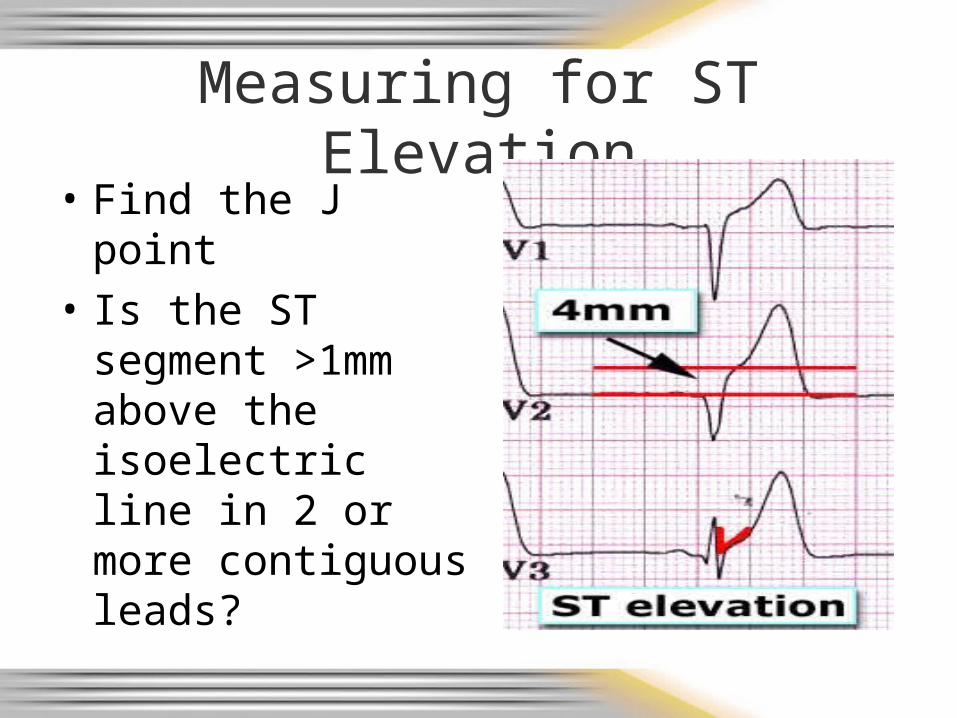
Measuring for ST Elevation
• Find the J point
• Is the ST segment >1mm above the isoelectric line in 2 or more contiguous leads?
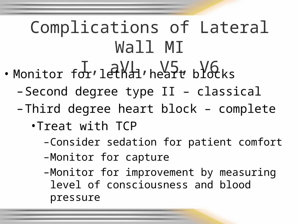
Complications of Lateral Wall MI I, aVL, V5, V6
• Monitor for lethal heart blocks
– Second degree type II – classical
– Third degree heart block – complete
• Treat with TCP–Consider sedation for patient comfort–Monitor for capture–Monitor for improvement by measuring level
of consciousness and blood pressure
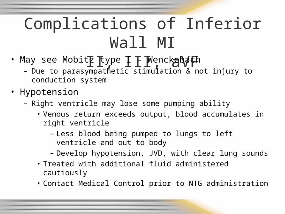
Complications of Inferior Wall MIII, III, aVF
• May see Mobitz type I – Wenckebach– Due to parasympathetic stimulation & not injury to conduction
system
• Hypotension– Right ventricle may lose some pumping ability
• Venous return exceeds output, blood accumulates in right ventricle
– Less blood being pumped to lungs to left ventricle and out to body
– Develop hypotension, JVD, with clear lung sounds• Treated with additional fluid administered cautiously• Contact Medical Control prior to NTG administration
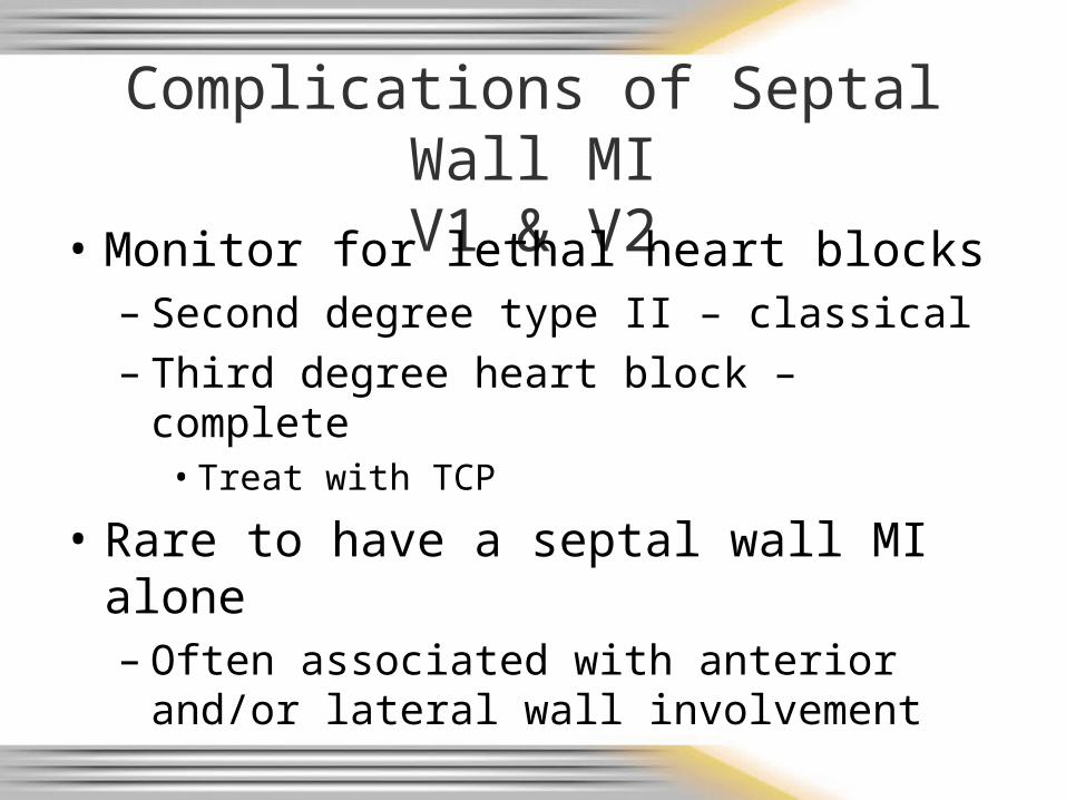
Complications of Septal Wall MIV1 & V2
• Monitor for lethal heart blocks– Second degree type II – classical– Third degree heart block – complete
• Treat with TCP
• Rare to have a septal wall MI alone– Often associated with anterior and/or lateral
wall involvement

Complications of Anterior Wall MIV3 & V4
• Occlusion of left main coronary artery – the “widow maker”– Cardiogenic shock and death without prompt reperfusion
• Second degree AV block type II– Often symptomatic– Often progress to 3rd degree heart block– Prepare to initiate TCP
• Third degree heart block – complete– Rhythm usually unstable– Rate usually less than 40 beats per minute– Prepare to initiate TCP
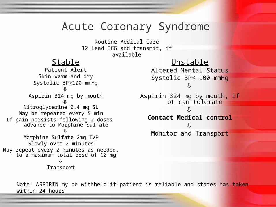
Acute Coronary Syndrome
StablePatient Alert
Skin warm and drySystolic BP>100 mmHg
Aspirin 324 mg by mouth
Nitroglycerine 0.4 mg SL
May be repeated every 5 minIf pain persists following 2 doses, advance to
Morphine Sulfate
Morphine Sulfate 2mg IVPSlowly over 2 minutes
May repeat every 2 minutes as needed, to a maximum total dose of 10 mg
Transport
UnstableAltered Mental Status
Systolic BP< 100 mmHg
Aspirin 324 mg by mouth, if pt can tolerate
Contact Medical control
Monitor and Transport
Note: ASPIRIN my be withheld if patient is reliable and states has taken within 24 hours
Routine Medical Care12 Lead ECG and transmit, if available
Patient Presenting with Coronary Chest Pain – AMI Until Proven Otherwise
• Oxygen– May limit ischemic injury– New trends/guidelines coming out in 2011 SOP’s
• Aspirin - 324 mg chewed (PO)– Blocks platelet aggregation (clumping) to keep
clot from getting bigger– Chewing breaks medication down faster & allows
for quicker absorption– Hold if patient allergic or for a reliable patient that
states they have taken aspirin within last 24 hours
• Nitroglycerin - 0.4 mg SL every 5 minutes– Dilates coronary vessels to relieve vasospams– Increases collateral blood flow– Dilates veins to reduce preload to reduce
workload of heart• Watch for hypotension• If inferior wall MI (II, III, aVF), contact Medical
Control prior to administration– If pain persists after 2 doses, move to Morphine– Check for recent male enhancement drug use
(ie: viagra, cialis, levitra)• Side effect could be lethal hypotension
Acute Coronary SyndromeMedications cont.
Acute Coronary SyndromeMedications cont.
• Morphine - 2 mg slow IVP– Decreases pain & apprehension– Mild venodilator & arterial dilator
• Reduces preload and afterload
– Given if pain level not changed after the 2nd dose of nitroglycerin
– Give 2mg slow IVP repeated every 2 minutes as needed
– Max total dose 10 mg
Practice Rhythms• Break into groups of 2 or 3 for rhythm quizzing• Instructor will use wristwatch or stopwatch to
give each group 30 seconds to determine strip– You don’t have 5 min in the field, you don’t get 5 min
in the classroom• Message to Instructor:
– Refer to slide notes for more information
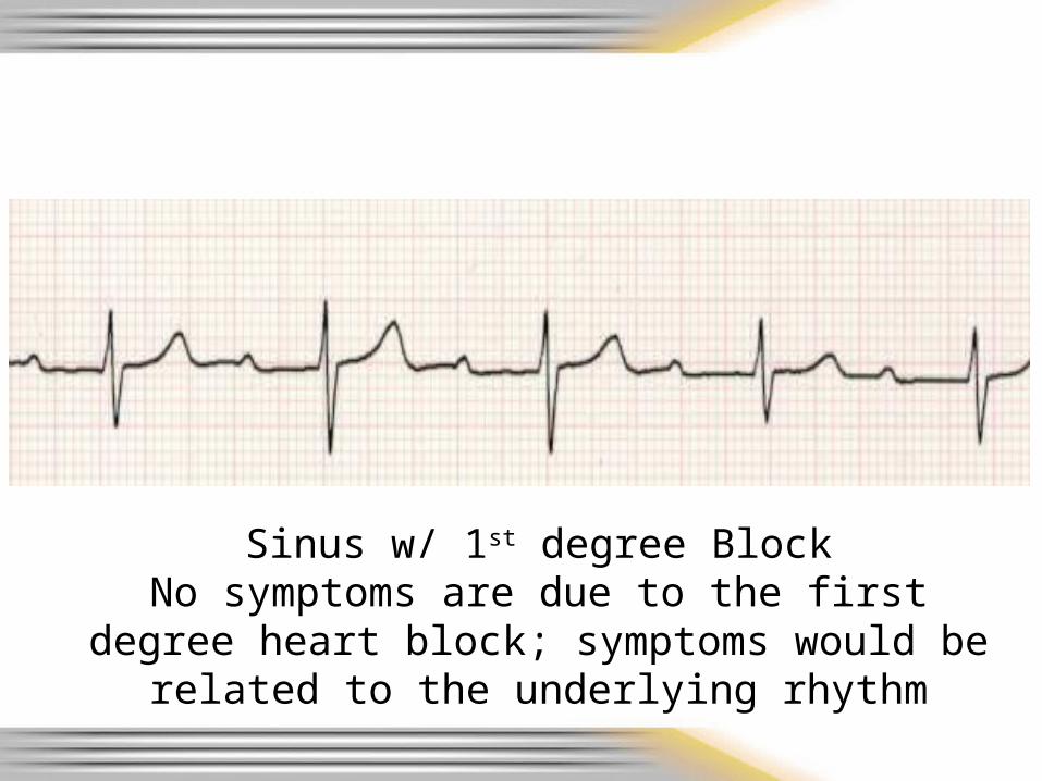
Sinus w/ 1st degree BlockNo symptoms are due to the first degree heart
block; symptoms would be related to the underlying rhythm
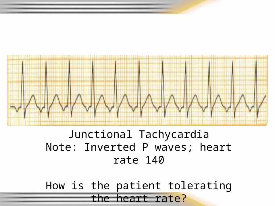
Junctional TachycardiaNote: Inverted P waves; heart rate 140
How is the patient tolerating the heart rate?
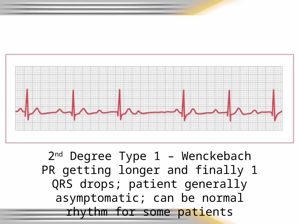
2nd Degree Type 1 – WenckebachPR getting longer and finally 1 QRS drops;
patient generally asymptomatic; can be normal rhythm for some patients
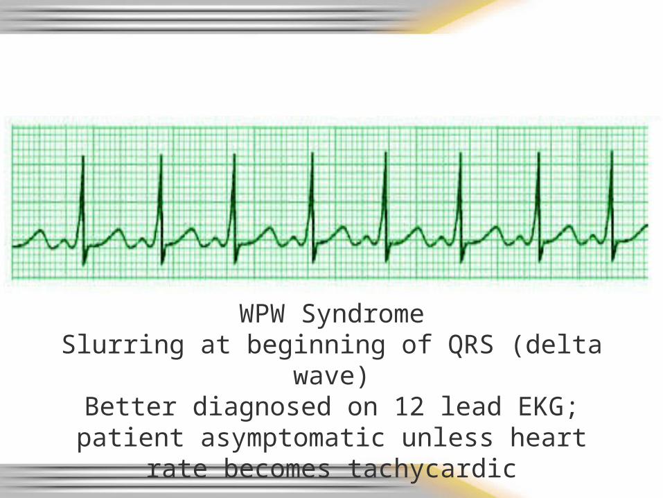
WPW SyndromeSlurring at beginning of QRS (delta wave)Better diagnosed on 12 lead EKG; patient asymptomatic unless heart rate becomes
tachycardic
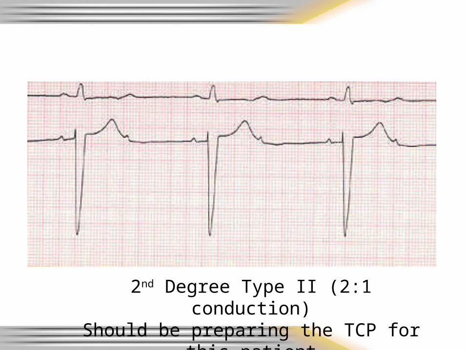
2nd Degree Type II (2:1 conduction)Should be preparing the TCP for this patient
3rd degree heart block (complete)with narrow QRS
Symptoms usually based on overall heart rate – the slower the heart rate the more
symptomatic the patient. Prepare the TCP.
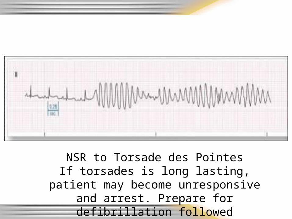
NSR to Torsade des PointesIf torsades is long lasting, patient may
become unresponsive and arrest. Prepare for defibrillation followed immediately with CPR
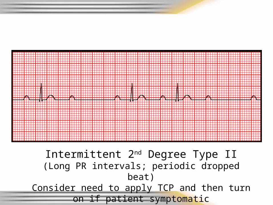
Intermittent 2nd Degree Type II(Long PR intervals; periodic dropped beat)
Consider need to apply TCP and then turn on if patient symptomatic
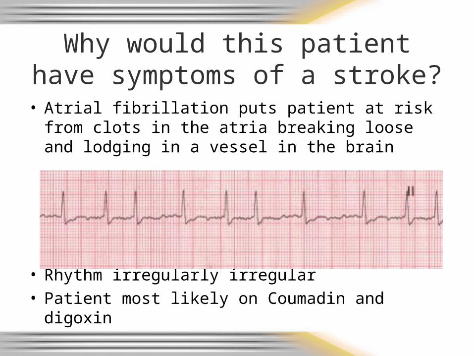
Why would this patient have symptoms of a stroke?
• Atrial fibrillation puts patient at risk from clots in the atria breaking loose and lodging in a vessel in the brain
• Rhythm irregularly irregular• Patient most likely on Coumadin and digoxin
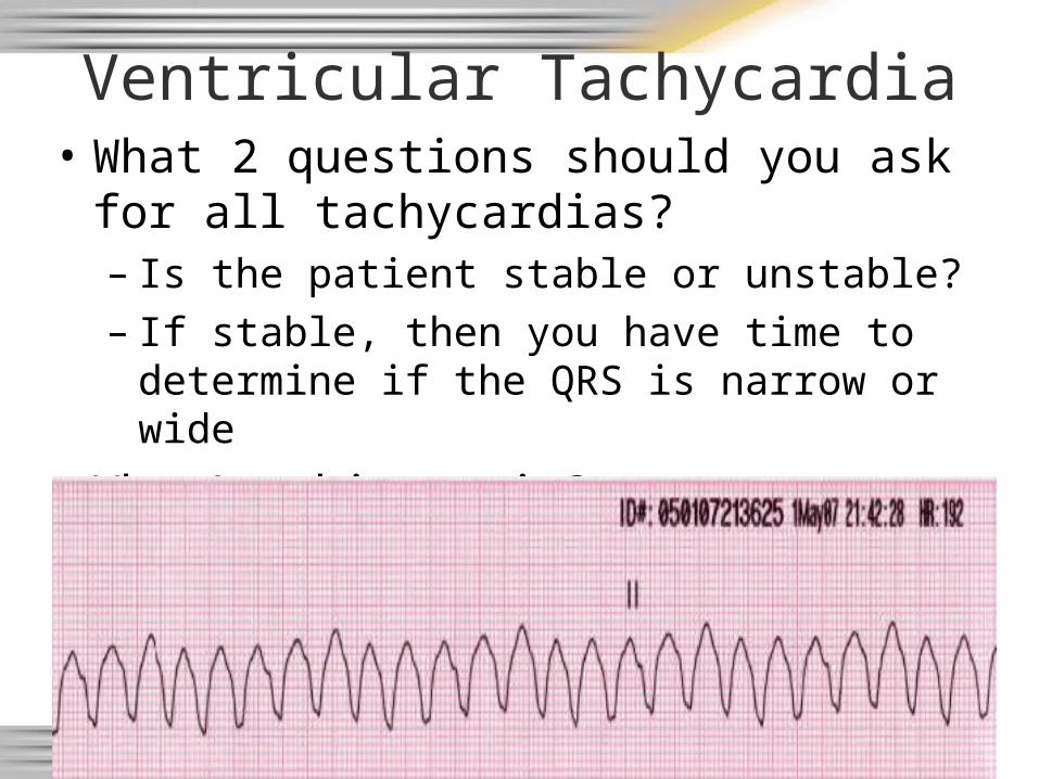
Ventricular Tachycardia• What 2 questions should you ask for all
tachycardias?– Is the patient stable or unstable?– If stable, then you have time to determine if
the QRS is narrow or wide
• What’s this strip?
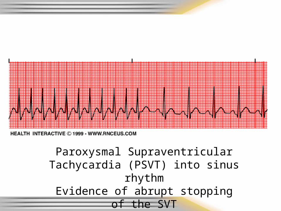
Paroxysmal Supraventricular Tachycardia (PSVT) into sinus rhythm
Evidence of abrupt stopping of the SVT
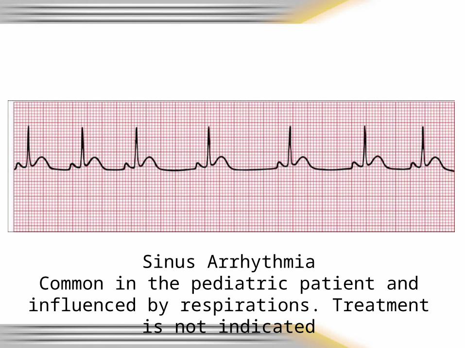
Sinus ArrhythmiaCommon in the pediatric patient and influenced by
respirations. Treatment is not indicated
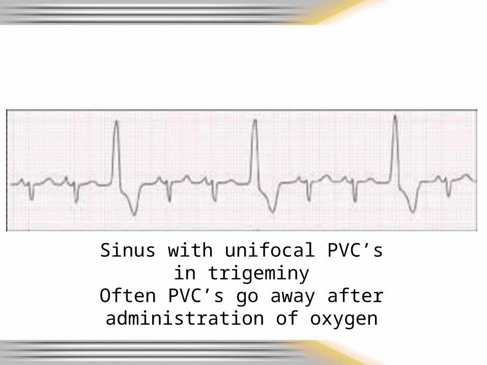
Sinus with unifocal PVC’sin trigeminy
Often PVC’s go away after administration of oxygen
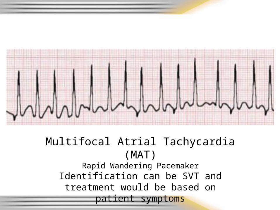
Multifocal Atrial Tachycardia(MAT)
Rapid Wandering Pacemaker
Identification can be SVT and treatment would be based on patient symptoms

12 – Lead Time!
• Same as Lead II strips– Identify St elevation and try to give anatomical
locations– Remember to be watchful for typical
complications based on location of infarct and blocked coronary vessel
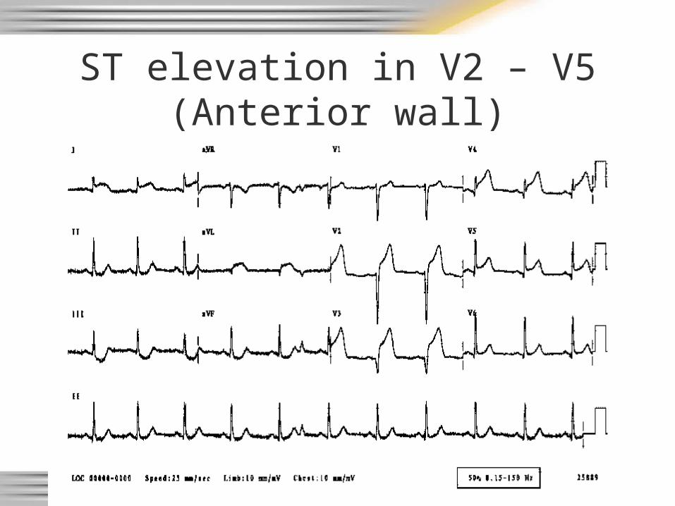
ST elevation in V2 – V5(Anterior wall)
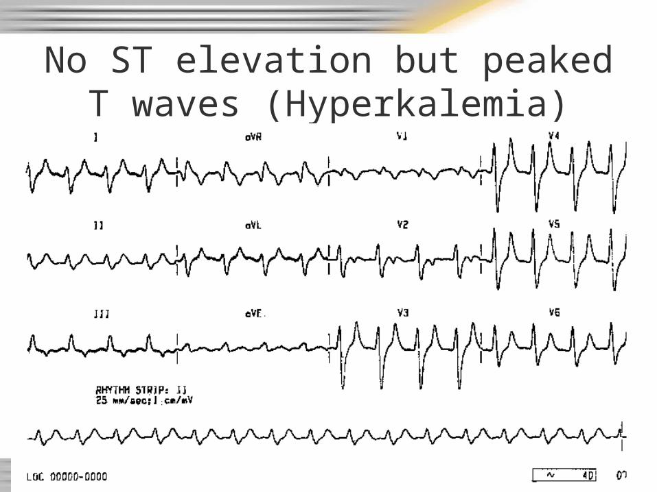
No ST elevation but peaked T waves (Hyperkalemia)
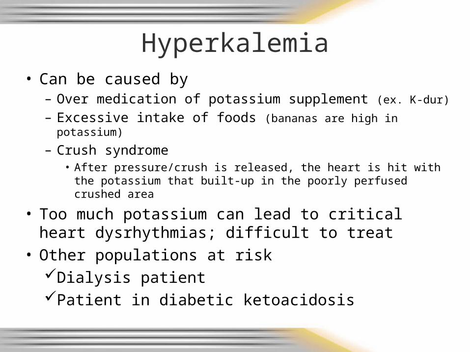
Hyperkalemia• Can be caused by
– Over medication of potassium supplement (ex. K-dur)
– Excessive intake of foods (bananas are high in potassium)
– Crush syndrome• After pressure/crush is released, the heart is hit with the
potassium that built-up in the poorly perfused crushed area
• Too much potassium can lead to critical heart dysrhythmias; difficult to treat
• Other populations at riskDialysis patientPatient in diabetic ketoacidosis
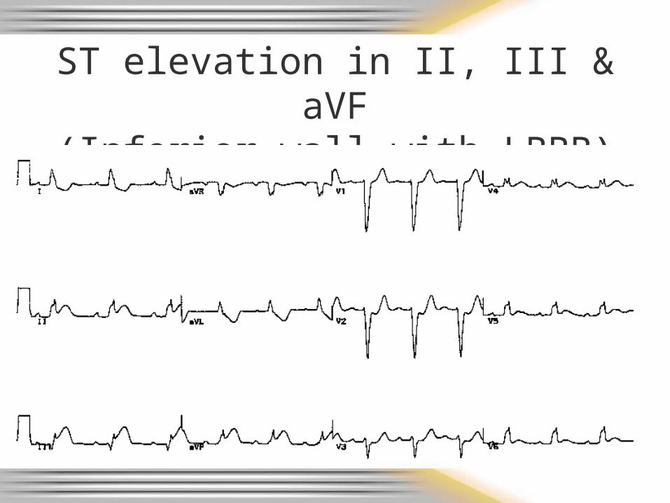
ST elevation in II, III & aVF(Inferior wall with LBBB)
Watch for hypotension
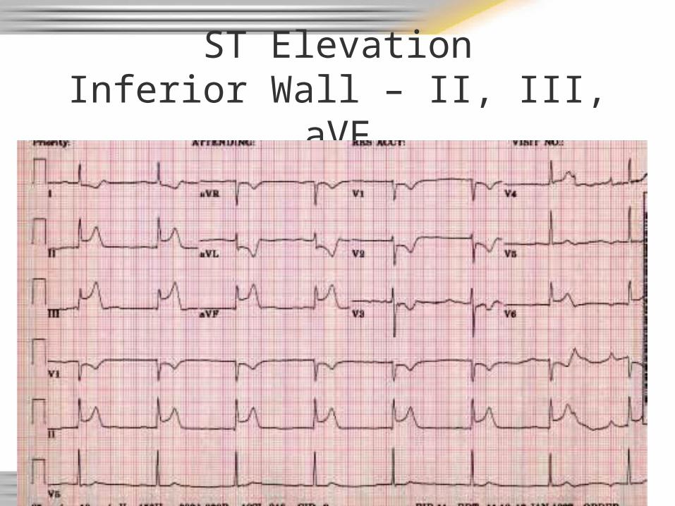
ST ElevationInferior Wall – II, III, aVF
Watch for hypotension

ST elevation in II, III, aVF(Inferior wall - note reciprocal changes)
Watch for hypotension
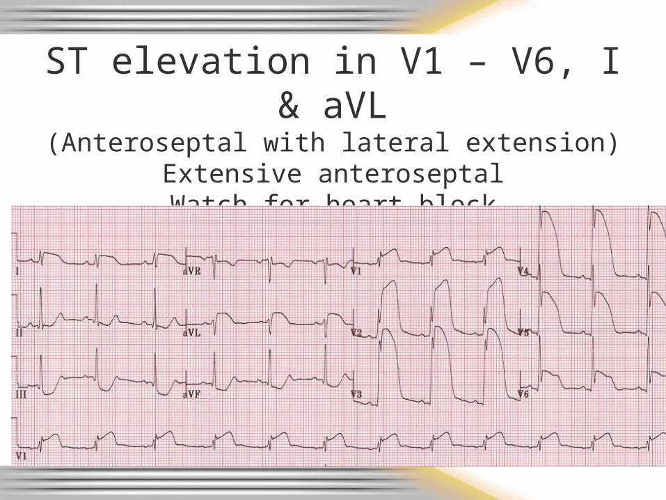
ST elevation in V1 – V6, I & aVL(Anteroseptal with lateral extension)
Extensive anteroseptalWatch for heart block
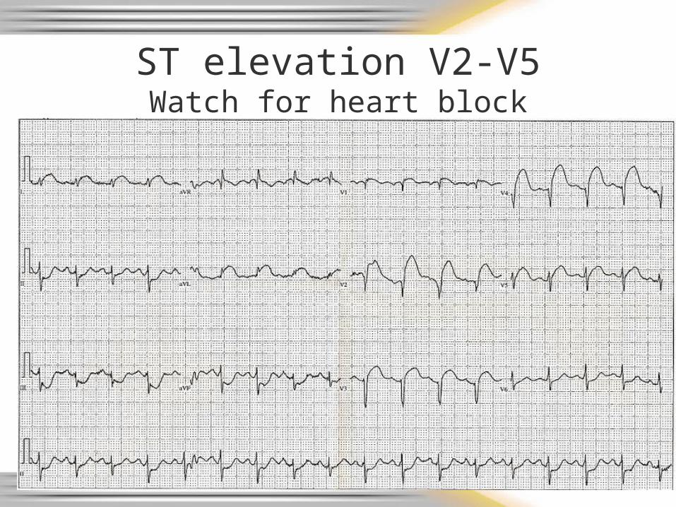
ST elevation V2-V5Watch for heart block
Documentation
• Rhythm strip interpretation
• If 12 lead EKG obtained:Note presence or absence of ST elevationIf ST elevation noted, in which leadsIf EKG was transmitted to hospital
Hospital Notification
• Notify the receiving hospital as soon as possible about a cardiac alert
• How did you determine this may be a cardiac alert?– Your general impression was made based on:
Gathering patient historyPerforming a cardiac assessmentObtaining a 12 lead EKG as quickly as possible
after first patient contact12 lead EKG evaluated for presence of ST
elevation
Field Trip• If your department can obtain
12 lead EKG’s review the process for marking – Patient age and race– Your department name on the 12 lead
• If your department can fax, review the process– Go to the ambulance to review the equipment
and process
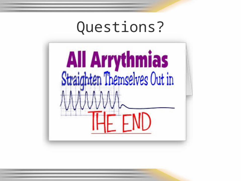
Questions?
Bibliography• Atwood, S., Stanton, C., Storey-Davenport, J. Introduction to
Basic Cardiac Dysrhythmia 3rd Edition. MosbyJems. 2003.• Bledsoe, B., Porter, R., Cherry, R. Paramedic Care Principles
& Practices Third Edition. Brady. 2009. • Page, B. 12 Lead ECG for Acute and Critical Care Providers.
Brady. 2005.• Previous CMC Cardiac CEs• Region X SOP March 2007; amended January 1, 2008• Various webpages
– For pictures, rhythms, and graphs
• Walraven, G. Basic Arrhythmia 7th Edition. Brady. 2011.
• www.MikeCowley.co.uk/leads.htm