Embed Size (px)
Citation preview
INFECTION AND IMMUNITY, Mar. 1973, p. 487-492Copyright © 1973 American Society for Microbiology
Vol. 7, No. 3P'rinted in U.S.A.
Immunosorbent for the Isolation of SpecificAntibodies Against Mannan: Localization of
Antigens in Yeast Cell WallsH. GERBER, M. HORISBERGER, AND H. BAUER
Research and Development Department, Nestle Products Technical Assistance Ltd.,1800 Vevey, Switzerland
Received for publication 29 August 1972
A procedure is giveii for the insolubilizationi of yeast maiiiaii by entrapment ina polyacrylamide gel. Specific anti-mannan antibodies were separated by immu-nosorption from all other aiitibodies of a rabbit antiserum against whole, Formalin-inactivated Candida utilis cells. Colloidal gold coated with the anti-mannlan andanti-non-mannan antibody fractions was used for the ultrastructural visualizationof cell wall antigens. A control was performed using normal rabbit serum. Inhibi-tioIn of anti-mannan antibodies was obtained with mannail oligosaccharides, andnone of the antibody fractions showed marking of Saccharomyces cerevisiae cell walls.
Techniques using insolubilized antigens havethe advantage over conventional absorptionmethods that antibodies free of serum compo-nents and antigens can be obtained; frequently aninsoluble antigen can be reused many times.Insolubilization of proteins by immobilization inthe lattice of polyacrylamide gels has been de-scribed (e.g., 3, 4, 12). In the present paper wedescribe the extension of this method to the in-solubilization of mannan, a neutral solublepolysaccharide. Colloidal gold coated with specificanti-mannan antibody prepared by this tech-nique was successfully used for marking isolatedCandida utilis cell walls.
MATERIALS AND METHODSChemicals. Acrylamide, N,N'-methylene bis-
acrylamide (bis) and N,N,N',N'-tetramethylene-diamine (TEMED) were from Serva, Heidelberg,Germany. Tris (hydroxymethyl)aminomethane(Tris) was from Merck, Darmstadt, Germany. Allother chemicals were purchased from Fluka AG,Buchs, Switzerland. The reagents were of analyti-cal purity and were used without further purifica-tion.The phosphate-buffered saline, pH 7.2 (PBS),
was made by dissolving NaCl (36.0 g), anhydrousNa2HPO4 (7.4 g), and anhydrous KH2PO4 (2.15 g)in water up to 5 liters.Preparation of the immunosorbent. The
method published by Carrel et al. (4) was modifiedas follows. In a 12- by 100-mm test tube, 1 ml ofmannan solutionl (50 mg of mannan in 1 ml ofwater) was mixed with 1 ml of buffer (0.05 M Tris-hydrochloride containing 0.20 ml of TEMED/100 ml, final pH 7.5) and 1 ml of monomer solution(2.40 g of acrylamide and 0.80 g of bis dissolved in
water to 10 ml). Finally, 1 ml of a freshly preparedcatalyst solution (0.14 g of ammonium persulfatein 50 ml of water) was added, and the mixturewas left undisturbed at room temperature (20-25 C). The white, nontransparent polyacrylamidegel formed within 30 min after addition of thepersulfate and was left 18 to 24 h to allow polymeri-zation to go to completion.The gel block was granulated by forcing it suc-
cessively through a 20- and 40-mesh stainless steelsieve with a spatula. The particles retained on an80-mesh sieve were filled to a 9-by 150-mm chroma-tography column (Pharmacia, Sweden) on top of a5-mm layer of glass beads (1 mm diameter).The immunosorbent was then precycled at 5 C
by washing at a flow rate of 10 to 20 ml/h withTris-buffered saline (0.05M Tris-hydrochloride -0.15 M NaCl, pH 7.5) and acid buffer (0.2 M gly-cine-hydrochloride, pH 2.3) until the effluentsshowed no optical absorbancy at 280 nm and werefree of mannan as compared to the eluting buffers.Finally, the gel was neutralized to pH 7.5 bywashing with Tris-buffered saline.
Isolation of anti-mannan antibodies. A4-ml portion of rabbit antiserum against C. utiliswas pumped through the precycled immuno-sorbent at a flow rate of 10 ml/h, and the unre-tained material was recovered by washing withTris-buffered saline until the optical density of theefflent at 280 nm returned to the value of thebuffer.The bounid ainti-manniian anitibodies were eluted
with the acid buffer at a flow rate of 10 to 20 ml/h.The fractions absorbing at 280 nm were pooled andimmediately adjusted to pH 7.5 with 2M Tris.The effluents collected at neutral and acid pH
were reconcentrated to the original volume of 4ml by pressure dialysis against Tris-buffered
487
on May 27, 2019 by guest
http://iai.asm.org/
Dow
nloaded from

4GERBER, HORISBEl{GER., ANI) BAUER I
saline. All manipulations were carried out at 5 C.The immunosorbent could be reused after washingwith Tris-buffered saline.Preparation of anti-C. utilis antiserum.
Two rabbits (2.5-3 kg) were immunized with asuspension (1.6 X 10⁸ cells per ml) of heat-inacti-vated (15 min at 70 C) C. utilis cells in physio-logical saline without adjuvant. Each rabbit wasgiven six injections spaced over 1 month of 1.5 to5 ml of suspension per injection by the intravenousor intravenous and subcutaneous route. Theanimals were bled by heart puncture and the serawere lyophilized.Agglutination assay. Samples (0.5 ml) of
double dilutions of antiserum (starting at 1:10dilution) were mixed with 0.5-ml portions of asuspension of Formalin-inactivated C. utilis cells(1.4 X 10⁸ cells per ml). Agglutination readingswere taken after 6 and 20 h of incubation at 50 C(Table 1).Double diffusion assay in agarose. Wells
(6.4-mm diameter) in a 1.5-mm thick layer ofagarose (1% in 0.05 M barbital buffer, pH 8.3)were filled with antisera or mannan solution (0.25mg of mannan per ml). Photographs were takenafter 22 h of diffusion at 18 to 20 C.Electrophoresis on cellulose acetate. The
Beckman Microzone Electrophoresis System(model R-101) was used.Electrophoresis was carried out in barbital
buffer, pH 8.6, g 0.075, at 250 V for 30 min. Theproteins were stained with Ponceau BS.Sugar analysis. Total sugars were estimated
by the phenol-sulfuric acid method (6).The amount of mannan retained in the poly-
acrylamide gel was determined either indirectlyfrom the difference between added or unboundmannan or by measuring mannan directly in thegel, or both. For the latter, precycled immuno-sorbent was first centrifuged (1,000 X g for 5 min)on a filter to remove liquid between the gel parti-cles. Samples (10-20 mg) of this gel were sus-pended in 1 ml of Tris-buffered saline and werereacted with phenol-sulfuric acid as with liquidsamples. Controls with buffers and polyacryla-mide gel without mannan were included.The sugars present in mannan after acid hy-
drolysis (2 N H2SO4, 100 C, 4 h) were quantita-tively determined as their alditol acetates by gasliquid chromatography at 210 C using a glass
TABLE 1. Agglutination of C. utilis cells by nativeantiserum against C. utilis, neutral and acideluates from mannan immunosorbent, and normal
rabbit serum
Sample Agglutina-tion titer
Native anti-C. utilis antiserum........ 1:320Neutral eluate (pH 7.5) ............... 1:80Acid eluate (pH 2.3) .................. 1:160Normal rabbit serum................. <1:10
column (0.3 by 200 cm) containing 3% (wt/wt)OV-225 (Applied Science Labs, State College,Pa.) on Gas-Chrom Q (100-120 mesh) (10).Determination of anti-mannan antibodies.
Samples (0.1 ml) of antisera were incubated at37 C for 30 min with optimal proportions of man-nan (15 ug) and left 24 to 48 h at 5 C. The washedprecipitates were dissolved in 0.1 M NaOH (1 ml),and the protein content was estimated spectro-photometrically at 280 nm, using an Elcm17 of 13.0for rabbit immunoglobulin.
Isolation of yeast cell walls. C. utilis CBS567 was grown at 30 C for 24 h in a 2% sucrosemedium supplemented with 0.3% yeast extract(Difco, Detroit, Mich.) and mineral salts as inor-ganic nitrogen source.
Cell walls were prepared from pressed baker'syeast and from fresh cells of C. utilis by themethod of Mill (11). They were judged free fromcytoplasmic contamination by light and electronmicroscope examination.
Isolation of mannan. Mannan from C. utiliswas prepared as described by Peat et al. (13) andwas purified by three precipitations with Fehlingsolution. The mannan contained mannose andglucose in the molar ratio 4:1. The mannan com-position was found unchanged by further precipi-tation with Fehling solution. The mannan had[a]f + 100° (water, c 1.2).Antibody labeling. Antibody fractions were
adsorbed onto colloidal gold by the procedure ofFaulk and Taylor (7) with one modification. Toprevent adsorption of the colloid onto the walls ofthe polyallomer centrifuge tubes, the colloid wascentrifuged twice in PBS containing 0.2% Tween80. This detergent has been shown not to interferewith antigen-antibody interaction (5). Prior tolabeling, the protein content of the anti-mannanantibody fraction was adjusted with normalrabbit serum to the protein level of the originalantiserum.Marking of yeast cell walls with antiserum-
coated colloidal gold. Yeast cell walls weresuspended in PBS to a final optical absorbancyof 1.0 at 420 nm. The antiserum-coated colloidalgold (0.2 ml) and PBS (0.1 ml) were added to thecell wall suspension (0.2 ml). After incubating at25 C for 2 h with gentle shaking, the suspensionwas centrifuged at 2,800 X g for 5 min, washedthree times with PBS (5 ml), and finally suspendedin the same buffer (0.3 ml).Inhibition studies. A mixture of oligosaccha-
rides from C. utilis mannan was prepared accord-ing to Kocourek and Ballou (9). C. utilis cell wallswere marked as described above with colloidalgold coated anti-mannan antibodies in the pres-ence of 1.2, 0.12, and 0.012 mg of oligosaccharidesin the incubation mixture (0.5 ml).Preparation for electron microscopy. A
drop of the prelabeled cell wall suspension wasplaced on a Formvar-carbon-coated grid. After afew minutes, the excess material was removed witha micropipette or a filter paper. The grids weredried in a vacuum desiccator overnight andexamined in a Philips EM300 electron microscope.
488 INFECT. IMMUNITY
on May 27, 2019 by guest
http://iai.asm.org/
Dow
nloaded from
VOL. 7, 1973 ISOLATION OF SPECIFIC ANTIBODIES AGAINST MANNAN
RESULTS AND DISCUSSION
Entrapment of mannan in polyacrylamidegel. Mannan analysis by the direct or indirect.method showed that 9.2 to 10.7 mg of mannanwere retained in the granulate from a 4-ml gelblock. This corresponds to 20% of the totalamount of mannan added to the monomer mix-ture.
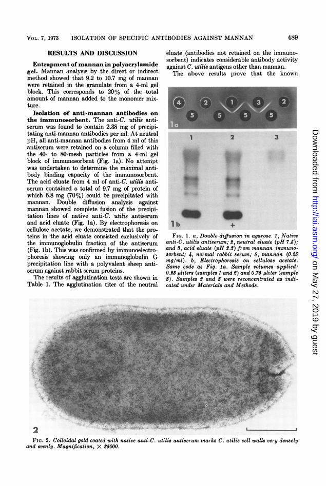
Isolation of anti-mannan antibodies onthe immunosorbent. The anti-C. utilis anti-serum was found to contain 2.38 mg of precipi-tating anti-mannan antibodies per ml. At neutralpH, all anti-mannan antibodies from 4 ml of thisantiserum were retained on a column filled withthe 40- to 80-mesh particles from a 4-ml gelblock of immunosorbent (Fig. la). No attemnptwas undertaken to determine the maximal anti-body binding capacity of the immunosorbent.The acid eluate from 4 ml of anti-C. utilis anti-serum contained a total of 9.7 mg of protein ofwhich 6.8 mg (70%) could be precipitated withmannan. Double diffusion analysis againstmannan showed complete fusion of the precipi-tation lines of native anti-C. utilis antiserumand acid eluate (Fig. la). By electrophoresis oncellulose acetate, we demonstrated that the pro-teins in the acid eluate consisted exclusively ofthe immunoglobulin fraction of the antiserum(Fig. lb). This was confirmed by immunoelectro-phoresis showing only an immunoglobulin Gprecipitation line with a polyvalent sheep anti-serum against rabbit serum proteins.The results of agglutination tests are shown in
Table 1. The agglutination titer of the neutral
2t, 5s
eluate (antibodies not retained on the immuno-sorbent) indicates considerable antibody activityagainst C. utilis antigeIns other than mannan.The above results prove that the known
1 2 3
m+
FIG. 1. a, Double diffusion in agarose. 1, Nativeanti-C. utilis antiserum; £, neutral eluate (pH 7.5);and 3, acid eluate (pH 2.3) from mannan immuno-8orbent; 4, normal rabbit serum; 5, mannan (0.25mg/ml). b, Electrophoresis on cellulose acetate.Same code as Fig. la. Sample volumes applied:0.25 uliters (samples 1 and 2) and 0.75 ;liter (sample3). Samples 2 and 3 were reconcentrated as indi-cated under Materials and Methods.
l b
FIG. 2. Colloidal gold coated with native anti-C. utilis antiserum marks C. utilis cell walls very denselyand evenly. Magnification, X £5000.
489
L- I
on May 27, 2019 by guest
http://iai.asm.org/
Dow
nloaded from

GERIBER,, HOR6ISBEIRC;GEItl1, AND BAUELlk,
g
I' %
3bJV_3cbm lt_
INFECT. IMMUNITY
A4A
' -T $ _
4v
'4 "t.
4S4oCtW,
Nt4 ..1
3*;:
490
3d
3e.!-. ., ";., I',."
I ;. "i, " til -. ....." W. "' .*' a... zI.eM21. . I i- :..
II
on May 27, 2019 by guest
http://iai.asm.org/
Dow
nloaded from

VOL. 7, 1973 ISOLATION OF SPECIFIC ANTIBODIES AGAINST MANNAN
method for insolubilization of protein antigens byentrapment into polyacrylamide gel can success-fully be extended to neutral polysaccharides likemannan. On the other hand, the use of an im-munosorbent technique in the present studyal4pwed the simple and clear-cut separation ofan ibodies directed against mannan and non-mannan antigenic determinants.Electron microscopy. A whole C. utilis cell
wall marked with colloidal gold coated withnative anti-C. utilis antiserum is seen in Fig. 2.The marking is dense and homogenous. Differentdensities of marking can be recognized in thesequence of electron micrographs represented inFig. 3a-f. The highest density is obtained withthe native anti-C. utilis antiserum (Fig. 3a).A weaker but still important and random mark-ing can be observed with the neutral eluate fromthe immunosorbent (anti-nonmannan anti-bodies; Fig. 3b). An almost identical density anddistribution of gold granules is observed with thespecific anti-mannan antibodies (acid eluate;Fig. 3c). The two dense lines of gold particles(arrows) correspond to the region of the bud scar,indicating a certain accumulation of mannan atthis site. A practically total inhibition of markingcan be achieved by the addition of oligosaccha-rides to the anti-mannan antibody fraction (Fig.3d). This is in agreement with the fact that sidechain oligosaccharides resulting from acetolysisof various yeast mannans strongly inhibit theantiserum-mannan precipitin reaction (1, 14, 15).Total inhibition was achieved with the highestconcentration of oligosaccharides of C. utilismannan (1.2 mg), partial inhibition at lowerconcentration.The weak marking obtained with normal
rabbit serum must be due to nonspecific adsorp-tion of the colloidal gold (Fig. 3e). The sameweak, nonspecific adsorption is observed withcolloidal gold coated with native anti-C. utilisantiserum when used against Saccharomycescerevisiae cell walls (Fig. 3f).The results of the marking experiments only
indicate a random distribution of the mannan atthe cell wall surface. To determine the location ofthis polysaccharide within the cell wall, thetechnique of thin sectioning has to be applied(studies are being undertaken in our laboratory).
Nevertheless, the results obtained with thespecific anti-mannan antibodies confirm the ob-servations with mercury-labeled concanavalini A(8), i.e., an even distribution of the mannan atthe cell wall surface. The dense marking of thebud scar region (accumulation of ma%nan) is inexcellent agreement with concanavalin A markingexperiments of isolated bud scars of S. cerevi8iae(2).
ACKNOWLEDGMENTS
We thank H. Hilpert for the antiserum against, Cutilis, D. A. Bush for preparing the yeast cell walls,and M. Weber for photographic work.
LITERATURE CITED
1. Ballou, C. E. 1970. A study of the immunochem-istry of three yeast mannans. J. Biol. Chem.245:1197-1203.
2. Bauer, H., M. Horisberger, D. A. Bush, and E.Sigarlakie. 1972. Mannan as a major componentof the bud scars of Saccharomyces cerevi8iae.Arch. Mikrobiol. 85:202-208.
3. Bernfeld, P., and J. Wan. 1963. Antigens and en-zymes made insoluble by entrapping them intolattices of synthetic polymers. Science 142:678-679.
4. Carrel, S., H. Gerber, and S. Barandun. 1969.Preparation of polyacrylamide gels which con-tain protein and their use as high capacity im-munosorbents. Nature (London) 221:385-386.
5. Crumpton, M. J., and R. M. E. Parkhouse. 1972.Comparison of the effects of various detergentson antigen-antibody interaction. FEBS Lett.22:210-212.
6. Dubois, M., K. A. Gilles, J. K. Hamilton, P. A.Rebers, and F. Smith. 1956. Colorimetric methodfor determination of sugars and related sub-stances. Anal. Chem. 28:350-356.
7. Faulk, W. P., and G. M. Taylor. 1971. An immu-nocolloid method for the electron microscope.Immunochemistry 8:1081-1083.
8. Horisberger, M., H. Bauer, and D. A. Bush. 1971.Mercury-labelled concanavalin A as a marker inelectron microscopy; localisation of mannan inyeast cell walls. FEBS Lett. 18:311-314.
9. Kocourek, J., and C. E. Ballou. 1969. Method forfingerprinting yeast cell wall mannans. J. Bac-teriol. 100:1175-1181.
10. Lonngren, J., and A. Pilotti. 1971. Gas-liquidchromatography of partially methylated alditolsas their acetates. II. Acta Chem. Scand. 25:1144-1145.
11. Mill, P. J. 1966. Phosphomannans and other com-ponents of flocculent and non-flocculent walls ofSaccharomyces cerevisiae. J. Gen. Microbiol. 44329-341.
FIG. 3. The densest marking of C. utilis cell walls is obtained with the native anti-C. utilis antiserunm(a). A randomly distributed marking is observed with the anti-nonmannan fraction (b). About the samedensity of marking can be detected with the specific anti-mannan antibodies. Note the increased density ofgold granules in the bud scar region (arrows) indicating mannan accumulation (c). Oligosaccharides addedto anti-mannan antibodies almost totally inhibit marking (d). Some nonspecific adsorption of gold granulescan be recognized with normal rabbit serum (e). The same is observed with native anti-e. utilis antiserumwhen used against Saccharomyces cerevisiae cell walls (f). Magnification, X40,000.
491
on May 27, 2019 by guest
http://iai.asm.org/
Dow
nloaded from

492 GERBER, HORISBEItGER, AND BAUERt
12. Nagai, Y., and H. Hori. 1972. Entrapment of col-lagen in a polyacrylamide matrix and its appli-cation in the purification of animal collagenases.Biochim. Biophys. Acta 263:564-573.
13. Peat, S., W. J. Whelan, and T. E. Edwards. 1961.Polysaccharides of baker's yeast. IV. Mannan.J. Chem. Soc. Part 1:29-34.
14. Suzuki, S., H. Sunayama, and T. Saito. 1968. An-
INFECT. IMMUNITY
tigenic activity of yeasts. I. Analysis of the de-terminant groups of the mannan of Saccharo-myces cerevisiae. Jap. J. Microbiol. 12:19-24.
15. Suzuki, S., anid H. Sunayama. 1968. Antigeinicactivities of yeasts. II. Isolation and inhibitionassay of oligosaccharides from acetolysate ofmannan of Candida albicans. Jap. J. Microbiol.12:413-422.
on May 27, 2019 by guest
http://iai.asm.org/
Dow
nloaded from