Embed Size (px)
Citation preview

ava i l ab l e a t www.sc i enced i r ec t . com
C l i n i ca l Immuno logy
www.e l sev i e r . com/ loca te /yc l im
Clinical Immunology (2011) 141, 49–57
MAP3738c andMptD are specific tags ofMycobacteriumavium subsp. paratuberculosis infection in type Idiabetes mellitusAndrea Cossu, Valentina Rosu, Daniela Paccagnini,Davide Cossu, Adolfo Pacifico, Leonardo Antonio Sechi⁎
Department of Biomedical Sciences, Division of Experimental and Clinical Microbiology, University of Sassari, Sassari, Italy
Received 10 March 2011; accepted with revision 5 May 2011Available online 14 May 2011
⁎ Corresponding author at: DepartmeViale San Pietro 43/b, 07100 Sassari, I
E-mail address: [email protected] (L.
1521-6616/$ - see front matter © 201doi:10.1016/j.clim.2011.05.002
KEYWORDS:Mycobacterium aviumsubsp. paratuberculosis;Autoimmune diseases;Type I Diabetes mellitus(T1DM);Recombinant proteins;Seroactivity
Abstract Mycobacterium avium subsp. paratuberculosis (Map) is the causative agent of Johne'sdisease, a chronic inflammation of ruminants' intestine. Recent studies have linked Map to type IDiabetes mellitus (T1DM). We searched the presence of antibodies against two specific proteinsof Map (MptD and MAP3738c) in sera of patients affected by T1DM and type II Diabetes mellitus(T2DM). MptD protein (MAP3733c) has been recognized as a Map virulent factor whereasMAP3738c has not yet been studied. Both proteins are encoded by genes belonging to a Mapspecific pathogenicity island. Forty three T1DM patients' sera, 56 T2DM patients' sera and 48healthy subjects' sera were screened by ELISA to evaluate the immunoresponse against MptD orMAP3738c recombinant proteins. Results showed a positive response to both proteins in T1DM
patients whereas no difference with controls was found for T2DM patients. Results suggest apotential relation between T1DM and the bacterial infection.© 2011 Elsevier Inc. All rights reserved.nt of Biomedical Sciences, Ditaly. Fax: +39 079 212345.A. Sechi).
1 Elsevier Inc. All rights reserv
1. Introduction
Mycobacterium avium subsp. paratuberculosis (Map) is theetiological agent of Johne's disease (Paratuberculosis), achronic granulomatous inflammatory pathology that affectsbig and small ruminants.
visi
ed.
Since Map has been found in retail and commercializedmilk, its consumption by children may be a risk factor forMapinfection [1]. Moreover, different reports suggest that thebacterium may be involved in inflammatory disease devel-opment, such Crohn's disease in genetically susceptiblepeople [2].
on of Experimental and Clinical Microbiology, University of Sassari,

50 A. Cossu et al.
Paratuberculosis is present in more than 50% of Sardinianherds, therefore Sardinia inhabitants may be exposed topotential contamination by Map, and it has been reportedthat occupational exposure is linked to increased humoralresponse against Map [3]. Moreover, genetic segregation ofthe Sardinian population [4] together with NOD2/CARD15 [5]as well as SLC11A1 [6] allele polymorphisms (both involved inpathogen detection and clearance) may create the idealsetting for Map infection in susceptible individuals.
Some authors speculated on the mimicry betweenbacterial and host heat shock proteins that share commonepitopes, during infection [7], and it was assumed that hostresponse triggered by microbial antigens would activate animmune response against self-epitopes.
In particular, infections caused by pathogens thatshare antigens with high homology to proteins of thehost, may activate T cells specific immune response toself antigens, causing autoimmune disease. The mecha-nism is due to the properties of antigen recognition by aT cell receptor (TcR) that is degenerate and thusflexible. In this way, antigens belonging to infectiousorganisms that have cross-reactivity with self-epitopescan also activate T cells that have a low affinity for self-molecules and escaped negative selection [8].
In line with this view, experimental analysis of thecorrelation between humoral response against Map and T1DMby using immunological assays with specific proteins has beenalready proposed [9].
Within this scenario, the purification of mycobacterialantigens highly likely unique in sequence and preferentiallylocated on the cell surface of the bacterium, may open upnew horizons for the development of immunodiagnosticpreparations or vaccines against Map.
For this reason, the goal of this study was to test Sardiniantype I and type II diabetic sera compared to healthy controlsin ELISA (Enzyme-Linked-Immunosorbent-Assay) experi-ments against two new specific recombinant proteins ofMap [MAP3733c (MptD) and MAP3738c], respectively anexpected membrane protein and a factor involved in thebiosynthesis of cell surface components, in order tothoroughly investigate the linkage between Map and theautoimmune disease T1DM.
2. Materials and Methods
2.1. Diabetic patients' sera
Sera from 43 Sardinian T1DM patients, 56 T2DM subjects, and48 healthy controls were collected to perform ELISA tests asalready described [9]. All patient donors gave specificinformed consent for the study.
2.2. Bacterial Cultures and Growth Media
Mycobacterium avium subsp. paratuberculosis strain 1515(MAP ATCC43015) was grown in 7 H9 medium (Sigma-Aldrich,St. Louis, MO, USA) supplemented with 10% ADC enrichment(Sigma), 0.05% Tween80 (Sigma-Aldrich) and Mycobactin J(2 μg/ml) (Allied Monitors, Fayette, MO, USA) at 37 C.
E. coli strainsM15 (pREP4) (Qiagen, Hilden, Germany) andBL21(DE3) codon plus (Stratagene, La Jolla, CA, USA), were
grown at 37 C in LB broth (Microbiol, Cagliari, Italy) or LBagar (Agar 1.5% (Invitrogen, Carlsbad, CA, USA) added to LB)with supplementation of desired antibiotics as kanamycin(Sigma) 30 μg/ml or ampicillin (Sigma) 100 μg/ml.
2.3. MAP3733c (MptD) and MAP3738c Cloning andProtein Purification
Nucleotide sequences were originally retrieved from theGenBank MAP3733c sequence (GeneID: 2717486) and theMAP3738c sequence (GeneID: 2721098) of Map strain K-10whole genome (NC_002944) GenBank: AE016958 [10]. Geneswere amplified using the genomic DNA of Map as templatepurified by phenol-chloroform extractionmethod [6]. Forwardprimer BamHI-MAP3733c-Fw (5′-GCGCGGATCCATGACGGCCACTAGCTCGACGACCCAGTCCAGTCGCCGC-3'), containingthe BamHI restriction site (underlined sequence), and reverseprimer PstI-MAP3733c-Rv (5'-GCCGCTGCAGTTATCAAGCTAGGCCGGCCCTCTG-3'), containing the PstI restriction site(underlined sequence), were used to amplify the completeMAP3733c Map gene using the proofreading Phusion™ High-Fidelity DNA Polymerase (New England Biolabs, Ipswich, MA,USA) with the following PCR mix: 5x Phusion HF Buffer (10 μl),dNTPs (200 μM), Primer Forward MAP3733c-BamHI (0.4 μM),Primer Reverse MAP3733c-PstI (0.4 μM), Phusion DNA Poly-merase (2U), genomicMap DNA (150 ng), H2O until a volume of50 μl; thermal cycling conditions were as follows: an initialdenaturation step at 98 C for 30 s followed by 35 cyclesconsisting in 98 °C for 10 s, 61 °C for 20 s and 72 °C for 30 s.Eventually, a final extension of 72 °C for 5 min has completedthe amplification.
Amplification product was cloned into the expressionvector pQE-30 (Qiagen) after double digestion with BamHIand PstI (New England Biolabs) restriction enzymes. Ligationwas performed with T4 DNA ligase (Invitrogen) for 2 h at37 °C and construct was electroporated into E. coli M15(pREP4). Kanamycin/ampicillin resistant clones were con-firmed by colony PCR and plasmid restriction analysis toassess MAP3733c fragment presence. Briefly, a PCR mixsolution (10x Taq buffer (3 μl), MgCl2 (1.5 mM), dNTPs(200 μM), forward primer BamHI-MAP3733c-Fw (0.3 μM),reverse primer PstI-MAP3733c-Rv (0.3 μM), DNAmize TaqEnzyme (1U) (Finnzymes, Woburn, MA, USA) in a final volumeof 30 μl) was used to amplify the fragment with the followingthermocycler parameters: initial denaturation at 95 °C for3 min and 35 cycles formed by 95 °C for 40 s, 65 °C for 40 s and72 °C for 1 min, endingwith a 72 °C final extension for 10 min.
Selected clones were grown until an exponential phase ofOD600=0.4 and 100 ml of culture were induced for expressionat 27 °C for 8 h by the addition of 0.5 mM isopropyl-α-D-thiogalactopyranoside (IPTG) (Sigma). Cells were harvested bycentrifugation at 4000×g for 20 min at 4 °C and pellet wasresuspended in 16 ml of denaturing Ni-NTA lysis buffer(Invitrogen) (6 M Guanidine Hydrochloride, 20 mM NaH2PO4,pH7.8, 500 mMNaCl) supplementedwithTriton X-100 (0.25%).Sample was homogenized by sonication (Bandelin, UW2070)and lysate was then clarified at 4.000×g for 30 min at 4 °C.Protein purification was performed by immobilized metalaffinity chromatography (IMAC) following the protocol of Ni-NTA resin purification system (Invitrogen) under hybridconditions with some modifications. Briefly, the lysate was
51M. paratuberculosis in type I diabetes mellitus
incubated in a column with the resin for 16 h at 4 °C withgentle agitation. The next day, the supernatant was removedand the resin was washed 2 times with 6 volumes of DenaturingBinding Buffer (8 M urea, 20 mM NaH2PO4 pH 7.8, 500 mMNaCl), 2 times with 5 volumes of Denaturing Wash Buffer (8 mUrea, 20 mMNaH2PO4, pH 6.0, 500 mMNaCl) and 5 timeswith 7volumes of Native wash buffer (50 mM NaH2PO4, pH 8.0,500 mM NaCl, 20 mM imidazole). Finally, the recombinantprotein was eluted with 10 ml of Native Elution Buffer (50 mMNaH2PO4, pH 8.0, 500 mM NaCl, 250 mM imidazole) splittingthe eluate in fractions of 1 ml each. Elution fractions wereanalyzed by sodium dodecyl sulfate (SDS)-polyacrylamide gelelectrophoresis (PAGE) to assess protein yield, purity and sizeby BSA standard (Invitrogen) comparison on gel. Fractioncontaing purified recombinant protein was concentrated anddialyzedagainst PBS supplementedwithPhenylmethanesulfonylfluoride (PMSF) 1 mM (Fluka, Biochemika) in Amicon centrifugalfilter devices (Millipore, Billerica, MA, USA).
Forward primer NdeI-MAP3738c-Fw (5'-GCGCCATATGAGCGTGACACCTGGCGCCGATC-3'), containing the NdeI restriction site(underlined sequence), and reverse primer XhoI-MAP3738c-Rv(5'-GCGCCTCGAGCCGTGGGTGCAGCACACCGATGA-3'), contain-ing the XhoI restriction site (underlined sequence), were usedto amplify MAP3738c gene as mentioned above for MAP3733cgene. TheMAP3738c PCR productwas cloned into the expressionvector pET-28a+(Novagen) and ligated construct was thenelectroporated into E. coli BL21(DE3)cp cells as host forexpression. Positive clones were selected by kanamycin resis-tance and confirmed by colony-PCR and restriction analysis.
Protein expression was induced at OD600=0.5 for 3 h at37 °C by the addition of 1 mM IPTG to 400 ml of cellculture. Cells were harvested by centrifugation, resus-pended in 30 ml of Profinia® native lysis buffer (Biorad,Hercules, CA, USA) (300 mM KCl, 50 mM KH2PO4, 5 mMimidazole, pH 8.0) and lysozime (1 mg/ml) to be subse-quently sonicated as above. Lysate was then clarified andMAP3738c fusion protein was purified by Profinia® proteinpurification system (Biorad) according to the manufac-turer's protocol for native conditions using solutionsprovided with the commercial kit. Gel electrophoresis,concentration and dialyzation to assess yield and sizewere performed as above.
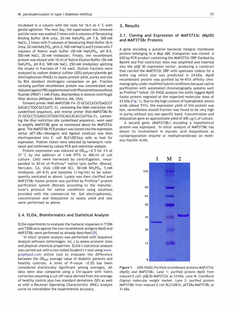
Figure 1 (SDS-PAGE) Purified recombinant proteins MAP3733c(MptD) and MAP3738c. Lane 1: purified protein MptD frominduced E.coli: pQE30-MAP3733c at 24 kDa. Lane M: ColorBurst(Sigma) molecular weight marker. Lane 2: purified proteinMAP3738c from induced E.coli BL21(DE3): pET28a-MAP3738c at31 kDa.
2.4. ELISA, Bioinformatics and Statistical Analysis
ELISA experiments to evaluate the humoral responses in T1DMand T2DM sera against the two recombinant antigens MptD andMAP3738c were performed as already described [9].
"In silico" protein analysis was performed with SequenceAnalysis software (Informagen, Inc.) to assess proteins' sizesand physical-chemical properties. ELISA′s statistical analysiswas carried out with a two-tailed Student's t-test using www.graphpad.com online tool to evaluate the differencebetween the OD405 average value of diabetic patients andhealthy controls. A level of P-value b0.05 has beenconsidered statistically significant among averages. Alldata were also compared using a Chi-square with Yate'scorrection assuming a cut-off value derived from the averageof healthy control plus two standard deviations (SD) as wellas with a Receiver Operating Characteristic (ROC) analysiscurve to convalidate the experimental accuracy.
3. Results
3.1. Cloning and Expression of MAP3733c (MptD)and MAP3738c Proteins
A gene encoding a putative bacterial integral membraneprotein belonging to a Map ABC transporter was cloned. A650 bp PCR product containing the MAP3733c ORF flanked byBamHI and PstI restriction sites was amplified and insertedinto the pQE-30 expression vector, producing a constructthat carried the MAP3733c ORF with upstream codons for a6xHis tag which size was predicted in 24 kDa. MptDrecombinant protein was purified by Ni-NTA affinity chro-matography under modified hybrid conditions because nativepurification with automated chromatography systems suchas Profinia® failed. On PAGE analysis the 6xHis tagged MptDfusion protein migrated at the expected molecular mass of24 kDa (Fig. 1). Due to the high content of hydrophobic aminoacids (about 51%), the expression yield of this protein waslow, nevertheless eluted fraction was found to be very highin purity without any non-specific band. Concentration anddialyzation gave an approximate yield of 300 μg/L of culture.
A second gene (MAP3738c) encoding a hypotheticalprotein was expressed. "In silico" analysis of MAP3738c hasshown its involvement in mycolic acid biosynthesis ascyclopropanation enzyme or methyltransferase on meth-oxy-mycolic acids.

52 A. Cossu et al.
MAP3738c was efficiently cloned into the pET-28a+vector.The cloned ORF was preceded at N-terminal by a sequenceencoding for MGSSHHHHHHSSGLVPRGSH with a histidine tag andfollowed by a second 6xHis tag at the C-terminal. The expressionof the 272 aa resulting protein was efficiently performed withhigh purity and optimal yield in native conditions. Afterconcentration and dialyzation a yield of 600 μg/L culture wasroughly assessed. SDS-PAGE analysis confirmed purity and anexpected size of 31 kDa (Fig. 1).
3.2. ELISA: Immunoreactivity of T1DM and T2DMPatients′ Sera Against MAP3733c (MptD) andMAP3738c Recombinant Fusion Proteins
Due to the lack of immunogenicity by the 6xHis tag, as theseresidues are uncharged at pH 7.2–7.4 and result in a poorlyimmunogenic action towards the majority of species [11],both fusion proteins were tested in ELISA for evaluation ofimmune response in diabetic sera against MptD and MAP3738cproteins without removing tags as well. Moreover, T1DM andcontrol sera were analyzed with a specific recombinantprotein belonging to Helicobacter pylori (HP0986) tagged
Figure 2 Evaluation of immunoreactivity against MAP3733c (MptD)in sera from 43 patients with type I diabetes mellitus and 48 healthy cperformed ELISA. The average value for each population is indicatindicates the AUC value for experiments with MptD (C) and MAP373
with 6xHis tag, which gave a negative result for the presenceof a significant immune response against it (data not shown).
We performed ELISA tests with 43 T1DM sera, 56 T2DM seraand 48 healthy control sera against MAP3733c (MptD) andMAP3738c recombinant proteins to investigate the presence ofhumoral response against these antigens. ELISA results showedan increased humoral response in T1 diabetic sera against bothMAP3733c and MAP3738c proteins compared to healthy sera(Fig. 2) [MAP3733c (MptD); t student = 7.70; FD= 89;p=0.000000000017 (Pb0.0001); 95%]. [MAP3738c; t student=6.49; FD=89; p=0.0000000045; (Pb0.0001); 95%]. Area UnderROC Curve (AUC) (Fig. 2) confirmed the accuracy of thedifference between averages for both proteins' experiments[MAP3733c (MptD); AUC=0,8576]. (MAP 3738c; AUC=0,8278).
Assuming a cut-off value of 0.4 absorbance units atOD405 (average of healthy controls +2SD) (Table 1), muchgreater than the ROC analysis curve's cut-off, set toapproximately 0.2 absorbance units, MptD protein's ELISAresults shown as a statistical analysis by the Chi-squarewith Yate's correction, generated χ equal to 21.657with 1 degree of freedom and a two-tailed P value of0.0001 showing a statistically significant differencebetween T1 diabetic patients and healthy controls. In
recombinant protein (A), and MAP3738c recombinant protein (B)ontrols. Data are shown as values of OD405 nm for each serum ined by the bolded horizontal line. ROC curve statistical analysis8c (D).

53M. paratuberculosis in type I diabetes mellitus
the same way, data for MAP3738c recombinant proteinwith a 0.4 cut-off gave a Chi-square with Yate'scorrection equal to 12.344 with 1 degree of freedomand a two-tailed P value equals to 0.0004.
Data from T2 diabetic sera pool (Fig. 3) (Table 1) showed nodifference between T2 sera and controls for both proteins:[(MAP3733c (MptD); t student=1,89; FD=102; p=0.0612; 95%;AUC=0,5822] and (MAP3738c; t student=0,11; FD=102;p=0.9108; 95%; AUC=0,5456). Similarly, Chi-square analysis ofT2DM ELISA data gave a χ value of 0.329 (MptD) and 1.581(MAP3738c) with a P value of 0.5662 and 0.2086 respectively,showing no correlation between humoral immune responseagainst Map and T2DM.
In conclusion, both ELISA data showed an increasedantibody presence against the twoMap specific antigens inT1DM sera, unlike sera of healthy controls and T2DMpatients.
4. Discussion
Iron uptake is essential for microbial growth because theacquisition of this metal is an important feature for theprocess of bacterial pathogenesis [12]. Metal absorption iscarried out by uptake factors such as the ABC transportersamong which is numbered a specific operon denominatedMpt.
The Mpt operon belongs to a specific 38 kb pathogenic-ity island of Map and a constitutive element of itssequence, the MAP3733c gene, encodes for the proteinMptD that was firstly described as a membrane proteinexpressed during infection stages [13] and further identi-fied as a virulent factor as well [14].
Because the process of iron acquisition might be a possibletarget for chemotherapeutic agents [12] or immunoprophylactictools, Heinzmann and collegues cloned mpt operon's fragmentsinto the integrative pMV306 expression plasmid. Among thesefragments alsomptDORFwas present and has been expressed inBCG host with the aim of creating a vaccine candidate againstMap [14]. Despite the authorswere successfull to demonstrate asurface exposure, MptD protein was not purified.
We had tried to clone the single MptD ORF in a pMAL-seriesplasmid or in pMV261 vector with M. smegmatis as host;unfortunately, purification of expressed protein failed (datanot shown). Subsequently, we cloned it into pQE-30 expressionvector, with a 27 °C expression temperature and a lesser IPTGconcentration during induction to avoid the formation ofinclusion bodies, making it easy to perform the 6xHis tag-mediatedpurificationalthoughundermodifiedhybrid conditionswith Triton X-100detergentwhich led to a lowpurification yield.
The second cloned protein, MAP3738c, seems to be thesmallest among the hypothetical proteins of the specific 38 kbpathogenical island ofMap [13]. Theprotein contains a putativecyclopropane mycolic acid synthase motif (CMAS) involved inbiosynthesis of methoxy and cyclopropyl mycolic acids as for asimilar domain found in Mycobacterium tuberculosis [15].
The analysis of the primary structure of MAP3738cshowed a less hydrophobic character compared to theMptD protein, a detail that might explain the ease of itsexpression and purification. Enzymes involved in additionor degradation of mycolic acid components alter thelipidic composition and the hydrophobic balance of cell
walls leading to a change in the ability to adhere to hostcell surfaces [16], furthermore cyclopropanated mycolicacid is involved in host cell entry.
Miltner and collegues cloned an homologue of Rv3720, acyclopropane fatty acyl-phospholipid synthase gene, asses-sing that its constitutive expression in M. avium host wasfundamental for the invasion of epithelial cells [16] while in asimilar study the inactivation of pcaA, another cyclopropanesynthase, dramatically decreased the virulence of M.tuberculosis [17].
With the emergence of reports about the zoonotic danger ofMap, many investigations had focused on the presence ofhumoral response against different Map antigens in patientsafflicted by autoimmune diseases [1,8,18]. In this study, astrong humoral immune response in T1DM sera againstMAP3733c (MptD) and MAP3738c proteins was detected. Anearlier study following a similar approach found this evidenceusing different specific antigens involved in bacteria survivalandvirulence [9]. Theproteins of this studywerenot recognizedby sera from patients with T2DM and controls. These resultssupport the hypothesis proposed where Map is indicated as apotential trigger of the autoimmunity that characterize T1DMrather than to a non-autoimmune disease as T2DM.
Immunological assays on autoimmune diseases havealready focused on MptD protein using a small peptide calledaMptD derived from a phage display study [13]. This peptidemimed an ideotype sequence that recognized a small part ofthe MptD protein, and its use in serological assays forautoimmune diseases such as T1DM [19] showed resultssimilar to the present results.
The direct identification of MptD protein has demon-strated the presence of intact Map cells in infected bulkmilk [13], in addition electron microscopy experiments[20] revealed that in human host Map is characterized ashaving a spheroplastic form with a partial degradation ofcell wall, which is also corroborated by the loss of theZiehl-Neelsen staining positiveness. This does not excludethe presence of the MptD antigen in Map infecting humans;therefore MptD remains a potential surface marker in theimmunodiagnostic of Map.
Concerning immune response of T1DM patients againstMAP3738c, proteins previously screened that are similar infunction or cellular localization would make easier theunderstanding of our results. Wu and colleagues showed theimportanceof pstAprotein in the addition of aminoacids to thelipopeptidic core of Map demonstrating a significant role forpstA in the development of an immune response in infectedcattle [21]. Differently, Bannantine and Stabel had cloned theHspX protein of Map and revealed its immunogenicity ininfected cows, although it was not a secreted antigen butrather a protein belonging to the soluble fraction [22].
As for HspX antigen, MAP3738c protein may not be exposed,it is in any case involved in the biosynthesis of cell surfacecomponents as the pstA antigen. This line of evidence suggeststhat even if MAP3738c is not exposed on the surface ofMap, it could arouse a humoral response in T1DM patientsas MptD, suggesting that the presence of the antigen onthe outer surface of a cell is not an absolute prerequisitefor the induction of an immune response by the host.
Our data showed a strong humoral response against twospecific Map antigens strengthening the hypothesis about acorrelation between T1DM and Map, as previously reported

Table 1 Characteristics and ELISA results of healthy controls′ sera (left), type I diabetes patients′ sera (center), and type II diabetes patients′ sera (right) against recombinantantigens MAP3733c (MptD) and MAP3738c. M, male; F, female; ND, not determined; I, type I diabetes; II, type II diabetes. Arbitrary values were taken depending on the readingvalues in relation to a cut-off set at OD405=0.4 (control average plus 2 SD). Values are the following: − indicates a value less than 0.4, + indicates a value of 0.4–0.5, ++ indicates avalue of 0.5–0.6, +++ indicates a value of 0.6–0.7, and ++++ indicates a value of 0.7–0.8.
Healthycontrol(samplename)
Sex Age atbloodsample(yr)
Familyhistory ofdiabetes(type)
Seropositivity for: DiabeticpatientT1DM(samplename)
Sex Age at bloodsample (yr)
Familyhistory ofdiabetes(type)
Seropositivity for: DiabeticpatientT2DM(samplename)
Sex Age atbloodsample(yr)
Familyhistory ofdiabetes(type)
Seropositivity for:
MAP3733c(MptD)
MAP3738c MAP3733c(MptD)
MAP3738c MAP3733c(MptD)
MAP3738c
1c F 33 ND – – 1d1 M 21 – – 1d2 M 62 – –2c M 25 ND – ++ 2d1 F 31 ++ ++ 2d2 F 66 – –3c F 50 ND – – 3d1 M 36 I +++ +++ 3d2 M 66 II – –4c F 36 ND – – 4d1 F 36 – – 4d2 M 81 – –5c F 67 ND – – 5d1 M 37 – – 5d2 M 74 ND – –6c M 45 ND – – 6d1 M 26 II – – 6d2 M 62 II – –7c M 45 ND – – 7d1 M 30 I/II + + 7d2 F 50 II – –8c M 53 ND – – 8d1 F 37 I – – 8d2 M 53 II – –9c M 37 ND – – 9d1 F 37 I + ++ 9d2 M 75 – –10c M 63 ND – – 10d1 F 27 I + + 10d2 F 72 ND – +11c M 63 ND – – 11d1 M 31 I – – 11d2 F 40 ND – –12c M 45 ND – – 12d1 M 40 I – + 12d2 F 76 ND – –13c F 60 ND – – 13d1 M 38 ++ – 13d2 M 57 ND – –14c F 43 ND – – 14d1 F 37 I + ++ 14d2 M 62 II – –15c F 34 ND – – 15d1 F 35 + + 15d2 F 77 – –16c F 25 ND – – 16d1 F 40 II – – 16d2 M 56 – –17c M 57 ND – – 17d1 F 34 – + 17d2 M 71 I – –18c F 26 ND – – 18d1 M 41 I – – 18d2 M 65 – –19c F 41 ND – + 19d1 F 36 I – – 19d2 F 66 – –20c M 37 ND – – 20d1 F 37 + – 20d2 M 69 – –21c M 48 ND – – 21d1 F 32 + + 21d2 M 75 – –22c F 57 ND – – 22d1 M 43 – – 22d2 M 63 ND – –23c M 31 ND – ++ 23d1 F 33 I ++ ++ 23d2 F 60 – –
54A.Cossu
etal.

24c M 37 ND – – 24d1 F 33 +++ +++ 24d2 M 68 I – –25c M 39 ND – – 25d1 M 38 + + 25d2 M 57 II – –26c M 28 ND – – 26d1 M 33 ++ ++++ 26d2 M 66 ND – ++27c F 35 ND – + 27d1 M 32 – – 27d2 M 76 II – –28c F 21 ND – – 28d1 M 26 II + +++ 28d2 F 74 ND – –29c M 45 ND – – 29d1 F 32 I ++ +++ 29d2 F 76 ND – –30c M 39 ND – – 30d1 F 38 + – 30d2 F 64 ND – –31c M 46 ND – + 31d1 M 34 – + 31d2 M 69 – –32c F 19 ND – – 32d1 M 94 +++ ++++ 32d2 M 66 ND – –33c M 35 ND – – 33d1 M 36 – – 33d2 F 65 ND + –34c M 49 ND – – 34d1 M 27 I +++ ++++ 34d2 M 65 ND – –35c F 25 ND – – 35d1 M 33 II + ++ 35d2 F 73 II – –36c M 42 ND – – 36d1 F 33 – – 36d2 M 80 ND – –37c F 61 ND – ++ 37d1 F 23 I – + 37d2 M 62 II – –38c M 31 ND – – 38d1 M 43 II – + 38d2 M 70 ND – –39c F 29 ND – – 39d1 M 34 +++ – 39d2 M 74 ND – –40c M 53 ND – – 40d1 F 59 I – – 40d2 M 78 + –41c M 25 ND + + 41d1 F 42 I – – 41d2 M 78 ND – –42c M 23 ND – – 42d1 F 47 II – – 42d2 F 53 – –43c M 28 ND – – 43d1 ND ND ND – – 43d2 F 49 – –44c F 35 ND – – 44d2 M 69 – –45c M 21 ND – – 45d2 M 70 – –46c M 28 ND + – 46d2 M 72 ++ –47c M 29 ND – – 47d2 M 60 II – –48c M 23 ND – – 48d2 F 67 ND – –
49d2 M 53 II – –50d2 M 56 – –51d2 M 62 ND + +52d2 F 73 ND – –53d2 M 66 – –54d2 F 60 – –55d2 F 62 ND + –56d2 F 55 ND – –
55M.paratuberculosis
intype
Idiabetes
mellitus

Figure 3 Evaluation of immunoreactivity against MAP3733c (MptD) recombinant protein (A), and MAP3738c recombinant protein (B)in sera from 56 patients with type II diabetes mellitus and 48 healthy controls. Data are shown as values of OD405 nm for each serum inperformed ELISA. The average value for each population is indicated by the bolded horizontal line. ROC curve statistical analysisindicates the AUC value for experiments with MptD (C) and MAP3738c (D).
56 A. Cossu et al.
by other studies [6,9,19], confirmed by IS900 PCR results forthe presence of Map DNA in the blood of T1DM patients.Furthermore, a similar study with T2DM patients did notshow any significant link between T2DM and Map [23] ascorroborated by this study.
In conclusion, we presented data suggesting that twospecific Map proteins, (MAP3733c) MptD and MAP3738c,may be involved in triggering an humoral immuneresponse only in T1DM patients and not in T2DM subjects.These results reinforce the hypothesis that the bacteriumhas a role, as an environmental factor in triggering T1DMand other autoimmune diseases [24], however, furtherstudies should evaluate if infection is an etiological factorfor the onset of the autoimmune disease or is merely aconsequence of the pathology.
5. Transparency Declaration
All authors have read and agreed to this version of themanuscript. The study does not present any conflict ofinterest for the authors.
Acknowledgment
The study was supported by the Sardinian Region L.R. 7and by the EU grant TBUSGENT.
References
[1] C.T. Dow, Paratuberculosis and type I diabetes: is this thetrigger? Med. Hypotheses 67 (2006) 782–785.
[2] R.J. Greenstein, Is Crohn's disease caused by a mycobacterium?Comparisons with leprosy, tuberculosis, and Johne's disease,Lancet Infect. Dis. 3 (2003) 507–514.
[3] R.J. Chiodini, W.R. Thayer, J.A. Coutu, Presence of Mycobac-terium paratuberculosis antibodies in animal healthcareworkers, in: R.J. Chiodini, M.E. Hines, M.T. Collins, M.A.Rehoboth (Eds.), Proceedings of the Fifth InternationalColloquium on Paratuberculosis, International Association ofParatuberculosis, 1996, pp. 324–328.
[4] G. Passarino, P.A. Underhill, L.L. Cavalli-Sforza, O. Semino,G.M. Pes, C. Carru, L. Ferrucci, M. Bonafe, C. Franceschi, L.Deiana, G. Baggio, G. De Benedictis, Y chromosome binarymarkers to study the high prevalence of males in Sardinian

57M. paratuberculosis in type I diabetes mellitus
centenarians and the genetic structure of the Sardinianpopulation, Hum. Hered. 52 (2001) 136–139.
[5] L.A. Sechi, M. Gazouli, L.E. Sieswerda, P. Molicotti, N. Ahmed,J. Ikonomopoulos, A.M. Scanu, D. Paccagnini, S. Zanetti,Relationship between Crohn's disease, infection with Mycobac-terium avium subspecies paratuberculosis and SLC11A1 genepolymorphisms in Sardinian patients, World J. Gastroenterol.12 (2006) 7161–7164.
[6] D. Paccagnini, L. Sieswerda, V. Rosu, S. Masala, A. Pacifico, M.Gazouli, J. Ikonomopoulos, N. Ahmed, S. Zanetti, L.A. Sechi,Linking chronic infection and autoimmune diseases: Myco-bacterium avium subspecies paratuberculosis, SLC11A1polymorphisms and type-1 diabetes mellitus, PLoS One 4(2009) e7109.
[7] C.T. Dow, Cows, Crohn's and more: is Mycobacterium para-tuberculosis a superantigen? Med. Hypotheses 71 (2008)858–861.
[8] M.T. Getts, S.D. Miller, 99th Dahlem conference on infection,inflammation and chronic inflammatory disorders: triggering ofautoimmune diseases by infections, Clin. Exp. Immunol. 160(2010) 15–21.
[9] L.A. Sechi, V. Rosu, A. Pacifico, G. Fadda, N. Ahmed, S.Zanetti, Humoral immune responses of type 1 diabetes patientsto Mycobacterium avium subsp. paratuberculosis lend supportto the infectious trigger hypothesis, Clin. Vaccine Immunol. 15(2008) 320–326.
[10] L. Li, J.P. Bannantine, Q. Zhang, A. Amonsin, B.J. May, D. Alt,N. Banerji, S. Kanjilal, V. Kapur, The complete genomesequence of Mycobacterium avium subspecies paratuberculosis,Proc. Natl. Acad. Sci. U. S. A. 102 (2005) 12344–12349.
[11] J. Crowe, B.S. Masone, J. Ribbe, One-step purification ofrecombinant proteins with the 6xHis tag and Ni-NTA resin,Methods Mol. Biol. 58 (1996) 491–510.
[12] C. Ratledge, Iron, mycobacteria and tuberculosis, Tuberculosis84 (2004) 110–130.
[13] J. Stratmann, B. Strommenger, R. Goethe, K. Dohmann, G.F.Gerlach, K. Stevenson, L.L. Li, Q. Zhang, V. Kapur, T.J. Bull, A38-kilobase pathogenicity island specific for Mycobacteriumavium subsp. paratuberculosis encodes cell surface pro-teins expressed in the host, Infect. Immun. 72 (2004)1265–1274.
[14] J. Heinzmann, M. Wilkens, K. Dohmann, G.F. Gerlach,Mycobacterium avium subsp. paratuberculosis-specific mptoperon expressed in M. bovis BCG as vaccine candidate, Vet.Microbiol. 130 (2008) 330–337.
[15] Y. Yuan, C.E. Barry, A common mechanism for the biosynthesisof methoxy and cyclopropyl mycolic acids in Mycobacteriumtuberculosis, Proc. Natl. Acad. Sci. U. S. A. 93 (1996)12828–12833.
[16] E. Miltner, K. Daroogheh, P.K. Mehta, S.L. Cirillo, J.D. Cirillo,L.E. Bermudez, Identification of Mycobacterium avium genesthat affect invasion of the intestinal epithelium, Infect.Immun. 73 (2005) 4214–4221.
[17] M.S. Glickman, J.S. Cox, W.R. Jacobs Jr., A novel mycolic acidcyclopropane synthetase is required for cording, persistence,and virulence of Mycobacterium tuberculosis, Mol. Cell 5(2000) 717–727.
[18] D. Polymeros, D.P. Bogdanos, R. Day, D. Arioli, D. Vergani,A. Forbes, Does cross-reactivity between Mycobacteriumavium paratuberculosis and human intestinal antigenscharacterize Crohn's disease? Gastroenterology 131 (2006)85–96.
[19] V. Rosu, N. Ahmed, D. Paccagnini, G. Gerlach, G. Fadda, S.E.Hasnain, S. Zanetti, L.A. Sechi, Specific immunoassays confirmassociation of Mycobacterium avium subsp. paratuberculosiswith type-1 but not type-2 diabetes mellitus, PLoS One 4 (2009)e4386.
[20] L.A. Sechi, A.M. Scanu, P. Molicotti, S. Cannas, M. Mura, G.Dettori, G. Fadda, S. Zanetti, Detection and isolation ofMycobacterium avium subspecies paratuberculosis from intes-tinal mucosal biopsies of patients with and without Crohn′sdisease in Sardinia, Am. J. Gastroenterol. 100 (2005)1529–1536.
[21] C.W. Wu, S.K. Schmoller, J.P. Bannantine, T.M. Eckstein, J.M.Inamine, M. Livesey, R. Albrecht, A.M. Talaat, A novel cell walllipopeptide is important for biofilm formation and pathogenicityof Mycobacterium avium subspecies paratuberculosis, Microb.Pathog. 46 (2009) 222–230.
[22] J.P. Bannantine, J.R. Stabel, HspX is present within Mycobacte-rium paratuberculosis-infectedmacrophages and is recognized bysera fromsome infected cattle, Vet.Microbiol. 76 (2000) 343–358.
[23] V. Rosu, N. Ahmed, D. Paccagnini, A. Pacifico, S. Zanetti, L.A.Sechi,Mycobacterium avium subspecies paratuberculosis is notassociated with type-2 diabetes mellitus, Ann. Clin. Microbiol.Antimicrob. 7 (2008) 9.
[24] D. Cossu, E. Cocco, D. Paccagnini, S. Masala, N. Ahmed, J. Frau,M.G. Marrosu, L.A. Sechi, Association of Mycobacterium aviumsubsp. paratuberculosis with multiple sclerosis in Sardinianpatients, PLoS One (2011), doi:10.1371/journal.pone.0018482Research Article, published 13 Apr.


![Presence of Mycobacterium avium Subspecies ... · 1. Introduction Mycobacterium avium subspecies paratuberculosis (Map) is a very slow growing member of the Mycobacteriumaviumcomplex[1,2]](https://img.dokumen.tips/doc/110x75/600962ca54e6680b3669b7b3/presence-of-mycobacterium-avium-subspecies-1-introduction-mycobacterium-avium.jpg)


