Embed Size (px)
Citation preview

Infectious diseases are major threats to human health worldwide, and tremendous effort has gone into understanding various infectious agents and their mechanisms of virulence. One theme that emerged early from studies of bacterial pathogens is that many inject pathogenic factors (also known as effectors or virulence factors) directly into host cells as part of their pathogenic strategy, and these factors specifically target crucial intracellular pathways in the host. This pathogenic strategy was shown to be used by certain extracellular and intracellular pathogens. These findings led to an exciting and dynamic crossover between related dis-ciplines, including microbiology, cell biology, biochemistry and immu-nology. And from such interdisciplinary studies arose the central tenet that bacterial pathogens specifically attack key intracellular-signalling and cytoskeletal pathways to alter host responses in a way that favours the pathogen. Another important finding of these studies was that the same pathways are often targeted by different bacterial effectors and that these pathways can be attacked at several points by a single pathogen, ensuring an override of important cellular functions.
In this review, we provide an overview of the current understanding of how bacterial pathogens interact with host cells. How these microorgan-isms exploit host cells is discussed in terms of both promoting the bacterial life cycle and evading the host immune response. We focus on examples of pathogenic bacteria that interact with mammalian intracellular-signalling, vesicular-trafficking and cytoskeletal pathways, because these are some of the best studied or most rapidly advancing areas.
Cell biology of bacterial infectionBacterial pathogens use a range of effectors to subvert and control nor-mal cellular functions. Effectors are usually specialized proteins that are injected directly into the cytosol of the host cell by a type III secretion system (T3SS) or a type IV secretion system (T4SS). These secretion sys-tems consist of a structurally conserved proteinaceous apparatus that is shaped like a needle1.
The concept of secreted proteins functioning as agents of microbial virulence is not new. Toxins have been recognized in this capacity for decades. But the ability to inject effectors directly into mammalian or plant cells is repeatedly encountered when considering the cell biology of the infectious process. Therefore, it is an important process in microbial pathogenesis as it is understood at present, and an important interface between pathogens and their hosts.
To promote their life cycle, bacterial pathogens can use host cells to aid their own adherence, replication and/or dissemination. The initial step in colonization by bacteria is adherence. Bacterial pathogens have a large variety of cell-surface adhesins, including fimbriae and afimbrial adhesins (see ref. 2 for a review), that enable them to attach to host cells. Some of these adhesins also have a further role: they bind to their cognate receptors on non-phagocytic cells, thereby allowing bacteria to be taken up by these cells. Such adhesins with dual roles include the invasins of Yersinia spp. and the internalins of Listeria spp.3. The internalization mechanisms of intracellular pathogens differ on the basis of the effector involved, and they require that complementary host intracellular-signalling pathways are commandeered.
The most commonly described cellular target of pathogens is the cytoskeleton. Various intracellular microorganisms harness cytoskeletal components to gain entry to, and to propel themselves within, host cells (see ref. 4 for a review) (Fig. 1). The cytoskeleton of eukaryotic cells is composed of actin filaments, microtubules and intermediate filaments. In terms of bacterial pathogenesis, the most extensively studied of these are actin filaments. Bacterial pathogens do not usually interact directly with actin filaments themselves. Instead, they subvert and control the polymerization of actin filaments by modulating cellular regulators of this process, such as small Rho-like G proteins5, through the action of delivered effectors (Fig. 1a).
Some bacterial pathogens remain in vacuoles after internalization, and these microorganisms often use effectors to modulate vesicular traf-ficking, providing a protective niche within host cells (Fig. 1c) (inclu-ding in macrophages and neutrophils, which normally kill bacteria)6. In addition, pathogens can interact with cell-death pathways (including apoptosis), modulating host-cell death to facilitate pathogen survival in the host7. Moreover, one of the key ways that pathogens evade or subvert the host immune response (both the innate and the adaptive immune mechanisms)8 is to secrete effector proteins (Fig. 1d). These steps of the colonization and immune-evasion processes are discussed in more detail later.
Manipulation of the cytoskeleton and membranous structures How pathogenic bacteria exploit the host-cell cytoskeleton, membra-nous structures and key signalling pathways to their advantage is dis-cussed in this section, together with the strategies and rationales these
1The University of British Columbia, Michael Smith Laboratories, 301-2185 East Mall, Vancouver, British Columbia V6T 1Z4, Canada. 2Present address: Simon Fraser University, 8888 University Drive, Department of Biological Sciences, Room B8276, Shrum Science Centre, Burnaby, British Columbia V5A 1S6, Canada.
Manipulation of host-cell pathways by bacterial pathogensAmit P. Bhavsar1, Julian A. Guttman1,2 & B. Brett Finlay1
Bacterial pathogens operate by attacking crucial intracellular pathways in their hosts. These pathogens usually target more than one intracellular pathway and often interact at several points in each of these pathways to commandeer them fully. Although different bacterial pathogens tend to exploit similar pathway components in the host, the way in which they ‘hijack’ host cells usually differs. Knowledge of how pathogens target distinct cytoskeletal components and immune-cell signalling pathways is rapidly advancing, together with the understanding of bacterial virulence at a molecular level. Studying how these bacterial pathogens subvert host-cell pathways is central to understanding infectious disease.
827
INSIGHT REVIEWNATURE|Vol 449|18 October 2007|doi:10.1038/nature06247
������������������� �� ��������������������
microorganisms use to invade, survive intracellularly and replicate in the host.
Interactions of bacteria with the actin cytoskeleton Bacterial pathogens manipulate the cytoskeleton to help invade a host cell and/or to gain motility in the cell, as mentioned earlier. They often interact with actin filaments in particular, and they do so by modulating G proteins. This process is exemplified by the interaction of the invasive bacterium Salmonella enterica with mammalian cells. During this pro-cess, S. enterica delivers the T3SS effector proteins SopE and SopE2 into the host cell. These effectors function as guanine-nucleotide-exchange factors for G proteins, activating the G protein CDC42 and the RAC family of G proteins in the target cell9–11. This G-protein activation, in turn, induces the generation of actin-rich membrane ruffles that engulf and internalize the bacteria (Fig. 1a). An interesting alternative strategy has recently been reported12: bacterial effector proteins that contain a Trp-X-X-X-Glu motif suppress the signalling of active G proteins and mimic these active G proteins themselves, thereby obviating the need for modulating the GTPase activity of G proteins (Box 1).
After invasion and escape from membrane-enclosed vesicles into the cytosol, many pathogens also manipulate actin-filament dynamics so that they can move within the infected host cell (see ref. 4 for a review). They do so by recruiting actin to just one of their poles, through bac t-erial-protein-mediated nucleation of actin. For example, the intracellular motility of Shigella flexneri is mediated by the bacterial effector IcsA. IcsA
interacts directly with the host protein N-WASP (neural Wiskott–Aldrich syndrome protein; also known as WASL), which in turn recruits a com-plex known as the Arp2/3 complex (consisting of seven host proteins, inclu ding actin-related protein 2 (ARP2) and ARP3).This complex polym-erizes actin filaments behind the advancing bacterium13. By contrast, the cytosolic motility of Listeria spp. is mediated by the bacterial protein ActA, which binds directly to both the Arp2/3 complex and the actin-associated protein VASP (vasodilator-stimulated phosphoprotein)14,15.
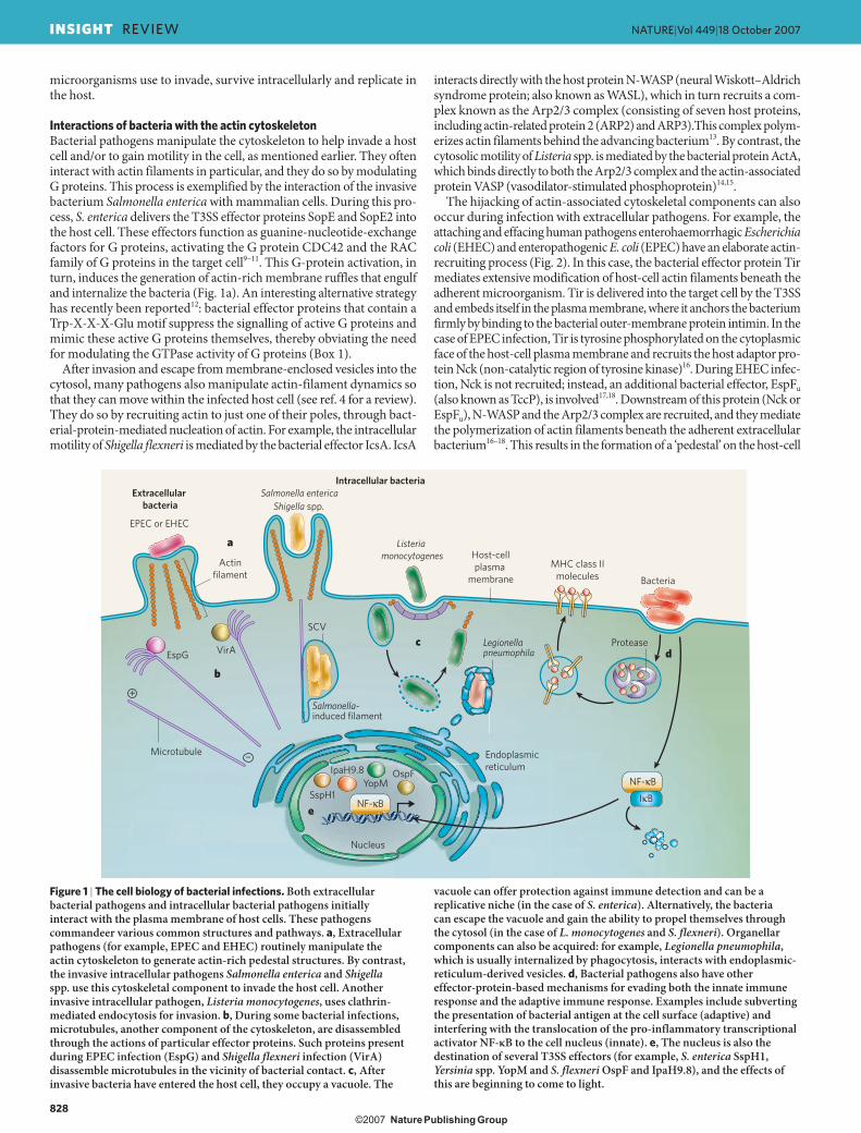
The hijacking of actin-associated cytoskeletal components can also occur during infection with extracellular pathogens. For example, the attaching and effacing human pathogens enterohaemorrhagic Escherichia coli (EHEC) and enteropathogenic E. coli (EPEC) have an elaborate actin-recruiting process (Fig. 2). In this case, the bacterial effector protein Tir mediates extensive modification of host-cell actin filaments beneath the adherent microorganism. Tir is delivered into the target cell by the T3SS and embeds itself in the plasma membrane, where it anchors the bac terium firmly by binding to the bacterial outer-membrane protein intimin. In the case of EPEC infection, Tir is tyrosine phosphorylated on the cytoplasmic face of the host-cell plasma membrane and recruits the host adaptor pro-tein Nck (non-catalytic region of tyrosine kinase)16. During EHEC infec-tion, Nck is not recruited; instead, an additional bacterial effector, EspFu (also known as TccP), is involved17,18. Downstream of this protein (Nck or EspFu), N-WASP and the Arp2/3 complex are recruited, and they mediate the polymerization of actin filaments beneath the adherent extracellular bact erium16–18. This results in the formation of a ‘pedestal’ on the host-cell
Extracellular bacteria
EPEC or EHEC
Actinfilament
Nucleus
EspG
Microtubule
SspH1YopM
IpaH9.8 OspF
VirA
Intracellular bacteria
MHC class IImolecules
Salmonella enterica
Listeria monocytogenes
Shigella spp.
Bacteria
Protease
Endoplasmicreticulum
SCV
NF-κB
NF-κB IκB
+
–
Host-cellplasma
membrane
Salmonella-induced filament
Legionellapneumophila
a
b
cd
e
Figure 1 | The cell biology of bacterial infections. Both extracellular bacterial pathogens and intracellular bacterial pathogens initially interact with the plasma membrane of host cells. These pathogens commandeer various common structures and pathways. a, Extracellular pathogens (for example, EPEC and EHEC) routinely manipulate the actin cytoskeleton to generate actin-rich pedestal structures. By contrast, the invasive intracellular pathogens Salmonella enterica and Shigella spp. use this cytoskeletal component to invade the host cell. Another invasive intracellular pathogen, Listeria monocytogenes, uses clathrin-mediated endocytosis for invasion. b, During some bacterial infections, microtubules, another component of the cytoskeleton, are disassembled through the actions of particular effector proteins. Such proteins present during EPEC infection (EspG) and Shigella flexneri infection (VirA) disassemble microtubules in the vicinity of bacterial contact. c, After invasive bacteria have entered the host cell, they occupy a vacuole. The
vacuole can offer protection against immune detection and can be a replicative niche (in the case of S. enterica). Alternatively, the bacteria can escape the vacuole and gain the ability to propel themselves through the cytosol (in the case of L. monocytogenes and S. flexneri). Organellar components can also be acquired: for example, Legionella pneumophila, which is usually internalized by phagocytosis, interacts with endoplasmic-reticulum-derived vesicles. d, Bacterial pathogens also have other effector-protein-based mechanisms for evading both the innate immune response and the adaptive immune response. Examples include subverting the presentation of bacterial antigen at the cell surface (adaptive) and interfering with the translocation of the pro-inflammatory transcriptional activator NF-κB to the cell nucleus (innate). e, The nucleus is also the destination of several T3SS effectors (for example, S. enterica SspH1, Yersinia spp. YopM and S. flexneri OspF and IpaH9.8), and the effects of this are beginning to come to light.
828
NATURE|Vol 449|18 October 2007INSIGHT REVIEW
������������������� � ��������������������
surface, where the pathogen resides. These pedestals can move (‘surf ’) on the cell surface19: the actin-disassembly proteins cofilin and gelsolin have been identified in pedestals, and these proteins pres umably regu-late actin-filament dynamics in conjunction with the actin-assembly protein profilin. Numerous other actin-associated proteins — inclu-ding cortactin20, GRB2 (growth-factor-receptor-bound protein 2), LPP (lipoma-preferred partner), SHC (SRC-homology-2-domain-containing transforming protein C), vinculin and zyxin21 — have been found in pedestals, although their precise organization during pedestal genera-tion has not yet been determined. The protein α-actinin has also been identified in EPEC-induced pedestals, where it specifically interacts with the amino terminus of Tir22. Surprisingly, the endocytosis-associated pro-tein dynamin has been found in EPEC-induced pedestals23, as have the intermediate-filament proteins cytokeratin 8 and cytokeratin 18 (ref. 24) and the tight-junction component ZO1 (also known as TJP1)25. Therefore, pedestals are useful sites at which to study the interplay of cytoskeletal systems with components of signalling pathways, endocytosis pathways and intercellular junctions.
Interactions of microtubules with effectorsMicrotubules are also commonly targeted by microorganisms. These polarized structures are normally used for structural support and as tracks to guide and transport intracellular cargo, with the aid of microtubule-associated molecular-motor proteins. During certain infections, both the cargo transport and the microtubule assembly and/or disassembly dynamics can be modified and controlled by the pathogen. For exam-ple, on invasion by Shigella spp., the VirA protein interacts directly with heterodimers of α-tubulin and β-tubulin, promoting destabilization of the microtubules26 (Fig. 1b). This results in a localized absence of microtubules near the invading bacteria, thus aiding invasion by Shigella spp. A similar phenotype is seen during EPEC infections (Fig. 1b). In this case, localized microtubule depolymerization depends on the bac-terial effector EspG, which, similarly to VirA, interacts directly with tubulin27.
Whereas Shigella spp. and pathogenic E. coli disassemble microtubules, a strain of Campylobacter jejuni has been shown to use microtubules and their associated molecular motors to aid invasion. Microtubules are polar structures that have distinct fast-growing (plus) and slow-growing (minus) ends (Fig. 1b), allowing the directional transport of cargo in cells. There are two general types of microtubule-based molecular motor found in the host-cell cytosol: kinesins and cytoplasmic dynein. Members of the kinesin superfamily generally transport cargo towards the plus ends of microtubules. By contrast, cytoplasmic dynein is thought to be a minus-end-directed motor. Microtubules, and particularly dynein, have been implicated in invasion by a strain of C. jejuni28. Given that the minus ends of microtubules in cultured, non-polarized cells are directed towards the interior of the cell (Fig. 1b), this model seems plausible. However, in polarized cells (such as those present in intestinal epithelial barriers), microtubule polarity is reversed, so dynein would not transport C. jejuni towards the cell interior. Therefore, further investigation into the uptake of C. jejuni by host cells is required.
Life in a host cellAfter entry to host cells, invasive pathogens are either localized in the cytosol or sequestered in vesicular structures. Presumably, all intracellular pathogens occupy a membrane-enclosed compartment at some point of their intracellular phase, even if only transiently. The initial compart-ments after internalization (vacuoles and modified phagosomes) are com-posed of membranous host-cell components; therefore, internalization often generates protected areas. Pathogens have adopted various strategies to multiply in, or escape from, these structures before surviving in the cytosol and then being disseminated throughout the host.
The ability to occupy a protected intracellular niche contributes to the pathogenesis of both S. enterica and Legionella spp. (see refs 29 and 30 for reviews). On passive internalization by phagocytic cells, Legionella spp. occupy a compartment known as a Legionella-containing vacuole (LCV). This phagosome is modified by the Dot/Icm secretion system (a T4SS).
T4SS effectors enable LCVs both to evade common phagocytic-degrada-tion pathways (by preventing the acidification of vacuoles and the associa-tion of proteins found in late endosomes and lysosomes with LCVs) and to acquire components commonly found in secretory pathways. One of these acquired components is the GTPase ADP-ribosylation factor 1 (ARF1), the function of which is mediated by the bacterial effector RalF31. Legionella spp. further modify the phagosome by using the T4SS effector SidJ to recruit small endoplasmic-reticulum-derived vesicles to the phagosomal membrane, and then mediate fusion with these vesicles32, potentially pro-viding a nutrient-rich resource for the bacteria (Fig. 1c). In addition, the GTPase RAB1 is found at the LCV membrane and has a role in the fusion of endoplasmic-reticulum-derived vesicles with the LCV. The function of RAB1 is controlled by the bacterial effector DrrA (also known as SidM),
One of the central themes of bacterial pathogenesis is the manipulation of host-cell cytoskeletal components by injected effector proteins, which mediate this effect by subverting host G-protein signalling through their guanine-nucleotide-exchange and GTPase-activating activities. However, new data show that bacterial effector proteins can also catalyse novel, diverse and ingenious biochemical reactions that contribute to pathogenesis. For example, several effector proteins contain a Trp-X-X-X-Glu amino-acid motif, which enables them to ‘mimic’ the function of active (GTP-bound) G proteins, allowing downstream signalling and cytoskeletal remodelling12. This novel biochemical activity, the mechanism of which remains unknown, bypasses the requirement for G proteins.
Insights into the biochemistry of effector proteins that target host-cell ubiquitylation pathways are also emerging. The Shigella flexneri effector IpaH9.8 and the Salmonella enterica effector SspH1 were recently shown to have E3 ubiquitin–protein ligase activity66. IpaH9.8 catalyses the transfer of ubiquitin to the yeast mitogen-activated protein kinase (MAPK)-signalling-cascade member Ste7, presumably inducing its degradation and abrogating MAPK signalling. SspH1 catalyses the transfer of ubiquitin to the mammalian protein kinase PKN1, although the effect on PKN1-mediated signalling is unclear. By contrast, the S. enterica effector SseL was recently shown to catalyse the removal of polyubiquitin chains that had been attached to host proteins during infection67. It is intriguing to consider that the opposing biochemical activities of SspH1 and SseL might reflect the necessity of coordinating effector functions (discussed later).
MAPK-signalling pathways have an important role in immunity of the host to bacterial pathogens. Consequently, these canonical phosphorylation cascades are subject to attack by multiple bacterial effector proteins. Recently, two novel and diverse biochemical activities were identified for effectors targeting this pathway. Proteomic analyses indicate that the Yersinia spp. effector YopP/J is an acetylase68. The authors of this study propose that the transfer of acetyl moieties to key residues on MAPK substrates competes effectively with phosphorylation at these sites, thereby blocking signal transduction (Fig. 3). By contrast, the S. flexneri effector OspF has been shown to irreversibly dephosphorylate specific MAPKs — extracellular-signal-regulated kinase 2 (ERK2; also known as MAPK1), p38 MAPK (also known as MAPK14) and Jun amino-terminal kinase (JNK; also known as MAPK8) — by an elimination reaction that chemically modifies the key threonine residue of the substrate so that this MAPK cannot function in the signalling pathway69 (Fig. 3). This enzymatic activity is called phosphothreonine-lyase activity, and it also seems to be present in other pathogenic bacteria. Interestingly, the authors of this study found that OspF had no phosphotyrosine-phosphatase activity, in contrast to another report that claimed OspF was a dual-specificity phosphatase48. The dephosphorylation of phosphotyrosine by an elimination reaction is a highly improbable mechanism, so to determine whether OspF is a dual-specificity phosphatase requires further study. Nevertheless, the identification of irreversible phosphothreonine-lyase activity opens the door to debate about the benefits for the infecting bacteria of irreversible modification of host-cell biology compared with those of reversible modulation.
Box 1 | Novel biochemical activities of translocated bacterial effectors
829
NATURE|Vol 449|18 October 2007 INSIGHT REVIEW
������������������� �� ��������������������

which has guanine-nucleotide-exchange-factor activity for RAB1 during infection with Legionella spp.33. As the infection progresses, these vesicles disappear (as assessed by morphological characteristics), and ribosomes are found to interact with the LCV membrane, thus placing the bacteria within a rough-endoplasmic-reticulum-like vacuole.
Similarly, S. enterica modifies its phagosome-like vacuole, by using a set of T3SS effectors, to provide a protective niche where the bac t-eria survive and replicate30 (Fig. 1c). It accomplishes this by selectively interacting with components of the endocytic machinery of the host
cell, thereby acquiring molecules such as early endosome antigen 1 and lysosomal-associated membrane protein 1 (refs 34, 35). Although it has long been accepted that lysosomes are inhibited from fusion with Salmonella-containing vacuoles (SCVs), recent advances have shown that lysosomes can readily fuse with SCVs during S. enterica infection36, raising numerous questions about the exact mechanisms used to evade destruction by the host.
Active invasion by S. flexneri produces a vacuole around the invading microorganism. However, during invasion of epithelial cells, S. flexneri occupies this vacuole only briefly. This escape from the vacuole allows the bacterium to replicate in the host-cell cytosol and, eventually, to spread from cell to cell37. Listeria monocytogenes, a pathogen that is inter-nalized through clathrin-mediated endocytosis38, is also initially found in a vacuole (Fig. 1c). In a similar manner to invasion by S. flexneri, these vacuoles are short lived. L. monocytogenes uses the membrane-pore-forming toxin listeriolysin O, as well as the enzymes PlcA and PlcB, to destroy the surrounding membrane, thereby allowing escape from the vacuole and, subsequently, replication within the host-cell cytosol and actin-mediated spreading from cell to cell39.
Interactions of bacterial pathogens with signalling pathwaysA beneficial strategy used by many pathogens is to interfere with the phosphorylation cascades in the intracellular-signalling pathways of the host cell. Phosphorylation states are usually controlled by protein kinases and protein phosphatases, and the functions of these enzymes are mimicked by certain bacterial effector proteins. Evidence for this comes from the study of T3SS effectors from S. enterica and Yersinia spp. The S. enterica effector SigD (also known as SopB) functions as a phosphoinositide phosphatase that catalyses the dephosphorylation of host phosphatidylinositol-4,5-bisphosphate and phosphatidyl-inositol-3,4,5-trisphosphate40. As a result, membrane-fission dynam-ics are altered and probably affect SCV formation. The effector YpkA (and its homologue YopO), produced by Yersinia spp., has structural and functional similarities to serine/threonine kinases. This effector is secreted by the bacteria in an inactive form and is autophosphorylated, and thereby activated, in the host cell, where it modulates the actin cytoskeleton through a direct interaction with the GTPase RAC1 (ref. 41). An interesting variation of manipulating host intracellular-signalling pathways involves another effector produced by Yersinia spp., YopM, which simultaneously binds to (and thereby activates) two host protein kinases42. However, the functional significance of the formation of this complex remains poorly understood (Box 2).
Pathogen preservationIt is therefore clear that bacterial pathogens use diverse mechanisms to accomplish a similar goal — to interact with and, potentially, alter the host cell. However, bacterial pathogens must also ensure their own preservation in the host: they need to evade the immune response, and this facilitates replication and spread, which are essential for the suc-cess of any pathogen. The following examples highlight the incredibly diverse mechanisms that bacterial pathogens use to evade both innate immune responses and adaptive immune responses.
Inflammation and nuclear factor-κB A cornerstone of innate immunity is the expression of genes that are responsive to the transcription factor nuclear factor-κB (NF-κB)43 (Fig. 3). This process is induced after bacterial pathogen-associated molecular patterns (PAMPs) are detected by pattern-recognition receptors (PRRs), including Toll-like receptors and NOD (nucleotide-binding oligomerization-domain protein)-like receptors (NLRs) (see ref. 44 for a review and see page 819). NF-κB-responsive genes include those that encode pro-inflammatory cytokines, anti-apoptotic factors (such as Bcl-2) and defensins (a class of antimicrobial peptide). Before these genes can be transcribed, NF-κB needs to be activated, and this occurs when its cytoplasmic binding partner, inhibitor of NF-κB (IκB), is degraded, enabling NF-κB to translocate to the nucleus. The degrada-tion of IκB occurs after it is phosphorylated by the protein IκB kinase
Intimin
Tir
α-Actinin
EPEC or EHEC
Host-cell plasmamembrane
CortactinCytokeratin 8Cytokeratin 18DynaminGRB2LPPSHCViniculinZyxin
P
P
Arp2/3 complex
N-WASP
Profilin
Cofilin
Actinfilament
ZO1
Fyn
PAbl
Nck
+
–
Tyr 474
EspFU
Figure 2 | Generation of pedestals by EPEC and EHEC. During infection with the extracellular bacterium EPEC, the intimin receptor (Tir) translocates into the host cell and inserts itself into the host-cell plasma membrane (a process mediated by the T3SS). This receptor interacts with intimin on the bacterial surface, thereby firmly anchoring the bacterium to the host cell. The carboxy terminus of EPEC Tir becomes phosphorylated on the tyrosine residue at position 474 by at least two host protein kinases, Fyn and Abl, resulting in host adaptor protein Nck being recruited and binding directly to Tir. During infection with EHEC, by contrast, the tyrosine-phosphorylation event is subverted by the EHEC effector EspFu, so Nck is not required. During EPEC or EHEC infection, N-WASP and the Arp2/3 complex (which consists of seven host proteins) are recruited downstream of the Tir-interacting protein (Nck or EspFu), leading to the generation of actin filaments beneath the attached bacteria and the formation of the pedestal structure. Numerous proteins are found in EPEC pedestals (some of which are listed in the shaded box); however, the precise organization of these proteins in EPEC- and EHEC-induced pedestal generation has not been clearly shown. It has been demonstrated that the tight-junction-associated protein ZO1 localizes to the distal portion of the actin filaments of the EPEC pedestal. In addition, the actin-disassembly protein cofilin has been shown to localize to pedestals and presumably, together with the actin-assembly protein profilin, regulates the actin-filament dynamics in pedestals. Also, the amino terminus of Tir has been shown to bind directly to α-actinin, but the effect of this interaction is unknown.
830
NATURE|Vol 449|18 October 2007INSIGHT REVIEW
������������������� �� ���������������� ���
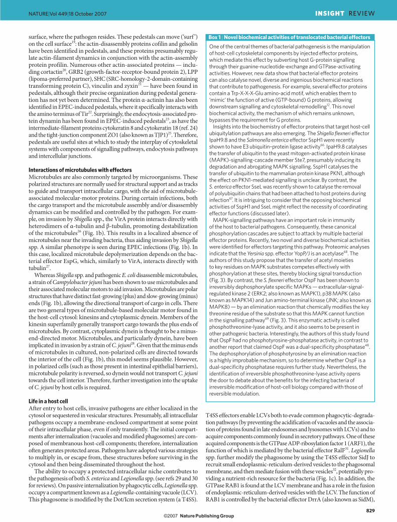
(IKK), the activity of which is stimulated by PRRs. Phosphorylated IκB is then modified with ubiquitin and undergoes proteolytic degradation (see ref. 45 for a review).
Pathogenic microorganisms have come to ‘understand’ the NF-κB-activation pathway and have developed strategies to circumvent it (Fig. 3). For example, both S. flexneri and Yersinia spp. can prevent IκB from being ubiquitylated and therefore prevent its degradation, causing NF-κB to remain inactive in the cell cytoplasm46. These bacteria effect this through the T3SS effector proteins OspG (from S. flexneri) and YopP/J (YopP and YopJ being orthologous proteins from different species of Yersinia). OspG binds to the ubiquitylated form of the E2 ubiquitin-conjugating enzyme UBCH5B (also known as UBE2D2) and prevents the trans-fer of ubiquitin to IκB by an E3 ubiquitin–protein ligase, even though IκB phosphorylation still occurs47. By contrast, until recently, it was known that YopP/J inhibits NF-κB signalling, but it was unclear whether this results from the inhibition of IκB phosphorylation or from the de-ubiquitylation of IκB. Intriguing new biochemical data indicate that inhibition of phosphorylation is the mechanism of action (Box 1).
Interestingly, S. flexneri ensures evasion of innate immune responses by altering the NF-κB-activation pathway at several points. Recent work has shown that S. flexneri uses the T3SS effector OspF to manipulate the physical and spatial context of DNA encoding NF-κB-responsive genes48 (Fig. 3). Epigenetic regulation through DNA modifications such as methylation can have marked effects on gene expression49. OspF func-tions as a unique phosphatase (Box 1). It dephosphorylates the mitogen-activated protein kinase (MAPK) ERK2 in the nucleus (Box 2), so ERK2 cannot then activate mitogen- and stress-activated kinase 1 (MSK1) and MSK2 (ref. 50). This, in effect, prevents histone phosphorylation, which is a prerequisite for NF-κB-dependent transcription48. Therefore, genes that are usually transcriptionally activated by NF-κB in response to the detection of S. flexneri remain silent.
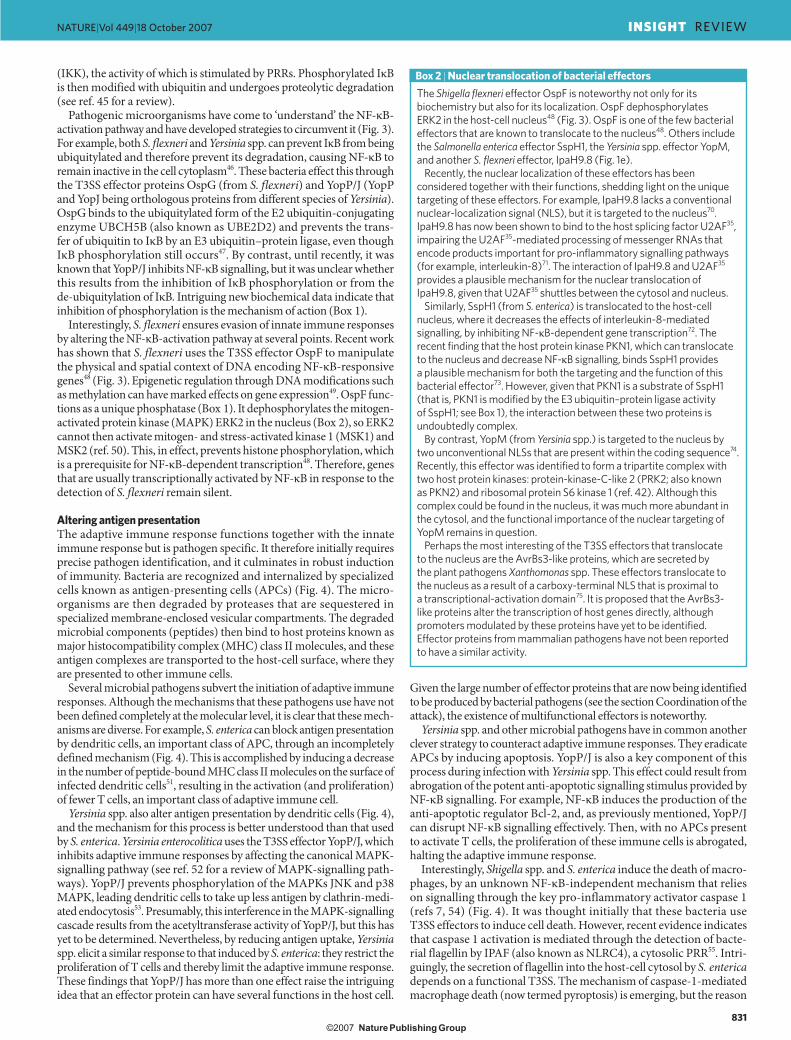
Altering antigen presentationThe adaptive immune response functions together with the innate immune response but is pathogen specific. It therefore initially requires precise pathogen identification, and it culminates in robust induction of immunity. Bacteria are recognized and internalized by specialized cells known as antigen-presenting cells (APCs) (Fig. 4). The micro-organisms are then degraded by proteases that are sequestered in specialized membrane-enclosed vesicular compartments. The degraded microbial components (peptides) then bind to host proteins known as major histocompatibility complex (MHC) class II molecules, and these antigen complexes are transported to the host-cell surface, where they are presented to other immune cells.
Several microbial pathogens subvert the initiation of adaptive immune responses. Although the mechanisms that these pathogens use have not been defined completely at the molecular level, it is clear that these mech-anisms are diverse. For example, S. enterica can block antigen presentation by dendritic cells, an important class of APC, through an incompletely defined mechanism (Fig. 4). This is accomplished by inducing a decrease in the number of peptide-bound MHC class II molecules on the surface of infected dendritic cells51, resulting in the activation (and proliferation) of fewer T cells, an important class of adaptive immune cell.
Yersinia spp. also alter antigen presentation by dendritic cells (Fig. 4), and the mechanism for this process is better understood than that used by S. enterica. Yersinia enterocolitica uses the T3SS effector YopP/J, which inhibits adaptive immune responses by affecting the canonical MAPK-signalling pathway (see ref. 52 for a review of MAPK-signalling path-ways). YopP/J prevents phosphorylation of the MAPKs JNK and p38 MAPK, leading dendritic cells to take up less antigen by clathrin-medi-ated endocytosis53. Presumably, this interference in the MAPK-signalling cascade results from the acetyltransferase activity of YopP/J, but this has yet to be determined. Nevertheless, by reducing antigen uptake, Yersinia spp. elicit a similar response to that induced by S. enterica: they restrict the proliferation of T cells and thereby limit the adaptive immune response. These findings that YopP/J has more than one effect raise the intriguing idea that an effector protein can have several functions in the host cell.
Given the large number of effector proteins that are now being identified to be produced by bacterial pathogens (see the section Coordination of the attack), the existence of multifunctional effectors is noteworthy.
Yersinia spp. and other microbial pathogens have in common another clever strategy to counteract adaptive immune responses. They eradicate APCs by inducing apoptosis. YopP/J is also a key component of this process during infection with Yersinia spp. This effect could result from abrogation of the potent anti-apoptotic signalling stimulus provided by NF-κB signalling. For example, NF-κB induces the production of the anti-apoptotic regulator Bcl-2, and, as previously mentioned, YopP/J can disrupt NF-κB signalling effectively. Then, with no APCs present to activate T cells, the proliferation of these immune cells is abrogated, halting the adaptive immune response.
Interestingly, Shigella spp. and S. enterica induce the death of macro-phages, by an unknown NF-κB-independent mechanism that relies on signalling through the key pro-inflammatory activator caspase 1 (refs 7, 54) (Fig. 4). It was thought initially that these bacteria use T3SS effectors to induce cell death. However, recent evidence indicates that caspase 1 activation is mediated through the detection of bacte-rial flagellin by IPAF (also known as NLRC4), a cytosolic PRR55. Intri-guingly, the secretion of flagellin into the host-cell cytosol by S. enterica depends on a functional T3SS. The mechanism of caspase-1-mediated macrophage death (now termed pyroptosis) is emerging, but the reason
The Shigella flexneri effector OspF is noteworthy not only for its biochemistry but also for its localization. OspF dephosphorylates ERK2 in the host-cell nucleus48 (Fig. 3). OspF is one of the few bacterial effectors that are known to translocate to the nucleus48. Others include the Salmonella enterica effector SspH1, the Yersinia spp. effector YopM, and another S. flexneri effector, IpaH9.8 (Fig. 1e).
Recently, the nuclear localization of these effectors has been considered together with their functions, shedding light on the unique targeting of these effectors. For example, IpaH9.8 lacks a conventional nuclear-localization signal (NLS), but it is targeted to the nucleus70. IpaH9.8 has now been shown to bind to the host splicing factor U2AF35, impairing the U2AF35-mediated processing of messenger RNAs that encode products important for pro-inflammatory signalling pathways (for example, interleukin-8)71. The interaction of IpaH9.8 and U2AF35 provides a plausible mechanism for the nuclear translocation of IpaH9.8, given that U2AF35 shuttles between the cytosol and nucleus.
Similarly, SspH1 (from S. enterica) is translocated to the host-cell nucleus, where it decreases the effects of interleukin-8-mediated signalling, by inhibiting NF-κB-dependent gene transcription72. The recent finding that the host protein kinase PKN1, which can translocate to the nucleus and decrease NF-κB signalling, binds SspH1 provides a plausible mechanism for both the targeting and the function of this bacterial effector73. However, given that PKN1 is a substrate of SspH1 (that is, PKN1 is modified by the E3 ubiquitin–protein ligase activity of SspH1; see Box 1), the interaction between these two proteins is undoubtedly complex.
By contrast, YopM (from Yersinia spp.) is targeted to the nucleus by two unconventional NLSs that are present within the coding sequence74. Recently, this effector was identified to form a tripartite complex with two host protein kinases: protein-kinase-C-like 2 (PRK2; also known as PKN2) and ribosomal protein S6 kinase 1 (ref. 42). Although this complex could be found in the nucleus, it was much more abundant in the cytosol, and the functional importance of the nuclear targeting of YopM remains in question.
Perhaps the most interesting of the T3SS effectors that translocate to the nucleus are the AvrBs3-like proteins, which are secreted by the plant pathogens Xanthomonas spp. These effectors translocate to the nucleus as a result of a carboxy-terminal NLS that is proximal to a transcriptional-activation domain75. It is proposed that the AvrBs3-like proteins alter the transcription of host genes directly, although promoters modulated by these proteins have yet to be identified. Effector proteins from mammalian pathogens have not been reported to have a similar activity.
Box 2 | Nuclear translocation of bacterial effectors
831
NATURE|Vol 449|18 October 2007 INSIGHT REVIEW
������������������� �� ��������������������

for its existence is enigmatic. Indeed, the induction of cell death as a bac-terial immune-evasion strategy is controversial. However, the outcome of pathogen-induced host-cell death is context specific and therefore might either harm or benefit the host (see ref. 56 for a review).
The above examples illustrate that bacterial pathogens have evolved different strategies to ensure their preservation when they interact with host cells, and these most commonly involve the manipulation of host-cell intracellular-signalling pathways by secreted bacterial effectors. Considering the coverage of discrete steps targeted in a given signalling pathway, microbial ‘understanding’ of eukaryotic signalling pathways is truly astounding.
Unresolved issuesDespite progress towards understanding the molecular mechanisms that underlie microbial pathogenesis, much remains to be learned about the bacterial effector proteins that are central to this process. In this section, we outline some of the important unanswered questions about effector acquisition, evolution and coordination.
Selection and evolution of virulence factorsIt is clear that microbial pathogens have diverse mechanisms for inter-acting with, and manipulating, host cells and for evading host immune responses. Presumably, bacterial pathogens have evolved these processes because they provide a selective advantage. Curiously, pathogens often encode several related copies of effectors: for example, EHEC produces at least 14 variants of the T3SS effector NleG, and L. monocytogenes produces several internalin proteins. Although, in some cases, these proteins might target related variants of host-cell components, they are presumably redundant in other cases. Why bacterial pathogens main-tain such a vast collection of seemingly redundant effectors is controver-sial, although recent evidence indicates that bacterial effector proteins are also crucial for successful transmission between hosts57.
It is now understood that the genetic transfer of effector-encoding genes between bacteria (by bacteriophages, conjugation or transforma-tion) has a key role in generating diversity in pathogens (and in gen-erating new diseases). For example, it has recently been shown that a bacterial nucleoid protein, H-NS, can silence genes when they are ini-tially acquired by horizontal transfer. These genes are then integrated into various regulatory pathways, including virulence regulons (in which the silencing is presumably removed), allowing the encoded molecules to participate in virulence pathways58.
Coordination of the attackPathogens with a T3SS commonly have a large repertoire of effec-tors (often tens to hundreds). For example, recent studies have shown that EHEC has at least 40 T3SS effectors, whereas the plant pathogen Pseudomonas syringae has 190 T3SS effectors59,60. Similar numbers of effectors are also present in pathogens with a T4SS, such as Legionella pneumophila. This raises an important question: how are the expres-sion, secretion and functional delivery of these effectors regulated and, perhaps more crucially, coordinated? Virulence-factor coordination was recently reported in the Gram-positive pathogen Staphylococcus aureus: the production of its pore-forming toxin Panton–Valentine leukocidin was shown to increase the production of Spa, a known S. aureus viru-lence factor61. The authors of this study suggested that Panton–Valen-tine leukocidin and Spa function together to exacerbate pathogenesis, although how they do so is unclear. Such effector coordination is also likely to be found in other pathogens with a T3SS or T4SS.
There is no doubt that specialized secretion systems need to coordinate the delivery of effectors to maintain an overall virulence strategy; however, this has not been documented. It also has not been documented, although it is presumed, that adherence precedes effector delivery. In theory, patho-gens could deliver different specialized sets of effectors to different types of host cell (for example, a macrophage and an epithelial cell) or to different tissue sites in a host, and different sets of effectors could also be used to target different host species. Moreover, it is conceivable that secretion sys-tems could work in reverse: that is, they could acquire host-cell molecules,
P
P
P
P
Ac
Ub
Ub
NF-κB
IκB
IKK
E2
E3
E3
P P
ERK2
MSK
YopP/J
OspG
OspF
Nucleus
Host-cell plasmamembrane
Bacterium
TLR
NLR
Bacterialcomponent
NF-κB
Figure 3 | Subversion of NF-κB-mediated signalling. The transcription factor NF-κB initiates the expression of genes that encode many innate immune factors. The NF-κB-signalling pathway is therefore crucial to the host. Various pathogens can subvert this pathway at different points. After pathogenic bacteria are detected by PRRs (TLRs and/or NLRs), a signalling cascade is triggered, resulting in the phosphorylation of the protein-kinase complex IKK. Activated IKK then catalyses the phosphorylation of the inhibitor of NF-κB, IκB. Ubiquitin, carried by an E2 ubiquitin-conjugating enzyme, is attached to phosphorylated IκB by an E3 ubiquitin–protein ligase, marking IκB for degradation in the cytosol and releasing NF-κB to translocate to the nucleus. The induction of gene expression by NF-κB requires remodelling of chromatin through the phosphorylation of histones. This is mediated by activated MSK, a protein kinase that is activated by the MAPK ERK2. The points at which protein effectors secreted by pathogenic bacteria interfere with NF-κB-mediated gene expression are indicated in red. YopP/J, produced by Yersinia spp., disarms IKK by competitively acetylating key amino-acid residues, thereby preventing their phosphorylation. Shigella flexneri subverts this signalling pathway at two points, in the cytosol and in the nucleus. The S. flexneri effector OspG ‘derails’ IκB ubiquitylation by binding the ubiquitylated E2 molecule. By contrast, another S. flexneri effector, OspF, prevents chromatin remodelling, through dephosphorylating activated ERK2.
832
NATURE|Vol 449|18 October 2007INSIGHT REVIEW
������������������� �� ��������������������

signals or even energy and nutrients for the bacteria, although there is no evidence to support this idea at present.
A recent bioinformatic study59 shows that the complexity of these viru-lence systems might be even greater than has been thought. The authors of this study proposed that pathogenic microorganisms can instan taneously produce novel chimaeric hybrids of T3SS effectors through a pro cess referred to as terminal reassortment59. By fusing new protein-coding sequences to sequences that control the expression and secretion of T3SS effectors, microorganisms can ‘sample’ new combinations of secreted effectors. This seemingly hastened effector evolution, coupled with the propensity of the genes encoding these ‘shuffled’ effectors to be located on mobile genetic elements and to confer strong selective pressure through either virulence or transmission, suggests that pathogenic micro organisms can coordinate the induced host-cell biology so that pathogenesis is optimized for the benefit of the microorganism. How induced functional responses are coordinated in the host cell is poorly understood.
Pathogenesis in the bigger picturePathogenic bacteria have the arduous task of interacting with host cells and reprogramming the complex molecular and cellular networks of these cells to allow bacterial replication and spread, while countering host-defence strategies. Evolution and transmission have shaped this bacterial pursuit through the accumulation of (sometimes) vast arse-nals of genes that encode effector proteins, which are probably subject to complex regulation. Piecemeal study of these arsenals might help to define them, but a deeper understanding of their mechanisms and of potential intervention points would best be achieved by considering the delivery, coordination and mechanistic functions of these arsenals as a whole. The current challenge is to assemble a cross-disciplinary toolbox that will enable pathogenesis to be studied at the ‘systems’ level.
ConclusionsThe field of bacterial pathogenesis is a rapidly evolving and expand-ing one. As the vastness of effector functions is being realized, it is a considerable challenge to integrate the numerous host-cell targets and to translate this knowledge into an accurate understanding of the mechanisms by which effector proteins cause disease.
Moving from studying cultured cells to relevant animal disease models is crucial for understanding disease, yet such studies are often neglected,
because cell-culture-based systems are easier to manipulate. However, the opportunity to study pathogenesis in relevant animal models is now within reach, because the current understanding of the mechanistic details of the host–pathogen interface, some of which have been outlined in this article, allows a directed approach to the problem. An elegant example is the recent re-engineering of an L. monocytogenes internalin protein to extend the host range of this bacterium to include mice62. Conversely, the host can now be engineered such that it is susceptible to infection63. These two studies present a glorious opportunity to probe the host–pathogen interface during disease. Similarly, genomic stud-ies have led to the identification of mutations in humans that alter the outcome of bacterial infections64. The recent realization that the host microbiota has a crucial role in mediating the outcome of disease adds another layer of complexity65.
Recognizing that pathogens can overrun crucial host-cell pathways by using a myriad of mechanisms has led to an increased understanding of microbiology, cell biology, biochemistry and immunology. However, this knowledge now needs to be advanced to the point at which it can be translated into a true understanding of disease. This remains the crucial challenge to all who are involved in this field. Only then will it be pos-sible to target these effector mechanisms rationally as a preventive or therapeutic strategy. ■
1. Galan, J. E. & Wolf-Watz, H. Protein delivery into eukaryotic cells by type III secretion machines. Nature 444, 567–573 (2006).
2. Pizarro-Cerda, J. & Cossart, P. Bacterial adhesion and entry into host cells. Cell 124, 715–727 (2006).
3. Cossart, P. & Sansonetti, P. J. Bacterial invasion: the paradigms of enteroinvasive pathogens. Science 304, 242–248 (2004).
4. Stevens, J. M., Galyov, E. E. & Stevens, M. P. Actin-dependent movement of bacterial pathogens. Nature Rev. Microbiol. 4, 91–101 (2006).
5. Finlay, B. B. Bacterial virulence strategies that utilize Rho GTPases. Curr. Top. Microbiol. Immunol. 291, 1–10 (2005).
6. Meresse, S. et al. Controlling the maturation of pathogen-containing vacuoles: a matter of life and death. Nature Cell Biol. 1, E183–E188 (1999).
7. Gao, L. & Abu Kwaik, Y. Hijacking of apoptotic pathways by bacterial pathogens. Microbes Infect. 2, 1705–1719 (2000).
8. Finlay, B. B. & McFadden, G. Anti-immunology: evasion of the host immune system by bacterial and viral pathogens. Cell 124, 767–782 (2006).
9. Hardt, W. D., Chen, L. M., Schuebel, K. E., Bustelo, X. R. & Galan, J. E. S. typhimurium encodes an activator of Rho GTPases that induces membrane ruffling and nuclear responses in host cells. Cell 93, 815–826 (1998).
10. Stender, S. et al. Identification of SopE2 from Salmonella typhimurium, a conserved guanine nucleotide exchange factor for Cdc42 of the host cell. Mol. Microbiol. 36, 1206–1221 (2000).
Salmonella entericaYersinia
enterocolitica
Clathrin
Bacteria
MHC class IImolecule
Flagellin
T3SS
MAPK signalling
Caspase 1
YopP/J
Cell death
IPAF
Protease
Unidentified effector
Host-cellplasma
membrane
Proteolyticallyprocessed bacterialcomponent
Figure 4 | Prevention of antigen presentation by APCs. Components of the adaptive immune system are activated after they recognize fragments of pathogens that are presented at the surface of infected host cells. Antigens from pathogenic bacteria are presented in this way after the bacteria have been taken up into endocytic vesicles that then fusewith cellular-protease-containing vesicles, in which the bacterial pathogens are degraded. Proteolytically processed bacterial components bind to MHC class II molecules, and the vesicles containing these complexes are transported to the cell surface, where the bacterial peptides are displayed to immune cells. Bacterial pathogens interfere with this antigen processing and presentation pathway at several points, which are indicated in red. Yersinia enterocolitica produces YopP/J, which subverts antigen presentation by inhibiting the MAPK-signalling pathway and thereby preventing clathrin-mediated endocytosis. Salmonella enterica secretes an unidentified effector that prevents antigen presentation, although the targeted step also remains unidentified (dashed blocking arrows). In addition, S. enterica can prevent antigen presentation by inducing cell death. This is thought to occur through the T3SS-mediated delivery of flagellin into the cytosol. Flagellin is subsequently detected by the PRR known as IPAF, which initiates a signalling cascade that results in the activation of caspase 1. Caspase 1 then mediates macrophage death by the newly defined mechanism called pyroptosis. Shigella spp. are also thought to induce macrophage death in the same manner.
833
NATURE|Vol 449|18 October 2007 INSIGHT REVIEW
������������������� �� ��������������������
11. Zhou, D., Chen, L. M., Hernandez, L., Shears, S. B. & Galan, J. E. A Salmonella inositol polyphosphatase acts in conjunction with other bacterial effectors to promote host cell actin cytoskeleton rearrangements and bacterial internalization. Mol. Microbiol. 39, 248–259 (2001).
12. Alto, N. M. et al. Identification of a bacterial type III effector family with G protein mimicry functions. Cell 124, 133–145 (2006).
13. Egile, C. et al. Activation of the CDC42 effector N-WASP by the Shigella flexneri IcsA protein promotes actin nucleation by Arp2/3 complex and bacterial actin-based motility. J. Cell Biol. 146, 1319–1332 (1999).
14. Chakraborty, T. et al. A focal adhesion factor directly linking intracellularly motile Listeria monocytogenes and Listeria ivanovii to the actin-based cytoskeleton of mammalian cells. EMBO J. 14, 1314–1321 (1995).
15. Welch, M. D., Iwamatsu, A. & Mitchison, T. J. Actin polymerization is induced by Arp2/3 protein complex at the surface of Listeria monocytogenes. Nature 385, 265–269 (1997).
16. Gruenheid, S. et al. Enteropathogenic E. coli Tir binds Nck to initiate actin pedestal formation in host cells. Nature Cell Biol. 3, 856–859 (2001).
17. Campellone, K. G., Robbins, D. & Leong, J. M. EspFU is a translocated EHEC effector that interacts with Tir and N-WASP and promotes Nck-independent actin assembly. Dev. Cell 7, 217–228 (2004).
18. Garmendia, J. et al. TccP is an enterohaemorrhagic Escherichia coli O157:H7 type III effector protein that couples Tir to the actin-cytoskeleton. Cell. Microbiol. 6, 1167–1183 (2004).
19. Shaner, N. C., Sanger, J. W. & Sanger, J. M. Actin and α-actinin dynamics in the adhesion and motility of EPEC and EHEC on host cells. Cell Motil. Cytoskeleton 60, 104–120 (2005).
20. Cantarelli, V. V. et al. Cortactin is necessary for F-actin accumulation in pedestal structures induced by enteropathogenic Escherichia coli infection. Infect. Immun. 70, 2206–2209 (2002).
21. Goosney, D. L., DeVinney, R. & Finlay, B. B. Recruitment of cytoskeletal and signaling proteins to enteropathogenic and enterohemorrhagic Escherichia coli pedestals. Infect. Immun. 69, 3315–3322 (2001).
22. Goosney, D. L. et al. Enteropathogenic E. coli translocated intimin receptor, Tir, interacts directly with α-actinin. Curr. Biol. 10, 735–738 (2000).
23. Unsworth, K. E. et al. Dynamin is required for F-actin assembly and pedestal formation by enteropathogenic Escherichia coli (EPEC). Cell. Microbiol. 9, 438–449 (2007).
24. Batchelor, M. et al. Involvement of the intermediate filament protein cytokeratin-18 in actin pedestal formation during EPEC infection. EMBO Rep. 5, 104–110 (2004).
25. Hanajima-Ozawa, M. et al. Enteropathogenic Escherichia coli, Shigella flexneri, and Listeria monocytogenes recruit a junctional protein, zonula occludens-1, to actin tails and pedestals. Infect. Immun. 75, 565–573 (2007).
26. Yoshida, S. et al. Shigella deliver an effector protein to trigger host microtubule destabilization, which promotes Rac1 activity and efficient bacterial internalization. EMBO J. 21, 2923–2935 (2002).
27. Hardwidge, P. R. et al. Modulation of host cytoskeleton function by the enteropathogenic Escherichia coli and Citrobacter rodentium effector protein EspG. Infect. Immun. 73, 2586–2594 (2005).
28. Hu, L. & Kopecko, D. J. Campylobacter jejuni 81-176 associates with microtubules and dynein during invasion of human intestinal cells. Infect. Immun. 67, 4171–4182 (1999).
29. Roy, C. R. & Tilney, L. G. The road less traveled: transport of Legionella to the endoplasmic reticulum. J. Cell Biol. 158, 415–419 (2002).
30. Knodler, L. A. & Steele-Mortimer, O. Taking possession: biogenesis of the Salmonella-containing vacuole. Traffic 4, 587–599 (2003).
31. Nagai, H. et al. A C-terminal translocation signal required for Dot/Icm-dependent delivery of the Legionella RalF protein to host cells. Proc. Natl Acad. Sci. USA 102, 826–831 (2005).
32. Robinson, C. G. & Roy, C. R. Attachment and fusion of endoplasmic reticulum with vacuoles containing Legionella pneumophila. Cell. Microbiol. 8, 793–805 (2006).
33. Murata, T. et al. The Legionella pneumophila effector protein DrrA is a Rab1 guanine nucleotide-exchange factor. Nature Cell Biol. 8, 971–977 (2006).
34. Steele-Mortimer, O., Meresse, S., Gorvel, J. P., Toh, B. H. & Finlay, B. B. Biogenesis of Salmonella typhimurium-containing vacuoles in epithelial cells involves interactions with the early endocytic pathway. Cell. Microbiol. 1, 33–49 (1999).
35. Cuellar-Mata, P. et al. Nramp1 modifies the fusion of Salmonella typhimurium-containing vacuoles with cellular endomembranes in macrophages. J. Biol. Chem. 277, 2258–2265 (2002).
36. Drecktrah, D., Knodler, L. A., Howe, D. & Steele-Mortimer, O. Salmonella trafficking is defined by continuous dynamic interactions with the endolysosomal system. Traffic 8, 212–225 (2007).
37. Sansonetti, P. J., Ryter, A., Clerc, P., Maurelli, A. T. & Mounier, J. Multiplication of Shigella flexneri within HeLa cells: lysis of the phagocytic vacuole and plasmid-mediated contact hemolysis. Infect. Immun. 51, 461–469 (1986).
38. Veiga, E. & Cossart, P. Listeria hijacks the clathrin-dependent endocytic machinery to invade mammalian cells. Nature Cell Biol. 7, 894–900 (2005).
39. Shaughnessy, L. M., Hoppe, A. D., Christensen, K. A. & Swanson, J. A. Membrane perforations inhibit lysosome fusion by altering pH and calcium in Listeria monocytogenes vacuoles. Cell. Microbiol. 8, 781–792 (2006).
40. Terebiznik, M. R. et al. Elimination of host cell PtdIns(4,5)P2 by bacterial SigD promotes membrane fission during invasion by Salmonella. Nature Cell Biol. 4, 766–773 (2002).
41. Prehna, G., Ivanov, M. I., Bliska, J. B. & Stebbins, C. E. Yersinia virulence depends on mimicry of host Rho-family nucleotide dissociation inhibitors. Cell 126, 869–880 (2006).
42. McDonald, C., Vacratsis, P. O., Bliska, J. B. & Dixon, J. E. The Yersinia virulence factor YopM forms a novel protein complex with two cellular kinases. J. Biol. Chem. 278, 18514–18523 (2003).
43. Hayden, M. S., West, A. P. & Ghosh, S. NF-κB and the immune response. Oncogene 25, 6758–6780 (2006).
44. Akira, S., Uematsu, S. & Takeuchi, O. Pathogen recognition and innate immunity. Cell 124, 783–801 (2006).
45. Perkins, N. D. Post-translational modifications regulating the activity and function of the nuclear factor κB pathway. Oncogene 25, 6717–6730 (2006).
46. Angot, A., Vergunst, A., Genin, S. & Peeters, N. Exploitation of eukaryotic ubiquitin signaling pathways by effectors translocated by bacterial type III and type IV secretion systems. PLoS Pathog. 3, e3 (2007).
47. Kim, D. W. et al. The Shigella flexneri effector OspG interferes with innate immune responses by targeting ubiquitin-conjugating enzymes. Proc. Natl Acad. Sci. USA 102, 14046–14051 (2005).
48. Arbibe, L. et al. An injected bacterial effector targets chromatin access for transcription factor NF-κB to alter transcription of host genes involved in immune responses. Nature Immunol. 8, 47–56 (2007).
49. Kouzarides, T. Chromatin modifications and their function. Cell 128, 693–705 (2007).
50. Soloaga, A. et al. MSK2 and MSK1 mediate the mitogen- and stress-induced phosphorylation of histone H3 and HMG-14. EMBO J. 22, 2788–2797 (2003).
51. Cheminay, C., Mohlenbrink, A. & Hensel, M. Intracellular Salmonella inhibit antigen presentation by dendritic cells. J. Immunol. 174, 2892–2899 (2005).
52. Ashwell, J. D. The many paths to p38 mitogen-activated protein kinase activation in the immune system. Nature Rev. Immunol. 6, 532–540 (2006).
53. Autenrieth, S. E. et al. Yersinia enterocolitica YopP inhibits MAP kinase-mediated antigen uptake in dendritic cells. Cell. Microbiol. 9, 425–437 (2007).
54. Scott, A. M. & Saleh, M. The inflammatory caspases: guardians against infections and sepsis. Cell Death Differ. 14, 23–31 (2007).
55. Miao, E. A. et al. Cytoplasmic flagellin activates caspase-1 and secretion of interleukin-1β via Ipaf. Nature Immunol. 7, 569–575 (2006).
56. DeLeo, F. R. Modulation of phagocyte apoptosis by bacterial pathogens. Apoptosis 9, 399–413 (2004).
57. Wickham, M. E., Brown, N. F., Boyle, E. C., Coombes, B. K. & Finlay, B. B. Virulence is positively selected by transmission success between mammalian hosts. Curr. Biol. 17, 783–788 (2007).
58. Navarre, W. W. et al. Selective silencing of foreign DNA with low GC content by the H-NS protein in Salmonella. Science 313, 236–238 (2006).
59. Stavrinides, J., Ma, W. & Guttman, D. S. Terminal reassortment drives the quantum evolution of type III effectors in bacterial pathogens. PLoS Pathog. 2, e104 (2006).
60. Tobe, T. et al. An extensive repertoire of type III secretion effectors in Escherichia coli O157 and the role of lambdoid phages in their dissemination. Proc. Natl Acad. Sci. USA 103, 14941–14946 (2006).
61. Labandeira-Rey, M. et al. Staphylococcus aureus Panton–Valentine leukocidin causes necrotizing pneumonia. Science 315, 1130–1133 (2007).
62. Wollert, T. et al. Extending the host range of Listeria monocytogenes by rational protein design. Cell 129, 891–902 (2007).
63. Lecuit, M. et al. A transgenic model for listeriosis: role of internalin in crossing the intestinal barrier. Science 292, 1722–1725 (2001).
64. Casanova, J. L. & Abel, L. Human genetics of infectious diseases: a unified theory. EMBO J. 26, 915–922 (2007).
65. Lupp, C. et al. Host-mediated inflammation disrupts the intestinal microbiota and promotes the overgrowth of Enterobacteriaceae. Cell Host Microbe 2, 119–129 (2007).
66. Rohde, J. R., Breitkreutz, A., Chenal, A., Sansonetti, P. J. & Parsot, C. Type III secretion effectors of the IpaH family are E3 ubiquitin ligases. Cell Host Microbe 1, 77–83 (2007).
67. Rytkonen, A. et al. SseL, a Salmonella deubiquitinase required for macrophage killing and virulence. Proc. Natl Acad. Sci. USA 104, 3502–3507 (2007).
68. Mukherjee, S. et al. Yersinia YopJ acetylates and inhibits kinase activation by blocking phosphorylation. Science 312, 1211-1214 (2006).
69. Li, H. et al. The phosphothreonine lyase activity of a bacterial type III effector family. Science 315, 1000–1003 (2007).
70. Toyotome, T. et al. Shigella protein IpaH9.8 is secreted from bacteria within mammalian cells and transported to the nucleus. J. Biol. Chem. 276, 32071–32079 (2001).
71. Okuda, J. et al. Shigella effector IpaH9.8 binds to a splicing factor U2AF35 to modulate host immune responses. Biochem. Biophys. Res. Commun. 333, 531–539 (2005).
72. Haraga, A. & Miller, S. I. A Salmonella enterica serovar Typhimurium translocated leucine-rich repeat effector protein inhibits NF-κB-dependent gene expression. Infect. Immun. 71, 4052–4058 (2003).
73. Haraga, A. & Miller, S. I. A Salmonella type III secretion effector interacts with the mammalian serine/threonine protein kinase PKN1. Cell. Microbiol. 8, 837–846 (2006).
74. Benabdillah, R., Mota, L. J., Lutzelschwab, S., Demoinet, E. & Cornelis, G. R. Identification of a nuclear targeting signal in YopM from Yersinia spp. Microb. Pathog. 36, 247–261 (2004).
75. Schornack, S., Meyer, A., Romer, P., Jordan, T. & Lahaye, T. Gene-for-gene-mediated recognition of nuclear-targeted AvrBs3-like bacterial effector proteins. J. Plant Physiol. 163, 256–272 (2006).
Acknowledgements We thank members of B.B.F.’s laboratory for helpful discussions and critical reading of the manuscript. We gratefully acknowledge F. Ness for assistance with the preparation of figures. We apologize to authors whose work could not be cited as a result of space restrictions. Work in B.B.F.’s laboratory is supported by grants from the Canadian Institutes of Health Research (CIHR), the Howard Hughes Medical Institute (HHMI), the Foundation for the National Institutes of Health, and Genome Canada. A.P.B. is supported by fellowships from the CIHR and the Michael Smith Foundation for Health Research (MSFHR). J.A.G. is supported by a Canadian Association for Gastroenterology/CIHR/AstraZeneca fellowship and a fellowship from the MSFHR. B.B.F. is a CIHR Distinguished Investigator, an HHMI International Research Scholar, and the Peter Wall Distinguished Professor, at the University of British Columbia.
Author Information Reprints and permissions information is available at npg.nature.com/reprints. Correspondence should be addressed to B.B.F. ([email protected]).
834
NATURE|Vol 449|18 October 2007INSIGHT REVIEW
������������������� �� ��������������������


![SMS[1] - TCCP](https://img.dokumen.tips/doc/110x75/5476ac3cb4af9fbe0a8b6489/sms1-tccp.jpg)
![From Ritual to Record [Allen Guttman]](https://img.dokumen.tips/doc/110x75/557210e1497959fc0b8dd792/from-ritual-to-record-allen-guttman.jpg)