Embed Size (px)
Citation preview

Managing Sclerotherapy Complications
Barbara Deusterman, RN
Objectives
The learner will be able to describe the most common complications of visually-guided sclerotherapy of the lower extremities
The learner will be able to identify prevention techniques or techniques that may minimize complications.
The learner will be able to list treatment strategies of the most common complications of visually-guided sclerotherapy of the lower extremities

Complications……
“We look for medicine to be an orderly field of knowledge and procedure. But it is not. It is an imperfect science, an enterprise of constantly changing knowledge, uncertain information, fallible individuals, and at the same time lives on the line. There is science in what we do, yes, but also habit, intuition, and sometimes plain old guessing. The gap between what we know and what we aim for persists. And this gap complicates everything we do.”
-Atul Gawande, Complications: A Surgeon’s Notes on an Imperfect Science
Know who you are treating!
• Physical assessment
• Through and current history
• Duplex ultrasound (if needed)
• Know risk factors that would increase likelihood of complication
• Before photos
• Consent form
• Manage patient expectations
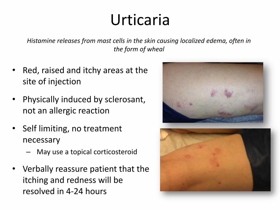
Urticaria
• Red, raised and itchy areas at the site of injection
• Physically induced by sclerosant, not an allergic reaction
• Self limiting, no treatment necessary– May use a topical corticosteroid
• Verbally reassure patient that the itching and redness will be resolved in 4-24 hours
Histamine releases from mast cells in the skin causing localized edema, often in the form of wheal
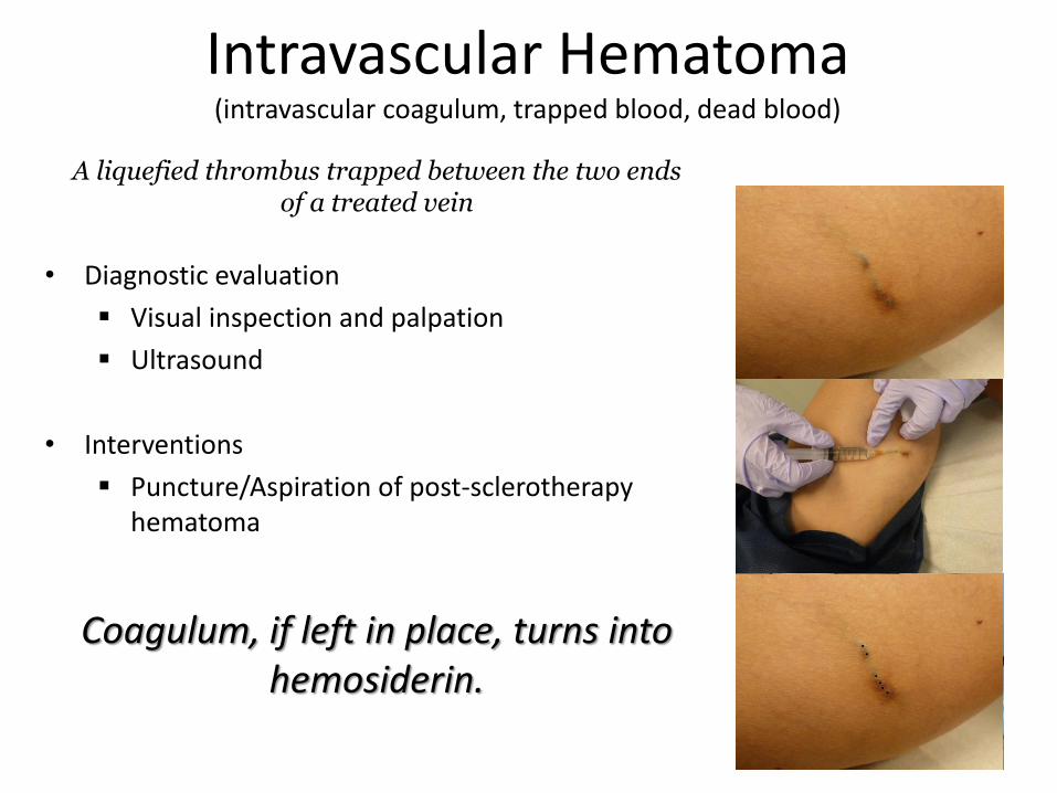
Intravascular Hematoma(intravascular coagulum, trapped blood, dead blood)
• Diagnostic evaluation
Visual inspection and palpation
Ultrasound
• Interventions
Puncture/Aspiration of post-sclerotherapy hematoma
A liquefied thrombus trapped between the two ends of a treated vein
Coagulum, if left in place, turns into hemosiderin.

Intravascular Hematoma(intravascular coagulum, trapped blood, dead blood)
Intervene with in 2-4 weeks
Choose most efficient/effective sclerosant concentration for location and size of vein
Injection of small volumes from single points of entry
Encourage compliance with graduated compression stockings and ambulation
Considerations
Coagulum, if left in place, turns into hemosiderin.

Hyperpigmentation
• Incidence- 10-30%
• Occurs within 4-6 Weeks
• 60% resolve within 6 months
• 90% resolve within 1 year
• 1-10% have pigment > 1 year
• Possible risk factors
• Taking Minocycline
• History of high iron stores
• Intense, persistent sun exposure during treatment process
• Possible link to those patients with dark hair and darker pigmented skin tones (controversial)
Hemosiderin staining happens when blood leaves a ruptured blood vessel, the red blood cell dies and the hemoglobin from the red blood cell is released into
the dermis.
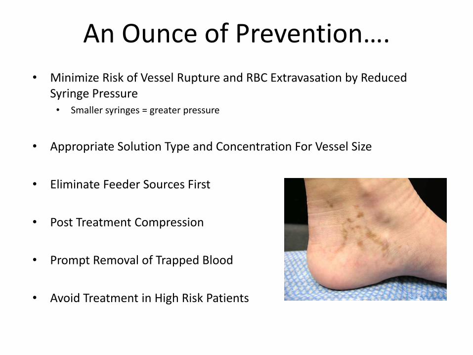
An Ounce of Prevention….
• Minimize Risk of Vessel Rupture and RBC Extravasation by Reduced Syringe Pressure • Smaller syringes = greater pressure
• Appropriate Solution Type and Concentration For Vessel Size
• Eliminate Feeder Sources First
• Post Treatment Compression
• Prompt Removal of Trapped Blood
• Avoid Treatment in High Risk Patients

Hyperpigmentation
• Topical agents
• Bleaching creams
• Exfoliants
• IPL laser
• Iron Chelation
Time is the first line intervention!
Interventions
Hyperpigmentation
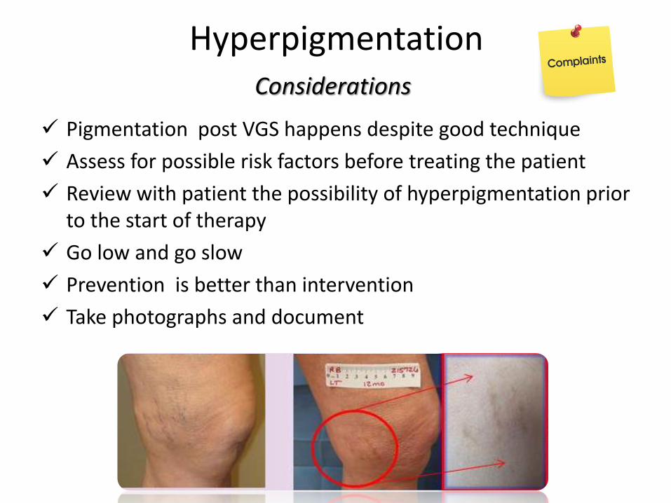
Pigmentation post VGS happens despite good technique
Assess for possible risk factors before treating the patient
Review with patient the possibility of hyperpigmentation prior to the start of therapy
Go low and go slow
Prevention is better than intervention
Take photographs and document
Considerations
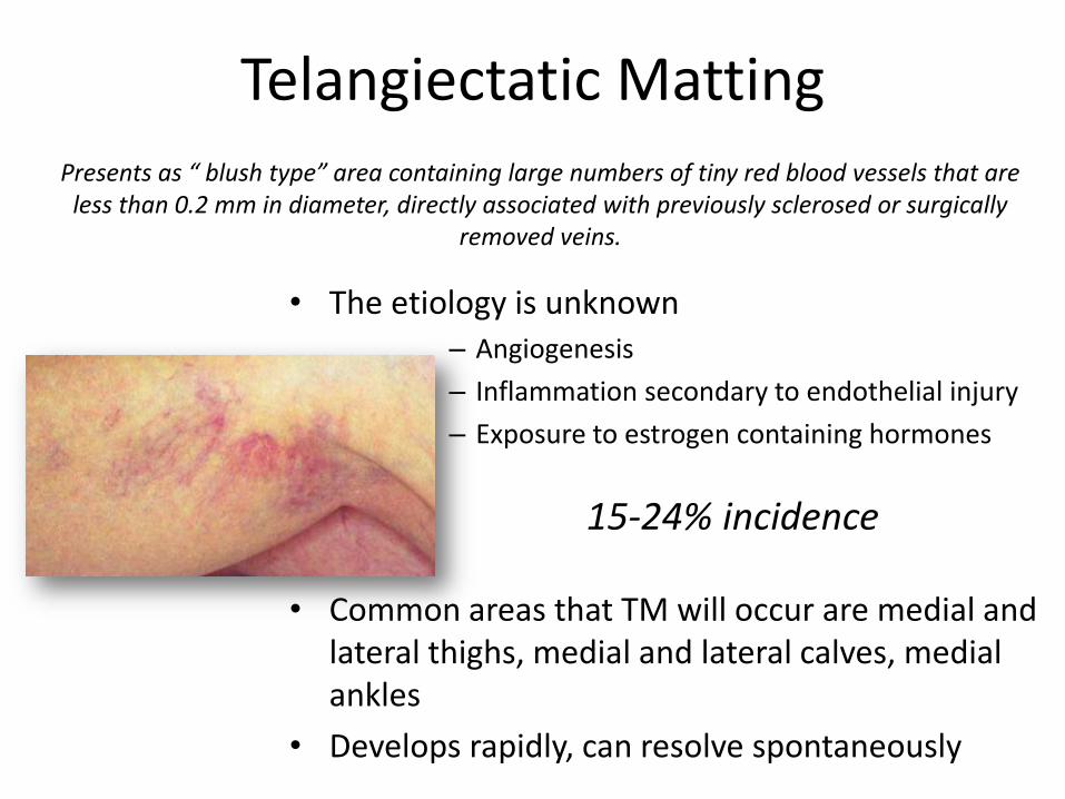
Telangiectatic Matting
• The etiology is unknown– Angiogenesis
– Inflammation secondary to endothelial injury
– Exposure to estrogen containing hormones
• Common areas that TM will occur are medial and lateral thighs, medial and lateral calves, medial ankles
• Develops rapidly, can resolve spontaneously
Presents as “ blush type” area containing large numbers of tiny red blood vessels that are less than 0.2 mm in diameter, directly associated with previously sclerosed or surgically
removed veins.
15-24% incidence

Telangiectatic Matting
• Reevaluate possible sources of reflux not recognized on initial exam.
• Time – TM may resolve on own with out any further intervention, use verbal reassurance liberally
• Inject remaining TM with Glycerin or low concentration of detergent sclerosant if not resolved after all sources of reflux are treated and the area was given time to resolve on own.
• Consider discontinuing contraceptive therapy for 1 month prior to beginning sclerotherapy and delaying reinstitution of such therapy for at least 2 months following the last treatment session
• Encourage weight loss in obese individuals prior to institution of therapy
• IPL laser
Treatment options

Telangiectatic Matting
Educate patient of the possibility of complication prior start of treatment
Assess for risk factors prior to treatment
Use the lowest concentration and lowest volume of the chosen sclerosant that will effectively obliterate the vein
Use low pressure when injecting the sclerosant to minimize excessive vessel injury
Do not use stronger sclerosant or multiple treatments in effort to resolve TM.
Document progress or resolution by photographing patient every 6 to 8 weeks
Consider stopping Estrogen therapy only after careful consideration and consultation with both supervising physician and patients prescribing physician.
Considerations

Cutaneous NecrosisLocalized superficial tissue damage as a direct result of sclerotherapy.
Ischemic necrosis that is caused by the arterial component of the vascular bed becoming occluded
• Bright erythema may be seen immediately after injection
• Prolonged blanching, can also be described as porcelain-white appearance immediately after injection
• Pain can be immediate or delayed
• Dermis can turn pale or dusky
• Dermal sloughing starts 24-72 hours after the ischemic event
• Ulcer can occur at site

Day 7
Day 12
Day 20
Day 29
Day 52
Cutaneous Necrosis
Measure and document and monitor
Cutaneous Necrosis
• Vigorous massage of extravasated area may decrease tissue damage
• If arteriolar injection or spasm suspected vigorously massage using 2% nitroglycerine ointment
• Hyaluronidase– Enzyme that rapidly diffuses extravasated solution thus minimizing tissue damage
– Dose 75 units re-constituted in 0.9% sodium chloride
– Must be injected into affected area with in 60 minutes to be effective
• Occlusive dressings are used to speed healing process and decrease pain
• Debridement of wound using either hydrocolloid dressings or surgical excision of necrotic tissue will promote granulation tissue formation.
• Short stretch wraps can be used if edema present
Interventions

Cutaneous Necrosis
• Stop injecting if you feel resistance
• Stop injecting if a bleb or wheal forms
• Stop injecting if prolonged blanching occurs
• Use the lowest volume and weakest concentration of sclerosing agent needed to close down target vein
• Inject using slow steady pressure • Smaller syringes = greater pressure
• Ultrasound guidance should be used for foam sclerotherapy of deeper reticular veins
Careful and methodical technique must be used

Cutaneous Necrosis
Areas of skin necrosis after Sclerotherapy take 4-12 weeks to heal
Explain length of healing process, frequent follow-up in the beginning
Reassure patient that the wound will heal, usually with a cosmetically acceptable scar
Photo document ulcer at discovery and continue to photo document progress of healing at follow up appointments
Document location and size of necrosis, and note the amount and concentration of sclerosant used in last treatment session prior to discovery.
Monitor patient for signs and symptoms of infection
High incidence of litigation with this complication
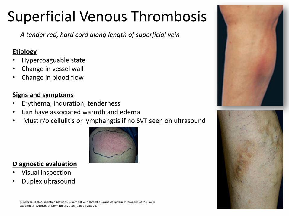
Superficial Venous Thrombosis
(Binder B, et al. Association between superficial vein thrombosis and deep vein thrombosis of the lower extremities. Archives of Dermatology 2009; 145(7): 753-757.)
A tender red, hard cord along length of superficial vein
Etiology• Hypercoaguable state • Change in vessel wall • Change in blood flow
Signs and symptoms• Erythema, induration, tenderness• Can have associated warmth and edema• Must r/o cellulitis or lymphangtis if no SVT seen on ultrasound
Diagnostic evaluation• Visual inspection• Duplex ultrasound

Superficial Venous Thrombosis
Rule out DVT in a patient with superficial phlebitis
• Patients with SVT have a 5%-40% chance of developing DVT 1,3
• One study showed SVT patients have concurrent DVT in 28% of cases 2
1. Hanson, J.N., Ascher, E., DePippo, P., Lorensen, E., Scheinman, M., Yorkovich, W., Hingorani , A. Saphenous vein thrombophlebitis (SVT): a deceptively benign disease J Vasc Surg. 1998 April; 27(4): 677–680.
2. Galanaud, J. P., Gentry, C., Sevestre, M. A., & Bisot, D. , et al .Predictive factors for concurrent deep-vein thrombosis and symptomatic venous thrombotic recurrence in case of superficial venous thrombosis. The OPTIMEV study. Thrombosis and haemostasis 2011, 105(1), 31-39.
3. Gloviczki, P., Dalsing, M. C., Eklof, B., Moneta, G. L., & Wakfield, T. W. (2009). Handbook of Venous Disorders (3rd ed.). London: Hodder Education
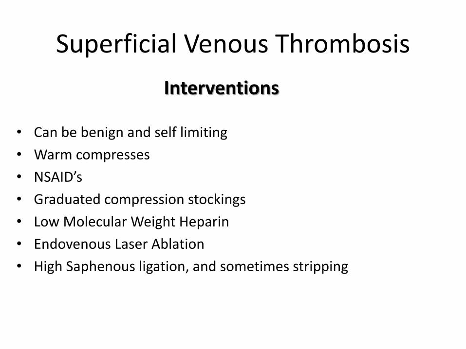
Superficial Venous Thrombosis
• Can be benign and self limiting
• Warm compresses
• NSAID’s
• Graduated compression stockings
• Low Molecular Weight Heparin
• Endovenous Laser Ablation
• High Saphenous ligation, and sometimes stripping
Interventions
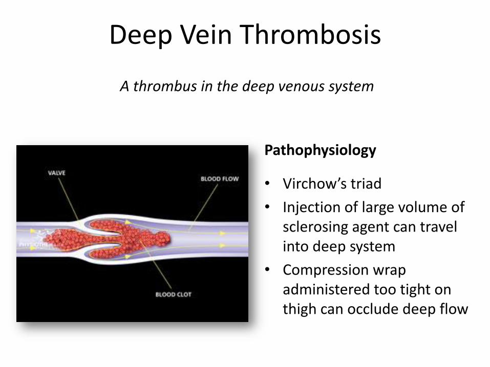
Deep Vein Thrombosis
A thrombus in the deep venous system
Pathophysiology
• Virchow’s triad
• Injection of large volume of sclerosing agent can travel into deep system
• Compression wrap administered too tight on thigh can occlude deep flow
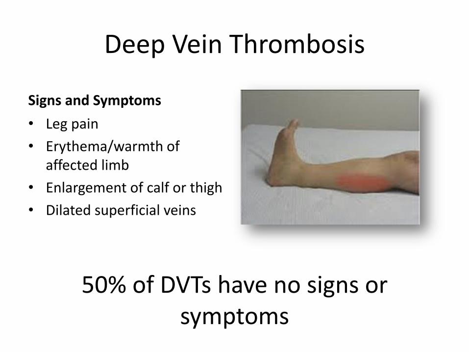
Deep Vein Thrombosis
Signs and Symptoms
• Leg pain
• Erythema/warmth of affected limb
• Enlargement of calf or thigh
• Dilated superficial veins
50% of DVTs have no signs or symptoms
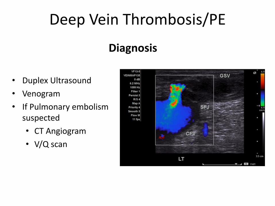
Deep Vein Thrombosis/PE
• Duplex Ultrasound
• Venogram
• If Pulmonary embolism suspected
• CT Angiogram
• V/Q scan
Diagnosis
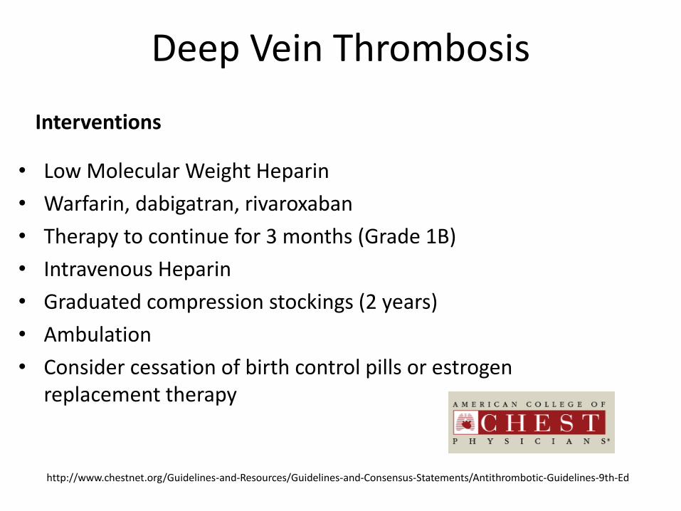
Deep Vein Thrombosis
Interventions
• Low Molecular Weight Heparin
• Warfarin, dabigatran, rivaroxaban
• Therapy to continue for 3 months (Grade 1B)
• Intravenous Heparin
• Graduated compression stockings (2 years)
• Ambulation
• Consider cessation of birth control pills or estrogen replacement therapy
http://www.chestnet.org/Guidelines-and-Resources/Guidelines-and-Consensus-Statements/Antithrombotic-Guidelines-9th-Ed
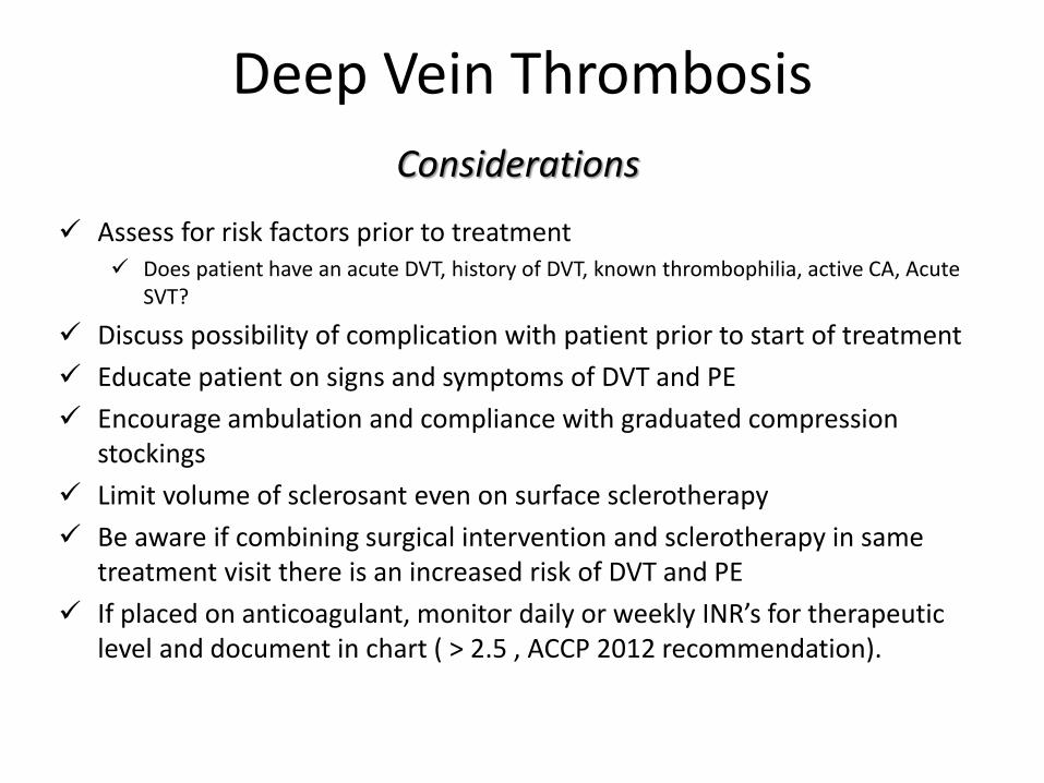
Deep Vein Thrombosis
Assess for risk factors prior to treatment Does patient have an acute DVT, history of DVT, known thrombophilia, active CA, Acute
SVT?
Discuss possibility of complication with patient prior to start of treatment
Educate patient on signs and symptoms of DVT and PE
Encourage ambulation and compliance with graduated compression stockings
Limit volume of sclerosant even on surface sclerotherapy
Be aware if combining surgical intervention and sclerotherapy in same treatment visit there is an increased risk of DVT and PE
If placed on anticoagulant, monitor daily or weekly INR’s for therapeutic level and document in chart ( > 2.5 , ACCP 2012 recommendation).
Considerations

Nerve Injury
The Saphenous nerve is the largest and longest branch of the femoral nerve and supplies the skin over the medial side of the leg
The sural nerve runs with the small saphenous vein on the posterior leg just lateral to the Achilles tendon. The sural nerve innervates lateral & posterior third of leg and lateral aspect of foot & heel, & lateral portion of the ankle
Almeida, Jose. Atlas Of Endovascular Venous Surgery. Philadelphia, PA: Elsevier, 2011. 16,18. Print.

Nerve Injury
• Diagnostic evaluation
• History and physical
• Pin prick test
• “Numbness” versus Hypoesthesia
• Intervention
• Usually self limiting
• NSAIDs
• Considerations Assess for loss of sensory and/or motor function
Document location and size of surface area affected
Verbally reassure patient that nerves can regenerate over time
High incidence of litigation with this complication
Irritation or injury to the Saphenous or Sural nerves or cutaneous branches after sclerotherapy

Neurological Complications
• Visual disturbances
• Blurred vision
• Pulsatile headache
• Visual aura preceding migraine
• Migraine
• Monocular blindness
• Transient Ischemic attack (rare)
• Cerebral Vascular Accident (rare)
Variety of neurologic events occurring after injection of liquid sclerosant, foamed sclerosant, or use of “ air block” technique

Neurological Complications
Patent Foramen Ovale
Air bubbles from foam sclerotherapy can travel from the right atrium to the left atrium and out to the brain. This can cause a variety of neurological symptoms from mild to severe

Neurological Complications
Interventions
• May be self limiting and no treatment necessary
• Neurological exam
• 100% oxygen
• Transfer to acute care facility
• Hyperbaric chamber indicated if symptomatic cerebral artery air embolus
Considerations
Assess patient for history of cardiac abnormalities or migraines
Limit the volume of foamed sclerosant on first visit
Keep patient supine for 5 minutes after injection of foam sclerosant
Document onset of neurological event and associated symptoms, progression or resolution of symptoms, vital signs, also note volume and concentration of sclerosant used

Allergic Reaction/ Anaphylaxis
Mild vs. Severe
Close observation
Be prepared!
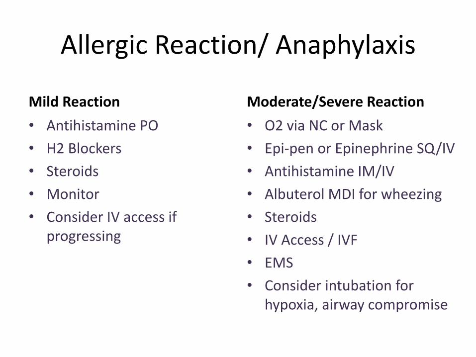
Allergic Reaction/ Anaphylaxis
Mild Reaction
• Antihistamine PO
• H2 Blockers
• Steroids
• Monitor
• Consider IV access if progressing
Moderate/Severe Reaction
• O2 via NC or Mask
• Epi-pen or Epinephrine SQ/IV
• Antihistamine IM/IV
• Albuterol MDI for wheezing
• Steroids
• IV Access / IVF
• EMS
• Consider intubation for hypoxia, airway compromise

Emergency Drug box or Crash Cart

Allergic Reaction
An allergic or anaphylaxis reaction can happen on the first exposure or on any re-exposure to a sclerosant Assess for previous know allergy to sclerosant or history of multiple allergies
Rapid recognition of an allergic reaction is essential
Patients should be observed 30 minutes in office after injections
Patients should be educated on signs and symptoms of allergic reactions in case of delayed onset
An emergency plan and emergency kit including medications, supplies and oxygen should be available in all practice settings
All staff should be properly trained to alert the emergency medical system
Document vital signs, onset of symptoms, medications and response to medications given if an allergic reaction should occur
Considerations
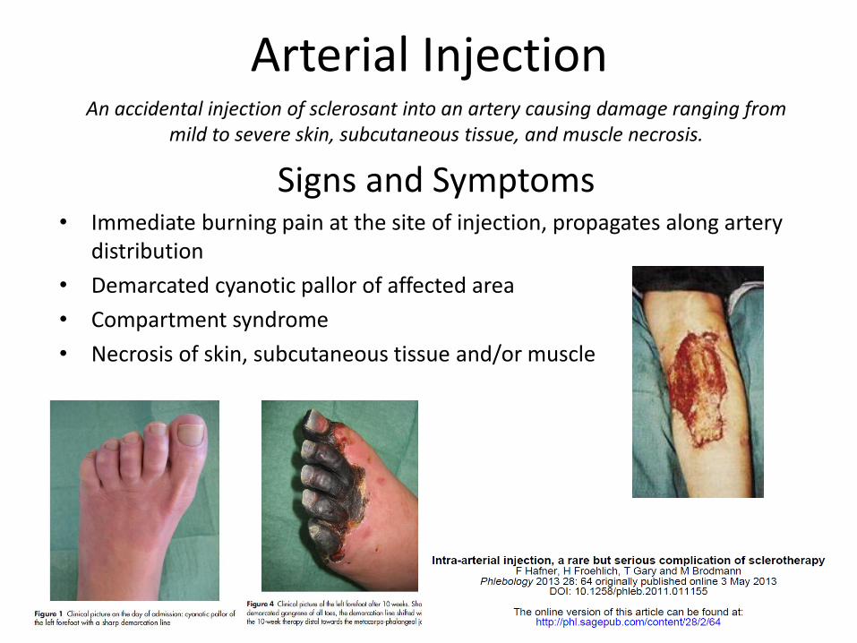
Arterial Injection
Signs and Symptoms• Immediate burning pain at the site of injection, propagates along artery
distribution
• Demarcated cyanotic pallor of affected area
• Compartment syndrome
• Necrosis of skin, subcutaneous tissue and/or muscle
An accidental injection of sclerosant into an artery causing damage ranging from mild to severe skin, subcutaneous tissue, and muscle necrosis.
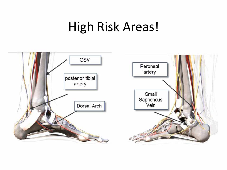
High Risk Areas!

Arterial Injection
Absence of consistent guidelines!
• Aspirate sclerosant if noted immediately
• Ice?
• Heparin
• Fibrinolytic therapy
• Vasodilators
• Catheter-directed thrombolysis
• Mechanical thrombectomy

Arterial Injection
Pain can happen quickly, or progress over several hours
Polidocanol has a local anesthetic effect; pain may not be appreciated until several days later
Document name of sclerosing agent volume of sclerosant used, location of injection, vital signs, skin findings, and presence of pedal pulses
Place ice at site of skin pallor to help decrease tissue damage
Patient must be transported to hospital
Vascular surgery and interventional radiology consult needed
This is a medical emergency!!

The most feared complication….
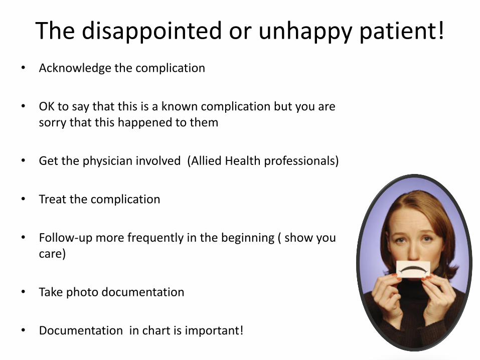
The disappointed or unhappy patient!• Acknowledge the complication
• OK to say that this is a known complication but you are sorry that this happened to them
• Get the physician involved (Allied Health professionals)
• Treat the complication
• Follow-up more frequently in the beginning ( show you care)
• Take photo documentation
• Documentation in chart is important!

Take Aways……
• Complications can happen even to the most experienced practitioner
• Know what they are and how to manage them
• Prevention is better then intervention!

Thank You!

ReferencesAlmeida, J. I., Raines, J.K. (2008). Laser ablation of cutaneous leg veins. Perspectives in Vascular Surgery and Endovascular Therapy, 20(4), 358-366.
Bergan, J. (2006). The vein book . Amsterdam: Elsevier.
Bergan, J. J., Weiss, R. A., & Goldman, M. P. (2000). Extensive tissue necrosis following high-concentration sclerotherapy for varicose veins. Dermatol Surg, 26, 535-542.
Bihari, I., & Magyar, E. (2001). Reasons for ulceration after injection treatment of telangiectasia. Dermatol Surg, 27, 133-136.
Bihari, I., Muranyi, A., & Bihari, P. (2005). Laser-doppler examination shows high flow in some common telangiectasias of the lower limb. Dermatol Surg, 31, 388-390.
Brzoza, Z., Kasperska-Zajac, A., Rogala, E., & Rogala, B. (2007, October/November). Anaphylactoid reaction after the use of sodium tetradecyl sulfate: A case report. Angiology, 58, 644-646.
Bush, R.G., Derrick, M., Manjoney, D. (2008). Major neurological events following foam Sclerotherapy. Phlebology, 23, 189-192.
Cavezzi, A., & Parsi, K. (2012). Complications of foam sclerotherapy. Phlebology, 27(), 46. http://dx.doi.org/10.1258/phleb.2012.012S09
Conrad, P., Malouf, G. M., & Stacey, M. C. (1995). The Australian Polidocanol (Aethoxysklerol) study. Dermatol Surg, 21, 334-336.
Gloviczki, P. (2009) Handbook of Venous Disorders (3rd ed).:London:Hodder Arnold.
Feied, C.F. (2007), MD. Sclerosing Solutions. In: Helane Fronek MD. The Fundamentals of Phlebology, Venous Disease for Clinicians. 2nd. American College of Phlebology:P. 23, Ch.5.
Galanaud, J. P., Gentry, C., Sevestre, M. A., & Bisot, D. , et al .Predictive factors for concurrent deep-vein thrombosis and symptomatic venous thrombotic recurrence in case of superficial venous thrombosis. The OPTIMEV study. Thrombosis and haemostasis 2011, 105(1), 31-39.
Gloviczki, P., Dalsing, M. C., Eklof, B., Moneta, G. L., & Wakfield, T. W. (2009). Handbook of Venous Disorders (3rd ed.). London: Hodder Education
Goldman, M. P., Bergan, J. J., Guex, J. J. (2006) Sclerotherapy: Treatment of varicose and telangiectatic leg veins (4th ed.). Amsterdam: Mosby
Guex, J. (1993). Indication for the sclerosing agent polidocanol. J Dermatol Surg Oncol, 19, 959-961.
Guex, J., Allaert, F., Gillet, J., & Chleir, F. (2005). Immediate and midterm complications of sclerotherapy: Report of a prospective multicenter registry of 12,173 sclerotherapy sessions. Dermatol Surg, 31, 123-128.
ReferencesGuex, J. J. (2009). Complications and side-efects of foam sclerotherapy. Phlebology, 24, 270. http://dx.doi.org/10.1258/phleb.2009.009049
Hafner, F., Froehlich, H., Gary, T., & Brodmann, M. (2013, May 3). Intra-arterial injection, a rare but serious complication of sclerotherapy. Phlebology, 28(), 64. http://dx.doi.org/10.1258/phleb.2011.011155
Hanson, J.N., Ascher, E., DePippo, P., Lorensen, E., Scheinman, M., Yorkovich, W., Hingorani , A. Saphenous vein thrombophlebitis (SVT): a deceptively benign disease J Vasc Surg. 1998 April; 27(4): 677–680.
Kern, P., Ramelet, A. A., Wutschert, R., & Bounameaux, H. (2004). Single-blind, randomized study comparing chromated glycerin, polidocanol solution, and polidocanol foam for treatment of telangiectatic leg veins. Dermatologic Surgery, 30(3), 367-372.
Leach, B. C., & Goldman, M. P. (2003). Comparative trial between sodium tetradecyl sulfate and glycerin in the treatment of telangiectatic leg veins. Dermatol Surg, 29, 612-615.
Miyake, R. L., King, J. T., Kikuchi, R., Duarte, F. H., Davidson, J. R., & Oba, C. (2012). Role of injection pressure, flow and sclerosant viscosity in causing cutaneous ulceration during sclerotherapy. Phlebology, 27, 383. http://dx.doi.org/10.1258/phleb.2011.011076
Munavalli, G. S., & Weiss, R. A. (2007). Complications of sclerotherapy. Semin Cutan Med Surg, 26, 22-28.
Ratinahirana, H., Benigni, J. P., & Bousser, M. G. (2003). Injection of polidocanol foam (PF) in varicose veins as a trigger for attacks of migraine with visual aura. Cephalalgia, 23, 850-851.
Sadick, N. S., & Sorhaindo, L. (2006). An evaluation of post-sclerotherapy laser compression and its efficacy in the treatment of leg telangiectasias. Phlebology, 21,191. http://dx.doi.org/10.1258/026835506779115771
Sibbitt, R. R., Palmer, D. J., & Sibbitt.Jr., W. L. (2008, October/November). Integration of patient safety technologies into sclerotherapy for varicose veins. Vascular and Endovascular Surgery, 42(5), 446-455.
Thibault, P. K. (n.d). Sclerotherapy of varicose veins and telangiectasias: A 2-year experience with sodium tetradecyl sulphate. Retrieved March 29, 2009, from http://www.cavezzi.it/thibauly.html
Tretbar, L. L. (1989, February). Injection sclerotherapy for spider telangiectasias: A 20-year experience with sodium tetradecyl sulfate. J Dermatol Surg Oncol, 15(2), 223-225.
Weiss, R. A., Feied, C.F., Weiss, M.A. (2000). Vein diagnosis and treatment. Mcgraw-Hill Professional
Zimmet, S. (2009). Sclerotherapy complications. Retrieved March 29, 2009, from http://www.veindirectory.org/news/news_details.asp?ID=18
Zimmet, S. E. (1996). Hyaluronidase in the prevention of sclerotherapy-induced extravasation necrosis: A dose-
response study. Dermatol Surgery