Embed Size (px)
Citation preview

MANAGEMENT OF TRAUMATIC SPLEEN INJURY
Michael LeCompte

History
Spleen was the source of emotion Aristotle challenged this idea and argued that
the spleen was unnecessary Gaius Pliny Secundus (Natural History) –
remove the spleen to improve the speed of athletes.
1549 – first splenectomy performed by Adrian Zacarellie for hypersplenism
1590 – first traumatic splenectomy performed by Franciscus Rosetti. (Partial Splenectomy)

History Cont…
Senn, N. 1903 – Described non-operative management of the spleen.
Kocher 1911 – described high failure rate of NOP and advocated for surgical management for blunt traumatic injury
1950s-1970s – high OPSI rates in children following splenectomy. Moved towards NOP in children
1990s – successful NOP in adults

Trauma Patient
Traumatic injury to the LUQ
Rib fractures 9-12 on the left
“Seat belt sign” Hypotension Pain referred to the left
shoulder increased with inspiration (Kehr’s sign)
Penetrating trauma
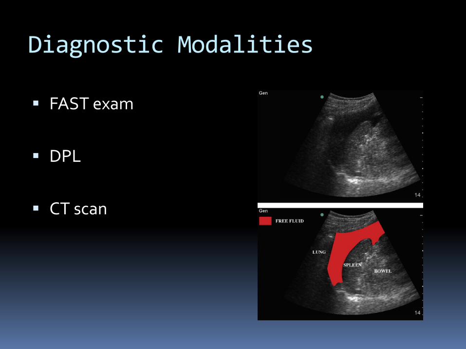
Diagnostic Modalities
FAST exam
DPL
CT scan

Grade I Injury Subcapsular Hematoma: < 10% of surface area Laceration: < 1cm into parenchyma WWW.TRAUMA.ORG

Grade II Injury Subcapsular Hematoma: 10-50% of surface area
Laceration: 1-3cm in depth. Does not involve trabecular vessel
WWW.TRAUMA.ORG

Grade III Injury Subcapsular Hematoma: >50% of surface
area Ruptured >5cm
Intraparenchymal Laceration: > 3cm Involving trabecular
vessel
WWW.TRAUMA.ORG

Grade IV Injury Laceration: Involves segmental or hilar vessels. >25%
devascularization of the spleen
Malays J Med Sci. 2011 Jan-Mar; 18(1): 60–67.
PMCID: PMC3216201
Computed Tomography of Blunt Spleen Injury: A Pictorial Review

Grade V Injury Shatter Spleen Laceration of hilar vasculature. Devascularized Spleen Avulsion Malays J Med Sci. 2011 Jan-Mar; 18(1): 60–67.
PMCID: PMC3216201
Computed Tomography of Blunt Spleen Injury: A Pictorial Review

AAST Grading Chart

To Operate or Not to Operate
80% of blunt splenic injuries can be managemed non-operatively.

Eastern Association of Surgery for Trauma Guidlines Level 1 Data:
1. Patient’s with hemodynamic instability or diffuse peritonitis should be taken urgently for laparotomy
Level 2 Data:
1. A routine laparotomy is not indicated in the hemodynamically stable patient presenting with an isolated spleen injury
2. The severity of the injury, suggested by grade CT grade, neurologic status, age >55 and or the presence of associated injuries are not contraindications to a trial of NOP management.
3. NOP should only be considered in an environment
that has capabilities to monitoring, serial exams and
available OR.

Failure of Non-operative Management Based on CT Grade

Non- operative Management
Serial Exams
Serial hemoglobin/hematocrit
Monitor vitals and urine output
Period of immobility or bed rest +/- Follow up CT scan 24-48 hours ( Weinberg et al showed repeat CT scan at 24-48 hours for grades II and above diagnosed
latent pseudoaneurysm that underwent embolization with improved outcomes.)

Embolization
1. AAST Grade III or Higher
2. Contrast Blush
3. Moderate Hemoperitoneum
4. Evidence of Ongoing Bleeding

Pros and Cons of Embolization
Cooney et al – showed use of selective arterial embolization to improve non-operative rates
Preservation of functional splenic tissue Hann et al – 20% complication rate including
failure to control bleeding, need for repeat embolization.
Failure to identify concurrent injuries Splenic Abscess

Surgical Management
1. Midline incision
2. Evacuation of blood
Packing of LUQ
Medial/Downward rotation of spleen with posterior packing
Pinch Hilum for control of bleeding.
Exploration for concurrent injury.

Spleen Mobilization

Splenorrhaphy
Linear lacerations or capusular tears may be managed by suture ligation of bleeding vessels, figure of eight closure, pledget butress or omental patch.

Splenic Mesh
Useful with stellate or multiple lacerations to the spleen
Specialized mesh bags Wrap spleen in
conventional mesh. Tighten mesh around
the spleen with stay sutures to tamponade bleeding.