Embed Size (px)
Citation preview
Listen to this manuscript’s
audio summary by
JACC Editor-in-Chief
Dr. Valentin Fuster.
J O U R N A L O F T H E A M E R I C A N C O L L E G E O F C A R D I O L O G Y V O L . 6 7 , N O . 8 , 2 0 1 6
ª 2 0 1 6 B Y T H E A M E R I C A N C O L L E G E O F C A R D I O L O G Y F O U N D A T I O N I S S N 0 7 3 5 - 1 0 9 7 / $ 3 6 . 0 0
P U B L I S H E D B Y E L S E V I E R h t t p : / / d x . d o i . o r g / 1 0 . 1 0 1 6 / j . j a c c . 2 0 1 5 . 1 1 . 0 6 1
THE PRESENT AND FUTURE
STATE-OF-THE-ART REVIEW
Management of Pulmonary Embolism
An UpdateStavros V. Konstantinides, MD, PHD,a,b Stefano Barco, MD,a Mareike Lankeit, MD,a Guy Meyer, MDc
ABSTRACT
Fro
Ge
Ge
Ko
an
ho
acc
an
an
pa
be
Ph
Ma
Pulmonary embolism (PE) remains a major contributor to global disease burden. Risk-adapted treatment and follow-up
contributes to a favorable outcome. Age-adjusted cutoff levels increase D-dimer specificity and may decrease overuse
of imaging procedures and overdiagnosis of PE. Primary systemic fibrinolysis has an unfavorable risk–benefit ratio in
intermediate-risk PE; catheter-directed techniques are an option for patients with hemodynamic decompensation and
high bleeding risk. New oral anticoagulant agents are effective and safe alternatives to standard anticoagulation
regimens. Recent trial data do not support insertion of cava filters in patients who can receive anticoagulant treat-
ments. Remaining areas of uncertainty include the therapeutic implications of subsegmental PE, the optimal diagnostic
approach to the pregnant patient with suspected PE, and the efficacy and safety of new oral anticoagulant agents in
patients with cancer. Campaigns to increase awareness combined with strategies to implement guideline recommen-
dations will be crucial steps towards further optimizing management of acute PE. (J Am Coll Cardiol 2016;67:976–90)
© 2016 by the American College of Cardiology Foundation.
V enous thromboembolism (VTE), which en-compasses deep vein thrombosis (DVT) andits most dangerous complication, acute pul-
monary embolism (PE), represents a major threat tothe health, the well-being, and occasionally, the livesof a large number of patients worldwide. The annualincidence rate of VTE ranges between 75 and 269cases per 100,000 persons, as shown by studiesin Western Europe, North America, Australia, andsouthern Latin America, with subjects 70 years ofage or older having an incidence of up to 700 per100,000 (1). As the risk of VTE approximately doubleswith each decade after the age of 40 years, it is to beexpected that an increasing number of people in
m the aCenter for Thrombosis and Hemostasis, University Medical Cen
rmany; bDepartment of Cardiology, Democritus University of Thrace, A
orges Pompidou, AP-HP, Université Paris Descartes, Sorbonne Paris Cité, a
nstantinides, Barco, and Lankeit was supported by the German Federal M
d 01EO1503). The authors are responsible for the contents of this paper. Dr.
noraria from Bayer HealthCare, Boehringer Ingelheim, Daiichi-Sankyo, a
ommodation/meeting expenses from Bayer HealthCare; and institutional
d Daiichi-Sankyo. Dr. Barco has received an educational travel grant from D
d lecture honoraria from Actelion, Bayer HealthCare, Daiichi-Sankyo, and
yment for travel accommodation/meeting expenses from Bayer HealthC
rship, consultancy, and lecture honoraria to his institution from Bayer H
arma, and Daiichi-Sankyo; and institutional grants from Boehringer Ingel
nuscript received October 14, 2015; revised manuscript received Novemb
aging societies throughout the world will be diag-nosed with the disease in the years to come. Despitethe epidemiological relevance of PE and its highshort-term mortality, a relative lack of public aware-ness was demonstrated by a global survey thatincluded more than 7,200 responders (2). In partic-ular, the level of awareness was clearly lower thanthat for other thrombotic disorders, such as heartattack and stroke, or compared with diseases previ-ously addressed by sensitization campaigns, such asbreast cancer, prostate cancer, and acquired immuno-deficiency syndrome (2).
The present paper critically reviews recent datathat have contributed to substantial improvement of
ter of the Johannes Gutenberg University, Mainz,
lexandroupolis, Greece; and the cHôpital Européen
nd GIRC Thrombose, Paris, France. The work of Drs.
inistry of Education and Research (BMBF 01EO1003
Konstantinides has received consultancy and lecture
nd Pfizer–Bristol-Myers Squibb; payment for travel
grants from Boehringer Ingelheim, Bayer HealthCare,
aiichi-Sankyo. Dr. Lankeit has received consultancy
Pfizer–Bristol-Myers Squibb. Dr. Meyer has received
are, Leo Pharma, and Daiichi-Sankyo; board mem-
ealthCare, Pfizer–Bristol-Myers Squibb, Sanofi, Leo
heim, and Leo Pharma.
er 11, 2015, accepted November 17, 2015.
AB BR E V I A T I O N S
AND ACRONYM S
ASO = antisense
oligonucleotide
CI = confidence interval
CT = computed tomography
CTEPH = chronic
thromboembolic pulmonary
hypertension
DVT = deep vein thrombosis
ESC = European Society of
Cardiology
FXI = factor XI
NOAC = non–vitamin
K-dependent oral
anticoagulant(s)
NT-proBNP = N-terminal pro–
B-type natriuretic peptide
OR = odds ratio
PE = pulmonary embolism
PESI = Pulmonary Embolism
Severity Index
RR = relative risk
rtPA = recombinant tissue-
type plasminogen activator
SPECT = single-photon
emission computed
tomography
sPESI = simplified Pulmonary
Embolism Severity Index
VKA = vitamin K antagonist
V/Q = ventilation/perfusion
VTE = venous
boembolism
J A C C V O L . 6 7 , N O . 8 , 2 0 1 6 Konstantinides et al.M A R C H 1 , 2 0 1 6 : 9 7 6 – 9 0 Pulmonary Embolism Update
977
management strategies for acute PE in past years,while also highlighting areas that remain to be clari-fied or resolved by ongoing and future research.
EVOLVING STRATEGIES FOR DIAGNOSIS AND
RISK ASSESSMENT
APPROPRIATE TRIAGE OF PATIENTS FOR IMAGING
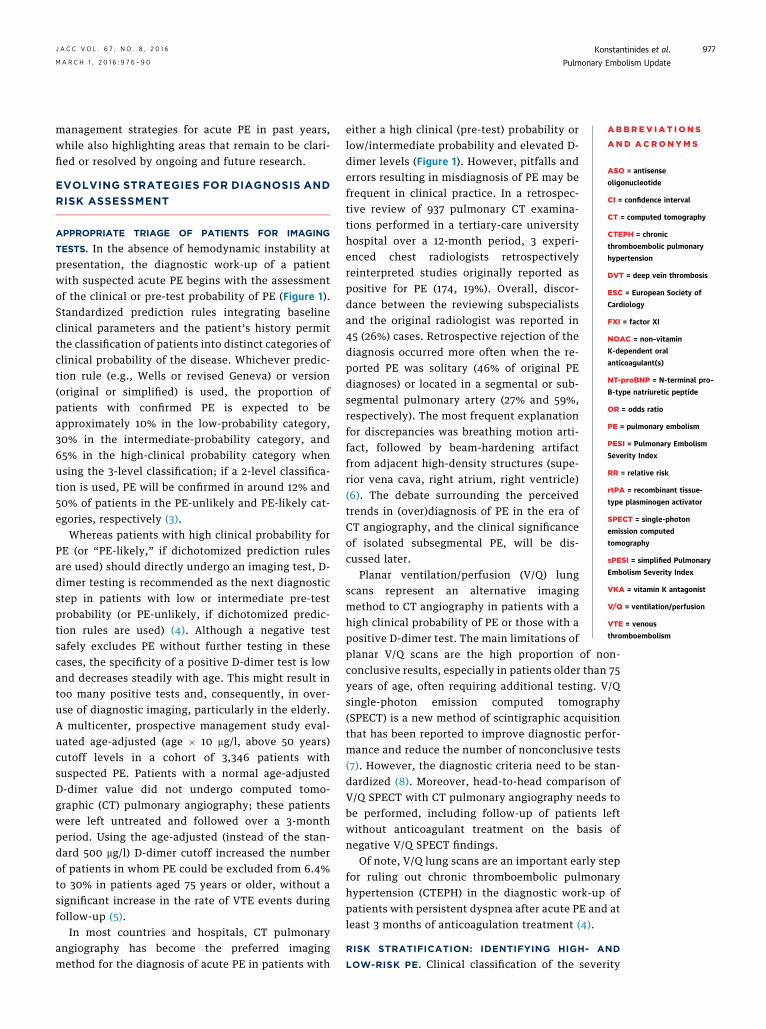
TESTS. In the absence of hemodynamic instability atpresentation, the diagnostic work-up of a patientwith suspected acute PE begins with the assessmentof the clinical or pre-test probability of PE (Figure 1).Standardized prediction rules integrating baselineclinical parameters and the patient’s history permitthe classification of patients into distinct categories ofclinical probability of the disease. Whichever predic-tion rule (e.g., Wells or revised Geneva) or version(original or simplified) is used, the proportion ofpatients with confirmed PE is expected to beapproximately 10% in the low-probability category,30% in the intermediate-probability category, and65% in the high-clinical probability category whenusing the 3-level classification; if a 2-level classifica-tion is used, PE will be confirmed in around 12% and50% of patients in the PE-unlikely and PE-likely cat-egories, respectively (3).
Whereas patients with high clinical probability forPE (or “PE-likely,” if dichotomized prediction rulesare used) should directly undergo an imaging test, D-dimer testing is recommended as the next diagnosticstep in patients with low or intermediate pre-testprobability (or PE-unlikely, if dichotomized predic-tion rules are used) (4). Although a negative testsafely excludes PE without further testing in thesecases, the specificity of a positive D-dimer test is lowand decreases steadily with age. This might result intoo many positive tests and, consequently, in over-use of diagnostic imaging, particularly in the elderly.A multicenter, prospective management study eval-uated age-adjusted (age � 10 mg/l, above 50 years)cutoff levels in a cohort of 3,346 patients withsuspected PE. Patients with a normal age-adjustedD-dimer value did not undergo computed tomo-graphic (CT) pulmonary angiography; these patientswere left untreated and followed over a 3-monthperiod. Using the age-adjusted (instead of the stan-dard 500 mg/l) D-dimer cutoff increased the numberof patients in whom PE could be excluded from 6.4%to 30% in patients aged 75 years or older, without asignificant increase in the rate of VTE events duringfollow-up (5).
In most countries and hospitals, CT pulmonaryangiography has become the preferred imagingmethod for the diagnosis of acute PE in patients with
either a high clinical (pre-test) probability orlow/intermediate probability and elevated D-dimer levels (Figure 1). However, pitfalls anderrors resulting in misdiagnosis of PE may befrequent in clinical practice. In a retrospec-tive review of 937 pulmonary CT examina-tions performed in a tertiary-care universityhospital over a 12-month period, 3 experi-enced chest radiologists retrospectivelyreinterpreted studies originally reported aspositive for PE (174, 19%). Overall, discor-dance between the reviewing subspecialistsand the original radiologist was reported in45 (26%) cases. Retrospective rejection of thediagnosis occurred more often when the re-ported PE was solitary (46% of original PEdiagnoses) or located in a segmental or sub-segmental pulmonary artery (27% and 59%,respectively). The most frequent explanationfor discrepancies was breathing motion arti-fact, followed by beam-hardening artifactfrom adjacent high-density structures (supe-rior vena cava, right atrium, right ventricle)(6). The debate surrounding the perceivedtrends in (over)diagnosis of PE in the era ofCT angiography, and the clinical significanceof isolated subsegmental PE, will be dis-cussed later.
Planar ventilation/perfusion (V/Q) lungscans represent an alternative imagingmethod to CT angiography in patients with ahigh clinical probability of PE or those with apositive D-dimer test. The main limitations of
planar V/Q scans are the high proportion of non-conclusive results, especially in patients older than 75years of age, often requiring additional testing. V/Qsingle-photon emission computed tomography(SPECT) is a new method of scintigraphic acquisitionthat has been reported to improve diagnostic perfor-mance and reduce the number of nonconclusive tests(7). However, the diagnostic criteria need to be stan-dardized (8). Moreover, head-to-head comparison ofV/Q SPECT with CT pulmonary angiography needs tobe performed, including follow-up of patients leftwithout anticoagulant treatment on the basis ofnegative V/Q SPECT findings.Of note, V/Q lung scans are an important early stepfor ruling out chronic thromboembolic pulmonaryhypertension (CTEPH) in the diagnostic work-up ofpatients with persistent dyspnea after acute PE and atleast 3 months of anticoagulation treatment (4).
RISK STRATIFICATION: IDENTIFYING HIGH- AND
LOW-RISK PE. Clinical classification of the severity
throm
FIGURE 1 PE: Risk-Adjusted Management in the Acute Phase and Over the Long Term
PREDICTORS OFEARLY ADVERSE OUTCOME
RISK FACTORS FORRECURRENT VTERISK FACTORS
FOR FIRST VTE
HORMONALCONTRACEPTION
TRAUMA OR FRACTURE
SURGERY
HORMONAL REPLACEMENTTREATMENT
PREGNANCYAND POSTPARTUM
IMMOBILIZATION
AGE
INFLAMMATORYBOWEL DISEASE
THROMBOPHILIA†
OBESITYCANCERCHEMOTHERAPY
PRIOR VTED-DIMERS
FIRST UNPROVOKEDVTE EVENT
MALE SEXCHRONIC HEART FAILURE
CHRONIC LUNG DISEASEACTIVE CANCER
VITAL SIGNSHYPOXEMIA
RV DYSFUNCTION(CT/ECHO)
BIOCHEMICAL MARKERS*
PRE-TESTCLINICAL ASSESSMENT DIAGNOSIS
ACUTE RISKSTRATIFICATION TREATMENT
LONG-TERMCLINICAL COURSE
Revised Geneva scoreWells ruleEmpirical assessment
(Age-adjusted) D-dimersCTPAV/Q scanEchocardiographyCUS
PESI and sPESIBiochemical markers*RV dysfunction(echocardiography)RV enlargment (CTPA)
Parenteral anticoagulantsOral anticoagulantsFibrinolyticsCatheter-directedtechniquesSurgical embolectomyVena cava filters
Assess bleeding riskPredict VTErecurrenceFocused screeningfor CTEPH insymptomatic patients
HIGH CLINICALPROBABILITY
LOW ORINTERMEDIATE
CLINICALPROBABILITY
Hemodynamicinstability
Absence ofhemodynamic
instability
Age-adjustedpositive
D-dimers
ALGORITHM FORHIGH-RISK PE
CTPA
CTPA
ALGORITHM FORNON HIGH-RISK PE
V/Q scan
HIGH RISK
INTERMEDIATE RISK
LOW RISK
PRIMARYREPERFUSION
ANTICOAGULANTTHERAPY
ANTICOAGULANTTHERAPY
ANTICOAGULANTTHERAPY
BLEEDING
RECURRENT VTE
CTEPH
Echocardiography (ifCTPA not readilyavailable or uncontrolledhypotension)
CUS-based algorithms
Hemodynamicinstability
INTERMEDIATE-HIGHINTERMEDIATE-LOW
plus
(Rescue reperfusion)
(Early discharge)
No validated predictionmodels for VTE patients
Standard-duration vs.extended (indefinite)treatment
Individualized follow-upprograms and intervals
*Biochemical markers include markers of myocardial injury (troponins, heart-type fatty acid-binding protein) and markers of heart failure (BNP or N-terminal-proBNP).
†Only antiphospholipid syndrome and high-risk inherited thrombophilia (i.e., homozygosity for factor V Leiden, homozygosity for prothrombin G20210A mutation,
double heterozygosity, antithrombin deficiency) are considered. Nevertheless, routine thrombophilia testing is not indicated in PE patients. BNP ¼ B-type natriuretic
peptide; CT ¼ computed tomography; CTEPH ¼ chronic thromboembolic pulmonary hypertension; CTPA ¼ computed tomographic pulmonary angiogram; CUS ¼compression ultrasound; Echo ¼ echocardiography; N-terminal-proBNP ¼ N-terminal pro–B-type natriuretic peptide; PE ¼ pulmonary embolism; PESI ¼ pulmonary
embolism severity index; RV ¼ right ventricular; sPESI ¼ simplified pulmonary embolism severity index; V/Q scan ¼ ventilation/perfusion lung scan; VTE ¼ venous
thromboembolism.
Konstantinides et al. J A C C V O L . 6 7 , N O . 8 , 2 0 1 6
Pulmonary Embolism Update M A R C H 1 , 2 0 1 6 : 9 7 6 – 9 0
978
of acute PE is on the basis of the estimated earlydeath risk (Figure 1). It has been established that thepresence of right ventricular dysfunction and failureresulting from acute pressure overload is the prin-cipal determinant of the patient’s early clinicalcourse and risk of an adverse outcome (reviewedin [4,9]). Accordingly, high-risk or massive PE refersto the presence of shock or persistent arterialhypotension as a result of overt right ventricularfailure. This is clearly a life-threatening situation, inwhich prompt reperfusion treatment (as discussedlater) is needed, along with circulatory and res-piratory support in order to break the spiral of
hemodynamic deterioration and to increase thechances of survival (4,10,11).
More than 95% of patients with acute PE are (orappear to be) hemodynamically stable at presentationand are thus not considered to be at high risk (12).Within this large group, the next challenging step isto determine which patients will need hospitalizationand possibly initial monitoring, and to distinguishthem from those who are at truly low risk and mayqualify for early discharge and outpatient treatment.To be used as risk stratification tools for this purpose,baseline clinical parameters and prediction scoresderived from them should reliably exclude severe

J A C C V O L . 6 7 , N O . 8 , 2 0 1 6 Konstantinides et al.M A R C H 1 , 2 0 1 6 : 9 7 6 – 9 0 Pulmonary Embolism Update
979
acute illness and the presence of significant comor-bidity. The Pulmonary Embolism Severity Index(PESI) has been extensively validated and shown tofulfill these requirements; patients in PESI risk strata Iand II were at low risk of 30-day mortality (13). Thesimplified version of the Pulmonary EmbolismSeverity Index (sPESI) also possessed a high negativepredictive value for ruling out an adverse earlyoutcome (14,15). Thus, a substantial proportion (be-tween 25% and 46%, depending on the cohort stud-ied) of all patients with acute PE can be classified asbeing at low risk on the basis of a PESI risk class of I orII, or a sPESI score of 0 (14–16). Although the negativepredictive value of the index may rise even furtherwhen it is combined with the (negative) result of ahigh-sensitivity cardiac troponin assay (17), it is un-certain how often this extra reassurance is reallyneeded in clinical practice.
Despite the uncontested prognostic value of thePESI, it should be kept in mind that this was primarilydesigned as an epidemiological tool, and not as a directguide to PE management. The only prospective trialthat used this severity index to randomize patients tooutpatient versus in-hospital treatment of PE requirednumerous additional eligibility criteria, including asupportive social environment (18). Other groups,particularly in the Netherlands, chose to developexplicit home treatment–oriented clinical criteria,either alone (Hestia criteria) (19) or in combinationwith biomarker testing (N-terminal pro–B-type natri-uretic peptide [NT-proBNP] plasma levels<500 pg/ml)(20), which they tested successfully in small- tomedium-sized (150 to 300 patients) prospective cohorttrials. These criteria await validation in larger cohortsand further countries. Importantly, and in view of firstreports that severe right ventricular dysfunction mayoccasionally be present in a patient with a negativesPESI (21), it will also need to be determined in thiscontext whether CT or echocardiographic imaging ofthe right ventricle should be added to clinical eligi-bility criteria for immediate or early discharge in orderto maximize patient safety.EVOLVING DEFINITION OF INTERMEDIATE-RISK PE.
What are the next steps if a normotensive patient isnot classified into the low-risk category on the basisof the clinical criteria described previously? With theobjective of further developing the concept ofintermediate-risk PE, the 2014 European Society ofCardiology (ESC) guidelines critically reviewed thecombinations of imaging (echocardiographic or CTangiographic) parameters and laboratory biomarkersthat can be used to detect right ventricular dysfunc-tion and/or myocardial injury (4). Taking into accountthat imaging and laboratory tests have consistently
been shown to have prognostic values additive toeach other and to those of clinical parameters (22,23),and aiming to discourage uncritical time- andresource-consuming laboratory and/or echocardio-graphic testing in every patient with confirmed PEwithout prior clinical triage, the updated ESC guide-lines proposed a stepwise classification of early risk,as displayed in the Central Illustration (4). Althoughsupported by evidence from cohort studies and vali-dated (at least in a modified form) in a large ran-domized therapeutic trial (24), the current riskstratification scheme will almost certainly needfurther improvement in the following years. Inparticular, the definition and positive prognosticvalue of the intermediate-high-risk class must beoptimized to better identify candidates for reperfu-sion treatment among normotensive patients withPE. Promising steps in this direction include the useof age-adjusted cutoff values for high-sensitivitycardiac troponin T in patients age 75 years or older(25) and laboratory biomarkers more specific forrelevant neurohumoral activation or for myocardialinjury (26). In 2,874 normotensive PE patientsderived from 6 cohort studies, a multidimensionalprognostic model on the basis of 4 variables (systolicblood pressure 90 to 100 mm Hg; heart rate $110beats/min; elevated cardiac troponin; right ventricu-lar dysfunction on imaging) was constructed, yielding3 risk strata (27). The rate of an adverse 30-dayoutcome was 29% in the high-risk stratum (>4points) of the derivation population (27), and up to42% in a validation cohort of 1,083 patients (28).External validation and implementation of new pre-diction rules in prospective management trials arenecessary steps before these can be integrated intofuture risk stratification algorithms.
ADVANCES IN ANTICOAGULATION
TREATMENT
In patients with acute VTE (presenting either as PE orproximal DVT), the duration of anticoagulation treat-ment should cover at least 3 months (4,10,11,29).Within this period, traditional regimens of acute-phase treatment consist of parenteral anticoagulation(intravenous unfractionated heparin, subcutaneouslow-molecular-weight heparin, or fondaparinux) overthe first 5 to 10 days, overlapping and followed by avitamin K antagonist (VKA), which is adjusted toobtain a therapeutic (2.0 to 3.0) international normal-ized ratio. Advances in knowledge and the remainingopen questions related to determining the optimalduration (beyond thefirst 3months) of anticoagulationafter PE are discussed separately in this review, in the
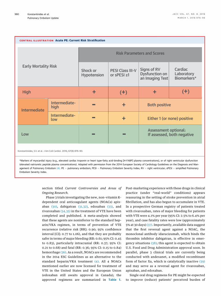
CENTRAL ILLUSTRATION Acute PE: Current Risk Stratification
Risk Parameters and Scores
Early Mortality Risk
+
+
+
+ (+)(+)
Either 1 (or none) positive
Assessment optional:If assessed, both negative-
Both positive
Shock orHypotension
PESI Class III-Vor sPESI ≥1
Signs of RVDysfunction onan Imaging Test
CardiacLaboratoryBiomarkers*
--
-Low
Intermediate-high
Intermediate-low
High
Intermediate
Konstantinides, S.V. et al. J Am Coll Cardiol. 2016; 67(8):976–90.
*Markers of myocardial injury (e.g., elevated cardiac troponin or heart type-fatty acid-binding [H-FABP] plasma concentrations), or of right ventricular dysfunction
(elevated natriuretic peptide plasma concentrations). Adapted with permission from the 2014 European Society of Cardiology Guidelines on the Diagnosis and Man-
agement of Pulmonary Embolism (4). PE ¼ pulmonary embolism; PESI ¼ Pulmonary Embolism Severity Index; RV ¼ right ventricular; sPESI ¼ simplified Pulmonary
Embolism Severity Index.
Konstantinides et al. J A C C V O L . 6 7 , N O . 8 , 2 0 1 6
Pulmonary Embolism Update M A R C H 1 , 2 0 1 6 : 9 7 6 – 9 0
980
section titled Current Controversies and Areas ofOngoing Research.
Phase 3 trials investigating the new, non–vitamin K-dependent oral anticoagulant agents (NOACs) apix-aban (30), dabigatran (31,32), edoxaban (33), andrivaroxaban (34,35) in the treatment of VTE have beencompleted and published. A meta-analysis showedthat these agents are noninferior to the standard hep-arin/VKA regimen, in terms of prevention of VTErecurrence (relative risk [RR]: 0.90; 95% confidenceinterval [CI]: 0.77 to 1.06), and that they are probablysafer in terms of major bleeding (RR: 0.61; 95% CI: 0.45to 0.83), particularly intracranial (RR: 0.37; 95% CI:0.21 to 0.68) and fatal (RR: 0.36; 95% CI: 0.15 to 0.84)hemorrhage (36). As a result, NOACs are recommendedin the 2014 ESC Guidelines as an alternative to thestandard heparin/VKA treatment (4). All 4 NOACsmentioned earlier are now licensed for treatment ofVTE in the United States and the European Union(edoxaban still awaits approval in Canada); theapproved regimens are summarized in Table 1.
Post-marketing experience with these drugs in clinicalpractice (under “real-world” conditions) appearsreassuring in the setting of stroke prevention in atrialfibrillation, and has also begun to accumulate in VTE.In a prospective German registry of patients treatedwith rivaroxaban, rates of major bleeding for patientswith VTE were 4.1% per year (95% CI: 2.5% to 6.4% peryear), and case fatality rates were low (approximately5% at 30 days) (37). Importantly, available data suggestthat the first reversal agent against a NOAC, themonoclonal antibody idarucizumab, which binds thethrombin inhibitor dabigatran, is effective in emer-gency situations (38); this agent is expected to obtainU.S. Food and Drug Administration approval soon. Inparallel, phase 3 clinical trials are currently beingconducted with andexanet, a modified recombinantform of factor Xa, which is catalytically inactive (39)and may serve as a reversal agent for rivaroxaban,apixaban, and edoxaban.
Single oral drug regimens for PE might be expectedto improve (reduce) patients’ perceived burden of

TABLE 1 Non–Vitamin K-Dependent Oral Anticoagulant Agents in the Treatment and Secondary Prevention of VTE
Dosage and Interval
Not Recommended or Contraindicated*Initial Phase Long-Term Phase Extended Phase
Rivaroxaban† 15 mg twice daily withfood for 21 days
20 mg once daily with food � CrCl <30 ml/min� Moderate or severe hepatic impairment
(Child-Pugh B and C), or hepatic diseaseassociated with coagulopathy
� Concomitant use of combined P-gp andstrong CYP3A4 inhibitors or inducers
Dabigatranetexilate‡
Initial therapy withparenteralanticoagulation for5–10 days shouldprecedeadministration ofdabigatran etexilate
150 mg twice daily � CrCl <30 ml/min� Concomitant treatment with P-gp inhibitors
in patients with CrCl <50 ml/min� Concomitant treatment with P-gp inducers
(i.e., rifampin)
Apixaban 10 mg twice dailyfor 7 days
5 mg twice daily 2.5 mg twice daily after atleast 6 months of treatment
� CrCl <15 ml/min� Severe hepatic impairment (Child-Pugh C),
or hepatic disease associated withcoagulopathy
� Strong dual inhibitors or inducers ofCYP3A4 and P-gp
Edoxaban§ Initial therapy withparenteralanticoagulation for5–10 days shouldprecedeadministrationof edoxaban
60 mg once daily30 mg once daily can be considered in patientswith $1 of the following factors: CrCl15–50 ml/min; body weight #60 kg;concomitant use of P-gp inhibitors,cyclosporin, dronedarone, erythromycin,or ketoconazole
� CrCl <15 ml/min� Moderate or severe hepatic impairment
(Child-Pugh B and C), or hepatic diseaseassociated with coagulopathy
� Concomitant treatment with rifampin
The table displays drugs and regimens on the basis of U.S. FDA approval for the treatment of acute VTE. *In addition to the specific conditions listed here, all mentionedanticoagulant agents should be avoided in patients: 1) with hemodynamically unstable acute pulmonary embolism for whom thrombolysis or pulmonary embolectomy may berequired; 2) requiring dialysis; 3) at significant risk of bleeding or with active pathological bleeding; 4) treated with a concomitant anticoagulant agent; 5) with known hy-persensitivity to the agent; and 6) in pregnant women or during breast feeding. Moreover, all mentioned anticoagulant agents should be administered with caution in patientswith an increased bleeding risk, including those receiving concomitant treatment with NSAIDs, acetylsalicylic acid, and platelet aggregation inhibitors. †According to the EMAproduct information, rivaroxaban 15 mg should be considered for the long-term phase if the patient’s assessed risk for bleeding outweighs the risk for recurrent venousthromboembolism. In the European Union, rivaroxaban is contraindicated in patients with CrCl <15 ml/min and should be used with caution in patients with CrCl 15-30 ml/min.‡According to the EMA product information, dabigatran etexilate 110 mg twice daily can be considered in patients $80 years of age; for those under concomitant treatmentwith moderate P-gp inhibitors (i.e., amiodarone, quinidine, verapamil); at higher risk of bleeding, including elderly patients>75 years of age with >1 risk factor for bleeding; andwith CrCl 30-50 ml/min. In the European Union, dabigatran etexilate is not recommended in patients with elevated liver enzymes >2� upper limit of normal or with liverdisease expected to have any impact on survival. §Although a separate extension trial was not conducted for edoxaban, >40% of patients included in the HOKUSAI-VTE studyreceived an extended anticoagulant treatment with edoxaban for up to 12 months.
CrCl ¼ creatinine clearance; CYP3A4 ¼ cytochrome P450-3A4; EMA ¼ European Medicines Agency; FDA ¼ Food and Drug Administration; NSAID ¼ nonsteroidal anti-inflammatory drug(s); P-gp ¼ P-glycoprotein; VTE ¼ venous thromboembolism.
J A C C V O L . 6 7 , N O . 8 , 2 0 1 6 Konstantinides et al.M A R C H 1 , 2 0 1 6 : 9 7 6 – 9 0 Pulmonary Embolism Update
981
anticoagulation therapy and, possibly, the costsrelated to prolonged hospitalization and bleedingcomplications. As part of the open-label EINSTEIN-PE(Oral Direct Factor Xa Inhibitor Rivaroxaban inPatients With Acute Symptomatic Pulmonary Embo-lism) rivaroxaban phase 3 trial, 2,397 patients in7 countries completed a validated measure of treat-ment satisfaction, the Anti-Clot Treatment Scale(ACTS); rivaroxaban treatment was reported to resultin improved treatment satisfaction compared withenoxaparin/VKA, particularly by reducing the patient-reported anticoagulation burden (40). In an analysis ofdata from the EINSTEIN trials, rivaroxaban was asso-ciated with greater discounted quality-adjusted life-expectancy, as well as per-patient cost savings for eachtreatment duration modeled (3, 6, and 12 months); thebenefits were greatest with shorter durations (41).
Further specific aspects of NOAC treatment thatwere beyond the scope of the phase 3 trials arecurrently under investigation. For example, the
safety and efficacy of NOACs in patients withintermediate-risk PE have not been systematicallyaddressed thus far (42). One of the phase 3 clinicaltrials investigating the use of NOACs in patients withVTE reported efficacy results for the subgroup ofpatients with acute PE and right ventriculardysfunction; the latter was defined either as NT-proBNP levels >500 pg/ml, or as a right-to-leftventricular dimension ratio >0.9 on CT pulmonaryangiography (33). Among patients with elevatedNT-proBNP levels, recurrent VTE occurred in 15 of 454patients in the edoxaban arm and in 30 of 484 patientsin the warfarin arm, with a hazard ratio of 0.52(95% CI: 0.28 to 0.98) (33). These findings are to beregarded as hypothesis-generating at the presentstage; a prospective, multicenter management trialwill focus on the safety, efficacy, and cost-effectiveness of dabigatran in the treatment of pa-tients with acute intermediate-risk PE, defined byimaging (echocardiographic or CT) and laboratory
Konstantinides et al. J A C C V O L . 6 7 , N O . 8 , 2 0 1 6
Pulmonary Embolism Update M A R C H 1 , 2 0 1 6 : 9 7 6 – 9 0
982
(circulating levels of cardiac troponins and natriureticpeptides) parameters, and their combinations, asproposed by the ESC guidelines (4). The study plan-ned to enroll its first patient in the fourth quarter of2015 (EudraCT 2015-001830-12).
The available data from cohort studies suggest, as awhole, that a shift towards ambulatory treatmentmight affect a substantial proportion (up to 50%) ofpatients with PE (reviewed in [43]). A prospectivemulticenter management trial has set out to deter-mine whether early discharge and out-of-hospitaltreatment with rivaroxaban of patients with “low-risk” PE (on the basis of the Hestia criteria (19),combined with the exclusion of right ventriculardysfunction and intracardiac thrombi) is feasible andsafe; the trial will also obtain health economic vari-ables as the basis for description of resource utiliza-tion (EudraCT 2013-001657-28).
The advances and outlook of anticoagulation inpatients with PE and cancer are discussed separatelyin the section Current Controversies and Areas ofOngoing Research.
REPERFUSION STRATEGIES
SYSTEMIC FIBRINOLYTIC TREATMENT. Fibrinolyticagents have been tested in randomized trials overalmost one-half a century, and those currentlyapproved for clinical use were recently reviewed (4).A meta-analysis of 15 trials involving a total of2,057 patients showed that fibrinolysis reduced overallmortality (odds ratio [OR]: 0.59; 95% CI: 0.36 to 0.96)and achieved a significant reduction in the combinedendpoint of death or treatment escalation (OR: 0.34;95% CI: 0.22 to 0.53), PE-related mortality (OR: 0.29;95% CI: 0.14 to 0.60), and PE recurrence (OR: 0.50;95%CI: 0.27 to 0.94). At the same time, however,majorhemorrhage (OR: 2.91; 95% CI: 1.95 to 4.36) and fatal orintracranial bleeding (OR: 3.18; 95% CI: 1.25 to 8.11)were significantly more frequent among patientsreceiving thrombolysis (44). Of note, interpretation ofmeta-analyses in this field should be extremelycautious, considering the marked heterogeneityof: 1) trial size and patient selection (PE severity)criteria; 2) the fibrinolytic agents, doses, and regimenstested; and 3) drug application modalities and dura-tion of treatment. These differences become evenmore pronounced and critical if trials on full-dose orreduced-dose (see later discussion) fibrinolysis, andon systemically or locally infused fibrinolytic agents,are analyzed together (45).
With all of the previously mentioned limitations inmind, there is consensus that immediate reperfusiontreatment using systemic fibrinolysis is indicated in
patients who present with high-risk or massive PE,that is, those with persistent arterial hypotension orshock (4,10,11). This is in contrast to the controversythat, until recently, surrounded the possible netclinical benefit of fibrinolysis in apparently stablepatients with intermediate-risk or submassive PE.The international PEITHO (Pulmonary EmbolismThrombolysis) trial (24) compared a single intrave-nous bolus of tenecteplase plus heparin with placeboplus heparin in 1,006 patients with confirmed PE,right ventricular dysfunction detected by echocardi-ography or CT, and a positive troponin I or T test(partly corresponding to the ESC category ofintermediate-high-risk PE [4]). In the fibrinolysisgroup, the primary outcome of all-cause deathor hemodynamic decompensation/collapse within7 days occurred less frequently than in the groupreceiving heparin alone (2.6% vs. 5.6%; OR: 0.44;95% CI: 0.23 to 0.88). In parallel, a higher incidence ofhemorrhagic stroke (2.0%) and major nonintracranialbleeding (6.3%) was observed in patients allocated totenecteplase than in the placebo group (0.2% and1.5%, respectively) (24). In view of these latter data, itbecomes clear that full-dose systemic thrombolysiscannot be recommended as routine primary treat-ment for patients with intermediate-risk or sub-massive PE, even if signs of both right ventriculardysfunction and myocardial injury are initially pre-sent. Patients belonging to this risk group shouldreceive parenteral heparin anticoagulation and bemonitored closely over at least 48 to 72 h, and rescuefibrinolysis should be considered if clinical signs ofhemodynamic decompensation appear (4).WEAK EVIDENCE FOR REDUCED-DOSE FIBRINOLYSIS.
The life-threatening bleeding complications thathave consistently been associated with full-dosesystemic fibrinolysis (reviewed in [46]) raise thequestion of whether reduced doses might improvesafety without loss of efficacy. In a randomized pilottrial of 118 patients with either hemodynamic insta-bility or “massive” pulmonary artery obstruction(without a standardized definition of clinicalseverity), half-dose recombinant tissue-type plas-minogen activator (rtPA) was noninferior to the fulldose in terms of improving pulmonary vascularobstruction, and it appeared to cause less bleeding(47). In another small study of 121 patients with“moderate” PE (also without a standardized defini-tion of clinical severity), reduced-dose rtPA appearedto be safe in the acute phase and for reducing thepersistence of echocardiographically assessed pul-monary hypertension at 28 � 5 months of follow-up(48). The safety and efficacy of reduced-dose intra-venous fibrinolytic regimens in patients 75 years of

J A C C V O L . 6 7 , N O . 8 , 2 0 1 6 Konstantinides et al.M A R C H 1 , 2 0 1 6 : 9 7 6 – 9 0 Pulmonary Embolism Update
983
age or older are also indirectly supported by thefindings of a randomized controlled trial on acuteST-segment elevation myocardial infarction (49).
Although “half-dose” systemic fibrinolytic treat-ment appears appealing to many physicians, the ev-idence in its favor should be considered preliminaryat best, and such off-label regimens cannot be rec-ommended at the present stage. As an alternativeoption for PE patients who need reperfusion treat-ment due to initial or evolving hemodynamicdecompensation, but present with absolute or rela-tive contraindications to systemic fibrinolysis,catheter-based techniques may be considered, as willbe explained later.CATHETER-DIRECTED REPERFUSION TECHNIQUES, WITH
OR WITHOUT FIBRINOLYSIS. Catheter-directed reper-fusion techniques for removal of obstructing thrombifrom the main pulmonary arteries may be an alter-native to surgical embolectomy for patients withabsolute or relative contraindications to thrombolysis(50). The phase 2 ULTIMA (Ultrasound AcceleratedThrombolysis of Pulmonary Embolism) trial random-ized 59 patients with acute main- or lower-lobePE and an echocardiographic right-to-left ventriculardimension ratio $1.0 to receive unfractionatedheparin plus a catheter-directed, ultrasound-assistedthrombolytic regimen of 10 to 20 mg rtPA over 15 h, asopposed to heparin alone (51). Reduced-dose localthrombolysis significantly reduced the subannularright-to-left ventricular dimension ratio betweenbaseline and 24-h follow-up, without an increase inbleeding complications (51). The efficacy and safetyof pharmacomechanical thrombolysis is further sup-ported by the results of a prospective, single-arm,multicenter trial from the United States thatenrolled 150 patients with submassive or massivePE (52). A recent multicenter registry oncatheter-directed mechanical or pharmacomechanicalthrombectomy reported clinical success (defined asall of the following: stabilization of hemodynamics;improvement in pulmonary hypertension and/orright heart strain; and survival to hospital discharge)in 86% of 28 included patients with massive PE and97% of 73 patients with submassive PE. No hemor-rhagic strokes were observed (53).
The obvious need for local expertise and a highinstitutional volume to ensure satisfactory outcomesof catheter-directed treatment (54), along with thehigh costs of the equipment for ultrasound-assistedpharmacomechanical fibrinolysis and the lack ofreimbursement by the health systems of severalcountries, still limit the widespread use of this tech-nique outside of selected specialized centers. More-over, it remains to be confirmed that ultrasound is
indeed necessary to enhance the efficacy of the locallydelivered low-dose fibrinolytic agent. In a recentlypublished controlled clinical trial, 48 patients withacute iliofemoral DVT (but not PE) were randomized toreceive ultrasound-assisted catheter-directed fibri-nolysis versus catheter-directed fibrinolysis alone(55). The thrombolysis regimen (20 mg alteplase over15 h) was identical in all patients. The percentage ofthrombus load reduction was 55 � 27% in theultrasound-assisted versus 54 � 27% in the conven-tional catheter-directed thrombolysis group (p¼ 0.91).At the 3-month follow-up, primary venous patencywas 100% in the ultrasound-assisted and 96% in theconventional catheter-directed thrombolysis group(p ¼ 0.33), and there was no difference in the severityof the post-thrombotic syndrome (55).
CONTINUING DISCUSSION ON THE UTILITY
OF INFERIOR VENA CAVA FILTERS
Venous filters are usually placed in the infrarenalportion of the inferior vena cava. A reasonable generalrecommendation is to use them in patients with acutePE who have absolute contraindications to anticoag-ulant drugs, in those experiencing major bleedingevents during the acute phase, and in patients withobjectively confirmed recurrent PE, despite adequateanticoagulation treatment (4). However, in somecountries, particularly in the United States, a growingliberalization of indications for both permanent andretrievable filters is observed; this trend is highlightedby a 3-fold increase in their use between 2001 and2006 according to data from the National HospitalDischarge Survey (56). Epidemiological data from theU.S. Nationwide Inpatient Sample, analyzing almost298,000 filter implantations, suggest that cava filtersmay be associated with an improved outcome (57);registry data from Europe (albeit with fewer patients)have been less convincing (58). The PREPIC (Préven-tion du Risque d’Embolie Pulmonaire par InterruptionCave) 2 trial, a randomized, open-label, blindedendpoint trial with a 6-month follow-up, was morerecently published (59). Hospitalized patients withacute, symptomatic PE associated with lower-limbvein thrombosis and at least 1 criterion for severitywere assigned to retrievable inferior vena cava filterimplantation plus anticoagulation (n ¼ 200) or anti-coagulation alone with no filter implantation (n¼ 199).Anticoagulant treatment was not interrupted duringfilter placement, and access site hematomas wereobserved in only 2.6% of the patients. By 3 months,recurrent PE had occurred in 6 patients (3.0%; allevents fatal) in the filter group and in 3 patients(1.5%; 2 fatal) in the control group (RR with filter: 2.0;

FIGURE 2 Global Trends in PE Incidence and Case Fatality Rates
120
110
100
90
80
70
60
50
40
30
20
10
1997 1999 2001 2003 2005 2007 2009 2011 2013Year
Num
ber o
f Pul
mon
ary
Embo
lism
Dia
gnos
es /
100,
000
Inha
bita
nts
1997 1999 2001 2003 2005 2007 2009 2011 2013Year
26
24
22
20
18
16
14
12
10
8
6
4
2
Num
ber o
f In-
hosp
ital D
eath
s / 10
0 Pu
lmon
ary
Embo
lism
Dia
gnos
es (%
)
Incidence Rate Case Fatality Rate
U.S. (66)†
U.S. (66)*Italy (62)*
Australia (61)*
Spain (68)*
China (64)*
Italy (62)*
Spain (68)*U.S. (66)†
U.S. (70)†
U.S. (66)*U.S. (70)*
U.S. (63)*
(Left) Pulmonary embolism (PE) incidence. (Right) Case fatality rates. Data shown here were retrieved from studies of trends in pulmonary embolism (61–64,66,68,70).
In case of duplicate or overlapping data, only the most recent publication was included. *Pulmonary embolism was listed as principal diagnosis. †Any listed code for
pulmonary embolism was considered.
Konstantinides et al. J A C C V O L . 6 7 , N O . 8 , 2 0 1 6
Pulmonary Embolism Update M A R C H 1 , 2 0 1 6 : 9 7 6 – 9 0
984
95% CI: 0.51 to 7.89); results were similar at 6 months(59). In this context, it should also be borne inmind that cava filter placement is not free of compli-cations, which may include penetration of the cavalwall or embolization to the right heart cavitiesand occasionally require emergency treatment (60).Moreover, and importantly, the high success rates offilter retrieval (153 of 164 patients in whom it wasattempted) reported in the PREPIC 2 trial (59) willbe very difficult to reproduce in the real world,probably increasing the rate of long-term complica-tions. In conclusion, the evidence derived fromtrial data does not support the liberalization ofcava filter use beyond the strict indications listedpreviously.
IMPACT OF EVOLVING MANAGEMENT
STRATEGIES: TRENDS IN MORTALITY
AND THE ECONOMIC BURDEN OF
PULMONARY EMBOLISM
Evidence published in the past decade and continuingto accumulate consistently indicates a progressivereduction of case fatality rates among patients withacute PE (Figure 2). Data obtained from the U.S.Nationwide Inpatient Sample during the 8-year period
between 1998 and 2005 were used to investigate theoutcomes of patients with a primary or secondary PEdiagnosis who had been discharged from acute carehospitals. The number of patients increased from126,546 to 229,637 annually during that period; at thesame time, in-hospital case fatality rates for these pa-tients decreased from 12.3% to 8.2%, and the length ofhospital stay decreased from 9.4 to 8.6 days (65).Another study, using both the U.S. Nationwide Inpa-tient Sample cohort and the Multiple Cause-of-Deathdatabase, reported that the incidence of diagnosedPE increased by as much as 81% (from 62.1 to 112.3 per100,000) following the introduction of CT angiog-raphy, in comparison to the earlier reference period(1998 to 2006 vs. 1993 to 1998); in parallel, case fatalityrates decreased before (from 13.2% to 12.1%) and,particularly, in the era of CT angiography (from 12.1%to 7.8%). Over the entire observation period, mortalityrelated to PE dropped from 13.4 to 11.9 per 100,000(66). Similar trends were reported from Germany (67),and also on the basis of the National Hospital DischargeDatabase, covering the entire Spanish population (68).In the latter study, in-hospital case fatality rates of PEdecreased from 12.9% in 2002 to 8.3% in 2011 in parallelwith a decrease in mean length of hospital stay from12.7 to 10 days.

J A C C V O L . 6 7 , N O . 8 , 2 0 1 6 Konstantinides et al.M A R C H 1 , 2 0 1 6 : 9 7 6 – 9 0 Pulmonary Embolism Update
985
An obvious conclusion from dropping case-fatalityrates could be that diagnosis and treatment of PEhave both improved substantially, likely due to acombination of a higher level of suspicion, stan-dardized clinical prediction rules and use of D-dimertesting, high accuracy of multidetector CT angiog-raphy, and the efficacy of low-molecular-weightheparins (population data reflecting the impact ofNOACs on VTE-related mortality are not yet avail-able). Evolution and broad implementation of multi-disciplinary programs, such as the PulmonaryEmbolism Response Team (PERT), which bringtogether a team of specialists to rapidly evaluateintermediate- and high-risk patients with PE, formu-late a treatment plan, and mobilize the necessaryresources, may contribute to even better patientoutcomes in the future (69). It can also be argued thatthe parallel increase in the annual incidence of VTE(68), and the rather subtle (66) or absent (70) changesin PE-related annual mortality over time, reflect theincreasing comorbidity in aging populations. How-ever, overdiagnosis of PE with the liberal use ofmultidetector CT is an alarming alternative explana-tion (66,71), and it needs to be addressed byconcerted efforts to increase the implementation ofevidence-based guidelines.
Costs related to the management of acute PE (andVTE in general) have long been assumed to be sub-stantial, but it was only recently that they were sys-tematically calculated in the United States (72). In acost model built on adult incidence-based events andpotential complications, total annual VTE costsranged from $13.5 to $69.3 billion, with preventablecosts of $4.5 to $39.3 billion (72). These data highlightthe potential for cost savings in the future by:1) improving VTE preventive measures in hospital-ized patients; 2) implementing evidence-based, risk-adjusted management algorithms, as recommendedby current guidelines; 3) identifying candidates forearly discharge and ambulatory treatment; 4) usinganticoagulant agents with an improved safetyprofile; and 5) increasing VTE awareness in the pop-ulation (2).
CURRENT CONTROVERSIES AND AREAS
OF ONGOING RESEARCH
MAGNETIC RESONANCE IMAGING. Five years afterthe disappointingly low rate of technically adequateimages reported in the PIOPED (Prospective Investi-gation of Pulmonary Embolism Diagnosis) III study,an ongoing prospective management study, is inves-tigating the performance of magnetic resonance im-aging in combination with lower-limb compression
ultrasound in the diagnostic workup of suspected PE(NCT02059551). Beyond angiographic techniques andthe detection of filling defects, a study using anexperimental mouse model of VTE, as well asex vivo-generated human clots, investigated a noveltechnique for the sensitive and specific identificationof developing thrombi (73). The study usedbackground-free 19F magnetic resonance imaging,together with alpha2-antiplasmin peptide-targetedperfluorocarbon nanoemulsions (PFCs) as a contrastagent cross-linked to fibrin by active factor XIII.Developing thrombi with a diameter <0.8 mm couldbe visualized in vivo in the murine inferior vena cavaas hot spots by simultaneous acquisition of anatomicmatching (73). If further developed and tested inhumans, this method may offer the potential tovisualize fresh, developing thrombi that are stillsusceptible to pharmacological intervention.
SIGNIFICANCE OF SUBSEGMENTAL PE. The clinicalsignificance and therapeutic implications (i.e., needfor anticoagulation treatment) of isolated sub-segmental PE on CT pulmonary angiography remainsubjects of debate. A meta-analysis reported that therate of subsegmental PE diagnosis has doubled inparallel with advances in CT technology, rising from4.7% (95% CI: 2.5% to 7.6%) of patients undergoingsingle-detector CT angiography to 9.4% (5.5% to14.2%) of those submitted to multidetector CT (71).The 3-month VTE recurrence risk in patients whowere left untreated on the basis of a negative CTangiography remained unaffected by the use ofmultidetector CT (71), and it is possible that at leastsome of the tests were false positive because thepositive predictive value is low and the interobserveragreement poor at this distal level (6,74). Compres-sion ultrasound of the leg veins can be helpful in thissituation, because the confirmation of proximal DVTin a patient with isolated subsegmental PE sets theindication for anticoagulant treatment, whereas theexclusion of DVT could support a decision againsttreatment; these latter cases should be managed onan individual basis, taking the clinical probability andthe bleeding risk into account (4). An ongoing pro-spective cohort study is currently investigating thesafety of withholding anticoagulation in patients withsubsegmental PE and no cancer, who have negativeserial bilateral lower extremity ultrasound tests andare carefully followed over 3 months (NCT01455818).
SUSPECTED VTE IN PREGNANCY. PE is the leadingcause of pregnancy-related maternal death in devel-oped countries; at the same time, pregnancy is 1 ofthe main risk factors for inappropriate managementof suspected PE (75). These facts emphasize the
Konstantinides et al. J A C C V O L . 6 7 , N O . 8 , 2 0 1 6
Pulmonary Embolism Update M A R C H 1 , 2 0 1 6 : 9 7 6 – 9 0
986
importance of the recommendation that suspicion ofPE in pregnancy warrants formal diagnostic assess-ment with validated methods (4). This may be easy tosay, but it is much more difficult to implement inpractice, because individual symptoms and clinicalsigns are even less specific than in nonpregnant pa-tients, and clinical prediction rules are lacking. AsD-dimer levels increase during pregnancy, adaptedcutoff values might help increase specificity, butthese have not been tested in outcome trials. Finally,controversy persists on the appropriate imagingtest(s) for PE, and VTE in general, due to the ques-tionable performance of leg vein compression ultra-sound in this setting and the (partly justified) fears offetal and maternal irradiation (overview of absorbeddoses provided in [4]). An ongoing multicenteroutcome study in pregnant patients is currentlyinvestigating a PE diagnostic strategy on the basis of 4sequential steps: assessment of clinical probability;D-dimer measurement; compression ultrasonogra-phy; and CT pulmonary angiography (NCT00771303).OPTIMAL MANAGEMENT OF PATIENTS WITH
CANCER. The epidemiological and clinical relevanceof the association between VTE and cancer is welldocumented. Overall, the risk of VTE in cancer pa-tients is 4� as high as in the general population (76).Conversely, the proportion of a first VTE event thatcould be attributed to cancer was consistently around20% in population-based studies and registries(reviewed in [77]). Importantly, unprovoked VTE maybe the earliest sign of cancer, and up to 10% of pa-tients with VTE may be diagnosed with cancer within1 year following the index event. Nevertheless, amulticenter randomized open-label trial of 854 pa-tients with first unprovoked VTE recently showedthat extended occult cancer screening, includingcomprehensive CT of the abdomen and pelvis, doesnot reduce the number of missed occult cancers, themean time to cancer diagnosis, or cancer-relatedmortality by the end of the 1-year follow-up,compared with a limited screening strategy (78).
The consensus that weight-adjusted subcutaneouslow-molecular-weight heparin should be consideredfor the first 3 to 6 months, instead of oral anticoagu-lant agents, for patients with PE and cancer hasremained unchanged in the past years (4,10). Theresults of the recently published CATCH (Comparisonof Acute Treatments in Cancer Hemostasis) trial on900 patients randomized to tinzaparin (175 IU/kgonce daily) versus international normalized ratio–adjusted warfarin treatment over 6 months generallysupport the efficacy and safety of this approach,although the study did not, by itself, show superiorefficacy of tinzaparin over warfarin in preventing VTE
recurrence or overall mortality (79). Post hoc analysisof the patients with active cancer or history of cancerincluded in the EINSTEIN-PE and -DVT phase 3rivaroxaban trials (80), as well as a meta-analysis ofthe cancer patients included in all phase 3 NOAC trialson the treatment of VTE (81), suggest a good efficacyand safety profile for target-specific oral anticoagu-lant agents as compared with VKA. However, theexisting evidence must be considered preliminary,and further data, including a comparison betweenNOACs and low-molecular-weight heparins, areneeded to determine the optimal anticoagulationstrategy in this patient population. The ongoingHOKUSAI-cancer (Cancer Venous Thromboembolism[VTE]) randomized controlled trial is aiming to eval-uate whether the oral factor Xa inhibitor edoxaban isnoninferior to the low-molecular-weight heparindalteparin for treating acute VTE in cancer patientsover a 12-month period (NCT02073682) (82).ANTICOAGULATION IN THE POST-NOAC ERA. Theideal anticoagulant agent could be expected to effec-tively prevent or treat thrombosis without (signifi-cantly) increasing the risk of bleeding. Experimentaldata suggest that reduction of factor XI (FXI) levelsmight be a promising target in this regard (83,84). Anopen-label, parallel-group study randomly assigned300 patients, who were undergoing elective primaryunilateral total knee arthroplasty, to receive 1 of 2doses of FXI antisense oligonucleotide (ASO) (200 mgor 300 mg), or 40 mg of enoxaparin once daily (85).The primary efficacy outcome, incidence of VTE(assessed by bilateral venography or report of symp-tomatic events), occurred in 36 of 134 patients (27%)who received 200 mg and 3 of 71 (4%) who received300 mg of FXI-ASO, compared with 21 of 69 patients(30%) who received enoxaparin. The 200-mg regimenwas thus noninferior, and the 300-mg regimen wassuperior, to enoxaparin (p < 0.001). Bleeding occurredin 3%, 3%, and 8% of the patients in the 3 studygroups, respectively (85). These results await confir-mation in larger trials and in further prophylactic ortherapeutic indications.FOLLOW-UP AFTER PE: DURATIONOF ANTICOAGULATION
AND SEARCH FOR CHRONIC THROMBOEMBOLIC
PULMONARY VASCULAR DISEASE. The optimalduration of anticoagulation following the firstepisode of acute unprovoked VTE (i.e., VTE occurringin the absence of reversible risk factors suchas surgery, trauma, immobilization, or contra-ception/hormonal replacement therapy in the 3weeks to 6 months preceding diagnosis [29]) remainscontroversial. Beyond the “compulsory” 3-monthanticoagulation treatment, decisions on extendingVTE secondary prophylaxis for an indefinite period
J A C C V O L . 6 7 , N O . 8 , 2 0 1 6 Konstantinides et al.M A R C H 1 , 2 0 1 6 : 9 7 6 – 9 0 Pulmonary Embolism Update
987
should be made on an individual basis, after takinginto consideration the patient’s risk profile forrecurrence without anticoagulant treatment andweighing it against the bleeding risk under anti-coagulation (4,29) (Figure 1). Although this approachsounds quite reasonable, it is often difficult totranslate into clinical practice, because the proposedrecurrence scores (86–88) still lack external valida-tion, and preliminary data suggest that the bleedingscores derived from atrial fibrillation may not performadequately in patients with VTE (89,90). In this re-gard, it has been proposed that D-dimer testing mayidentify patients in whom anticoagulation can besafely discontinued, particularly with serial mea-surements combined with evidence of residualthrombosis (91) or when integrated into recurrencescores (86–88). However, a recently published pro-spective management study questioned the safety ofthis strategy (92). In 410 adults aged 75 years oryounger with a first unprovoked proximal DVT or PE,who had completed 3 to 7 months of anticoagulanttherapy, warfarin was stopped if D-dimer test resultswere negative and was not restarted if results werestill negative after 1 month. The study showed thatthe risk for recurrence was, at least in men, not lowenough to justify the discontinuation of anticoagu-lant therapy; there was imprecision and uncertaintyin the female population (92). An ongoing prospectivecohort study is investigating the value of a rulecombining clinical data and D-dimer levels for pre-diction of a low recurrence risk after unprovoked VTE(NCT00261014).
In summary, considering: 1) the inconsistent re-ports on the influence of specific baseline or follow-up parameters on the risk of VTE recurrence(reviewed in [29]); 2) the lack of externally validatedrecurrence or bleeding scores for VTE; 3) the risein recurrence risk as soon as anticoagulation is dis-continued, regardless of its previous duration(93–95); and 4) the good safety profile of extendedNOAC treatment (35,94,96), but also of contemporaryVKA-based anticoagulation (93), a progressive shift ofanticoagulation strategies towards indefinite treat-ment periods after a first unprovoked PE can beanticipated, provided that it can be confirmed withreal-world data that the favorable risk-to-benefit ratioof anticoagulation is maintained over the long term.An ongoing randomized double-blind study is inves-tigating the efficacy and safety of reduced-dose(10 mg once daily) versus standard-dose (20 mgonce daily) rivaroxaban versus aspirin in the long-term prevention of recurrent symptomatic VTE inpatients who have completed 6 or 12 months ofanticoagulation after DVT or PE (NCT02064439).
Long-term follow-up studies indicate that as manyas 50% of patients who have survived an acute PEepisode report persistent functional limitation and/orreduced quality of life for long periods (up to severalyears) after the index event. We are still far fromunderstanding how many of these patients experi-ence a “post-PE syndrome,” and what the definitionand pathophysiology of such a syndrome might be(97). So far, we have made progress in understandingand managing the far end of its severity spectrum,namely CTEPH. This debilitating and potentially life-threatening disease is caused by chronic obstructionof major pulmonary arteries and has a reported cu-mulative incidence of 0.1% to 9.1% within the first2 years after a symptomatic PE event (98). This largemargin of error is probably due to referral bias,absence of early symptoms, and the occasional diffi-culty in differentiating acute PE from an episodesuperimposed on pre-existing CTEPH (99). The 2014ESC guidelines specify that CTEPH should be ruledout in patients with persistent dyspnea after acute PEand at least 3 months of anticoagulation treatment;however, routine screening for CTEPH is not recom-mended in asymptomatic survivors of PE on the basisof currently available data (4). Adequately poweredmulticenter prospective cohort studies with system-atic, multimodality follow-up programs are neededto identify the determinants of progressive clinical,functional, and hemodynamic impairment after PE,and the population in whom early signs of developingCTEPH should be sought.
CONCLUSIONS
Recently published randomized trials and majorcohort studies were able to clarify several importantaspects related to the management of acute PE. Inparticular, age-adjusted D-dimer cutoff levels werehelpful in optimizing the use of imaging proceduresin patients with low pre-test clinical probabilityfor the disease; the risks of full-dose systemic fibri-nolysis, administered as primary treatment, wereshown to outweigh its benefits in patients withintermediate-risk PE; catheter-directed pharmaco-mechanical techniques emerged as a promising op-tion for patients with indications to reperfusiontreatment and a high bleeding risk; 4 new oral anti-coagulant agents were approved for treatment andsecondary prevention of VTE, showing noninferiorefficacy and probably superior safety compared withtraditional heparin/VKA regimens; retrievable venacava filters were not found to improve 3-month or 6-month prognoses when inserted on top of anti-coagulation treatment; and further evidence was

Konstantinides et al. J A C C V O L . 6 7 , N O . 8 , 2 0 1 6
Pulmonary Embolism Update M A R C H 1 , 2 0 1 6 : 9 7 6 – 9 0
988
provided in favor of extended or even indefiniteanticoagulation for secondary prophylaxis afterunprovoked PE. However, important issues thatremain to be resolved include the therapeutic impli-cations of subsegmental PE on CT pulmonary angi-ography; diagnostic algorithms adapted to pregnantpatients; the efficacy and safety of new oral antico-agulant agents in patients with cancer; and the elab-oration of follow-up strategies, in order to determinethe optimal duration of anticoagulation and identifypatients at risk of developing CTEPH. Overdiagnosisof PE is a growing challenge, as is the need todemonstrate the cost-effectiveness of new drugs and
interventions. Updated guidelines recommendingrisk-adjusted diagnostic and therapeutic algorithmsneed to be implemented in clinical practice andaccompanied by campaigns to increase the awarenessof the disease; these measures will provide the basisfor a sustainable decrease of both incidence and casefatality rates in the years to come.
REPRINT REQUESTS AND CORRESPONDENCE: Prof.Stavros V. Konstantinides, Center for Thrombosisand Hemostasis, University Medical Center Mainz,Langenbeckstrasse 1, Building403, 55131Mainz,Germany.E-mail: [email protected].
RE F E RENCE S
1. Raskob GE, Angchaisuksiri P, Blanco AN, et al.,for the ISTH Steering Committee for WorldThrombosis Day. Thrombosis: a major contributorto global disease burden. Arterioscler ThrombVasc Biol 2014;34:2363–71.
2. Wendelboe AM, McCumber M, Hylek EM,et al., for the ISTH Steering Committee for WorldThrombosis Day. Global public awareness ofvenous thromboembolism. J Thromb Haemost2015;13:1365–71.
3. Ceriani E, Combescure C, Le Gal G, et al. Clinicalprediction rules for pulmonary embolism: a sys-tematic review and meta-analysis. J ThrombHaemost 2010;8:957–70.
4. Konstantinides SV, Torbicki A, Agnelli G, et al.2014 ESC guidelines on the diagnosis and man-agement of acute pulmonary embolism. Eur HeartJ 2014;35:3033–69, 3069a–3069k.
5. Righini M, Van Es J, den Exter PL, et al. Age-adjusted D-dimer cutoff levels to rule out pul-monary embolism: the ADJUST-PE study. JAMA2014;311:1117–24.
6. Hutchinson BD, Navin P, Marom EM, et al.Overdiagnosis of pulmonary embolism by pulmo-nary CT angiography. AJR Am J Roentgenol 2015;205:271–7.
7. Phillips JJ, Straiton J, Staff RT. Planar andSPECT ventilation/perfusion imaging and com-puted tomography for the diagnosis of pulmonaryembolism: a systematic review and meta-analysisof the literature, and cost and dose comparison.Eur J Radiol 2015;84:1392–400.
8. Le Duc-Pennec A, Le Roux PY, Cornily JC, et al.Diagnostic accuracy of single-photon emissiontomography ventilation/perfusion lung scan in thediagnosis of pulmonary embolism. Chest 2012;141:381–7.
9. Konstantinides S, Goldhaber SZ. Pulmonaryembolism: risk assessment and management. EurHeart J 2012;33:3014–22.
10. Kearon C, Akl EA, Comerota AJ, et al. Antith-rombotic Therapy for VTE Disease: AntithromboticTherapy and Prevention of Thrombosis, 9th ed:American College of Chest Physicians Evidence-Based Clinical Practice Guidelines. Chest 2012;141:e419S–e94S.
11. Jaff MR, McMurtry MS, Archer SL, et al., for theAmerican Heart Association Council on Cardiopul-monary, Critical Care, Perioperative and Resusci-tation, Council on Peripheral Vascular Disease, andCouncil on Arteriosclerosis, Thrombosis andVascular Biology. Management of massive andsubmassive pulmonary embolism, iliofemoral deepvein thrombosis, and chronic thromboembolicpulmonary hypertension: a scientific statementfrom the American Heart Association. Circulation2011;123:1788–830.
12. Laporte S, Mismetti P, Décousus H, et al., forthe RIETE Investigators. Clinical predictors for fatalpulmonary embolism in 15,520 patients withvenous thromboembolism: findings from theRegistro Informatizado de la Enfermedad Trombo-Embolica venosa (RIETE) Registry. Circulation2008;117:1711–6.
13. Aujesky D, Obrosky DS, Stone RA, et al. Deri-vation and validation of a prognostic model forpulmonary embolism. Am J Respir Crit Care Med2005;172:1041–6.
14. Jiménez D, Aujesky D, Moores L, et al.Simplification of the pulmonary embolism severityindex for prognostication in patients with acutesymptomatic pulmonary embolism. Arch InternMed 2010;170:1383–9.
15. Righini M, Roy PM, Meyer G, et al. TheSimplified Pulmonary Embolism Severity Index(PESI): validation of a clinical prognostic model forpulmonary embolism. J Thromb Haemost 2011;9:2115–7.
16. Aujesky D, Roy PM, Le Manach CP, et al.Validation of a model to predict adverse outcomesin patients with pulmonary embolism. Eur Heart J2006;27:476–81.
17. Lankeit M, Jiménez D, Kostrubiec M, et al.Predictive value of the high-sensitivity troponin Tassay and the simplified Pulmonary EmbolismSeverity Index in hemodynamically stable patientswith acute pulmonary embolism: a prospectivevalidation study. Circulation 2011;124:2716–24.
18. Aujesky D, Roy PM, Verschuren F, et al.Outpatient versus inpatient treatment for patientswith acute pulmonary embolism: an international,open-label, randomised, non-inferiority trial.Lancet 2011;378:41–8.
19. Zondag W, Mos IC, Creemers-Schild D, et al.,for the Hestia Study Investigators. Outpatienttreatment in patients with acute pulmonary em-bolism: the Hestia Study. J Thromb Haemost 2011;9:1500–7.
20. Agterof MJ, Schutgens RE, Snijder RJ, et al.Out of hospital treatment of acute pulmonaryembolism in patients with a low NT-proBNP level.J Thromb Haemost 2010;8:1235–41.
21. Hellenkamp K, Kaeberich A, Schwung J, et al.Risk stratification of normotensive pulmonaryembolism based on the sPESI: does it work for allpatients? Int J Cardiol 2015;197:162–3.
22. Lankeit M, Jiménez D, Kostrubiec M, et al.Validation of N-terminal pro-brain natriuretic pep-tide cut-off values for risk stratification of pulmo-nary embolism. Eur Respir J 2014;43:1669–77.
23. Jiménez D, Kopecna D, Tapson V, et al., for thePROTECT Investigators. Derivation and validation ofmultimarker prognostication for normotensive pa-tients with acute symptomatic pulmonary embo-lism. Am J Respir Crit Care Med 2014;189:718–26.
24. Meyer G, Vicaut E, Danays T, et al., for thePEITHO Investigators. Fibrinolysis for patientswith intermediate-risk pulmonary embolism.N Engl J Med 2014;370:1402–11.
25. Kaeberich A, Seeber V, Jiménez D, et al. Age-adjusted high-sensitivity troponin T cut-off valuefor risk stratification of pulmonary embolism. EurRespir J 2015;45:1323–31.
26. Dellas C, Tschepe M, Seeber V, et al. A novelH-FABP assay and a fast prognostic score for riskassessment of normotensive pulmonary embolism.Thromb Haemost 2014;111:996–1003.
27. Bova C, Sanchez O, Prandoni P, et al. Identifi-cation of intermediate-risk patients with acutesymptomatic pulmonary embolism. Eur Respir J2014;44:694–703.
28. Fernández C, Bova C, Sanchez O, et al. Vali-dation of a model for identification of patients atintermediate to high risk for complications asso-ciated with acute symptomatic pulmonary embo-lism. Chest 2015;148:211–8.
29. Kearon C, Akl EA. Duration of anticoagulanttherapy for deep vein thrombosis and pulmonaryembolism. Blood 2014;123:1794–801.

J A C C V O L . 6 7 , N O . 8 , 2 0 1 6 Konstantinides et al.M A R C H 1 , 2 0 1 6 : 9 7 6 – 9 0 Pulmonary Embolism Update
989
30. Agnelli G, Buller HR, Cohen A, et al., for theAMPLIFY Investigators. Oral apixaban for thetreatment of acute venous thromboembolism.N Engl J Med 2013;369:799–808.
31. Schulman S, Kakkar AK, Goldhaber SZ, et al.,for the RE-COVER II Trial Investigators. Treatmentof acute venous thromboembolism with dabiga-tran or warfarin and pooled analysis. Circulation2014;129:764–72.
32. Schulman S, Kearon C, Kakkar AK, et al., forthe RE-COVER Study Group. Dabigatran versuswarfarin in the treatment of acute venous throm-boembolism. N Engl J Med 2009;361:2342–52.
33. Hokusai-VTE Investigators. Edoxaban versuswarfarin for the treatment of symptomatic venousthromboembolism. N Engl J Med 2013;369:1406–15.
34. EINSTEIN–PE Investigators. Oral rivaroxabanfor the treatment of symptomatic pulmonaryembolism. N Engl J Med 2012;366:1287–97.
35. EINSTEIN Investigators. Oral rivaroxaban forsymptomatic venous thromboembolism. N Engl JMed 2010;363:2499–510.
36. van Es N, Coppens M, Schulman S, et al. Directoral anticoagulants compared with vitamin K an-tagonists for acute venous thromboembolism:evidence from phase 3 trials. Blood 2014;124:1968–75.
37. Beyer-Westendorf J, Förster K, Pannach S,et al. Rates, management, and outcome of rivar-oxaban bleeding in daily care: results from theDresden NOAC registry. Blood 2014;124:955–62.
38. Pollack CV Jr., Reilly PA, Eikelboom J, et al.Idarucizumab for dabigatran reversal. N Engl JMed 2015;373:511–20.
39. Lu G, DeGuzman FR, Hollenbach SJ, et al.A specific antidote for reversal of anticoagulationby direct and indirect inhibitors of coagulationfactor Xa. Nat Med 2013;19:446–51.
40. Prins MH, Bamber L, Cano SJ, et al. Patient-reported treatment satisfaction with oral rivarox-aban versus standard therapy in the treatment ofpulmonary embolism; results from the EINSTEINPE trial. Thromb Res 2015;135:281–8.
41. Bamber L, Muston D, McLeod E, et al. Cost-effectiveness analysis of treatment of venousthromboembolism with rivaroxaban comparedwith combined low molecular weight heparin/vitamin K antagonist. Thromb J 2015;13:20.
42. Klok FA, Meyer G, Konstantinides S. Manage-ment of intermediate-risk pulmonary embolism:uncertainties and challenges. Eur J Haematol2015;95:489–97.
43. Meyer G, Planquette B, Sanchez O. Pulmonaryembolism: whom to discharge and whom tothrombolyze? J Thromb Haemost 2015;13 Suppl 1:S252–8.
44. Marti C, John G, Konstantinides S, et al. Sys-temic thrombolytic therapy for acute pulmonaryembolism: a systematic review and meta-analysis.Eur Heart J 2015;36:605–14.
45. Chatterjee S, Chakraborty A, Weinberg I, et al.Thrombolysis for pulmonary embolism and risk ofall-cause mortality, major bleeding, and intracra-nial hemorrhage: a meta-analysis. JAMA 2014;311:2414–21.
46. Konstantinides S. Clinical practice. Acute pul-monary embolism. N Engl J Med 2008;359:2804–13.
47. Wang C, Zhai Z, Yang Y, et al., for the ChinaVenous Thromboembolism (VTE) Study Group.Efficacy and safety of low dose recombinanttissue-type plasminogen activator for the treat-ment of acute pulmonary thromboembolism: arandomized, multicenter, controlled trial. Chest2010;137:254–62.
48. Sharifi M, Bay C, Skrocki L, et al., for the“MOPETT” Investigators. Moderate pulmonaryembolism treated with thrombolysis (from the“MOPETT” Trial). Am J Cardiol 2013;111:273–7.
49. Armstrong PW, Gershlick AH, Goldstein P,et al., for the STREAM Investigative Team. Fibri-nolysis or primary PCI in ST-segment elevationmyocardial infarction. N Engl J Med 2013;368:1379–87.
50. Engelberger RP, Kucher N. Ultrasound-assisted thrombolysis for acute pulmonary em-bolism: a systematic review. Eur Heart J 2014;35:758–64.
51. Kucher N, Boekstegers P, Müuller OJ, et al.Randomized, controlled trial of ultrasound-assisted catheter-directed thrombolysis for acuteintermediate-risk pulmonary embolism. Circula-tion 2014;129:479–86.
52. Piazza G, Hohlfelder B, Jaff MR, et al., for theSEATTLE II Investigators. A Prospective, Single-Arm, Multicenter Trial of Ultrasound-Facilitated,Catheter-Directed, Low-Dose Fibrinolysis forAcute Massive and Submassive Pulmonary Embo-lism: the SEATTLE II study. J Am Coll Cardiol Intv2015;8:1382–92.
53. Kuo WT, Banerjee A, Kim PS, et al. PulmonaryEmbolism Response to Fragmentation, Embolec-tomy, and Catheter Thrombolysis (PERFECT):initial results from a prospective multicenter reg-istry. Chest 2015;148:667–73.
54. Jarrett H, Zack CJ, Aggarwal V, et al. Impact ofinstitutional volume on outcomes of catheterdirected thrombolysis in the treatment of acuteproximal deep vein thrombosis: a 6-year UnitedStates experience (2005-2010). Circulation 2015;132:1127–35.
55. Engelberger RP, Spirk D, Willenberg T, et al.Ultrasound-assisted versus conventional catheter-directed thrombolysis for acute iliofemoral deepvein thrombosis. Circ Cardiovasc Interv 2015;8:e002027.
56. Stein PD, Matta F, Hull RD. Increasing use ofvena cava filters for prevention of pulmonaryembolism. Am J Med 2011;124:655–61.
57. Stein PD, Matta F, Keyes DC, et al. Impact ofvena cava filters on in-hospital case fatality ratefrom pulmonary embolism. Am J Med 2012;125:478–84.
58. Muriel A, Jiménez D, Aujesky D, et al. Survivaleffects of inferior vena cava filter in patients withacute symptomatic venous thromboembolism anda significant bleeding risk. J Am Coll Cardiol 2014;63:1675–83.
59. Mismetti P, Laporte S, Pellerin O, et al., for thePREPIC2 Study Group. Effect of a retrievableinferior vena cava filter plus anticoagulation vs
anticoagulation alone on risk of recurrent pulmo-nary embolism: a randomized clinical trial. JAMA2015;313:1627–35.
60. Jia Z, Wu A, Tam M, et al. Caval penetration byinferior vena cava filters: a systematic literaturereview of clinical significance and management.Circulation 2015;132:944–52.
61. Shiraev TP, Omari A, Rushworth RL. Trends inpulmonary embolism morbidity and mortality inAustralia. Thromb Res 2013;132:19–25.
62. Dentali F, Ageno W, Pomero F, et al. Timetrends and case fatality rate of in-hospital treatedpulmonary embolism during 11 years of observa-tion in Northwestern Italy. Thromb Haemost 2016;115:399–405.
63. Agarwal S, Clark D III, Sud K, et al. Genderdisparities in outcomes and resource utilization foracute pulmonary embolism hospitalizations in theUnited States. Am J Cardiol 2015;116:1270–6.
64. Yang Y, Liang L, Zhai Z, et al., for the In-vestigators for National Cooperative Project forPrevention and Treatment of PTE-DVT. Pulmonaryembolism incidence and fatality trends in Chinesehospitals from 1997 to 2008: a multicenterregistration study. PLoS One 2011;6:e26861.
65. Park B, Messina L, Dargon P, et al. Recenttrends in clinical outcomes and resource utilizationfor pulmonary embolism in the United States:findings from the nationwide inpatient sample.Chest 2009;136:983–90.
66. Wiener RS, Schwartz LM, Woloshin S. Timetrends in pulmonary embolism in the UnitedStates: evidence of overdiagnosis. Arch Intern Med2011;171:831–7.
67. Pütter C, von Beckerath O, Sobik HM, et al.Prescription of enoxaparin is associated withdecreasing pulmonary embolism mortality rate inGermany. J Thromb Thrombolysis 2015;40:468–73.
68. de Miguel-Diez J, Jiménez-Garcia R,Jiménez D, et al. Trends in hospital admissions forpulmonary embolism in Spain from 2002 to 2011.Eur Respir J 2014;44:942–50.
69. Provias T, Dudzinski DM, Jaff MR, et al. TheMassachusetts General Hospital Pulmonary Em-bolism Response Team (MGH PERT): creation of amultidisciplinary program to improve care of pa-tients with massive and submassive pulmonaryembolism. Hosp Pract (1995) 2014;42:31–7.
70. Tsai J, Grosse SD, Grant AM, et al. Trends in in-hospital deaths among hospitalizations with pul-monary embolism. Arch Intern Med 2012;172:960–1.
71. Carrier M, Righini M, Wells PS, et al. Sub-segmental pulmonary embolism diagnosed bycomputed tomography: incidence and clinical im-plications. A systematic review and meta-analysisof the management outcome studies. J ThrombHaemost 2010;8:1716–22.
72. Mahan CE, Borrego ME, Woersching AL, et al.Venous thromboembolism: annualised UnitedStates models for total, hospital-acquired andpreventable costs utilising long-term attack rates.Thromb Haemost 2012;108:291–302.
73. Temme S, Grapentin C, Quast C, et al. Nonin-vasive imaging of early venous thrombosis by 19Fmagnetic resonance imaging with targeted
Konstantinides et al. J A C C V O L . 6 7 , N O . 8 , 2 0 1 6
Pulmonary Embolism Update M A R C H 1 , 2 0 1 6 : 9 7 6 – 9 0
990
perfluorocarbon nanoemulsions. Circulation 2015;131:1405–14.
74. Stein PD, Goodman LR, Hull RD, et al. Diagnosisand management of isolated subsegmental pul-monary embolism: review and assessment of theoptions. Clin Appl Thromb Hemost 2012;18:20–6.
75. Le Gal G, Righini M. Controversies in thediagnosis of venous thromboembolism. J ThrombHaemost 2015;13 Suppl 1:S259–65.
76. Heit JA, Silverstein MD, Mohr DN, et al. Riskfactors for deep vein thrombosis and pulmonaryembolism: a population-based case-control study.Arch Intern Med 2000;160:809–15.
77. Timp JF, Braekkan SK, Versteeg HH, et al.Epidemiology of cancer-associated venousthrombosis. Blood 2013;122:1712–23.
78. Carrier M, Lazo-Langner A, Shivakumar S,et al., for the SOME Investigators. Screening foroccult cancer in unprovoked venous thromboem-bolism. N Engl J Med 2015;373:697–704.
79. Lee AY, Kamphuisen PW, Meyer G, et al., forthe CATCH Investigators. Tinzaparin vs warfarinfor treatment of acute venous thromboembolismin patients with active cancer: a randomized clin-ical trial. JAMA 2015;314:677–86.
80. Prins MH, Lensing AWA, Brighton TA, et al.Oral rivaroxaban versus enoxaparin withvitamin K antagonist for the treatment of symp-tomatic venous thromboembolism in patientswith cancer (EINSTEIN-DVT and EINSTEIN-PE): apooled subgroup analysis of two randomisedcontrolled trials. Lancet Haematol 2014;1:e37–46.
81. Vedovati MC, Germini F, Agnelli G, et al. Directoral anticoagulants in patients with VTE and can-cer: a systematic review and meta-analysis. Chest2015;147:475–83.
82. van Es N, Di Nisio M, Bleker SM, et al. Edox-aban for treatment of venous thromboembolism inpatients with cancer. Rationale and design of theHokusai VTE-cancer study. Thromb Haemost 2015;114:1268–76.
83. Crosby JR, Marzec U, Revenko AS, et al.Antithrombotic effect of antisense factor XIoligonucleotide treatment in primates. Arte-rioscler Thromb Vasc Biol 2013;33:1670–8.
84. Zhang H, Löwenberg EC, Crosby JR, et al. In-hibition of the intrinsic coagulation pathway factorXI by antisense oligonucleotides: a novel antith-rombotic strategy with lowered bleeding risk.Blood 2010;116:4684–92.
85. Büller HR, Bethune C, Bhanot S, et al., for theFXI-ASO TKA Investigators. Factor XI antisenseoligonucleotide for prevention of venous throm-bosis. N Engl J Med 2015;372:232–40.
86. Tosetto A, Iorio A, Marcucci M, et al. Predict-ing disease recurrence in patients with previousunprovoked venous thromboembolism: a pro-posed prediction score (DASH). J Thromb Haemost2012;10:1019–25.
87. Eichinger S, Heinze G, Jandeck LM, et al. Riskassessment of recurrence in patients with unpro-voked deep vein thrombosis or pulmonary embo-lism: the Vienna prediction model. Circulation2010;121:1630–6.
88. Rodger MA, Kahn SR, Wells PS, et al. Identi-fying unprovoked thromboembolism patients atlow risk for recurrence who can discontinue anti-coagulant therapy. CMAJ 2008;179:417–26.
89. Klok FA,NiemannC,Dellas C, et al. Performanceof five different bleeding-prediction scores in pa-tients with acute pulmonary embolism. J ThrombThrombolysis 2015 Jun 20 [E-pub ahead of print].
90. Riva N, Bellesini M, Di Minno MN, et al. Poorpredictive value of contemporary bleeding riskscores during long-term treatment of venousthromboembolism. A multicentre retrospectivecohort study. Thromb Haemost 2014;112:511–21.
91. Palareti G, Cosmi B, Legnani C, et al., for theDULCIS (D-dimer and ULtrasonography in Combi-nation Italian Study) Investigators. D-dimer toguide the duration of anticoagulation in patientswith venous thromboembolism: a managementstudy. Blood 2014;124:196–203.
92. Kearon C, Spencer FA, O’Keeffe D, et al., forthe D-Dimer Optimal Duration Study Investigators.D-dimer testing to select patients with a firstunprovoked venous thromboembolism who canstop anticoagulant therapy: a cohort study. AnnIntern Med 2015;162:27–34.
93. Couturaud F, Sanchez O, Pernod G, et al., forthe PADIS-PE Investigators. Six months vsextended oral anticoagulation after a first episodeof pulmonary embolism: the PADIS-PE random-ized clinical trial. JAMA 2015;314:31–40.
94. Schulman S, Kearon C, Kakkar AK, et al., forthe RE-MEDY Trial Investigators, RE-SONATE TrialInvestigators. Extended use of dabigatran,warfarin, or placebo in venous thromboembolism.N Engl J Med 2013;368:709–18.
95. Agnelli G, Prandoni P, Becattini C, et al.Extended oral anticoagulant therapy after a firstepisode of pulmonary embolism. Ann Intern Med2003;139:19–25.
96. Agnelli G, Buller HR, Cohen A, et al., forthe AMPLIFY-EXT Investigators. Apixaban forextended treatment of venous thromboembolism.N Engl J Med 2013;368:699–708.
97. Klok FA, van der Hulle T, den Exter PL, et al.The post-PE syndrome: a new concept for chroniccomplications of pulmonary embolism. Blood Rev2014;28:221–6.
98. Lang IM, Pesavento R, Bonderman D, et al.Risk factors and basic mechanisms of chronicthromboembolic pulmonary hypertension: acurrent understanding. Eur Respir J 2013;41:462–8.
99. Guérin L, Couturaud F, Parent F, et al. Preva-lence of chronic thromboembolic pulmonary hy-pertension after acute pulmonary embolism.Thromb Haemost 2014;112:598–605.
KEY WORDS anticoagulation, cava filters,prognosis, reperfusion, risk assessment,venous thromboembolism