Embed Size (px)
Citation preview

Learn and Live SM
ACC/AHA Pocket Guideline
Based on the ACC/AHA/ESC
Guidelines for the
Management of Patients With
Atrial Fibrillation
July 2007

iii
Managem
ent StrategiesR
ecomm
endations
Management of Patients With
Atrial FibrillationJuly 2007
ACC/AHA/ESC Writing Committee
Valentin Fuster, MD, PhD, FACC, FAHA, FESC, Co-Chair
Lars E. Rydén, MD, PhD, FACC, FESC, FAHA, Co-Chair
David S. Cannom, MD, FACC
Harry J. Crijns, MD, FACC, FESC*
Anne B. Curtis, MD, FACC, FAHA
Kenneth A. Ellenbogen, MD, FACC†
Jonathan L. Halperin, MD, FACC, FAHA
Jean-Yves Le Heuzey, MD, FESC
G. Neal Kay, MD, FACC
James E. Lowe, MD, FACC
S. Bertil Olsson, MD, PhD, FESC
Eric N. Prystowsky, MD, FACC
Juan Luis Tamargo, MD, FESC
Samuel Wann, MD, FACC, FESC
* European Heart Rhythm Association Official Representative;
† Heart Rhythm Society Official Representative.
Special thanks to
Distributed with the support of sanofi-aventis U.S. Inc.
sanofi-aventis U.S. Inc. was not involved in the development
of this publication and in no way influenced its content.

iv
Man
agem
ent
Stra
tegi
esR
ecom
men
dati
ons
ClassificationEpi/Prognosis
Clinical Eval.M
anagement Strategies
Recom
mendations
Contents
Introduction . . . . . . . . . . . . . . . . . . . . . . . . . . . . . . . . . . . . . . . . . . . . . . . . . . . . . 2
II. Classification of AF . . . . . . . . . . . . . . . . . . . . . . . . . . . . . . . . . . . . . . . . . . . . 6
III. Epidemiology and Prognosis . . . . . . . . . . . . . . . . . . . . . . . . . . . . . . . . . . . 8
IV. Clinical Evaluation . . . . . . . . . . . . . . . . . . . . . . . . . . . . . . . . . . . . . . . . . . . . 9
A. Clinical History and Physical Examination . . . . . . . . . . . . . . . . . . . . . . . . . . . . . . 9
V. Proposed Management Strategies . . . . . . . . . . . . . . . . . . . . . . . . . . . . . 11
A. Strategic Objectives . . . . . . . . . . . . . . . . . . . . . . . . . . . . . . . . . . . . . . . . . . . . . 11
B. Overview of Algorithms for Management of Patients With AF . . . . . . . . . . . . . . 12
C. Pharmacological Cardioversion . . . . . . . . . . . . . . . . . . . . . . . . . . . . . . . . . . . . . 17
D. Pharmacological Enhancement of Direct-Current Cardioversion . . . . . . . . . . . . 23
E. Echocardiography and Risk Stratification . . . . . . . . . . . . . . . . . . . . . . . . . . . . . 26
F. Risk Stratification . . . . . . . . . . . . . . . . . . . . . . . . . . . . . . . . . . . . . . . . . . . . . . . 28
G. Catheter Ablation. . . . . . . . . . . . . . . . . . . . . . . . . . . . . . . . . . . . . . . . . . . . . . . . 30
VI. Recommendations . . . . . . . . . . . . . . . . . . . . . . . . . . . . . . . . . . . . . . . . . . . 30
A. Pharmacological Rate Control During AF . . . . . . . . . . . . . . . . . . . . . . . . . . . . . 30
B. Preventing Thromboembolism . . . . . . . . . . . . . . . . . . . . . . . . . . . . . . . . . . . . . 33
C. Cardioversion of Atrial Fibrillation . . . . . . . . . . . . . . . . . . . . . . . . . . . . . . . . . . 37
D. Maintenance of Sinus Rhythm . . . . . . . . . . . . . . . . . . . . . . . . . . . . . . . . . . . . . 42
E. Postoperative Atrial Fibrillation . . . . . . . . . . . . . . . . . . . . . . . . . . . . . . . . . . . . . 44
F. Acute Myocardial Infarction . . . . . . . . . . . . . . . . . . . . . . . . . . . . . . . . . . . . . . . . 45
G. Management of Atrial Fibrillation Associated With
the Wolff-Parkinson-White Pre-excitation Syndrome . . . . . . . . . . . . . . . . . . . . 46
H. Hyperthyroidism . . . . . . . . . . . . . . . . . . . . . . . . . . . . . . . . . . . . . . . . . . . . . . . . 47
I. Management of Atrial Fibrillation During Pregnancy . . . . . . . . . . . . . . . . . . . . . 48
J. Management of Atrial Fibrillation in Patients With
Hypertrophic Cardiomyopathy . . . . . . . . . . . . . . . . . . . . . . . . . . . . . . . . . . . . . 49
K. Management of Atrial Fibrillation in Patients With Pulmonary Disease . . . . . . . 50
© 2007 American College of Cardiology Foundation
and American Heart Association, Inc.
The following material was adapted from the ACC/
AHA/ESC Guideline for the Management of Patients
With Atrial Fibrillation: Executive Summary (Journal
of the American College of Cardiology 2006;48:854–
906; Circulation 2006;114:700–52; and European Heart
Journal 2006;27:1979–2030).
For a copy of the full report or published executive
summary, visit our Web sites at http://www.acc.org,
http://www.americanheart.org or http://www.escardio.org
or call the ACC Resource Center at 1-800-253-4636,
ext. 5603.

Clin
ical
Eva
l.
2 3
Scope of the Pocket Guide
The 2006 Guidelines for the Management of Patients With Atrial
Fibrillation cannot be reproduced in their entirety in a pocket
guide format. For this reason, the pocket guide focuses on
issues most frequently encountered in clinical practice:
n Newly Discovered AF
n Recurrent Paroxysmal AF
n Recurrent Persistent AF
n Permanent AF
n Maintenance of Sinus Rhythm
Classification of Recommendations
A classification of recommendation and a level of evidence
have been assigned to each recommendation. Classifications
of recommendations and levels of evidence are expressed in
the ACC/AHA format as described in more detail in Table 1.
I. Introduction
Atrial fibrillation is a supraventricular tachyarrhythmia
characterized by uncoordinated atrial activation with
consequent deterioration of mechanical function. Atrial
fibrillation (AF) is the most common sustained cardiac rhythm
disturbance, increasing in prevalence with age. AF is often
associated with structural heart disease although a substantial
proportion of patients with AF have no detectable heart disease.
Hemodynamic impairment and thromboembolic events related
to AF result in significant morbidity, mortality, and cost.
Accordingly, the American College of Cardiology (ACC), the
American Heart Association (AHA), and the European Society
of Cardiology (ESC) created a committee to establish guidelines
for optimum management of this frequent and complex
arrhythmia.
The pocket guide is derived from the executive summary of
the ACC/AHA/ESC Guidelines for the Management of Patients
With Atrial Fibrillation. These guidelines were first published in
2001 and then revised in 2006. This text provides a more
detailed explanation of the management of atrial fibrillation,
along with appropriate caveats and levels of evidence. Both the
full-text guidelines and the executive summary are available
online, at http://www.acc.org, http://www.americanheart.org
or http://www.escardio.org. Users of this pocket guide should
consult those documents for additional information.

4 5
Recom
mendations
Class IIb
Benefit ≥ RiskAdditional studies with broad objectives needed; additional registry data would be helpful
Procedure/Treatment may be ConsIdered
n recommendation’s usefulness/efficacy less well established
n Greater conflicting evidence from multiple randomized trials or meta-analyses
n recommendation’s usefulness/efficacy less well established
n Greater conflicting evidence from single randomized trial or nonrandomized studies
n recommendation’s usefulness/efficacy less well established
n only diverging expert opinion, case studies, or standard-of-care
Class IIIRisk ≥ BenefitNo additional studies needed
Procedure/Treatment should noT be performed/adminis-tered sInCe IT Is noT helP-ful and may be harmful
n recommendation that procedure or treatment is not useful/effective and may be harmful
n sufficient evidence from multiple randomized trials or meta-analyses
n recommendation that procedure or treatment is not useful/effective and may be harmful
n limited evidence from single randomized trial or nonrandomized studies
n recommendation that procedure or treatment is not useful/effective and may be harmful
n only expert opinion, case studies, or standard-of-care
may/might be considered
may/might be reasonable
usefulness/effectiveness is unknown/unclear/uncertain or not well established
is not recommended
is not indicated
should not
is not useful/effective/beneficial
may be harmful
* Data available from clinical trials
or registries about the usefulness/
efficacy in different subpopulations,
such as gender, age, history of
diabetes, history of prior myo-
cardial infarction, history of heart
failure, and prior aspirin use. A
recommendation with Level of
Evidence B or C does not imply
that the recommendation is weak.
Many important clinical questions
addressed in the guidelines do not
lend themselves to clinical trials.
Even though randomized trials are
not available, there may be a very
clear clinical consensus that a
particular test or therapy is useful
or effective.
† In 2003 the ACC/AHA Task Force
on Practice Guidelines provided
a list of suggested phrases to use
when writing recommendations.
All recommendations in this
guideline have been written in full
sentences that express a complete
thought, such that a recommenda-
tion, even if separated and presented
apart from the rest of the document
(including headings above sets of
recommendations), would still
convey the full intent of the recom-
mendation. It is hoped that this will
increase readers’ comprehension of
the guidelines and will allow queries
at the individual recommenda-
tion level.
Table 1. Applying Classification of Recommendations and Level of Evidence†
LeveL A
multiple (3-5) population risk strata evaluated*
General consistency of direction and magnitude of effect
LeveL B
limited (2-3) population risk strata evaluated*
LeveL C
Very limited (1-2) population risk strata evaluated*
CLAss I
Benefit >>> Risk
Procedure/Treatment should be performed/ administered
n recommendation that procedure or treatment is useful/effective
n sufficient evidence from multiple randomized trials or meta-analyses
n recommendation that procedure or treatment is useful/effective
n limited evidence from single randomized trial or nonrandomized studies
n recommendation that procedure or treatment is useful/effective
n only expert opinion, case studies, or standard-of-care
CLAss IIA
Benefit >> Risk
Additional studies with focused objectives needed
IT Is reasonable to per-form procedure/administer treatment
n recommendation in favor of treatment or procedure being useful/effective
n some conflicting evidence from multiple randomized trials or meta-analyses
n recommendation in favor of treatment or procedure being useful/effective
n some conflicting evidence from single randomized trial or nonrandomized studies
n recommendation in favor of treatment or procedure being useful/effective
n only diverging expert opinion, case studies, or standard-of-care
should
is recommended
is indicated
is useful/effective/beneficial
Suggested phrases for writing recommendations†
is reasonable
can be useful/effective/beneficial
is probably recommended or indicated
S I z E o F T R E A T M E n T E F F E C T
ES
tim
At
E o
f C
Er
tA
int
y (
Pr
EC
iSio
n)
of
tr
EA
tm
En
t E
ff
EC
t

6 7
Managem
ent StrategiesR
ecomm
endationsClassification
This terminology applies to episodes lasting more than 30
seconds without a reversible cause. Secondary AF in the setting
of acute myocardial infarction (MI), cardiac surgery, pericarditis,
myocarditis, hyperthyroidism, or acute pulmonary disease is
considered separately. Then AF is not the primary problem,
and treatment of the underlying disorder usually terminates the
arrhythmia. Conversely, when AF occurs in the course of a
concurrent disorder like well-controlled hypothyroidism, the
general principles for management of the arrhythmia apply.
The term lone AF applies to individuals under 60 years old
without clinical or echocardiographic evidence of cardio-
pulmonary disease, including hypertension. These patients have
a favorable prognosis with respect to thromboembolism and
mortality. Over time, patients move out of the lone AF category
due to aging or development of cardiac abnormalities such
as enlargement of the left atrium, and the risks of thrombo-
embolism and mortality rise. The term nonvalvular AF refers to
cases without rheumatic mitral valve disease, prosthetic heart
valve or valve repair.
Figure 1. Patterns of Atrial Fibrillation
1 Episodes that generally last less than or equal to 7 days (most less than 24 h); 2 usually more than 7 days; 3 cardioversion failed or not attempted; and 4 both paroxysmal and persistent AF may be recurrent.
first detected
Paroxysmal1,4
(Self-terminating)Persistent2,4
(Not self-terminating)
Permanent3
II. Classification of AF
Various classification systems have been proposed for AF based
on the ECG pattern, epicardial or endocavitary recordings, map-
ping of atrial electrical activity or clinical features. Although the
pattern of AF can change over time, it may be helpful to char-
acterize the arrhythmia at a given moment. The classification
scheme recommended here represents a consensus driven by
a desire for simplicity and clinical relevance.
The clinician should distinguish a first-detected episode of
AF, whether or not symptomatic or self-limited, recognizing
the uncertainty about the actual duration of the episode and
about previous undetected episodes (Figure 1). After two or
more episodes, AF is considered recurrent. If the arrhythmia
terminates spontaneously, recurrent AF is designated paroxysmal;
when sustained beyond 7 days, it is termed persistent.
Termination with pharmacological therapy or direct-current
cardioversion does not alter the designation. First detected
AF may be either paroxysmal or persistent. The category of
persistent AF also includes cases of long-standing AF (e.g.,
greater than one year), usually leading to permanent AF, in
which cardioversion has failed or has been foregone.
These categories are not mutually exclusive. One patient may
have several episodes of paroxysmal AF and occasional persis-
tent AF, or the reverse. It is practical to categorize a given patient
by their most frequent presentation. The definition of permanent
AF is often arbitrary, and the duration refers both to individual
episodes and to how long the diagnosis has been present in a
given patient. Thus, in a patient with paroxysmal AF, episodes
lasting seconds to hours may occur repeatedly for years.
Clas
sifi
cati
on

Clinical Eval.
9
IV. Clinical Evaluation
A. Clinical History and Physical Examination
The diagnosis of AF requires confirmation by ECG,
sometimes in the form of bedside telemetry or
ambulatory Holter recordings. The initial evaluation
involves characterizing the pattern of the arrhythmia
as paroxysmal or persistent, determining its cause,
and defining associated cardiac and extracardiac
factors pertinent to the etiology, tolerability and
management. The workup and therapy can usually
be accomplished in a single outpatient encounter
(Table 2), unless the rhythm has not been specifically
documented and additional monitoring is necessary.
III. Epidemiology and Prognosis
AF is the most common arrhythmia in clinical
practice, accounting for approximately one-third of
hospitalizations for cardiac rhythm disturbances. An
estimated 2.3 million people in North America and
4.5 million in the European Union have paroxysmal
or persistent AF. During the last 20 years, hospital
admissions for AF have increased by 66% due to
the aging of the population, a rising prevalence of
chronic heart disease, more frequent diagnosis
through use of ambulatory monitoring devices and
other factors.
Epi/
Prog
nosi
s
8

Clin
ical
Eva
l.
10 11
Managem
ent StrategiesR
ecomm
endations
V. Proposed Management Strategies
A. Strategic objectives
Management of patients with AF involves three,
not mutually exclusive, objectives—rate control,
prevention of thromboembolism and correction
of the rhythm disturbance. The initial management
involves primarily a rate or rhythm control strategy.
Under the rate control strategy, the ventricular rate
is controlled with no commitment to restore or
maintain sinus rhythm while the rhythm control
strategy attempts restoration and/or maintenance
of sinus rhythm. The latter strategy also requires
attention to rate control. Depending on the patient’s
course, the strategy initially chosen may prove
unsuccessful and the alternate strategy is then
adopted. Regardless of whether the rate control or
rhythm control strategy is pursued, attention must
also be directed to antithrombotic therapy for
prevention of thromboembolism.
Table 2. Clinical Evaluation in Patients With AF
minimum evaluation
1. History and physical examination, to define
n Presence and nature of symptoms associated with AF
n Clinical type of AF (first episode, paroxysmal, persistent, or permanent)
n Onset of the first symptomatic attack or date of discovery of AF
n Frequency, duration, precipitating factors, and modes of termination of AF
n Response to any pharmacological agents that have been administered
n Presence of any underlying heart disease or other reversible conditions (e.g., hyperthyroidism or alcohol consumption)
2. Electrocardiogram, to identify
n Rhythm (verify AF)
n LV hypertrophy
n P-wave duration and morphology or fibrillatory waves
n Preexcitation
n Bundle-branch block
n Prior MI
n Other atrial arrhythmias
n To measure and follow the R-R, QRS, and QT intervals in conjunction with antiarrhythmic drug therapy
3. Transthoracic echocardiogram, to identify
n Valvular heart disease
n LA and RA atrial size
n LV size and function
n Peak RV pressure (pulmonary hypertension)
n LV hypertrophy
n LA thrombus (low sensitivity)
n Pericardial disease
4. Blood tests of thyroid, renal, and hepatic function
n For a first episode of AF, when the ventricular rate is difficult to control
additional testing
One or several tests may be necessary.
1. Six-minute walk test
n If the adequacy of rate control is in question
2. Exercise testing
n If the adequacy of rate control is in question (permanent AF)
n To reproduce exercise-induced AF
n To exclude ischemia before treatment of selected patients with a type IC antiarrhythmic drug
3. Holter monitoring or event recording
n If diagnosis of the type of arrhythmia is in question
n As a means of evaluating rate control
4. Transesophageal echocardiography
n To identify LA thrombus (in the LA appendage)
n To guide cardioversion
5. Electrophysiological study
n To clarify the mechanism of wide-QRS-complex tachycardia
n To identify a predisposing arrhythmia such as atrial flutter or paroxysmal supraventricular tachycardia
n To seek sites for curative ablation or AV conduction block/modification
6. Chest radiograph, to evaluate
n Lung parenchyma, when clinical findings suggest an abnormality
n Pulmonary vasculature, when clinical findings suggest an abnormality
Type IC refers to the Vaughan Williams classification of antiarrhythmic drugs (see Table 14 in the executive summary).
af indicates atrial fibrillation; aV, atrioventricular; la, left atrial; lV, left ventricular; mI, myocardial infarction; ra, right atrial; and rV, right ventricular.

12
Man
agem
ent
Stra
tegi
es
13
Managem
ent StrategiesR
ecomm
endations
aad indicates antiarrhythmic drugs; af indicates atrial fibrillation; hf, heart failure.
*See Figure 5
Figure 2. Pharmacological Management of Patients With newly Discovered Atrial Fibrillation
newly discovered af
Cardioversion
Long-term antiarrhythmic drug therapy unnecessary
PersistentParoxysmal
No therapy needed unless severe symptoms (e.g., hypotension, HF,
angina pectoris)
Anticoagulationas needed
Accept permanent AF Rate control and anticoagulation as needed
Anticoagulation and rate control*
as needed
Consider antiarrhythmic drug therapy
B. overview of Algorithms for Management of Patients With AF
Management of patients with AF requires knowledge
of its pattern of presentation (paroxysmal, persistent,
or permanent) underlying conditions and decisions
about restoration and maintenance of sinus rhythm,
control of the ventricular rate, and antithrombotic
therapy. These issues are addressed in the various
management algorithms for each presentation of AF
(see Figures 2, 3, 4, and 5).
Due to scarcity of data from randomized trials of
antiarrhythmic medications for treatment of patients
with AF, the drug-selection algorithms were
developed by consensus and are subject to revision
as additional evidence emerges.

14
Man
agem
ent
Stra
tegi
es
15
Managem
ent StrategiesR
ecomm
endations
Figure 4. Pharmacological Management of Patients With Recurrent Persistent or Permanent Atrial Fibrillation
recurrent Persistent af
Permanent af
Minimal or no symptoms
Disabling symptoms in AF
Anticoagulationand rate control*
as needed
Anticoagulationand rate control
Antiarrhythmic drug therapy*
Electrical cardioversion as needed
Anticoagulationand rate control*
as needed
Continue anticoagulation as needed and
therapy to maintainsinus rhythm*
Consider ablation for severely symptomatic
recurrent AF after failure of greater than or equal to 1 AAD plus rate control
aad indicates antiarrhythmic drugs; af indicates atrial fibrillation.
*See Figure 5. Initiate drug therapy before cardioversion to reduce the likelihood of early recurrence of AF.
Figure 3. Pharmacological Management of Patients With Recurrent Paroxysmal Atrial Fibrillation
Disabling symptoms in AF
Anticoagulation and rate control
as needed
Antiarrhythmic drug therapy*
Minimal or no symptoms
Anticoagulation and rate control
as needed
No drug for prevention of AF
recurrent Paroxysmal af
AF ablation if AAD treatment fails
aad indicates antiarrhythmic drugs; af indicates atrial fibrillation.
*See Figure 5

16
Man
agem
ent
Stra
tegi
es
17
Managem
ent StrategiesR
ecomm
endations
C. Pharmacological Cardioversion
A summary of recommendations concerning the
use of pharmacological agents for cardioversion
of AF is presented in Tables 3, 4, 5, and 6. Table 7
lists dosages and adverse effects. Algorithms for
pharmacological management of AF are given in
Figures 2, 3, 4 and 5. Throughout this document,
reference is made to the Vaughan Williams
classification of antiarrhythmic drugs, modified
to include drugs that became available after the
original classification was developed (Table 19 in
the full text and 14 in the executive summary.) The
recommendations given in this document are based
on published data and do not necessarily adhere
to the regulations and labeling requirements of
governmental agencies.
Figure 5. Antiarrhythmic Drug Therapy to Maintain Sinus Rhythm in Patients With Recurrent Paroxysmal or Persistent Atrial Fibrillation
maintenance of sinus rhythm
No (or minimal) heart disease Hypertension Coronary
artery disease
Flecainide Propafenone
Sotalol
Catheter ablation
Amiodarone Dofetilide
Substantial LVH
YesNo
Flecainide Propafenone
SotalolAmiodarone
Catheter ablation
Amiodarone Dofetilide
Catheter ablation
Dofetilide Sotalol
AmiodaroneCatheter ablation
Catheter ablation
Within each box, drugs are listed alphabetically and not in order of suggested use. The vertical flow indicates order of preference under each condition. The seriousness of heart disease proceeds from left to right, and selection of therapy in patients with multiple conditions depends on the most serious condition present. See Section 8.3.3 in the full-text guidelines for details.
lVh indicates left ventricular hypertrophy.
Heart failure
Amiodarone Dofetilide

18
Man
agem
ent
Stra
tegi
es
19
Managem
ent StrategiesR
ecomm
endations
route of Class of level of drug* administration recommendation evidence
agents with Proven efficacy
Dofetilide Oral I A
Amiodarone Oral or intravenous IIa A
Ibutilide Intravenous IIa A
less effective or incompletely studied agents
Disopyramide Intravenous IIb B
Flecainide Oral IIb B
Procainamide Intravenous IIb C
Propafenone Oral or intravenous IIb B
Quinidine Oral IIb B
should not be administered
Digoxin Oral or intravenous III B
Sotalol Oral or intravenous III B
* The doses of medications used in these studies may not be the same as those recommended by the manufacturers. Drugs are listed alphabetically within each category of recommendation and level of evidence.
Table 4. Recommendations for Pharmacological Cardioversion of Atrial Fibrillation Present for More Than 7 Days Duration
route of Class of level of drug* administration recommendation evidence
agents with proven efficacy
Dofetilide Oral I A
Flecainide Oral or intravenous I A
Ibutilide Intravenous I A
Propafenone Oral or intravenous I A
Amiodarone Oral or intravenous IIa A
less effective or incompletely studied agents
Disopyramide Intravenous IIb B
Procainamide Intravenous IIb B
Quinidine Oral IIb B
should not be administered
Digoxin Oral or intravenous III A
Sotalol Oral or intravenous III A
* The doses of medications used in these studies may not be the same as those recommended by the manufacturers. Drugs are listed alphabetically within each category of recommendation and level of evidence.
Table 3. Recommendations for Pharmacological Cardioversion of Atrial Fibrillation of up to 7 Days Duration

20
Man
agem
ent
Stra
tegi
es
21
Managem
ent StrategiesR
ecomm
endations
enhance Conversion suppress sraf by dC shock and recommendation level of and maintenance efficacy Prevent Iraf* Class evidence Therapy Class
Known Amiodarone IIa B All drugs in
Flecainide recommendation
Ibutilide Class I
Propafenone (except ibutilide) plus
Sotalol beta blockers
uncertain/unknown Beta-blockers IIb C Diltiazem
Diltiazem Dofetilide
Disopyramide Verapamil
Dofetilide
Procainamide
Verapamil
All drugs (except beta-blockers and amiodarone) should be initiated in the hospital.
Iraf indicates immediate recurrence of atrial fibrillation; sraf, subacute recurrence of atrial fibrillation; and dC, direct-current.
*Drugs are listed alphabetically within each class of recommendation.
Table 6. Pharmacological Treatment Before Cardioversion in Patients With Persistent AF: Effects of Various Antiarrhythmic Drugs on Immediate Recurrence, outcome of Transthoracic Direct-Current Shock, or Both
route of drug* administration
Amiodarone Oral
Intravenous/oral
Dofetilide Oral
Flecainide Oral
Intravenous
Ibutilide Intravenous
Propafenone Oral
Intravenous
Quinidine‡ Oral
dosage**
Inpatient: 1.2 to 1.8 g per day in divided dose until 10 g total, then 200 to 400 mg per day maintenance or 30 mg/kg as single dose
Outpatient: 600 to 800 mg per day divided dose until 10 g total, then 200 to 400 mg per day maintenance 5 to 7 mg/kg over 30 to 60 min, then 1.2 to 1.8 g per day continuous IV or in divided oral doses until 10 g total, then 200 to 400 mg per day maintenance
Creatinine clearance Dose(mL/min) (mcg BID)
>60 500
40 to 60 250
20 to 40 125
<20 Contraindicated
200 to 300 mg†
1.5 to 3.0 mg/kg over 10 to 20 min†
1 mg over 10 min; repeat 1 mg when necessary
600 mg
1.5 to 2.0 mg/kg over 10 to 20 min†
0.75 to 1.5 g in divided doses over 6 to 12 h, usually with a rate- slowing drug
Potential adverse effects
Hypotension, bradycardia, QT prolongation, torsades de pointes (rare), GI upset, constipation, phlebitis (IV)
QT prolongation, torsades de pointes; adjust dose for renal function, body size and age
Hypotension, atrial flutter with high ventricular rate
QT prolongation, torsades de pointes
Hypotension, atrial flutter with high ventricular rate
QT prolongation, torsades de pointes, GI upset, hypotension
Table 5. Recommended Doses of Drugs Proven Effective for Pharmacological Cardioversion of Atrial Fibrillation
GI indicates gastrointestinal; IV, intravenous; bId, twice a day.
* Drugs are listed alphabetically
** Dosages given in the table may differ from those recommended by the manufacturers. † Insufficient data are available on which to base specific recommendations for the use of one loading regimen over
another for patients with ischemic heart disease or impaired left ventricular function, and these drugs should be used cautiously or not at all in such patients. ‡ The use of quinidine loading to achieve pharmacological conversion of atrial fibrillation is controversial and safer methods are available with the alternative agents listed in the table. Quinidine should be used with caution.

22
Man
agem
ent
Stra
tegi
es
23
Managem
ent StrategiesR
ecomm
endations
When rapid control of the ventricular response of
AF is required or oral administration is not feasible,
medication may be administered parenterally.
In hemodynamically stable patients negative
chronotropic medication may be administered
orally (See Table 8).
D. Pharmacological Enhancement of Direct-Current Cardioversion
When given in conjunction with direct-current
cardioversion, the primary aims of antiarrhythmic
medication therapy are to increase the likelihood
of success and prevent early recurrence of AF.
The risks of pharmacological treatment include
the possibility of inducing ventricular arrhythmias.
Table 7. Typical Doses of Drugs Used to Maintain Sinus Rhythm in Patients With Atrial Fibrillation*
drug** daily dosage Potential adverse effects
Amiodarone† 100 to 400 mg Photosensitivity, pulmonary toxicity, polyneuropathy,
GI upset, bradycardia, torsades de pointes (rare),
hepatic toxicity, thyroid dysfunction, eye complications
Disopyramide 400 to 750 mg Torsades de pointes, HF, glaucoma, urinary retention,
dry mouth
Dofetilide‡ 500 to 1000 mcg Torsades de pointes
Flecainide 200 to 300 mg Ventricular tachycardia, HF, conversion to atrial flutter
with rapid conduction through the AV node
Procainamide 1000 to 4000 mg Torsades de pointes, lupus-like syndrome, GI symptoms
Propafenone 450 to 900 mg Ventricular tachycardia, HF, conversion to atrial flutter
with rapid conduction through the AV node
Quinidine 600 to 1500 mg Torsades de pointes, GI upset, enhanced
AV nodal conduction
Sotalol‡ 160 to 320 mg Torsades de pointes, HF, bradycardia, exacerbation
of chronic obstructive or bronchospastic lung disease
GI indicates gastrointestinal; aV, atrioventricular; hf, heart failure.
*The drugs and doses given here have been determined by consensus based on published studies.
**Drugs are listed alphabetically. † A loading dose of 600 mg per day is usually given for one month or 1000 mg per day for 1 week. ‡ Dose should be adjusted for renal function and QT-interval response during in-hospital initiation phase.

24
Man
agem
ent
Stra
tegi
es
25
Managem
ent StrategiesR
ecomm
endations
Table 8. Intravenous and orally Administered Pharmacological Agents for Heart Rate Control in Patients With Atrial Fibrillation
drug Class/loe recommendation loading dose onset maintenance dose major side effects
acute setting
heart rate Control in patients without accessory pathway
Esmolol*† Class I, LOE C 500 mcg/kg IV over 1 min 5 min 60 to 200 mcg/kg/min IV ↓BP, HB, ↓HR, asthma, HF
Metoprolol† Class I, LOE C 2.5 to 5 mg IV bolus over 2 min; up to 3 doses 5 min NA ↓BP, HB, HR, asthma, HF
Propranolol† Class I, LOE C 0.15 mg/kg IV 5 min NA ↓BP, HB, ↓HR, asthma, HF
Diltiazem Class I, LOE B 0.25 mg/kg IV over 2 min 2 to 7 min 5 to 15 mg/h IV ↓BP, HB, HF
Verapamil Class I, LOE B 0.075 to 0.15 mg/kg IV over 2 min 3 to 5 min NA ↓BP, HB, HF
heart rate Control in patients with accessory pathway§
Amiodarone‡|| Class IIa, LOE C 150 mg over 10 min Days 0.5 to 1 mg/min IV ↓BP, HB, Pulmonary toxicity, skin discoloration, hypothyroidism, hyperthyroidism, corneal deposits, optic neuropathy, warfarin interaction, sinus bradycardia
heart rate Control in patients with heart failure and without accessory pathway
Digoxin Class I, LOE B 0.25 mg IV each 2 h, up to 1.5 mg 60 min or more§ 0.125 to 0.375 mg daily IV or orally Digitalis toxicity, HB, ↓HR
Amiodarone‡ Class IIa, LOE C 150 mg over 10 min Days 0.5 to 1 mg/min IV ↓BP, HB, Pulmonary toxicity, skin discoloration, hypothyroidism, hyperthyroidism, corneal deposits, optic neuropathy, warfarin interaction, sinus bradycardia
non-acute setting and Chronic maintenance Therapy¶
heart rate Control
Metoprolol† Class I, LOE C Same as maintenance dose 4 to 6 h 25 to 100 mg twice a day, orally ↓BP, HB, ↓HR, asthma, HF
Propranolol† Class I, LOE C Same as maintenance dose 60 to 90 min 80 to 240 mg daily in divided doses, orally ↓BP, HB, ↓HR, asthma, HF
Diltiazem Class I, LOE B Same as maintenance dose 2 to 4 h 120 to 360 mg daily in divided doses; ↓BP, HB, HF slow release available, orally
Verapamil Class I, LOE B Same as maintenance dose 1 to 2 h 120 to 360 mg daily in divided doses; ↓BP, HB, HF, digoxin interaction slow release available, orally
heart rate Control in patients with heart failure and without accessory pathway
Digoxin Class I, LOE C 0.5 mg by mouth daily 2 days 0.125 to 0.375 mg daily, orally Digitalis toxicity, HB, ↓HR
Amiodarone‡ Class IIb, LOE C 800 mg daily for 1 wk, orally 1 to 3 wk 200 mg daily, orally ↓BP, HB, Pulmonary toxicity, skin discoloration, 600 mg daily for 1 wk, orally hypothyroidism, hyperthyroidism, corneal deposits, 400 mg daily for 4 to 6 wk, orally optic neuropathy, warfarin interaction, sinus bradycardia
*Onset is variable and some effect occurs earlier. †Only representative members of the type of beta-adrenergic antagonist drugs are included in the table, but other, similar agents could be used for this indication in appropriate doses. Beta blockers are grouped in an order preceding the alphabetical listing of drugs. ‡Amiodarone can be useful to control the heart rate in patients with atrial fibrillation (AF) when other measures are unsuccessful or contraindicated.
§Conversion to sinus rhythm and catheter ablation of the accessory pathway are generally recommended; pharmacological therapy for rate control may be appropriate in certain patients. ||If rhythm cannot be converted or ablated and rate control is needed, intravenous (IV) amiodarone is recommended.
¶Adequacy of heart rate control should be assessed during physical activity as well as at rest.
bP indicates hypotension; hr, bradycardia; hb, heart block; hf, heart failure; loe, level of evidence; and na, not applicable.
26
Man
agem
ent
Stra
tegi
es
27
Managem
ent StrategiesR
ecomm
endations
Chads2 risk Criteria score
Prior stroke or TIA 2
Age >75 years 1
Hypertension 1
Diabetes mellitus 1
Heart failure 1
Patients adjusted stroke rate (n=1733) (%/year)* (95% CI) Chads2 score
120 1.9 (1.2-3.0) 0
463 2.8 (2.0-3.8) 1
523 4.0 (3.1-5.1) 2
337 5.9 (4.6-7.3) 3
220 8.5 (6.3-11.1) 4
65 12.5 (8.2-17.5) 5
5 18.2 (10.5-27.4) 6
*The adjusted stroke rate was derived from multivariate analysis assuming no aspirin usage. Data from van Walraven C, Hart RG, Wells GA, et al. A clinical prediction rule to identify patients with atrial fibrillation and a low risk for stroke while taking aspirin. Arch Intern Med 2003; 163:936-43, et al. and Gage BF, Waterman AD, Shannon W, Boechler M, Rich MW, Radford MJ. Validation of clinical classification schemes for predicting stroke: results from the National Registry of Atrial Fibrillation. JAMA 2001; 285:2864-70.
af indicates atrial fibrillation; Chads2; Cardiac Failure, Hypertension, Age, Diabetes, and Stroke (Doubled); CI, confidence interval; and TIa, transient ischemic attack.
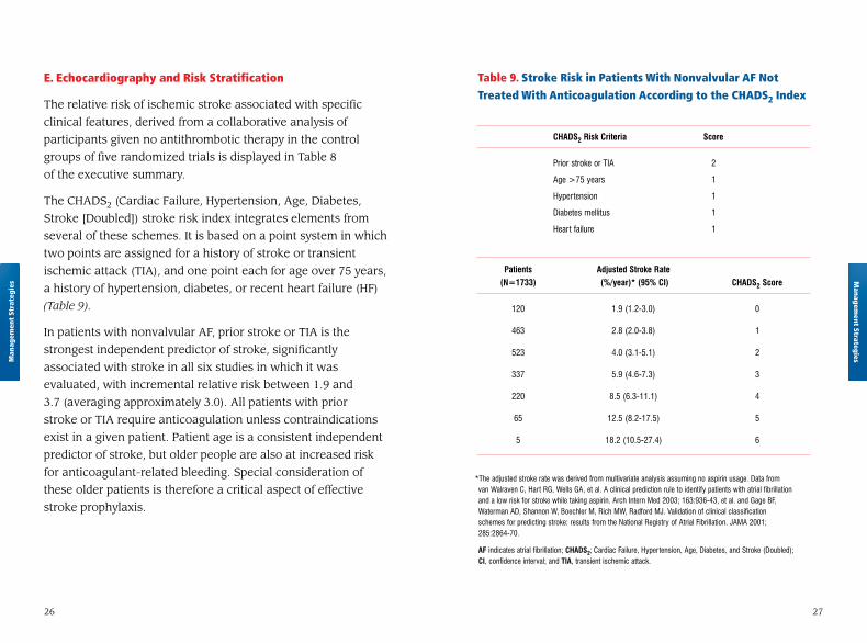
Table 9. Stroke Risk in Patients With nonvalvular AF not Treated With Anticoagulation According to the CHADS2 Index
E. Echocardiography and Risk Stratification
The relative risk of ischemic stroke associated with specific
clinical features, derived from a collaborative analysis of
participants given no antithrombotic therapy in the control
groups of five randomized trials is displayed in Table 8
of the executive summary.
The CHADS2 (Cardiac Failure, Hypertension, Age, Diabetes,
Stroke [Doubled]) stroke risk index integrates elements from
several of these schemes. It is based on a point system in which
two points are assigned for a history of stroke or transient
ischemic attack (TIA), and one point each for age over 75 years,
a history of hypertension, diabetes, or recent heart failure (HF)
(Table 9).
In patients with nonvalvular AF, prior stroke or TIA is the
strongest independent predictor of stroke, significantly
associated with stroke in all six studies in which it was
evaluated, with incremental relative risk between 1.9 and
3.7 (averaging approximately 3.0). All patients with prior
stroke or TIA require anticoagulation unless contraindications
exist in a given patient. Patient age is a consistent independent
predictor of stroke, but older people are also at increased risk
for anticoagulant-related bleeding. Special consideration of
these older patients is therefore a critical aspect of effective
stroke prophylaxis.

28
Man
agem
ent
Stra
tegi
es
29
Managem
ent StrategiesR
ecomm
endations
less validated or weaker risk factors moderate risk factors high risk factors
n Female gender n Age ≥75 years n Previous stroke, TIA or embolism
n Age 65-74 years n Hypertension n Mitral stenosis
n Coronary artery disease n Heart failure n Prosthetic heart valve*
n Thyrotoxicosis n LV ejection fraction ≤35%
n Diabetes mellitus
* indicates if mechanical valve, target INR greater than 2.5.
Inr indicates international normalized ratio; lV, left ventricular; TIa, transient ischemic attack.
Table 10. Antithrombotic Therapy for Patients With Atrial Fibrillation
risk Category recommended Therapy
No risk factors Aspirin, 81-325 mg daily
One moderate risk factor Aspirin, 81-325 mg daily or Warfarin
(INR 2.0 to 3.0, target 2.5)
Any high risk factor Warfarin
or more than 1 moderate risk factor (INR 2.0 to 3.0, target 2.5)*
F. Risk Stratification
Although these schemes for stratification of stroke
risk identify patients who benefit most and least
from anticoagulation, the threshold for use of
anticoagulation is still controversial. Our
recommendations for antithrombotic therapy
are summarized in Table 10.
Anticoagulation is recommended for 3 wk prior to
and 4 wk after cardioversion for patients with AF
of unknown duration or with AF for longer than
48 h. Although left atrial thrombus and systemic
embolism have been documented in patients with
AF of shorter duration, the need for anticoagulation
is less clear. When acute AF produces hemodynamic
instability in the form of angina pectoris, MI, shock,
or pulmonary edema, immediate cardioversion
should not be delayed to deliver therapeutic
anticoagulation, but intravenous unfractionated
heparin or subcutaneous injection of a low-
molecular-weight heparin should be initiated before
cardioversion by direct-current countershock or
intravenous antiarrhythmic medication.

30 31
Managem
ent StrategiesR
ecomm
endations
response to AF in the acute setting, exercising
caution in patients with hypotension or HF.
(Level of Evidence: B)
3. Intravenous administration of digoxin or
amiodarone is recommended to control the heart
rate in patients with AF and HF who do not have an
accessory pathway. (Level of Evidence: B)
4. In patients who experience symptoms related to
AF during activity, the adequacy of heart rate control
should be assessed during exercise, adjusting
pharmacological treatment as necessary to keep the
rate in the physiological range. (Level of Evidence: C)
5. Digoxin is effective following oral administration
to control the heart rate at rest in patients with
AF and is indicated for patients with HF or LV
dysfunction or for sedentary individuals.
(Level of Evidence: C)
Class IIa 1. A combination of digoxin and either a beta
blocker, diltiazem, or verapamil is reasonable
to control the heart rate both at rest and during
exercise in patients with AF. (Level of Evidence: B)
2. It is reasonable to use ablation of the arterioven-
tricular (AV) node or accessory pathway to control
heart rate when pharmacological therapy is insuffi-
cient or associated with side effects. (Level of Evidence: B)
3. Intravenous amiodarone can be useful to control
the heart rate in patients with AF when other measures
are unsuccessful or contraindicated. (Level of Evidence: C)
G. Catheter Ablation
Catheter-directed ablation of AF represents a substantial
achievement that promises better therapy for a large number
of patients presently resistant to pharmacological or electrical
conversion to sinus rhythm. The limited available studies
suggest that catheter-based ablation offers benefit to selected
patients with AF, but these studies do not provide convincing
evidence of optimum catheter positioning or absolute rates of
treatment success. Identification of patients who might benefit
from ablation must take into account both potential benefits
and short- and long-term risks. Rates of success and complica-
tions vary, sometimes considerably, from one study to another
because of patient factors, patterns of AF, criteria for definition
of success, duration of follow-up, and technical aspects.
VI. Recommendations
A. Pharmacological Rate Control During Atrial Fibrillation
Class I 1. Measurement of the heart rate at rest and control
of the rate using pharmacological agents are
recommended for patients with persistent or
permanent AF. (Level of Evidence: B)
2. In the absence of pre-excitation, intravenous
administration of a beta blocker, diltiazem, or
verapamil is recommended to slow the ventricular Rec
omm
enda
tion
s

32
Rec
omm
enda
tion
s
33
Managem
ent StrategiesR
ecomm
endations
3. In patients with decompensated HF and AF,
intravenous administration of a nondihydropyridine
calcium channel antagonist may exacerbate hemo-
dynamic compromise and is not recommended.
(Level of Evidence: C)
4. Intravenous administration of lidocaine, beta
blockers, or nondihydropyridine calcium channel
antagonists to patients with AF and pre-excitation
may accelerate the ventricular response and is not
recommended. (Level of Evidence: C)
B. Preventing Thromboembolism
Class I 1. Antithrombotic therapy to prevent thrombo-
embolism is recommended for all patients with
AF, except those with lone AF or contraindications.
(Level of Evidence: A)
2. The antithrombotic agent should be chosen
based upon the absolute risks of stroke and bleeding
and the relative risk and benefit for a given patient.
(Level of Evidence: A)
3. For patients at high risk of stroke, chronic oral
anticoagulant therapy with a vitamin K antagonist
(INR 2.0 to 3.0) is recommended, unless contra-
indicated. Factors associated with highest risk for
stroke in patients with AF are prior stroke, TIA, or
systemic embolism, rheumatic mitral stenosis and
a mechanical heart valve. (Level of Evidence: A).
4. When electrical cardioversion is not necessary
in patients with AF and an accessory pathway,
intravenous procainamide or ibutilide are
reasonable alternatives. (Level of Evidence: C)
Class IIb 1. When the rate of ventricular response to AF can-
not be adequately controlled using a beta blocker,
diltiazem, verapamil or digoxin, alone or in
combination, oral amiodarone may be administered
to control the heart rate. (Level of Evidence: C)
2. Intravenous procainamide, disopyramide,
ibutilide, or amiodarone may be considered for
hemodynamically stable patients with AF involv-
ing conduction over an accessory pathway.
(Level of Evidence: B)
3. When the rate of ventricular response to AF
cannot be controlled with pharmacological agents
or tachycardia-mediated cardiomyopathy is
suspected, catheter-directed ablation of the AV
node may be considered. (Level of Evidence: C)
Class III 1. Digitalis should not be used as the sole agent to
control the rate of ventricular response in patients
with paroxysmal AF. (Level of Evidence: B)
2. Catheter ablation of the AV node should not be
attempted without a prior trial of medication to
control the ventricular rate in patients with AF.
(Level of Evidence: C)

34
Rec
omm
enda
tion
s
35
Managem
ent StrategiesR
ecomm
endations
2. For patients with nonvalvular AF who have
1 or more of the less well-validated risk factors
(age 65-74 years, female gender, or CAD), treat-
ment with either aspirin or a vitamin K antagonist
is reasonable. (Level of Evidence: B)
3. It is reasonable to select antithrombotic therapy
using the same criteria irrespective of the pattern
(paroxysmal, persistent, or permanent) of AF.
(Level of Evidence: B)
4. In patients with AF without a mechanical heart
valve, it is reasonable to interrupt anticoagulation
for up to 1 week for procedures that carry a risk of
bleeding. (Level of Evidence: C)
5. It is reasonable to re-evaluate the need for anti-
coagulation at regular intervals. (Level of Evidence: C)
Class IIb 1. In patients 75 years of age and older at risk of
bleeding but without contraindications to anti-
coagulant therapy, and in patients who are unable
to safely tolerate standard anticoagulation (INR
2.0 to 3.0), a lower INR target (2.0; range 1.6 to 2.5)
may be considered for primary prevention of stroke
and systemic embolism. (Level of Evidence: C)
2. When interruption of oral anticoagulant therapy
for longer than 1 week is necessary in high-risk
patients, unfractionated or low-molecular-weight
heparin may be given by injection, although efficacy
is uncertain. (Level of Evidence: C)
4. Anticoagulation with a vitamin K antagonist is
recommended for patients with more than 1 moderate
risk factor (age greater than 75 years, hypertension,
diabetes mellitus, HF, or impaired LV systolic
function [ejection fraction 35% or less or fractional
shortening less than 25%]). (Level of Evidence: A)
5. INR should be determined at least weekly during
initiation of therapy and monthly when stable.
(Level of Evidence: A)
6. Aspirin, 81-325 mg daily, is recommended in
low-risk patients or in those with contraindications
to oral anticoagulation. (Level of Evidence: A)
7. For patients with AF who have mechanical heart
valves, the target intensity of anticoagulation should
be based on the type of prosthesis, maintaining an
INR of at least 2.5. (Level of Evidence: B)
8. Antithrombotic therapy is recommended for
patients with atrial flutter as for AF. (Level of Evidence: C)
Class IIa 1. For primary prevention of thromboembolism
in patients with nonvalvular AF who have just 1
of the validated risk factors (age greater than 75
years (especially in female patients), hypertension,
diabetes mellitus, HF, or impaired LV function),
antithrombotic therapy with either aspirin or a
vitamin K antagonist is reasonable, based upon an
assessment of the risk of bleeding complications,
ability to safely sustain anticoagulation, and patient
preferences. (Level of Evidence: A)

36
Rec
omm
enda
tion
s
37
Managem
ent StrategiesR
ecomm
endations
C. Cardioversion of Atrial Fibrillation
1.PharmacologicalCardioversion
Class I 1. Administration of flecainide, dofetilide,
propafenone, or ibutilide is recommended
for pharmacological cardioversion of AF.
(Level of Evidence: A)
Class IIa 1. Administration of amiodarone is reasonable
for pharmacological cardioversion of AF.
(Level of Evidence: A)
2. A single oral dose of propafenone or flecainide
(“pill-in-the-pocket”) can be used to terminate
persistent AF out of hospital for selected patients
once treatment has proved safe in hospital. Before
antiarrhythmic medication is initiated, a beta
blocker, diltiazem or verapamil should be given to
prevent rapid AV conduction. (Level of Evidence: C)
3. Amiodarone can be beneficial on an outpatient
basis in patients with paroxysmal or persistent
AF when rapid restoration of sinus rhythm is
unnecessary. (Level of Evidence: C)
Class IIb 1. Quinidine or procainamide might be considered
for cardioversion of AF, but their usefulness is not
well established. (Level of Evidence: C)
3. Following coronary revascularization in patients
with AF, low-dose aspirin (less than 100 mg daily)
and/or clopidogrel (75 mg daily) may be given
concurrently with anticoagulation, but these
strategies are associated with an increased risk
of bleeding. (Level of Evidence: C)
4. In patients undergoing coronary revascularization,
anticoagulation may be interrupted to prevent
bleeding, but should be resumed as soon as possible
after the procedure and the dose adjusted to achieve
a therapeutic INR. Aspirin may be given during the
hiatus. For patients undergoing percutaneous
intervention, the maintenance regimen should
consist of clopidogrel, 75 mg daily, plus warfarin
(INR 2.0 to 3.0). Clopidogrel should be given for a
minimum of 1 mo after a bare metal stent, at least
3 mo for a sirolimus-eluting stent, at least 6 mo for
a paclitaxel-eluting stent, and 12 mo or longer in
selected patients, followed by warfarin alone.
(Level of Evidence: C)
5. In patients with AF who sustain ischemic stroke
or systemic embolism during treatment with
anticoagulation (INR 2.0 to 3.0), it may be
reasonable to raise the intensity of anticoagulation
up to a target INR of 3.0 to 3.5. (Level of Evidence: C)
Class III 1. Long-term anticoagulation is not recommended for
primary stroke prevention in patients below age 60
years without heart disease (lone AF). (Level of
Evidence: C)

38
Rec
omm
enda
tion
s
39
Recom
mendations
2. Patient preference is a reasonable consideration
in the selection of infrequently repeated cardio-
versions for the management of symptomatic or
recurrent AF. (Level of Evidence: C)
Class III 1. Frequent direct-current cardioversion is not
recommended for patients with relatively short
periods of sinus rhythm after multiple cardioversion
procedures despite prophylactic antiarrhythmic drug
therapy. (Level of Evidence: C)
2. Electrical cardioversion is contraindicated in
patients with digitalis toxicity or hypokalemia.
(Level of Evidence: C)
3.PharmacologicalEnhancement ofDirect-CurrentCardioversion
Class IIa 1. Pretreatment with amiodarone, flecainide,
ibutilide, propafenone, or sotalol can be useful to
enhance direct-current cardioversion and prevent
recurrent AF. (Level of Evidence: B)
2. In patients who relapse to AF after successful
cardioversion, it can be useful to repeat the
procedure following administration of
antiarrhythmic medication. (Level of Evidence: C)
Class III 1. Digoxin and sotalol are not recommended for
pharmacological cardioversion of AF. (Level of
Evidence: A)
2. Quinidine, procainamide, disopyramide, and
dofetilide should not be started out of hospital for
conversion of AF. (Level of Evidence: B)
2.Direct-CurrentCardioversion
Class I 1. When a rapid ventricular response to AF does not
respond promptly to pharmacological measures,
immediate direct-current cardioversion is recom-
mended for patients with myocardial ischemia, symp-
tomatic hypotension, angina, or HF. (Level of Evidence: C)
2. Immediate direct-current cardioversion is recom-
mended for patients with pre-excitation when AF
occurs with extreme tachycardia or hemodynamic
instability. (Level of Evidence: B)
3. Cardioversion is recommended when symptoms
of AF are unacceptable to the patient. In case of
relapse, direct-current cardioversion may be
repeated following administration of antiarrhythmic
medication. (Level of Evidence: C)
Class IIa 1. Direct-current cardioversion can be useful
to restore sinus rhythm as part of a long-term
management strategy for patients with AF.
(Level of Evidence: B)

40
Rec
omm
enda
tion
s
41
Managem
ent StrategiesR
ecomm
endations
administered concurrently by an initial intravenous
injection followed by a continuous infusion
(aPTT 1.5 to 2 times control). Thereafter oral
anticoagulation (INR 2.0 to 3.0) should be provided
for at least 4 weeks, as for elective cardioversion.
Limited data support subcutaneous low-molecular-
weight heparin. (Level of Evidence: C)
3. For patients with AF of less than 48-h duration
associated with hemodynamic instability, cardio-
version should be performed immediately without
anticoagulation. (Level of Evidence: C)
Class IIa 1. During the 48 h after onset of AF, the need for
anticoagulation before and after cardioversion may
be based on the patient’s risk of thromboembolism.
(Level of Evidence: C)
2. As an alternative to anticoagulation prior to
cardioversion of AF, it is reasonable to perform
transesophageal echocardiography in search of
thrombus. (Level of Evidence: B)
2a. For patients with no identifiable thrombus,
cardioversion is reasonable immediately after
anticoagulation. (Level of Evidence: B)
Thereafter, continuation of oral anticoagulation
(INR 2.0 to 3.0) is reasonable for at least 4 weeks,
as for elective cardioversion. (Level of Evidence: B)
Limited data are available to support subcutaneous
low-molecular-weight heparin in this indication.
(Level of Evidence: C)
Class IIb 1. For patients with persistent AF, administration of
beta blockers, disopyramide, diltiazem, dofetilide,
procainamide, or verapamil may be considered,
although the efficacy of these agents to enhance the
success of direct-current cardioversion or to prevent
early recurrence of AF is uncertain. (Level of Evidence: C)
2. Out-of-hospital initiation of antiarrhythmic
medications may be considered in patients without
heart disease to enhance the success of cardio-
version of AF. (Level of Evidence: C)
3. Out-of-hospital administration of antiarrhythmic
medications may be considered to enhance the
success of cardioversion of AF in patients with certain
forms of heart disease, once the safety of the drug
has been verified for the patient. (Level of Evidence: C)
4.PreventionofThromboembolisminPatients WithAtrialFibrillationUndergoingCardioversion
Class I 1. For patients with AF of 48-h duration or longer,
or when the duration of AF is unknown, anti-
coagulation (INR 2.0 to 3.0) is recommended
for at least 3 weeks prior to and 4 weeks after
cardioversion, regardless of the method used to
restore sinus rhythm. (Level of Evidence: B)
2. For patients with AF of more than 48-h duration
requiring immediate cardioversion because of
hemodynamic instability, heparin should be

42
Rec
omm
enda
tion
s
43
Managem
ent StrategiesR
ecomm
endations
no associated heart disease when the agent is well
tolerated. (Level of Evidence: C)
4. In patients with lone AF without structural heart
disease, propafenone or flecainide can be beneficial
on an outpatient basis in patients with paroxysmal
AF who are in sinus rhythm at the time of drug
initiation. (Level of Evidence: B)
5. Sotalol can be beneficial in outpatients in sinus
rhythm with little or no heart disease prone to
paroxysmal AF if the baseline uncorrected QT
interval is less than 460 ms, electrolytes are normal,
and risk factors associated with proarrhythmia are
absent. (Level of Evidence: C)
6. Catheter ablation is a reasonable alternative to
pharmacological therapy to prevent recurrent AF
in symptomatic patients with little or no left atrial
enlargement. (Level of Evidence: C)
Class III 1. Antiarrhythmic therapy with a particular drug is
not recommended for maintenance of sinus rhythm
in patients with AF who have risk factors for
proarrhythmia with that agent. (Level of Evidence: A)
2. Pharmacological therapy is not recommended
for maintenance of sinus rhythm in patients
with advanced sinus node disease or AV node
dysfunction unless they have a functioning
pacemaker. (Level of Evidence: C)
2b. For patients in whom thrombus is identified, oral
anticoagulation (INR 2.0 to 3.0) is reasonable for
at least 3 weeks before and 4 weeks after restora-
tion of sinus rhythm, and longer anticoagulation
may be appropriate after apparently successful
cardioversion, because the risk of thrombo-
embolism often remains elevated in such cases.
(Level of Evidence: C)
3. For patients with atrial flutter undergoing
cardioversion, anticoagulation can be beneficial
according to the recommendations as for patients
with AF. (Level of Evidence: C)
D. Maintenance of Sinus Rhythm
Class I 1. Before initiating antiarrhythmic drug therapy,
treatment of precipitating or reversible causes of AF
is recommended. (Level of Evidence: C)
Class IIa 1. Pharmacological therapy can be useful in patients
with AF to maintain sinus rhythm and prevent
tachycardia-induced cardiomyopathy. (Level of
Evidence: C)
2. Infrequent, well-tolerated recurrence of AF
is reasonable as a successful outcome of
antiarrhythmic drug therapy. (Level of Evidence: C)
3. Outpatient initiation of antiarrhythmic drug
therapy is reasonable in patients with AF who have

44
Rec
omm
enda
tion
s
45
Managem
ent StrategiesR
ecomm
endations
F. Acute Myocardial Infarction
Class I 1. Direct-current cardioversion is recommended for
patients with severe hemodynamic compromise or
intractable ischemia, or when adequate rate control
cannot be achieved with pharmacological agents in
patients with acute MI and AF. (Level of Evidence: C)
2. Intravenous amiodarone is recommended to slow
a rapid ventricular response to AF and improve LV
function in patients with acute MI. (Level of Evidence: C)
3. Intravenous beta blockers and nondihydropyridine
calcium antagonists are recommended to slow a
rapid ventricular response to AF in patients with
acute MI who do not have LV dysfunction,
bronchospasm, or AV block. (Level of Evidence: C)
4. For patients with AF and acute MI, unfractionated
heparin is recommended (aPTT 1.5 to 2.0 times
control), unless contraindicated. (Level of Evidence: C)
Class IIa 1. Intravenous digitalis is reasonable to slow a rapid
ventricular response and improve LV function in
patients with acute MI and AF associated with
severe LV dysfunction and HF. (Level of Evidence: C)
Class III 1. Class IC antiarrhythmic drugs are not recom-
mended in patients with AF and acute MI.
(Level of Evidence: C)
E. Postoperative Atrial Fibrillation
Class I 1. Unless contraindicated, an oral beta blocker is
recommended to prevent postoperative AF for
patients undergoing cardiac surgery.
(Level of Evidence: A)
2. AV nodal blocking agent is recommended for rate
control in patients who develop postoperative AF.
(Level of Evidence: B)
Class IIa 1. Preoperative amiodarone reduces the incidence
of AF in patients undergoing cardiac surgery and
represents appropriate prophylactic therapy for
patients at high risk for postoperative AF. (Level of
Evidence: A)
2. It is reasonable to restore sinus rhythm by
pharmacological cardioversion with ibutilide or
direct-current cardioversion in patients who develop
postoperative AF. (Level of Evidence: B)
3. Antiarrhythmic medication is reasonable to
maintain sinus rhythm in patients with recurrent or
refractory postoperative AF. (Level of Evidence: B)
4. Antithrombotic medication is reasonable in patients
who develop postoperative AF. (Level of Evidence: B)
Class IIb 1. Prophylactic sotalol may be considered for
patients at risk of developing AF following cardiac
surgery. (Level of Evidence: B)

46
Rec
omm
enda
tion
s
47
Managem
ent StrategiesR
ecomm
endations
Class IIb 1. It may be reasonable to administer intravenous
quinidine, procainamide, disopyramide, ibutilide, or
amiodarone to hemodynamically stable patients
with AF involving an accessory pathway.
(Level of Evidence: B)
Class III 1. Intravenous beta-blocking agents, digitalis
glycosides, diltiazem, or verapamil is not recom-
mended in patients with WPW syndrome who
have pre-excited ventricular activation during AF.
(Level of Evidence: B)
H. Hyperthyroidism
Class I 1. A beta blocker is recommended to control the
heart rate in patients with AF complicating thyro-
toxicosis, unless contraindicated. (Level of Evidence: B)
2. When a beta blocker cannot be used, a
nondihydropyridine calcium channel antagonist
is recommended to control the ventricular rate
in patients with AF and thyrotoxicosis.
(Level of Evidence: B)
3. In patients with AF and thyrotoxicosis, oral
anticoagulation (INR 2.0 to 3.0) is recommended.
(Level of Evidence: C)
4. Once euthyroid state is achieved, antithrombotic
prophylaxis is the same as for patients without
hyperthyroidism. (Level of Evidence: C)
G. Management of Atrial Fibrillation Associated With the Wolff-Parkinson-White Pre-excitation Syndrome
Class I 1. Catheter ablation of the accessory pathway is
recommended in symptomatic patients with AF
who have WPW syndrome, particularly those with
syncope due to rapid rate or short bypass tract
refractory period. (Level of Evidence: B)
2. Immediate direct-current cardioversion is
recommended to prevent ventricular fibrillation
in patients with a short anterograde bypass tract
refractory period in whom AF occurs with a rapid
ventricular response associated with hemodynamic
instability. (Level of Evidence: B)
3. Intravenous procainamide or ibutilide is
recommended to restore sinus rhythm in patients
with WPW in whom AF occurs without hemo-
dynamic instability in association with a wide QRS
complex on the ECG (≥120-ms duration) or rapid
pre-excited ventricular response. (Level of Evidence: C)
Class IIa 1. Intravenous flecainide or direct-current
cardioversion is reasonable when very rapid
ventricular rates occur in patients with AF involving
an accessory pathway. (Level of Evidence: B)

48
Rec
omm
enda
tion
s
49
Managem
ent StrategiesR
ecomm
endations
2. During the first trimester and last month of
pregnancy subcutaneous low-molecular-weight
heparin may be considered for patients with AF and
risk factors for thromboembolism despite limited
data. (Level of Evidence: C)
3. During the second trimester, consider oral
anticoagulation for pregnant women with AF at high
thromboembolic risk. (Level of Evidence: C)
4. Quinidine or procainamide may be considered for
pharmacological cardioversion in hemodynamically
stable patients who develop AF during pregnancy.
(Level of Evidence: C)
J. Management of Atrial Fibrillation in Patients With Hypertrophic Cardiomyopathy
Class I 1. Oral anticoagulation (INR 2.0 to 3.0) is recom-
mended in patients with HCM who develop AF.
(Level of Evidence: B)
Class IIa 1. Antiarrhythmic medications can be useful to
prevent recurrent AF in patients with HCM. Either
disopyramide combined with a beta blocker or
nondihydropyridine calcium channel antagonist
or amiodarone alone is generally preferred.
(Level of Evidence: C)
I. Management of Atrial Fibrillation During Pregnancy
Class I 1. Digoxin, a beta blocker, or nondihydropyridine
calcium channel antagonist is recommended to
control the ventricular rate in pregnant patients
with AF. (Level of Evidence: C)
2. Direct-current cardioversion is recommended in
pregnant patients who become hemodynamically
unstable due to AF. (Level of Evidence: C)
3. Protection against thromboembolism is
recommended throughout pregnancy for patients
with AF except those at low thromboembolic risk.
The choice of anticoagulant or aspirin should be
chosen according to the stage of pregnancy.
(Level of Evidence: C)
Class IIb 1. During the first trimester and last month of
pregnancy for patients with AF and risk factors for
thromboembolism, consider administering
unfractionated heparin by continuous intravenous
infusion (aPTT 1.5 to 2 times control) or by
subcutaneous injection (10 000 to 20 000 units every
12 h, adjusted to prolong the aPTT 6 h after injection
to 1.5 times control). (Level of Evidence: B)

50
Rec
omm
enda
tion
s
K. Management of Atrial Fibrillation in Patients With Pulmonary Disease
Class I 1. For patients who develop AF during an acute pul-
monary illness or exacerbation of chronic pulmonary
disease, correction of hypoxemia and acidosis are
the primary therapeutic measures. (Level of Evidence: C)
2. Diltiazem or verapamil is recommended to
control the ventricular rate in patients with
obstructive pulmonary disease who develop AF.
(Level of Evidence: C)
3. Direct-current cardioversion should be attempted
in patients with pulmonary disease who become
hemodynamically unstable as a consequence of AF.
(Level of Evidence: C)
Class III 1. Theophylline and beta-adrenergic agonist
agents are not recommended in patients with
bronchospastic lung disease who develop AF.
(Level of Evidence: C)
2. Beta blockers, sotalol, propafenone, and
adenosine are not recommended in patients
with obstructive lung disease who develop AF.
(Level of Evidence: C)

XX%
Cert no. XXX-XXX-XXXXXX
11m
m
19mm
Space around logo has to be the height ofthe letters (FSC) in the logo times 2.
Ex.: 10 pt. - height of lettersx 2
= 20 pt. of space
Portrait - Logo widthminimum is19mm. XX%
Cert no. XXX-XXX-XXXXXX
NO TEXT WITHIN SPACE
NO TEXT WITHIN SPACE
NOTEXTWITHINSPACE
NO
TEX
TW
ITH
INSP
AC
E
NO
TEXT
WITH
INSPA
CE
NO
TEX
TW
ITH
INSP
AC
E NO
TEXT
WITH
INSPA
CENOTEXTWITHINSPACE
NO
TEX
TW
ITH
INSP
AC
E NO
TEXT
WITH
INSPA
CE
FSC LOGO
Landscape - Logo height minimumis 11mm.
Logo Usage Colors
Black/BlackPMS378 - only if PMS378
4 color - build 378
Cert no. XXX-XXX-XXXXXX
XX%
Cert no. XXX-XXX-XXXXXX Cert no. XXX-XXX-XXXXXX
Cert no. XXX-XXX-XXXXXX
XX%
Cert no. XXX-XXX-XXXXXX Cert no. XXX-XXX-XXXXXX
FSC_MS_1_LPBW_33_No# FSC_MS_2_LPBW_33_No# FSC_MS_3_LPBW_33_No#
FSC_MS_1_LPC_33_No# FSC_MS_2_LPC _33_No# FSC_MS_3_LPC_33_No#
FSC_MS_1_PPBW_33_No# FSC_MS_2_PPBW_33_No# FSC_MS_3_PPBW_33_No#
FSC_MS_3_PPC_33_No#
Cert no. XXX-XXX-XXXXXX
Cert no. XXX-XXX-XXXXXX
XX%
Cert no. XXX-XXX-XXXXXX
XX%
Cert no. XXX-XXX-XXXXXX
Cert no. XXX-XXX-XXXXXX
FSC_MS_1_PPC_33_No# FSC_MS_2_PPC_33_No#
Cert no. XXX-XXX-XXXXXX