Embed Size (px)
Citation preview
Menopause: The Journal of The North American Menopause SocietyVol. 13, No. 3, pp. 340 - 367DOI: 10.1097/01.gme.0000222475.93345.b3* 2006 by The North American Menopause Society5 Text printed on acid-free paper.
POSITION STATEMENT
Management of osteoporosis inpostmenopausal women: 2006 position statementof The North American Menopause Society
ABSTRACT
Objective: To update the evidence-based position statement published by The NorthAmerican Menopause Society (NAMS) in 2002 regarding the management of osteoporosis inpostmenopausal women.
Design: NAMS followed the general principles established for evidence-based guidelines tocreate this updated document. A panel of clinicians and researchers expert in the field ofmetabolic bone diseases and/or women`s health was enlisted to review the 2002 NAMS positionstatement, compile supporting statements, and reach consensus on recommendations. Thepanel`s recommendations were reviewed and approved by the NAMS Board of Trustees.
Results: Osteoporosis, whose prevalence is especially high among elderly postmenopausalwomen, increases the risk of fractures. Hip and spine fractures are associated with particularlyhigh morbidity and mortality in this population. Given the health implications of osteoporoticfractures, the primary goal of osteoporosis therapy is to prevent fractures, which is accomplishedby slowing or stopping bone loss, maintaining bone strength, and minimizing or eliminatingfactors that may contribute to fractures. The evaluation of postmenopausal women forosteoporosis risk requires a medical history, physical examination, and diagnostic tests. Majorrisk factors for postmenopausal osteoporosis (as defined by bone mineral density) includeadvanced age, genetics, lifestyle factors (such as low calcium and vitamin D intake, smoking),thinness, and menopause status. The most common risk factors for osteoporotic fracture areadvanced age, low bone mineral density, and previous fracture as an adult. Management focusesfirst on nonpharmacologic measures, such as a balanced diet, adequate calcium and vitamin Dintake, adequate exercise, smoking cessation, avoidance of excessive alcohol intake, and fallprevention. If pharmacologic therapy is indicated, government-approved options are bisphos-phonates, a selective estrogen-receptor modulator, parathyroid hormone, estrogens, and calcitonin.
Conclusions: Management strategies for postmenopausal women involve identifying those atrisk of low bone density and fracture, followed by instituting measures that focus on reducingmodifiable risk factors through lifestyle changes and, if indicated, pharmacologic therapy.
Key Words: Menopause Y Osteoporosis Y Fractures Y Bone mineral density Y Estrogentherapy Y Hormone therapy Y Bisphosphonate Y Selective estrogen-receptor modulator YCalcitonin Y Parathyroid hormone Y Calcium Y Vitamin D Y NAMS.
Received February 27, 2006.
The Board of Trustees of The North American Menopause Society(NAMS) developed this manuscript with assistance from an EditorialBoard composed of Bruce Ettinger, MD; Steven T. Harris, MD, FACP;David Kendler, MD; Bruce Kessel, MD; and Michael R. McClung, MD.It was edited, modified, and subsequently approved by the NAMSBoard of Trustees on February 24, 2006.
The development of this position statement was supported by unrestrictededucational grants from the Novartis Pharmaceuticals Corporation.
Address correspondence to: NAMS, P.O. Box 94527, Cleveland, OH44101. E-mail: [email protected].
340 Menopause, Vol. 13, No. 3, 2006
Osteoporosis becomes a serious healththreat for aging postmenopausal womenby predisposing them to an increased riskof fracture. Osteoporotic fractures are
associated with substantial morbidity and mortalityin postmenopausal women, especially older women.
In response to the need to define standards ofclinical practice in North America as they relate tomenopause-associated health conditions, The NorthAmerican Menopause Society (NAMS) has createdthis evidence-based position statement. The objectiveof this position statement is to provide guidance onthe diagnosis, prevention, and treatment of osteopo-rosis in postmenopausal women to physicians, physi-cian assistants, nurse practitioners, nurses, and otherhealthcare professionals caring for postmenopausalwomen, especially those in the clinical practice fieldsof obstetrics and gynecology, internal medicine,family medicine, and geriatrics.
This position statement is an update of the NAMSposition statement published in 2002.1 Since then, thepublication of additional scientific evidence hascreated a need to update the position statement.
For this revision, NAMS conducted a search of themedical literature published since the previous posi-tion statement was submitted for publication inNovember 2001. A search was made for clinicaltrials, meta-analyses, and clinical practice guidelinespublished in English and related to osteoporosis inpostmenopausal women using the database MED-LINE. The Medical Subject Headings (MeSH) usedfor the search were postmenopausal osteoporosis andbone loss with subheadings for epidemiology, eti-ology, diagnosis, prevention and control, and therapy.The National Guideline Clearinghouse was searchedfor relevant clinical practice guidelines and theCochrane Library was searched for relevant system-atic reviews. Priority was given to evidence fromrandomized controlled clinical trials and meta-analysesof such trials, followed by evidence from controlledobservational studies, using criteria described else-where.2<4 Conclusions from other evidence-basedguidelines also were reviewed. Because standards ofcare and available treatment options differ throughoutthe world, the focus is limited to therapies available inNorth America.
To help with this revision, NAMS enlisted a five-person Editorial Board composed of endocrinologistsand gynecologists from both clinical practice andresearch with expertise in metabolic bone diseasesand/or women`s health. The Editorial Board reviewedthe previous position statement and incorporated data
published since that statement, compiled supportingstatements, and made recommendations. Where theevidence was contradictory or inadequate to forma conclusion, a consensus-based opinion was estab-lished. (Practice parameter standards related toNAMS position statements have been described inan editorial.5) The NAMS Board of Trustees wasresponsible for the final review and approval of thisdocument. Updates to this revised position statementwill be published as developments occur in scientificresearch that substantially alter the conclusions.
BACKGROUND
OsteoporosisVthe most common bone disorderaffecting humansVis a skeletal disorder charac-terized by compromised bone strength predisposinga person to an increased risk of fracture.6 Bonestrength (and, hence, fracture risk) is dependent onboth bone quality and bone mineral density (BMD).6
Expressed as grams of mineral per area or volume,BMD at any given age is a function of peak bonemass (reached around age 30 years) and how muchbone is subsequently lost. Qualities of bone otherthan BMD (including degree of mineralization,hydroxyapatite crystal size, collagen structure, heter-ogeneity of bone microstructure, connectivity oftrabeculae, and microdamage) are difficult or impos-sible to measure in clinical practice.
To standardize values from different bone densi-tometry tests, results are reported as either a Z-score ora T-score, with both expressed as standard deviation(SD) units.
& T-score is calculated by comparing current BMDto the mean peak BMD of a normal, young adultpopulation of the same gender. For women, thereference database is white (nonYrace-adjusted)women aged 20 to 29 years. Use of T-scores isthe preferred choice for postmenopausal women.
& Z-score is based on the difference between thewoman`s BMD and the mean BMD of a referencepopulation of the same gender, age, and ethnicity.
NAMS supports the World Health Organization(WHO) definition7 of osteoporosis in a postmeno-pausal woman as a BMD T-score less than or equal toj2.5 at the total hip, femoral neck, or lumbar spine(posterior-anterior, not lateral) (see below). If ana-tomic factors such as obesity or arthritis makemeasurements invalid, the distal one-third radius bonedensity may be considered a diagnostic site. However,the relationship between the T-score at this site andfracture risk has not been systematically examined.
Menopause, Vol. 13, No. 3, 2006 341
NAMS POSITION STATEMENT
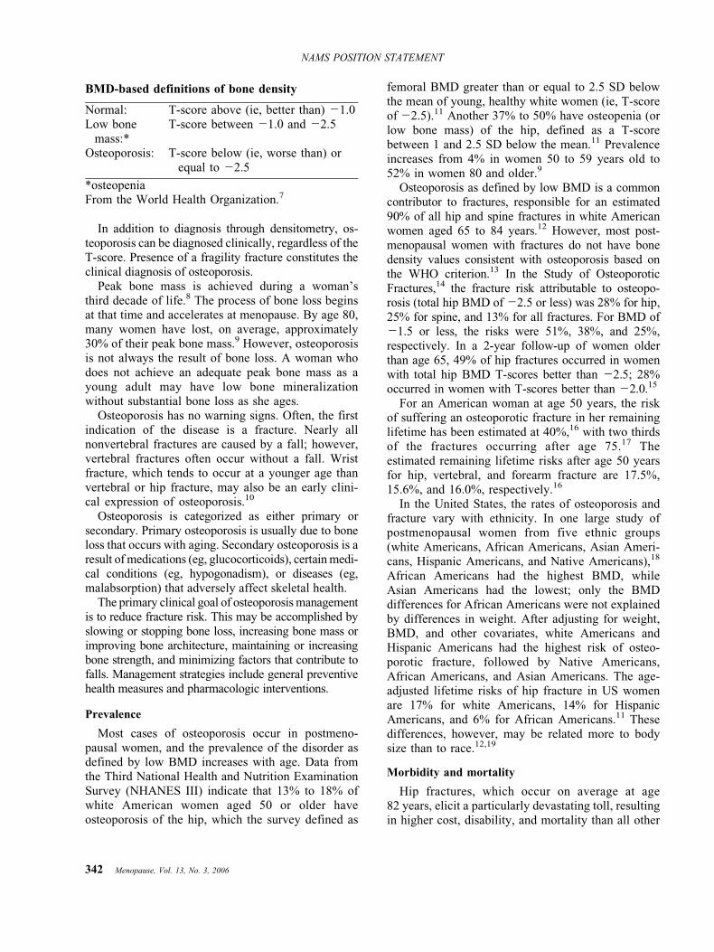
In addition to diagnosis through densitometry, os-teoporosis can be diagnosed clinically, regardless of theT-score. Presence of a fragility fracture constitutes theclinical diagnosis of osteoporosis.
Peak bone mass is achieved during a woman`sthird decade of life.8 The process of bone loss beginsat that time and accelerates at menopause. By age 80,many women have lost, on average, approximately30% of their peak bone mass.9 However, osteoporosisis not always the result of bone loss. A woman whodoes not achieve an adequate peak bone mass as ayoung adult may have low bone mineralizationwithout substantial bone loss as she ages.
Osteoporosis has no warning signs. Often, the firstindication of the disease is a fracture. Nearly allnonvertebral fractures are caused by a fall; however,vertebral fractures often occur without a fall. Wristfracture, which tends to occur at a younger age thanvertebral or hip fracture, may also be an early clini-cal expression of osteoporosis.10
Osteoporosis is categorized as either primary orsecondary. Primary osteoporosis is usually due to boneloss that occurs with aging. Secondary osteoporosis is aresult of medications (eg, glucocorticoids), certain medi-cal conditions (eg, hypogonadism), or diseases (eg,malabsorption) that adversely affect skeletal health.
The primary clinical goal of osteoporosis managementis to reduce fracture risk. This may be accomplished byslowing or stopping bone loss, increasing bone mass orimproving bone architecture, maintaining or increasingbone strength, and minimizing factors that contribute tofalls. Management strategies include general preventivehealth measures and pharmacologic interventions.
Prevalence
Most cases of osteoporosis occur in postmeno-pausal women, and the prevalence of the disorder asdefined by low BMD increases with age. Data fromthe Third National Health and Nutrition ExaminationSurvey (NHANES III) indicate that 13% to 18% ofwhite American women aged 50 or older haveosteoporosis of the hip, which the survey defined as
femoral BMD greater than or equal to 2.5 SD belowthe mean of young, healthy white women (ie, T-scoreof j2.5).11 Another 37% to 50% have osteopenia (orlow bone mass) of the hip, defined as a T-scorebetween 1 and 2.5 SD below the mean.11 Prevalenceincreases from 4% in women 50 to 59 years old to52% in women 80 and older.9
Osteoporosis as defined by low BMD is a commoncontributor to fractures, responsible for an estimated90% of all hip and spine fractures in white Americanwomen aged 65 to 84 years.12 However, most post-menopausal women with fractures do not have bonedensity values consistent with osteoporosis based onthe WHO criterion.13 In the Study of OsteoporoticFractures,14 the fracture risk attributable to osteopo-rosis (total hip BMD of j2.5 or less) was 28% for hip,25% for spine, and 13% for all fractures. For BMD ofj1.5 or less, the risks were 51%, 38%, and 25%,respectively. In a 2-year follow-up of women olderthan age 65, 49% of hip fractures occurred in womenwith total hip BMD T-scores better than j2.5; 28%occurred in women with T-scores better than j2.0.15
For an American woman at age 50 years, the riskof suffering an osteoporotic fracture in her remaininglifetime has been estimated at 40%,16 with two thirdsof the fractures occurring after age 75.17 Theestimated remaining lifetime risks after age 50 yearsfor hip, vertebral, and forearm fracture are 17.5%,15.6%, and 16.0%, respectively.16
In the United States, the rates of osteoporosis andfracture vary with ethnicity. In one large study ofpostmenopausal women from five ethnic groups(white Americans, African Americans, Asian Ameri-cans, Hispanic Americans, and Native Americans),18
African Americans had the highest BMD, whileAsian Americans had the lowest; only the BMDdifferences for African Americans were not explainedby differences in weight. After adjusting for weight,BMD, and other covariates, white Americans andHispanic Americans had the highest risk of osteo-porotic fracture, followed by Native Americans,African Americans, and Asian Americans. The age-adjusted lifetime risks of hip fracture in US womenare 17% for white Americans, 14% for HispanicAmericans, and 6% for African Americans.11 Thesedifferences, however, may be related more to bodysize than to race.12,19
Morbidity and mortality
Hip fractures, which occur on average at age82 years, elicit a particularly devastating toll, resultingin higher cost, disability, and mortality than all other
BMD-based definitions of bone density
Normal: T-score above (ie, better than) j1.0Low bone
mass:*T-score between j1.0 and j2.5
Osteoporosis: T-score below (ie, worse than) orequal to j2.5
*osteopeniaFrom the World Health Organization.7
342 Menopause, Vol. 13, No. 3, 2006
NAMS POSITION STATEMENT

osteoporotic fracture types combined. Hip fracturescause up to a 25% increase in mortality within 1 yearof the incident. Approximately 25% of women requirelong-term care after a hip fracture, and 50% will havesome long-term loss of mobility.20
Fractures at other sites can also result in seriousmorbidity. Vertebral fractures occur, on average, in awoman`s mid-70s. Multiple or severe vertebral frac-tures may cause substantial pain as well as loss ofheight and exaggerated thoracic kyphosis. Spinal painand deformity can greatly restrict normal movement,including bending and reaching. Importantly, existingvertebral fractures greatly increase (fivefold to seven-fold) the risk of subsequent vertebral fracture.21,22
Thoracic fractures may restrict lung function andcause digestive problems.23 In the Fracture InterventionTrial,24 after an average of 3.8 years of follow-up, therelative risk of mortality was 6.7 (95% CI, 3.08-14.52)for hip fracture and 8.64 (95% CI, 4.45-16.74) forvertebral fracture.
Osteoporotic fractures take a psychological toll aswell.25 Hip and vertebral fractures and the resultantpain, loss of mobility, changed body image, and lossof independence can have a significant impact onself-esteem and mood.
PATHOPHYSIOLOGY
Bone remodeling is the process of bone resorptionand bone formation. At the cellular level, osteoclastspromote bone resorption by stimulating the produc-tion of acid and enzymes that dissolve bone mineraland proteins. Osteoblasts promote bone formation bycreating a protein matrix consisting primarily ofcollagen, which is soon calcified, resulting in miner-alized bone.
In normal bone remodeling, bone resorption isbalanced by bone formation. Bone loss occurs whenthere is an imbalance between bone resorption andbone formation, resulting in a decrease in bone massand an increase in the risk of fracture.
Menopause is associated with a few years of rapidbone loss attributed to lower circulating levels of 17A-estradiol, related primarily to the loss of estrogen-mediated inhibition of bone resorption without a fullycompensatory increase in bone formation26 However,there is only a weak association between serumestradiol levels and rates of bone turnover in post-menopausal women.
RISK FACTORS
In determining risk factors, it is important to dis-tinguish between risk factors for osteoporosis as defined
by BMD (both primary and secondary causes) and riskfactors for osteoporotic fracture. For BMD-definedosteoporosis, major risk factors in postmenopausalwomen are advanced age, genetics, lifestyle factors(eg, low calcium and vitamin D intake, smoking),thinness, and menopause status. Risk factors forosteoporotic fracture are listed in Table 1; the mostcommon are advanced age, low BMD, and previousfracture (other than skull, facial bone, ankle, finger,and toe) as an adult.
Risk factors for BMD-defined osteoporosis andosteoporotic fracture overlap, given that BMD is arisk factor for fracture. Importantly, however, manyfracture risk factors are not related to BMD.
BMD and fracture risk
Bone density is an important determinant of fracturerisk, especially in women aged 65 and older.27,28
In general, lower BMD scores indicate more severeosteoporosis and higher risk of fracture. A decrease of1 SD in BMD represents a 10% to 12% decrease inBMD and an increase in fracture risk by a factor of1.5 to 2.6.29,30 BMD and fracture risk are mostclosely related when bone density is used to predictthe fracture risk at that same site. Risks for spinefracture and hip fracture increase 2.3-fold and 2.6-fold, respectively, for each decrease of 1 SD in age-adjusted BMD at spine and hip, respectively.30
Fracture risk, however, depends largely on factorsother than BMD. Furthermore, a reliable marker topredict fracture risk or determine fracture riskreduction from therapy is not currently available.The use of BMD scores to assess fracture risk can bemarkedly improved by combining BMD with infor-mation about other risk determinants, particularly the
TABLE 1. Risk factors for osteoporotic fracture
Advanced ageLow BMDPrevious fracture (other than skull, facial bone, ankle, finger, and toe)
as an adultHistory of hip fracture in a parentThinness [body weight G127 lb (57.7 kg) or low BMI (G21 kg/m2)]Current smoking, any amountLow calcium or vitamin D intakeMore than two alcoholic drinks per dayOral or intramuscular glucocorticoid use for 93 moIncreased fall risk
Impaired visionDementiaPoor health/frailtyLow physical activityHistory of recent falls
BMD, bone mineral density; BMI, body mass index.
Menopause, Vol. 13, No. 3, 2006 343
NAMS POSITION STATEMENT
woman`s age and fracture history. The WHO is currentlydeveloping a model to estimate the 10-year absolutefracture risk based on known risk factors. It will offersubstantial benefits to healthcare providers comparedwith current methods.
Treatment-induced changes in BMD do not alwayscorrelate well with reductions in vertebral fracturerisk.31<34 In addition, fracture risk reductions inresponse to antiresorptive therapy occur much morerapidly than discernible BMD changes. For example,significant fracture risk reduction has been reportedafter 6 months of risedronate therapy,35 althoughminimal BMD increases were observed at that time.36
Advanced age
As women age, their risk of fracture increases. Ingeneral, the risk of osteoporotic fracture doublesevery 7 or 8 years after age 50. The median age forhip fracture is 82 years. The median age for vertebralfracture is thought to occur in a woman`s 70s.12
Older women are at substantially greater risk offracture at any given BMD value.31 For example, atthe same T-score of j2.5, a 75-year-old woman hasabout 8 to 10 times the 10-year hip fracture risk of a45-year-old woman.
Fracture history
In two analyses of studies, a peri- or postmeno-pausal woman who has had a fracture has approx-imately a 2.0 increased risk of sustaining anotherfracture; adjustment for BMD did not significantlyaffect the risk.22,37 A study of older women (meanage, 74 years) with recent vertebral fracture foundthat approximately 20% of these women experiencedanother vertebral fracture within 1 year of an incidentvertebral fracture.21 However, the risk of recurrentfracture was significantly affected by the number ofexisting fracturesVwomen with two or more verte-bral fractures had a significantly increased risk(relative risk, 11.6) of another vertebral fracturewithin 1 year.
Genetics
The greatest influence on a woman`s peak bone mass(ie, the maximal BMD gained during the skeletaldevelopment and maturation phase) is heredity.Studies have suggested that up to 80% of thevariability in peak bone density might be attributableto genetic factors.38,39 Female children of women whohave osteoporotic fractures have lower bone densitythan would be expected for their age.40,41 First-degree relatives (ie, mother, sister) of women with
osteoporosis also tend to have lower bone densitythan those with no family history of osteoporosis.42
A history of fracture in a first-degree relative alsosignificantly increases the fracture risk. In a meta-analysis,43 a family history of fracture was found tobe associated with significant increases in anyosteoporotic fracture. Hip fracture risks were nearly50% higherV127% higher if a hip fracture hadoccurred in a parent.
Lifestyle factors
Several lifestyle factors are associated with the riskof low BMD and fracture. These include nutrition,physical activity, cigarette smoking, and heavyalcohol consumption.
Nutrition
A balanced diet plays an important role in bonedevelopment and maintenance of bone healththroughout life. Both calcium and vitamin D havewell-known roles in bone metabolism. Adequate intakeof calcium and vitamin D is required throughout life fora woman to achieve her genetically determined peakbone mass and to maintain optimal bone mass andstrength after peak bone mass is attained.44
Low vitamin D intake has been linked to impairedmuscle strength, increased fall risk, and increasedfracture risk along with increased rates of bone loss.45
Furthermore, treatment with vitamin D has beenfound to reduce fracture risk in elderly postmeno-pausal women,46<49 although not in all studies.50,51
Elderly postmenopausal women have an increasedrisk of hip fracture associated with low dietarycalcium intake.52
Physical activity
There is general agreement that weight-bearingexercises confer a positive effect on the musculo-skeletal system and that weight-bearing exercises (eg,walking, running, step aerobics, gymnastics) providethe greatest osteogenic stimulus.53<55 The effectsof exercise on bone mass could be caused by osteo-blast activity.56
Regular exercise has been associated with reducedfracture risk.6 Exercise also appears to reduce the riskof falls by increasing muscle mass, strength, andbalance, although it is unclear whether exerciseaffects the risk of fracture from falls that do occur.6
Long-term immobilization, such as prolonged bedrest, has been associated with rapid and significantbone loss.57,58 However, no evidence indicates that asedentary lifestyle increases the risk of bone loss.
344 Menopause, Vol. 13, No. 3, 2006
NAMS POSITION STATEMENT

Cigarette smoking
Compared with nonsmokers, women smokers tendto lose bone more rapidly, have lower bone mass, andreach menopause 2 years earlier, on average.59 <61 Inaddition, some data show that postmenopausalwomen who currently smoke have significantly higherfracture rates than nonsmokers.62 The risk impartedby smoking remains significant even after adjust-ing for BMD.63
The mechanisms by which smoking mightadversely affect bone mass are not known, althoughevidence suggests that cigarette smokers may haveimpaired calcium absorption59,64,65 and lower 17A-estradiol levels.66
Alcohol consumption
Heavy alcohol consumption [defined in the Fra-mingham Study as Q7 oz/wk (200 mL/wk)]67 hasbeen shown to increase the risk of falls and hipfracture. A recent meta-analysis68 showed that con-suming as few as two drinks per day significantlyincreases the fracture risk. Very heavy alcoholconsumption also has detrimental effects on BMD.However, moderate alcohol consumption [1-2 oz/wk(30-60 mL/wk)] in women 65 years of age and olderis associated with higher BMD69 and decreased riskof hip fracture.70
Thinness
Being thinVoften cited as body weight less than127 lb (57.7 kg), the lower quartile of weight for USwomen older than age 65, or a body mass index(BMI) less than 21 kg/m2Vis a risk factor for lowBMD.71 Thinness has also been associated withincreased fracture risk, especially in older women.72
In a meta-analysis of population-based cohort studies,73
a BMI of 20 kg/m2 conferred nearly a twofoldincreased risk of fracture compared with a BMI atthe upper range of normal (Q25 kg/m2). Being thinseems to have its primary effect on fracture risk by itsassociation with low bone density.
Menopause status
The increased rate of bone resorption immedi-ately after menopause clearly indicates a hormonalinfluence on bone density in women. The mostlikely explanation for this increased resorption is thedrop in ovarian estrogen production that accompa-nies menopause.
Bone loss begins to accelerate approximately 2 to3 years before the last menses, and this acceleration
ends 3 to 4 years after menopause. For an interval ofa few years around menopause, women lose 2% ofbone annually. Afterward, bone loss slows to about1% to 1.5% per year.74,75 A prospective, longitudinalstudy of white women reported BMD losses duringthis 5- to 7-year interval of 10.5% for the spine, 5.3%for the femoral neck, and 7.7% for the total body.74
Although some of the decline can be attributed toage-related factors, lower estrogen levels were impli-cated as the cause for approximately two thirds of thebone loss. Lower estrogen levels have also beensignificantly associated with increased fracture risk inolder women (mean age, 75 years).76
Women experiencing menopause at or before age40Veither spontaneously or induced (eg, throughbilateral oophorectomy, chemotherapy, pelvic radia-tion therapy)Vare at greater risk of low BMD thanother women of the same age who have not reachedmenopause.77 However, by age 70, when fractures aremore likely to occur, these women have the same riskof low BMD or fracture as women who reachedmenopause at the average age.78,79
Secondary causes of bone loss
Various medications, disease states, and geneticdisorders are associated with bone loss (Table 2). Oralglucocorticoid use causes the most common form ofdrug-related osteoporosis. Evidence suggests thathigh-dose inhaled glucocorticoids may cause boneloss.80,81 Other studies82,83 suggest that no effectoccurs with the approved doses of inhaled steroids. Ina meta-analysis of seven population-based cohortstudies (N = 42,500 men and women),84 current andprevious oral glucocorticoid use was found to besignificantly associated with increased risk of osteo-porotic fracture. This risk appears within 3 months ofbeginning corticosteroid use.
Current use of two drugs prescribed for premeno-pausal womenVgonadotropin-releasing hormone(GnRH) and intramuscular medroxyprogesteroneacetate (MPA)Vhas been associated with bone loss.Use of GnRH contributes to bone loss by creatingiatrogenic hypogonadism.85 Bone loss with short-term use of GnRH agonist therapy is reversible. Boneloss with long-term use can be ameliorated byBadding back^ low-dose estrogen therapy. Use ofdepot MPA (150 mg/3 months) as a contraceptive hasbeen associated with bone loss.86,87 This bone loss,which has never been linked to the occurrence ofosteoporotic fracture, has been shown to be reversiblein some studies; however, other studies have indi-cated that BMD only partially recovers.88
Menopause, Vol. 13, No. 3, 2006 345
NAMS POSITION STATEMENT

Medical conditions associated with bone lossinclude excess urinary calcium excretion, whichmay be caused by a renal calcium leak or hyper-thyroidism. Vitamin D deficiency, an especiallycommon condition in the older women, is a correct-able cause of secondary hyperparathyroidism andaccelerated bone loss. Other conditions that can havea detrimental effect on bone include multiple mye-loma, endocrine disorders such as hyperparathy-roidism and Cushing`s syndrome, and disorders ofcollagen structures. Renal failure can cause eitherincreased bone resorption (secondary/tertiary hyper-parathyroidism) or decreased bone formation, leadingto renal osteodystrophy.
EVALUATION
All postmenopausal women should be assessedfor risk factors associated with osteoporosis andfracture. This assessment requires a history, physicalexamination, and any necessary diagnostic tests. The
goals of this evaluation are to identify risk factors forfractures, including whether osteoporosis is present,and, if so, assessing its severity, ruling out secondarycauses for osteoporosis, and identifying modifiablerisk factors for falls and injuries.
History and physical examination
The medical history and physical examinationshould focus on the detection of clinical risk factorsfor osteoporosis and fracture. This includes a personalhistory of fracture as well as a history of hip fracturein a parent. Most of these risks can be uncovered witha simple questionnaire. Although risk factors mayhelp identify contributing causes of osteoporosis orhelp guide therapeutic recommendations, they cannotbe used to diagnose osteoporosis.
Loss of height may be a sign of vertebral fracture.After achieving maximal height, women (and men)can lose up to 1.0 to 1.5 inches (2-3 cm) of height aspart of the normal aging process, primarily as aresult of shrinkage of intervertebral disks. Heightloss greater than 1.5 inches (3 cm) increases thelikelihood that a vertebral fracture is present.89 Heightshould be measured annually with an accuratemethod, such as a wall-mounted ruler or a stadio-meter. Loss of 1.5 inches (3 cm) or more calls forevaluation by a lateral thoracolumbar radiograph toidentify silent vertebral fractures.
Weight also should be recorded to identify thosewith a body weight of 127 lb (57.7 kg) or lower andto calculate BMI.
The examination should include an assessment foracute or chronic back pain, especially in the middleback, which may indicate the presence of vertebralfractures. The midback vertebrae T11Y12 and L1 arethe most common fracture sites, followed by T6through T9.90<92 Multiple, severe vertebral compres-sion fractures ultimately result in kyphosis (abnormalcurvature of the thoracic spine), the most obvioussign of osteoporosis.
Because back pain, height loss, and kyphosis canoccur without osteoporosis, and two thirds of verte-bral fractures are asymptomatic,93,94 vertebral frac-ture must be confirmed, usually by lateral spineradiographs. In addition, some dual energy x-rayabsorptiometry (DXA) techniques (eg, instant verte-bral assessment, morphometric x-ray absorptiometry)allow vertebral fracture assessment and, hence, canbe used to visualize a fracture at the same time thatBMD is being measured.95,96 Height loss of morethan 20% (or 4 mm) of the anterior, mid, or posterior
TABLE 2. Secondary causes of bone loss
MedicationsOral or intramuscular use of glucocorticoids for 93 moExcessive thyroxine dosesAromatase inhibitorsLong-term use of certain anticonvulsants (eg, phenytoin)HeparinCytotoxic agentsGonadotropin-releasing hormone agonists or analoguesIntramuscular medroxyprogesterone contraceptiveImmunosuppressives (eg, cyclosporine)
Genetic disordersOsteogenesis imperfectaThalassemiaHypophosphatasiaHemochromatosis
Disorders of calcium balanceHypercalciuriaVitamin D deficiency
EndocrinopathiesCortisol excessCushing`s syndromeGonadal insufficiency (primary and secondary)HyperthyroidismType 1 diabetes mellitusPrimary hyperparathyroidism
Gastrointestinal diseasesChronic liver disease (eg, primary biliary cirrhosis)Malabsorption syndromes (eg, celiac disease, Crohn`s disease)Total gastrectomyBillroth I gastroenterostomy
Other disorders and conditionsMultiple myelomaLymphoma and leukemiaSystemic mastocytosisNutritional disorders (eg, anorexia nervosa)Rheumatoid arthritisChronic renal disease
346 Menopause, Vol. 13, No. 3, 2006
NAMS POSITION STATEMENT

dimension of a vertebra on spinal radiograph is alsoindicative of vertebral fracture.97,98
After menopause, a woman`s risk of falls should beassessed at least annually. Clinical factors related toan increased risk of falls include the following:
& A history of falls, fainting, or loss ofconsciousness
& Muscle weakness or coordination
& Dizziness or balance problems
& Difficulty standing or walking
& Arthritis
& Impaired vision
The risk of falls is also increased by use ofmedications that affect balance and coordination (eg,sedatives, narcotic analgesics, anticholinergics, anti-hypertensives) or by use of multiple medications.99
The greater the number of risk factors, the greaterthe risk is of falling. In one study, having four ormore of these risk factors increased the risk of fallsby nearly 80%.100
Safety hazards in the home and work environ-ment, such as obstacles and poor lighting, alsocontribute to the risk of falls. These hazards can beassessed by questioning the woman or through ahome and/or workplace visit by an occupationaltherapist or other healthcare professional knowledge-able in fall prevention.
Bone mineral density measurement
BMD testing is the technical standard for diagnos-ing osteoporosis. Determinants of bone strengthother than bone density cannot be measured in theclinical setting.
Indications for BMD testing
Testing of BMD should be performed based on awoman`s risk profile. Testing is not indicated unlessthe results will influence a treatment or managementdecision. Other factors such as availability of BMDtesting equipment and reimbursement by insurancealso affect the decision to measure BMD.
NAMS recommends that BMD be measured in thefollowing populations:
& Postmenopausal women with medical causes ofbone loss, regardless of age
& Postmenopausal women at least 65 years of age,regardless of additional risk factors
Testing should be considered for healthy post-menopausal women younger than age 65 when one ormore of the following risk factors for fracture have
been identified (the greater the number of risk factors,the greater is the need for testing):
& Fracture (other than skull, facial bone, ankle,finger, and toe) after menopause
& Thinness [body weight G127 lb (57.7 kg) or BMIG21 kg/m2]
& History of hip fracture in a parent
& Current smoker
BMD testing options
Several tests to measure BMD are available. DXAis the preferred technique for measuring central (eg,spine, hip) BMD and for diagnosing osteoporosisbecause it measures BMD at the important sites ofosteoporotic fractures.101
When BMD testing is indicated, NAMS recom-mends measuring the total hip, femoral neck, andposterior-anterior lumbar spine, and using the lowestof the three BMD scores. In some older patients(older than 60 years), there can be artifacts of thespine that make measurements unreliable. The spine,however, is a useful site for BMD measurement inearly postmenopausal women because they tend tolose bone faster in the spine than in the hip.
Although tests at peripheral sites (eg, wrist,calcaneus) can identify women with low bone mass,they are not as useful as central-site tests becausethe prediction of risk with the results is not welldetermined. WHO diagnostic criteria cannot be ap-plied to peripheral sites with the exception of thedistal radius, although BMD measurement has beenpredictive of fracture risk.102 Peripheral site mea-surements should be limited to the assessment offracture risk when DXA is not available. They cannotbe used to diagnose osteoporosis or to follow re-sponse to therapy.103
Follow-up BMD testing
In most cases, repeat DXA testing in untreatedpostmenopausal women is not useful until 3 to 5 yearshave passed, given the rate of bone loss of 1% to 1.5%per year. Postmenopausal women, after substantialBMD losses in early postmenopause, generally loseabout 0.5 T-score units every 5 years.74,104
For women receiving osteoporosis therapy, BMDmonitoring may not provide clinically useful infor-mation until after 2 years of treatment. The lack of anincrease in BMD is not evidence of treatment failure.In two randomized controlled trials, most women whoappeared to have lost more than 4% of BMD duringthe first year of treatment with either alendronate or
Menopause, Vol. 13, No. 3, 2006 347
NAMS POSITION STATEMENT
raloxifene showed substantial gains the secondand third years while remaining on the sametherapy.105,106 This variability was seen despite excel-lent quality assurance programs and is a consequenceof the imprecision of DXA testing.
A statistically insignificant decrease in BMD ontreatment may be related to imprecision in the DXAmeasurement rather than to treatment failure. How-ever, a statistically significant decrease in BMD(usually >4%-5%) would warrant further consider-ation of secondary causes of bone loss and evaluationof adherence to therapy.
Bone turnover markers
Biochemical markers of bone turnover cannotdiagnose osteoporosis and have varying ability topredict fracture risk. Nevertheless, these tests havebeen studied as a means to assess therapeuticresponse earlier than through BMD changes, some-times within a few months as opposed to the 1 to 3 yearsrequired with BMD.107<109 However, bone turnovermarkers vary from day to day, are affected by foodintake and time of day, and lack assay standardization,limiting their clinical utility.
The value of bone turnover markers in routineclinical practice has not been established. Althoughsome clinicians have found that these data canencourage adherence to therapy, several trials havefound no difference in adherence when marker valuesare communicated to women.110,111
Tests for secondary causes
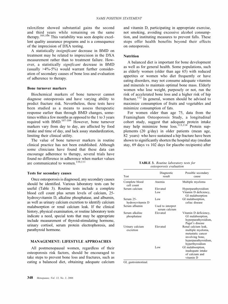
Once osteoporosis is diagnosed, any secondary causesshould be identified. Various laboratory tests can beuseful (Table 3). Routine tests include a completeblood cell count plus serum levels of calcium, 25-hydroxyvitamin D, alkaline phosphatase, and albumin,as well as urinary calcium excretion to identify calciummalabsorption or renal calcium leak. If the clinicalhistory, physical examination, or routine laboratory testsindicate a need, special tests that may be appropriateinclude measurement of thyroid-stimulating hormone,urinary cortisol, serum protein electrophoresis, andparathyroid hormone.
MANAGEMENT: LIFESTYLE APPROACHES
All postmenopausal women, regardless of theirosteoporosis risk factors, should be encouraged totake steps to prevent bone loss and fractures, such aseating a balanced diet, obtaining adequate calcium
and vitamin D, participating in appropriate exercise,not smoking, avoiding excessive alcohol consump-tion, and instituting measures to prevent falls. Thesesteps offer health benefits beyond their effectson osteoporosis.
Nutrition
A balanced diet is important for bone developmentas well as for general health. Some populations, suchas elderly women (older than age 65) with reducedappetites or women who diet frequently or haveeating disorders, may not consume adequate vitaminsand minerals to maintain optimal bone mass. Elderlywomen who lose weight, purposely or not, run therisk of accelerated bone loss and a higher risk of hipfracture.112 In general, women should be advised tomaximize consumption of fruits and vegetables andminimize consumption of fats.
For women older than age 75, data from theFramingham Osteoporosis Study, a longitudinalcohort study, suggest that adequate protein intakemay help minimize bone loss.113,114 Protein sup-plements (20 g/day) in older patients (mean age,82 years) who have sustained a hip fracture have beenshown to significantly shorten the hospital stay (medianstay, 69 days vs 102 days for placebo recipients) after
TABLE 3. Routine laboratory tests forosteoporosis evaluation
TestDiagnostic
resultPossible secondary
cause
Complete bloodcell count
Anemia Multiple myeloma
Serum calcium Elevated HyperparathyroidismLow Vitamin D deficiency,
GI malabsorptionSerum 25-
hydroxyvitamin DLow GI malabsorption,
celiac diseaseSerum albumin Used to interpret
serum calciumSerum alkaline
phosphataseElevated Vitamin D deficiency,
GI malabsorption,hyperparathyroidism,Paget`s disease
Urinary calciumexcretion
Elevated Renal calcium leak,multiple myeloma,metastatic cancerinvolving bone,hyperparathyroidism,hyperthyroidism
Low GI malabsorption,inadequate intakeof calcium andvitamin D
GI, gastrointestinal.
348 Menopause, Vol. 13, No. 3, 2006
NAMS POSITION STATEMENT

hip fracture and improve the clinical outcomes whilein the hospital.115 Compared with the controls, pro-tein recipients also had significantly lower rates ofcomplications and mortality 7 months after their hipfracture.
An adequate intake of both calcium and vitamin Dis important for bone health, and it is recognized asan important component of any osteoporosis pre-scription-drug regimen. For example, a review of 31clinical trials evaluating estrogen therapy with andwithout calcium supplements found annual BMDgains at the hip were significantly greater for thosereceiving estrogen plus calcium (2.4%) comparedwith estrogen alone (0.9%).116
Calcium, either alone or with vitamin D, is not aseffective as pharmacotherapy with either estrogenalone (ET) or estrogen plus progestogen (EPT), aselective estrogen-receptor modulator (SERM), or abisphosphonate. Nevertheless, calcium and vitamin Dare both important components of osteoporosistherapy in combination with all antiresorptive agents.
Calcium
Evidence has established the role of adequatecalcium intake on bone health, primarily in increasingBMD and improving the efficacy of therapeuticagents. Calcium has not been shown to have apositive effect on fracture risk;50,117 however, in theWomen`s Health Initiative (WHI) trial,117 hip frac-tures were significantly reduced in older women whowere adherent to the calcium regimen.
Calcium requirements increase with advancingage, particularly after menopause, owing in part toboth reduced intestinal calcium absorption and renalcalcium conservation. The primary factor influencingthe amount of calcium absorbed is the amount ofcalcium ingested.
Most experts support the published recommenda-tions for daily calcium consumption from either the
National Institutes of Health (revised in 1994)118 orthe National Academy of Sciences (revised in1997).119 Recommendations related to peri- andpostmenopausal women are presented in Table 4.
No single laboratory test can accurately detectcalcium deficiency. However, a 24-hour urine cal-cium level of less than 50 mg suggests eitherinsufficient intake or poor absorption. In general,postmenopausal women in the United States andCanada have dietary calcium intakes that are low,with median intakes of approximately 600 mg/day.120,121 Specific populations of postmenopausalwomen at increased risk of inadequate calcium intakeinclude women who are older, are lactose intolerant,follow a vegetarian diet, or have poor eating habits.
Dietary sources should be the primary source ofcalcium intake because of the other essential nutrientsfound in high-calcium food. Dairy products are thebest sources of calcium based on their high elementalcalcium content, high absorption rate, and low costrelative to total nutritional value. To achieve maximalcalcium absorption from food sources, food selectiondecisions should reflect the food`s calcium bioavail-ability and the presence in the meal of other foodsthat may inhibit calcium absorption (eg, oxalic acid-containing foods, such as spinach, and phytate-richgrains, such as wheat bran).122
Calcium supplements and calcium-fortified foodsare alternative sources for women unable to consumeenough dietary calcium; most women need an addi-tional 600 to 900 mg/day (two to three dairy portions)over their usual daily intake to reach recommendedlevels. Calcium citrate supplements are well absorbedwhen taken with meals or on an empty stomach;calcium carbonate is better absorbed when taken withfood. It is best to take calcium in divided doses forbetter absorption, although single doses of up to1,000 mg can be taken.
Total calcium intakes of up to 1,500 mg/day do notappear to increase the risk of developing renal calculiand may actually reduce it.123 Calcium supplementsare contraindicated in a woman with a calcium-containing renal calculus until her urinary biochemi-cal profile has been assessed. Larger amounts ofcalcium (>2,500 mg/day) should be avoided.
Calcium intervention trials have not reported anyserious adverse events. Nevertheless, some womenhave difficulty swallowing the large tablet or havegastrointestinal (GI) adverse effects (ie, gaseousness,constipation). Tolerability can be addressed byswitching the type of calcium or reducing the dose.GI adverse effects are often related to a woman`s
TABLE 4. Recommended daily elemental calcium intakein peri- and postmenopausal women
National Academy of SciencesAge 31-50 1,000 mgAge 51 and older 1,200 mg
National Institutes of HealthPremenopausal women aged 25-50 1,000 mgPostmenopausal women younger than age 65 and
using estrogen therapy1,000 mg
Postmenopausal women not using estrogen therapy 1,500 mgAll women aged 65 and older 1,500 mg
Adapted from the National Institutes of Health118 and the NationalAcademy of Sciences.119
Menopause, Vol. 13, No. 3, 2006 349
NAMS POSITION STATEMENT

taking more calcium than required, not dividingdoses, or perhaps confusing supplemental intake withrecommended total daily intake.
Vitamin D
The nutrient vitamin D is essential for the intestinalabsorption of calcium. The current National Academyof Sciences recommended dietary intake for vitaminD is 400 IU/day for women aged 51 to 70 years and600 IU/day for women older than age 70.119 Inaddition, NAMS recommends intake of 700 to800 IU/day for women at risk of deficiency becauseof inadequate sunlight exposure, such as older, frail,chronically ill, housebound, or institutionalizedwomen or those who live in northern latitudes.124
Doses as high as 2,000 IU/day are safe.119 Muchhigher doses may introduce risks such as hyper-calciuria and hypercalcemia.
Sources of vitamin D include sunlight, vitamin DYfortified dairy products, fatty fish, and supplements.Daily requirements can usually be met with a multi-vitamin supplement (typically containing 400 IUvitamin D), vitamin DYfortified foods (eg, milk,breakfast cereals providing 100 IU per serving), orcalcium supplements containing vitamin D (usually200 IU per tablet). Many women over the age of 65who have little or no sun exposure and rely onmultivitamins alone for vitamin D intake may havesuboptimal vitamin D levels.125 Currently, there is noworldwide consensus on criteria for acceptable serum25-hydroxyvitamin D values, but if parathyroid hor-mone concentration were used as an index of calciumabsorption, as some suggest, the lower end of thenormal 25-hydroxyvitamin D concentration would bein the range of 30 ng/mL (70-80 nmol/L).126 Becauseof the long half-life of vitamin D, taking vitamin D atthe same time as calcium is not necessary, although itcan be a convenient way to obtain adequate intake ofboth nutrients.
Regarding the effect of vitamin D on fracture risk,several large randomized controlled studies of vita-min D (400 and 800 IU/day) plus 1,000 mgcalcium50,51,117 have shown that the nutrients do nothave a significant effect on fracture risk. However, ameta-analysis49 of randomized clinical trials in post-menopausal women (mean ages, 71-85 years) foundthat a vitamin D dose of 700 to 800 IU/day wasassociated with significant reductions in the risk ofboth hip and nonvertebral fractures. No significantchanges were found in either fracture outcome intrials that used only 400 IU/day.
Studies have found that vitamin D (600-700 IU/day)with supplemental calcium can reduce the rate of post-menopausal bone loss, especially in older women.127
More recent results from the WHI117 found calcium(1,000 mg/day) plus vitamin D (400 IU/day) recipientshad a small but significant 1% improvement in hipBMD. Vitamin D supplementation also has been foundto improve muscle strength128 and balance,129 andreduce the risk of falling.130
Vitamin K
Supplementation with vitamin K (1 mg/day) appearsto be associated with beneficial effects on bone turnoverand bone density. Taking vitamin K as part of a dailymultivitamin supplement may contribute to reducingpostmenopausal bone loss, especially in the hip,131 butfurther validation is needed to define its contribution.Vitamin K supplements are contraindicated in womentaking warfarin.
Magnesium
Another nutrient, magnesium, is sometimes men-tioned as a necessary supplement for the protection ofbone health and/or for absorption of calcium. However,in most trials focused on BMD or osteoporotic fracture,benefits of calcium were observed without magnesiumsupplements. Moreover, a study with calcium absorptionas the end point found that adding 789 to 826 mg/dayof magnesium, more than double the daily average mag-nesium intake (280 mg) for postmenopausal women,had no effect on calcium absorption.132 Nevertheless, inwomen with excessive magnesium loss, usually due toGI disease (eg, diarrhea, vomiting), magnesium sup-plementation would be appropriate.133,134
Isoflavones
Clinical trial data do not support the use ofisoflavones (a class of phytoestrogens found in richsupply in soybeans and soy products as well as in redclover) to prevent or treat osteoporosis. Althoughsome data suggest that isoflavones may favorablyaffect bone health,135 accumulating data from severalmore recent studies indicate a lack of bone benefitsfrom isoflavones, regardless of the source (ie,extracted from red clover or soy or consumed in soyfoods).136<138 Ipriflavone, a synthetic isoflavone avail-able without a prescription in the United States andCanada, has not demonstrated a positive effect onbone density, bone turnover markers, or fracture riskin women with osteoporosis.139
350 Menopause, Vol. 13, No. 3, 2006
NAMS POSITION STATEMENT
Exercise
Local increases in bone mass occur in response toactivities that cause significant stress to bone. Activeexercises that involve weight training can increasebone mass if they increase muscle mass andstrength. Applying passive stress tests to bone alsoshows promise, with the most positive resultscoming from use of high-frequency, whole-bodyvibration systems.140,141
In early postmenopausal women, strength trainingprovides small but significant benefits on bonemass.142 A meta-analysis143 found that women whoexercised increased spinal BMD by approximately2%. For women who use menopausal estrogen-containing therapy, strength training provides addi-tional BMD benefits over hormone therapy alone.144
Most strength training studies have used progressiveresistance obtained with machines designed for thispurpose (eg, Nautilus). However, strength trainingcan be performed as few times as twice a week andneed not involve expensive equipment. Exercise forwomen with established osteoporosis should notinclude heavy weight-bearing exercises or activityso vigorous that it may trigger a fracture.
For elderly women with osteoporosis, physicalactivity plays an important role in reducing the riskof falls. Among women aged 75 and older, muscle-strengthening and balance exercises have beenshown to reduce the risk of falls and fall-relatedinjuries by 75%.145
Fall prevention
Falls are the precipitating factor in nearly 90% ofall fractures.146 In the United States and Canada,approximately one third of women older than age 60fall at least once a year.100,147 In nearly one half ofthese cases, it is a recurrent fall. The incidence offalls increases with age, rising to a 50% annual rate inpeople older than age 80. Elderly women have asignificantly higher risk of falls than do men of thesame age. As a result, prevention of falls that cancause fractures should be an aspect of routine care forall postmenopausal women.
Several healthcare interventions have proven effec-tive in reducing the risk of falls. These focusprimarily on exercises to improve balance and musclestrength, adjusting medication use (especially psycho-tropic drugs), and reducing fall hazards in thehome.148 Tapering or discontinuing use of benzo-diazepines, neuroleptic agents, and antidepressantshas been found to reduce the risk of falling by more
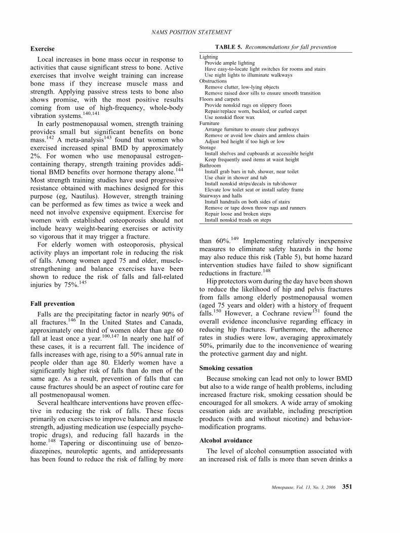
than 60%.149 Implementing relatively inexpensivemeasures to eliminate safety hazards in the homemay also reduce this risk (Table 5), but home hazardintervention studies have failed to show significantreductions in fracture.148
Hip protectors worn during the day have been shownto reduce the likelihood of hip and pelvis fracturesfrom falls among elderly postmenopausal women(aged 75 years and older) with a history of frequentfalls.150 However, a Cochrane review151 found theoverall evidence inconclusive regarding efficacy inreducing hip fractures. Furthermore, the adherencerates in studies were low, averaging approximately50%, primarily due to the inconvenience of wearingthe protective garment day and night.
Smoking cessation
Because smoking can lead not only to lower BMDbut also to a wide range of health problems, includingincreased fracture risk, smoking cessation should beencouraged for all smokers. A wide array of smokingcessation aids are available, including prescriptionproducts (with and without nicotine) and behavior-modification programs.
Alcohol avoidance
The level of alcohol consumption associated withan increased risk of falls is more than seven drinks a
TABLE 5. Recommendations for fall prevention
LightingProvide ample lightingHave easy-to-locate light switches for rooms and stairsUse night lights to illuminate walkways
ObstructionsRemove clutter, low-lying objectsRemove raised door sills to ensure smooth transition
Floors and carpetsProvide nonskid rugs on slippery floorsRepair/replace worn, buckled, or curled carpetUse nonskid floor wax
FurnitureArrange furniture to ensure clear pathwaysRemove or avoid low chairs and armless chairsAdjust bed height if too high or low
StorageInstall shelves and cupboards at accessible heightKeep frequently used items at waist height
BathroomInstall grab bars in tub, shower, near toiletUse chair in shower and tubInstall nonskid strips/decals in tub/showerElevate low toilet seat or install safety frame
Stairways and hallsInstall handrails on both sides of stairsRemove or tape down throw rugs and runnersRepair loose and broken stepsInstall nonskid treads on steps
Menopause, Vol. 13, No. 3, 2006 351
NAMS POSITION STATEMENT
week, as established by the Framingham HeartStudy.70 Postmenopausal women who drink shouldbe advised to drink moderately and not to exceedseven drinks a week. One drink is considered to beone 12-oz (360 mL) beer, 4 oz (120 mL) of wine, or1 oz (30 mL) of liquor.
MANAGEMENT:
PHARMACOLOGIC APPROACHES
A management strategy focused on lifestyleapproaches may be all that is needed for postmeno-pausal women who are at low risk of osteoporoticfracture. NAMS recommends adding osteoporosisdrug therapy in the following populations:
& All postmenopausal women who have had anosteoporotic vertebral fracture
& All postmenopausal women who have BMDvalues consistent with osteoporosis (ie, T-scoresequal to or worse than j2.5)
& All postmenopausal women who have T-scoresfrom j2.0 to j2.5 and at least one of thefollowing risk factors for fracture: thinness [bodyweight G127 lb (57.7 kg) or low BMI (G21 kg/m2)],history of fragility fracture since menopause, orhistory of hip fracture in a parent
The diagnostic categorization should be based onthe lowest of the BMD values among the threemeasured sites of total hip, femoral neck, andposterior-anterior lumbar spine. Current treatmentguidelines are based on specific bone density thresh-olds and risk factors. Available in the near future willbe an improved method from the WHO that usesalgorithms that incorporate estimates of absolutefracture risk.
Several pharmacologic options are available forosteoporosis therapy, including bisphosphonates, theSERM raloxifene, parathyroid hormone, estrogens,and calcitonin. No studies have compared thesetherapies for antifracture efficacy.
Adherence to therapy is poor. In studies of6 months to 1 year, adherence rates for prescriptiondrugs ranged from below 25% to 81%, dependingon the therapy.152 <154 Ensuring adherence to thetreatment plan is perhaps the most important follow-up measure for clinicians.
Bisphosphonates
This class of drugs works by inhibiting the activityof osteoclasts and shortening their life span, thereby
reducing bone resorption.155 Bisphosphonates do nothave known beneficial effects on the body other thanon bone. The most common adverse effect ofbisphosphonate therapy is esophageal and gastricirritation, particularly affecting individuals who doseinappropriately. Before starting bisphosphonatetherapy, serum creatinine should be used to estimatethe glomerular filtration rate; treatment may beinitiated only if the rate is 30 mL/min or greater.
Clinical trials have demonstrated that bisphospho-nates significantly increase BMD at the spine and hipin a dose-dependent manner in both younger andolder postmenopausal women. In women with osteo-porosis, bisphosphonates have reduced the risk ofvertebral fractures by 40% to 50% and reduced theincidence of nonvertebral fracture, including hipfracture, by about half this amount.106,155
All the bisphosphonates approved for osteoporosistherapy in both the United States (alendronate,ibandronate, and risedronate) and Canada (alendro-nate, etidronate, and risedronate) are available in oralformulations for daily and intermittent dosing regi-mens. Weekly oral dosing regimens of alendronateand risedronate and monthly oral dosing regimensand IV dosing of ibandronate have been approvedbased on clinical trials that showed BMD responsesequivalent to those observed with daily treat-ment.156 <159 All fracture data are from trials withdaily dosing; the bridging studies beyond dailydosing were not designed with fracture end points.
Alendronate
This bisphosphonate, marketed as Fosamax, isapproved in both the United States (as an oral tabletand liquid) and Canada (oral tablet only) for post-menopausal osteoporosis prevention (5 mg/day or35 mg/wk) and treatment (10 mg/day or 70 mg/wk).Alendronate is also available in a single weekly oraltablet of 70 mg with 2,800 IU of vitamin D (FosamaxPlus D).
For women in early postmenopause, 2 to 6 years oftreatment with alendronate (5 mg or more daily) hasbeen shown to significantly increase BMD at thespine and hip by approximately 1% to 4% frombaseline, whereas BMD in placebo recipientsdecreased by 2% to 4% during that time.160,161 Inolder women with osteoporosis,162 therapy with10 mg daily significantly increased BMD in the spine(8.8%) and the femoral neck (5.9%) after 3 years,compared with placebo. In 7- and 10-year trials inwomen with low bone density,163,164 alendronatetherapy resulted in increases from baseline of 5% to
352 Menopause, Vol. 13, No. 3, 2006
NAMS POSITION STATEMENT

10% at the spine and hip in postmenopausal womenwho had low BMD or established osteoporosis.Because placebo groups were not followed for theduration of the studies,163,164 the antifracture effectsof long-term alendronate therapy could not beadequately evaluated. However, there was no appa-rent increase in fracture risk over time.
The efficacy of alendronate in decreasing fracturerisk has been demonstrated only in postmenopausalwomen with osteoporosis. Similar to other bisphos-phonates, alendronate has shown lesser effects inwomen without osteoporosis.
In the Fracture Intervention Trial (FIT),165 dailyalendronate therapy for 2.9 years significantlyreduced the risk of vertebral fracture by 47% and ofhip fracture by 51% in women with low BMD andprevious vertebral fracture. The incidence of clinicalvertebral fractures was reduced by 59% within thefirst year.166 In a composite analysis of the two armsof the FIT study,166 3 years of alendronate therapy ina subgroup of women with osteoporosis (ie, vertebralfracture or T-score equal to or worse than j2.5)significantly reduced the risk of nonspine fracture by27% and new spine fracture by 50%.
Risedronate
This bisphosphonate, marketed as Actonel, isapproved for the prevention and treatment of post-menopausal osteoporosis in oral tablet doses of 5 mgdaily or 35 mg once weekly. Recently available inthe United States is a packet containing bothrisedronate and calcium (marketed as Actonel withCalcium) that provides 4 weeks of risedronatetherapy (35 mg/wk) and calcium carbonate (500 mgfor the no-risedronate days).
In a randomized clinical trial of early postmeno-pausal women (age range, 40-61 years; mean age,51-52 years) with normal bone density, risedronatedoses of 5 mg/day for 2 years produced significantBMD increases of 5.7% in the lumbar spine and5.4% in the hip greater than with placebo.167 In arandomized controlled trial in older postmenopausalwomen (mean age, 68-69 years),36 3 years ofrisedronate therapy (5 mg/day) resulted in significantBMD increases of 4.3% in the spine and 2.8% in thefemoral neck compared with placebo. Therapy for7 years resulted in progressive increases in BMD of11.5% from baseline (with no placebo group after5 years).168
Several randomized controlled trials have foundfracture risk reductions with risedronate. In two trialsof postmenopausal women with osteoporosis,36,169
1 to 3 years of treatment with 5 mg/day ofrisedronate significantly reduced the risk of vertebralfracture (41%- 49%) compared with placebo. Withinthe first year of therapy, the relative risk of vertebralfracture was reduced by 61% to 65%. After 3 yearsof therapy, vertebral fracture risk reductions werestill statistically significant relative to placebo. Inone of these trials,36 the risk of nonvertebral fracturewas significantly reduced by 39%. In the othertrial,169 nonvertebral fracture risk was reduced by33%, although this was not statistically significantversus placebo.
In the Hip Intervention Program Study Group,170 arandomized controlled trial of 5,445 postmenopausalwomen aged 70 to 79 years, daily risedronate therapysignificantly reduced the relative risk of hip fractureby 40% in women with BMD values consistent withosteoporosis. It reduced the risk of hip fracture by60% in the group with previous vertebral fractures.However, therapy did not significantly lower the hipfracture risk in women 80 years of age and older whohad risk factors for falling but who did not have BMDtesting performed to confirm osteoporosis.
In a randomized controlled trial of 265 postmeno-pausal women (mean age, 72 years), the incidence ofvertebral fractures in women treated with risedronate5 mg/day was significantly reduced during years 4and 5 compared with placebo,171 and appeared toremain reduced through 7 years of treatment (noplacebo group after 5 years).168 No new adverseevents were observed in these trials.
Ibandronate
Ibandronate, marketed as Boniva, is approved atan oral tablet daily dose of 2.5 mg as well as in aonce-monthly oral tablet dose of 150 mg for theprevention and treatment of postmenopausal osteo-porosis. It is also approved in an IV formulation at adose of 3 mg every 3 months (administered by ahealthcare professional) for the treatment of post-menopausal osteoporosis.
In early postmenopausal women (mean ages,57.6-58.8 years) without osteoporosis, those receiv-ing oral ibandronate at 2.5 mg/day had significantBMD increases of 1.9% in the lumbar spine (vsY1.9% for placebo) and 1.2% in the total hip (vsY0.6% for placebo) after 2 years.172 In older women(mean age, 69 years) with low spinal BMD andprevalent vertebral fractures, oral ibandronate at2.5 mg/day significantly increased BMD comparedwith placebo in the spine (5.2%) and femoral neck(4.1%) after 3 years.173 Daily oral ibandronate
Menopause, Vol. 13, No. 3, 2006 353
NAMS POSITION STATEMENT
therapy reduced morphometric vertebral fractures by52% over the 3 years, but there was no significanteffect on nonvertebral fracture risk in the overallstudy population. In a post hoc analysis, a 69%reduction of nonvertebral fracture risk was described,but only in the subgroup of study patients withbaseline femoral neck T-scores below j3.
Etidronate
The bisphosphonate etidronate, marketed as Didroneloral tablets, is approved in Canada for osteoporosisprevention and treatment in postmenopausal women. Inthe United States, it is approved only for treatment ofPaget`s disease, not for osteoporosis therapy.
A meta-analysis174 of 13 trials investigating inter-mittent cyclic etidronate therapy for postmenopausalosteoporosis found that, relative to control groups,1 to 3 years of therapy increased BMD by 4.1% in thelumbar spine and 2.3% in the femoral neck. Thisanalysis concluded that etidronate significantlyreduced the risk of vertebral fracture (37%) but notthe risk of nonvertebral fracture.
For osteoporosis therapy, etidronate is typicallyadministered at 400 mg/day for 14 days every 3 months,with calcium taken between the cycles. A cyclicregimen is used because daily high-dose use mayinterfere with bone mineralization.175 This is not theschedule for Paget`s disease.
Adverse events with bisphosphonate therapy
Bisphosphonates may cause upper GI disorderssuch as dysphagia, esophagitis, and esophageal andgastric ulcer, a contraindication in those with esoph-ageal abnormalities that delay esophageal emptyingor in those who are unable to stand or sit upright forat least 30 to 60 minutes after ingestion. Studies arenot adequate to determine upper GI adverse effectdifferences among oral bisphosphonates, althoughonce-quarterly IV ibandronate labeling does not carrythe warnings of the tablet formulations regardingupper GI adverse events.
IV ibandronate labeling includes an enhanced pre-caution on hypocalcemia and renal impairment; how-ever, no cases of acute renal failure have been observedin clinical trials. Patients who receive IV ibandronateshould have serum creatinine measured before eachdose administration.
Bisphosphonates are poorly absorbed; typically,approximately 0.5% of an oral dose is absorbed, evenwhen taken on an empty stomach with plain water.
Therefore, oral bisphosphonates must be taken thefirst thing in the morning when the stomach is empty.Food, drink, and medications (including supplements)must be avoided for 30 minutes (alendronate andrisedronate) to 60 minutes (ibandronate) after dosing;etidronate labeling recommends waiting 2 hours.
A transient flu-like illness, often called an acute-phase reaction, occurs infrequently with large dosesof oral or IV bisphosphonates. This has beenobserved infrequently after monthly oral or quarterlyIV dosing with ibandronate. Symptoms are generallymild, most often occur with the first, but notsubsequent, doses and are treated symptomatically.
A theoretical concern exists regarding possibleoversuppression of bone turnover with long-termbisphosphonate therapy, resulting in a more brittleskeleton. Individual cases with poor fracture healingafter alendronate therapy have been described,176 butmost of those patients were receiving combinedalendronate-estrogen therapy or had serious under-lying medical problems.
Jaw lesions, usually after dental extraction (oftendescribed as osteonecrosis of the jaw), have beenobserved with bisphosphonate use, most often inpatients treated with large intravenous doses forcancer-related bone diseases.177,178 The large doseamount, not the duration of therapy (12-25 months),was linked to the osteonecrosis. In 2- to 10-yearrandomized clinical trials of alendronate or risedro-nate using smaller oral doses appropriate for osteopo-rosis, no osteonecrosis of the jaw has been observed,although these lesions have been anecdotallyreported.179,180
Long-term safety of bisphosphonate therapy
Randomized clinical trials of more than 5 years`duration with alendronate or risedronate161,163,164,168
have demonstrated persistent reduction of bone turn-over without evidence of unexpected adverse effectsor abnormal bone histomorphometry. No data areavailable on effects of long-term (>3 years) iban-dronate or etidronate therapy. Current evidence doesnot support recommendations regarding the optimalduration of bisphosphonate therapy.
Discontinuation of bisphosphonate therapy
Following discontinuation of alendronate after 4 to5 years of therapy, bone turnover remains relativelysuppressed with BMD remaining stable or decreasingslowly.163,164,181 Bone turnover markers remain
354 Menopause, Vol. 13, No. 3, 2006
NAMS POSITION STATEMENT

suppressed but return to pretreatment levels overtime. Whether the fracture protection afforded byalendronate therapy persists after discontinuation isnot known, although there is no apparent abruptincrease in fracture rate upon treatment cessation.
Discontinuation of risedronate therapy after 2 yearsin young postmenopausal women (mean ages, 51-52 years) has been shown to result in significant boneloss at both the spine and hip during the first year aftertreatment is stopped.167 The effects of stopping therapyin older women or after longer treatment intervals arenot known.
No data are available regarding discontinuation ofibandronate or etidronate therapy.
SERMs
The SERMs are nonsteroidal agents of variouschemical structures that act as estrogen receptoragonists and/or antagonists. The SERM raloxifene(marketed as Evista oral tablets) is governmentapproved for the prevention and treatment of osteo-porosis at a dose of 60 mg/day. No other SERM isapproved for osteoporosis therapy, although severalare in clinical development.
Raloxifene has beneficial effects on BMD, and itdecreases bone turnover as assessed by biochemicalmarkers. In a 2-year randomized controlled trial of 601postmenopausal women without osteoporosis (meanage, 55 years), raloxifene at a dose of 60 mg/daysignificantly improved BMD at the lumbar spine(1.6%) and femoral neck (1.2%) compared withplacebo (decreases of 0.8% and 1.2%, respectively).182
In the randomized controlled Multiple Outcomes ofRaloxifene Evaluation (MORE) trial evaluating post-menopausal women with osteoporosis (mean age,67 years),183 3 years of raloxifene therapy at 60 mg/day significantly increased BMD versus placebo by2.6% at the spine and 2.1% at the femoral neck.
The efficacy of raloxifene in reducing osteoporoticfractures also was demonstrated in the MORE trial.183
After 3 years of therapy, raloxifene (60 mg/day)reduced the risk of vertebral fracture by 55% inwomen with a femoral neck or lumbar spine BMDT-score of j2.5 or below and by 30% in women withlow T-scores and a prevalent vertebral fracture; bothfindings were significant compared with placebo. A1-year blinded extension of the MORE trial184 foundpersistent vertebral fracture risk reductions of 50%and 38% in the two groups, respectively. A separateanalysis revealed that at 1 year, raloxifene (60 mg/day) reduced the risk of new clinical vertebralfracture by 68% in the overall study population.185
No raloxifene effect has been observed on hip orother nonvertebral fracture risk.
In addition to its effects on bone, raloxifene hasbeen associated with a reduced risk of invasive breastcancer in postmenopausal women with osteoporosis.In the MORE trial, the overall incidence of invasivebreast cancer was significantly reduced by 76% after3 years186 and 72% after 4 years.187 In a 4-yearextension of the MORE trialVthe Continuing Out-comes Relevant to Evista (CORE) trial188Vthe riskafter 8 years was 59% lower in raloxifene recipients;the risk of estrogen receptor (ER)-positive invasivebreast cancer was 66% lower. The combined resultsshow invasive breast cancer and ER-positive breastcancer risks were reduced by 66% and 76%,respectively. It should be noted that the MORE-CORE studies were conducted on postmenopausalwomen initially selected for risk of osteoporosis, notfor risk of breast cancer.
A significant increase in thromboembolic eventswas noted in the MORE trial.189 However, a secondaryanalysis of the MORE trial data190 found no overallsignificant differences in the number of coronary orcerebrovascular events between placebo and raloxi-fene, although in a subset of women with increasedcardiovascular risk at baseline, raloxifene significantlyreduced cardiovascular risk. Again, it should be notedthat the MORE trial was not designed with cardiovas-cular outcomes as the primary objective.
Randomized clinical trials of more than 5 years`duration have demonstrated no other significantadverse effects.189 Raloxifene therapy may be asso-ciated with an increase in vasomotor symptoms.However, it does not increase the risk of cataracts,gallbladder disease, endometrial hyperplasia, orendometrial cancer or cause vaginal bleeding orbreast pain.183,189
Bone loss often resumes when raloxifene therapyis stopped.191,192
Parathyroid hormone
The various chemical structures of parathyroidhormone (PTH) are anabolic agents that directlystimulate osteoblastic bone formation, resulting insubstantial increases in trabecular bone density andconnectivity in women with postmenopausal osteo-porosis. This mechanism of action is very differentfrom that of antiresorptive agents such as estrogenand bisphosphonates, which reduce bone resorption.
Teriparatide (recombinant human PTH 1-34), mar-keted as Forteo, is approved in both the United Statesand Canada for the treatment of postmenopausal
Menopause, Vol. 13, No. 3, 2006 355
NAMS POSITION STATEMENT

women with osteoporosis who are at high risk offracture. A full-length PTH (1-84) is being evaluatedin clinical trials.
Randomized controlled trials have shown that dailysubcutaneous injections of teriparatide stimulate boneformation and improve bone density in postmeno-pausal women, regardless of whether they are receiv-ing estrogen therapy.193<195 In postmenopausalwomen with previous vertebral fracture,195 19 monthsof teriparatide treatment (20 Kg/day) significantlyincreased bone density in the spine by 8.6% and inthe femoral neck by 3.5% compared with placebo.The incidence of new vertebral fractures was reducedby 65% and new nonvertebral fragility fractures by53%, although the study was not designed to examinethe effect on hip fractures.
Drug-related adverse effects include muscle crampsand infrequent hypercalcemia, nausea, and dizziness.High-dose teriparatide treatment has caused bonetumors (osteosarcoma) in a rat model at doses rangingfrom 3 to 60 times the 20 Kg/day dose in humans,196
although the significance of this finding in humans isuncertain. Teriparatide should not be administered topostmenopausal women with hypercalcemia, bonemetastases, or disorders that predispose them to bonetumors such as Paget`s disease or those who receivedprior skeletal irradiation. Therapy is indicated for nolonger than 18 to 24 months.
When PTH therapy is stopped, substantial bone losshas occurred within the first year.197 However, inrandomized controlled trials using PTH 1-84, admin-istering alendronate after discontinuing PTH therapywas shown to maintain or improve BMD,197,198 al-though previous alendronate treatment tends to slowbone turnover and delay the PTH-induced increases inBMD and bone turnover response by 3 to 6 months.199
It is unclear whether a second course of PTH can besafely restarted after a period without therapy orwhether regimens other than daily can be effective.
Estrogens
Systemic estrogen products (EPT or ET for womenwithout a uterus) are government approved in theUnited States and Canada for prevention, but nottreatment, of postmenopausal osteoporosis. A numberof randomized controlled trials have evaluated theeffect of systemic estrogen on BMD and fracture inpostmenopausal women.
Bone mineral density
The beneficial effects of systemic oral or trans-dermal ET/EPT at standard doses on BMD preservation
are well established. A 2002 meta-analysis200 of 57randomized clinical trials comparing ET/EPT withplacebo in postmenopausal women found consistentBMD increases with ET/EPT at all sites. In trials of2 years` duration, the mean difference in BMD afterET/EPT was 6.8% at the lumbar spine and 4.1% atthe femoral neck.
The two largest and best controlled trials supportthese findings. In the Postmenopausal Estrogen/Progestin Interventions trial201 (N = 875), standarddaily doses of 0.625 mg conjugated equine estrogens(CEE), with or without a progestogen [either medroxy-progesterone acetate (MPA) or micronized proges-terone], for 3 years significantly increased spinal BMDby 3.5% to 5.0%, with a 1.7% increase in hip BMD.More recently, the WHI,202 a 5-year randomizedcontrolled trial in postmenopausal women aged 50to 79 years (N = 16,608), reported that standard dosesof daily EPT (0.625 mg CEE plus 2.5 mg MPA)significantly increased spine and total hip BMD by4.5% and 3.7%, respectively, relative to placebo.
Effects of lower-than-standard doses of ET/EPT onBMD have been investigated. Randomized controlledtrials203 <207 using doses as low as 0.3 mg/day oralCEE, 0.25 mg/day oral micronized 17A-estradiol, and0.014 mg/day transdermal 17A-estradiol reported sig-nificant increases in spine and hip BMD relative toplacebo. These trials were conducted either in pop-ulations of early postmenopausal women (mean age,51-52 years) or in older postmenopausal women(mean ages, 67 and 74 years). Changes in lumbarspine BMD were in the range of 1% to 3%, sig-nificantly better than placebo.
Significant BMD improvements have also beennoted with systemic estrogen doses delivered via avaginal ring (Femring).208 In a randomized controlledtrial of 174 postmenopausal women younger than age65, daily doses of 0.05 and 0.1 mg of estradiol acetatedelivered via the ring significantly increased hip BMD(1.7% and 1.8%, respectively) and lumbar spine BMD(2.7% and 3.3%) compared with baseline.
Fracture
Evidence from both randomized controlled trialsand observational studies indicate that standarddoses of ET/EPT (including 0.625 mg CEE/day orthe equivalent) reduce fracture risk in postmeno-pausal women. Two meta-analyses have found thatET/EPT significantly reduces the risk of fracture byup to 27%.209,210
Two recent, large observational studies supportthese data. The National Osteoporosis Risk Assessment
356 Menopause, Vol. 13, No. 3, 2006
NAMS POSITION STATEMENT

study examined 200,160 postmenopausal women andreported that current estrogen use was associated witha significantly reduced risk of new fracture.102
Participants were at least 50 years old and had noprevious diagnosis of osteoporosis. The MillionWomen Study,211 a prospective observational studyof 138,737 postmenopausal women, reported thatcurrent ET/EPT use provided a significant relativerisk reduction in incidence of fracture.
Results were confirmed in the WHI. In both the EPTarm202 and the ET arm,212 significant risk reductionswere seen for hip fractures, vertebral fractures, andtotal fractures compared with placebo. The selectioncriteria and outcomes evaluated in the WHI (ie,women were not selected based on an establishedosteoporosis risk factor or BMD level; fracture out-comes included hip, wrist/lower arm, and clinicallyidentified vertebral and total fractures) are in contrastto the design of studies of fracture risk reduction withbisphosphonates or SERMs.36,163,166,169,170,183 In thosestudies, women were selected based on high risk ofosteoporosis (ie, prevalent vertebral fracture and/orlow BMD), and radiographically detected vertebralfractures were often a primary outcome.
The Million Women Study, although observationalin design, addressed issues related to ET/EPT and therisk of fracture that could not be ascertained in theWHI trials, such as comparisons between differentEPT formulations, doses, and routes of administra-tion. When the overall fracture-risk reduction wasexamined by type of hormone, no difference wasfound between ET and EPT. Sequential or continuousprogestin use also did not significantly affect theresults. Furthermore, the relative risk of fracture wasnot different when specific estrogen or progestinproducts were compared (ie, CEE versus estradiol; MPAversus norethisterone or norgestrel/levonorgestrel). Thisstudy did not specifically report on the possible fractureprotection afforded by a low estrogen dose (ie, 0.3 mg),but found that similar risk reductions for doses greaterthan 0.625 mg were similar to those for doses 0.625 mgor less.
Therapy management
The primary indication for systemic ET/EPT isfor women experiencing moderate to severe meno-pause symptoms (eg, vasomotor symptoms, vaginalatrophy).
In the WHI, systemic EPT (CEE plus MPA) atstandard doses for 5.6 years in postmenopausal womenaged 50 to 79 years was associated with a statisticallysignificant increased risk of breast cancer,213 stroke,214
coronary heart disease,215 and thromboembolic event.216
In women who had undergone a hysterectomy, ETalone for 6.8 years resulted in a statistically significantincreased risk of stroke and deep venous thrombosis,whereas total venous thromboembolic events andpulmonary embolism were not statistically increased.212
For postmenopausal women aged 65 to 79 yearsfollowed for a mean of 4.0 years, the Women`s HealthInitiative Memory Study217 found a statistically signifi-cant increase in probable dementia for those who startedEPT (CEE plus MPA). After a mean follow-up of5.2 years, there was a nonsignificant trend for in-creased probable dementia among women allocated toET alone.
NAMS recommends use of ET/EPT at the lowesteffective dose for the shortest time period consistentwith treatment goals.218 Lower-than-standard doses ofET/EPT, however, have not been examined with regardto fracture efficacy. Specific to the use of ET/EPT forits effects on osteoporosis, NAMS states that systemicET/EPT can still be considered, weighing its risks andbenefits as well as those of alternate therapies. Theoptimal time to initiate ET/EPT and the optimalduration of therapy have not been established.
Discontinuation of therapy
Studies have shown a BMD loss of 3% to 6%during the first year after cessation of systemicET/EPT.181,219 <222 Data also indicate that the fracturerisk reduction with ET/EPT use does not persist afterdiscontinuation of therapy. In the Million WomenStudy,210 past users of hormone therapy had noprotection against fracture, and incidence ratesreturned to those of never-users within about 1 yearof ceasing use. In the National Osteoporosis RiskAssessment study,223 clinical fractures of the hip,spine, forearm, wrist, or rib were reduced in currentET/EPT users but not in women who had stopped5 years previously. In a further analysis of hipfractures, women who had discontinued ET/EPTwithin the previous 5 years had a risk of hipfracture at least as high as that in women who hadnever used ET/EPT.224
Calcitonin
Salmon calcitonin is government approved forpostmenopausal osteoporosis treatment but not forprevention.225 It is available in the United States as anasal spray (marketed as Miacalcin Nasal Spray,Fortical Nasal Spray) and a subcutaneous injection(marketed as Miacalcin Injection). Available inCanada are a nasal spray (Miacalcin Nasal Spray)
Menopause, Vol. 13, No. 3, 2006 357
NAMS POSITION STATEMENT

and an injectable form (marketed as CalcimarSolution, Caltine), although these injectables are notindicated for osteoporosis.
Calcitonin is an inhibitor of bone resorption. Inclinical use, however, the reduction in bone turnoverwith calcitonin is much less than with other anti-resorptive agents. A small, dose-finding study ofintranasal calcitonin in postmenopausal women withosteoporosis showed significant increases in spinalBMD of 3% relative to baseline.226
In the randomized controlled Prevent Recurrenceof Osteoporotic Fractures (PROOF) trial,227 intra-nasal spray calcitonin doses of 200 IU/day for 5 yearssignificantly reduced the risk of new vertebralfracture by 33% compared with placebo in 1,255postmenopausal women with established osteopo-rosis. However, statistically significant reductionswere not observed at either 100 IU/day or 400 IU/day. After 5 years, a significant spinal BMD increasecompared with placebo was seen only for recipientsof the 400-IU dose. No significant effect on hip BMDoccurred at any dose. The absence of a dose responseand a 60% dropout rate have led some experts todoubt the reliability of these data.
Calcitonin has been shown to reduce bone painfrom osteoporotic vertebral compression fracturesmore quickly than placebo immediately after afracture;228,229 however, it has not been shown todecrease bone pain in other situations.230 Drug-related adverse effects include nausea, local inflam-mation, and flushing of the face or hands when givenas an injection and local nasal irritation with the nasalspray formulation.
Because calcitonin is a less potent agent than otherpharmacologic therapies for osteoporosis, it isreserved as an alternative for women who cannot orchoose not to take one of the other osteoporosis agents.The efficacy of calcitonin has not been observed inearly postmenopausal women. Thus, product labelingrecommends its use only in women with osteoporosiswho are at least 5 years beyond menopause.
Combination therapies
Combining potent antiresorptive agents results insmall additional increments in bone density. Inpostmenopausal women (mean age, 61-62 years) withlow bone mass, BMD improvements in the spine andhip with combined alendronate and ET were signifi-cantly greater (8.3%) than results for either agent alone(6.0%).231 Combined risedronate and ET/EPT alsohas shown favorable, although modest, BMD effectscompared with either agent alone.232 Whether
increases in BMD result in better fracture protectionis not known, and the long-term safety of combina-tion therapies has not been evaluated. One concern isthat combining two antiresorptive therapies mightoversuppress bone turnover, adversely affect bonequality, and thereby increase the likelihood offracture. Combining antiresorptive agents is not gen-erally recommended.
Combining an anabolic agent such as teriparatidewith an antiresorptive agent has been considered.Significant increases in BMD occurred in a randomizedcontrolled trial when teriparatide was added to ongoingET.194 When PTH 1-84 and alendronate were com-bined, the BMD response was less than that seen withPTH alone.197 Based on available data, recommenda-tions cannot be made for or against combiningantiresorptive and anabolic drugs.
Promising new therapies
Several new drugs show promise for the treatmentand/or prevention of osteoporosis. Some are nowavailable outside North America, while others are inclinical development. These include another IVbisphosphonate (zoledronic acid), additional SERMs(lasofoxifene, arzoxifene, and basodoxifene), PTH1-84, tibolone, denosumab, and strontium ranelate.Although some data on statins and thiazides suggestbeneficial bone effects, it is unlikely that randomizedcontrolled trials will be performed to evaluatepotential benefits.
This document summarizes data for the onlypromising therapy that has demonstrated fractureefficacy in published trials, oral strontium ranelate.Approved for the prevention and treatment ofosteoporosis in more than 25 countries outside NorthAmerica, strontium ranelate (marketed as Protelos),appears to have modest antiresorptive and mildanabolic effects, although its mechanism of action isunknown. Dosing involves dissolving 2 g of stron-tium ranelate in water and drinking it before bedtime.
Efficacy is best documented by fracture trials.BMD may show artifactual increases due to thehigher molecular weight of strontium compared withthe calcium it replaces in the skeleton.
In large randomized controlled trials in postmeno-pausal women in Europe and Australia,233,234 3 yearsof strontium therapy significantly increased bonedensity and reduced the incidence of both spine andnonspine fractures. Drug-related adverse effectsincluded significant increases in nausea and diarrheathat resolved after 3 months. Increases in boneformation markers (less than seen with PTH) and
358 Menopause, Vol. 13, No. 3, 2006
NAMS POSITION STATEMENT

decreases in bone resorption markers (less than withbisphosphonates or estrogen) have been observedwith strontium therapy.
A large randomized controlled trial in postmeno-pausal women with the primary end point of vertebralfractures233 demonstrated that 3 years of therapysignificantly increased bone density at the spine(14%) and femoral neck (8%). Compared withplacebo, the risk of spine fractures in strontium-treated women was significantly reduced by 49%after 1 year and 41% after 3 years compared withplacebo. A second randomized controlled trial inves-tigating nonvertebral fractures234 reported that after 3years, nonvertebral fractures were significantlyreduced by 16% in treated women compared withplacebo. In a subgroup of high-risk women (olderthan age 74 with a femoral neck T-score of less thanj2.4), there was a 36% decrease in hip fractures.Drug-related adverse effects in both trials wereinfrequent and included nausea and diarrhea thatusually resolved after 3 months.
RECOMMENDATIONS
Management strategies for osteoporosis in post-menopausal women require assessment of risk factorsfor BMD-defined osteoporosis and osteoporotic frac-ture, followed by institution of measures that focus onreducing risk factors through lifestyle changes and, ifindicated, pharmacologic therapy.
& All postmenopausal women should be encouragedto employ lifestyle practices that reduce the risk ofbone loss and osteoporotic fractures: maintaining ahealthy weight, eating a balanced diet, obtainingadequate calcium and vitamin D, participating inappropriate exercise, avoiding excessive alcoholconsumption, not smoking, and using measures toprevent falls. Periodic reviews of calcium andvitamin D intake and lifestyle behaviors are useful.After menopause, a woman`s risk of falls should beassessed at least annually.
& The physical examination should include anannual measurement of height and weight, alongwith an assessment for kyphosis and back pain.
& BMD testing is indicated for:YAll postmenopausal women with medicalcauses of bone lossYAll postmenopausal women aged 65 andolder
BMD testing should be considered for healthypostmenopausal women younger than age 65
who have one or more of the following riskfactors:
YPrevious fracture (other than skull, facial bone,ankle, finger, and toe) after menopauseYThinness [body weight G127 lb (57.7 kg) orBMI G21 kg/m2]YHistory of hip fracture in a parentYCurrent smoking
When BMD testing is indicated, DXA is the pre-ferred technique. The total hip, femoral neck, andposterior-anterior lumbar spine should be measured,using the lowest of the three BMD scores.
& The routine use of biochemical markers of bone turn-overinclinicalpractice isnotgenerallyrecommended.
& If osteoporosis is diagnosed, either clinically orby BMD, any secondary causes should beidentified, although the data defining the mostthorough or cost-effective workup are limited.
& Vertebral fracture must be confirmed, through eithera vertebral fracture assessment with DXA measure-ment of the spine or height loss of more than 20%(or 4 mm) of a vertebra on spinal radiograph.
& In postmenopausal women, the need for prescrip-tion osteoporosis therapy is based on a combinationof BMD and risk factors. Osteoporosis drug therapyis recommended in the following populations:
YAll postmenopausal women who have had anosteoporotic vertebral fractureYAll postmenopausal women who have BMDvalues consistent with osteoporosis (ie, T-scoreworse than or equal to Y2.5)YAll postmenopausal women who have a T-scorefrom Y2.0 to Y2.5 plus at least one of thefollowing risk factors for fracture: thinness,history of fragility fracture (other than skull,facial bone, ankle, finger, and toe) sincemenopause, and history of hip fracture in aparent
& Treatment recommendations are based on bothefficacy data and clinical parameters, whichinclude magnitude of fracture risk, side effectprofile, tolerability of specific drugs, extraskeletalrisks and potential benefits, confounding diseases,cost, and patient preference, including choice ofdosing. Selection of one therapy over anothercannot be made on the basis of clinical evidencebecause head-to-head trials comparing the effec-tiveness of pharmacologic therapies to reducefracture risk have not been conducted.
& Bisphosphonates are the first-line drugs for treat-ing postmenopausal women with osteoporosis.Alendronate and risedronate reduce the risk of
Menopause, Vol. 13, No. 3, 2006 359
NAMS POSITION STATEMENT

both vertebral and nonvertebral fractures.Whether there are differences in fracture protec-tion among the bisphosphonates is uncertain. It islikely that all produce greater relative and absolutefracture risk reductions in women with more severeosteoporosis.
& The SERM raloxifene is most often considered inpostmenopausal women with low bone mass oryounger postmenopausal women with osteoporo-sis who are at greater risk of spine fracture thanhip fracture. It prevents bone loss and reduces therisk of vertebral fractures, but its effectiveness inreducing other fractures is uncertain. Extraskel-etal risks and benefits are important when con-sidering raloxifene therapy.
& Teriparatide (PTH 1Y34) is reserved for treatingwomen at high risk of fracture, including thosewith very low BMD (T-score worse than Y3.0)with a previous vertebral fracture. PTH improvesBMD and reduces the risk of new vertebral andnonvertebral fractures. Dosage requirements (ie,daily subcutaneous injections) may limit use.
& The primary indication for systemic ET/EPT is totreat moderate to severe menopause symptoms (eg,vasomotor symptoms). When symptoms are con-trolled or cease, continued hormone therapy can stillbe considered for bone effects, weighing its benefitsand risks against those of alternate therapies.
& Calcitonin is not a first-line drug for postmeno-pausal osteoporosis treatment, as its fractureefficacy is not strong and its BMD effects areless than those of other agents. However, it is anoption for women with osteoporosis who are morethan 5 years beyond menopause. Calcitonin ther-apy may reduce vertebral fracture risk in womenwith osteoporosis, although the evidence docu-menting fracture protection is not strong. It is notrecommended for treating bone pain, except bonepain from acute vertebral compression fractures.
& Data are inadequate to make definitive recom-mendations regarding combination or serial anti-resorptive and anabolic drug therapy.
& During therapy, it is appropriate to reevaluate thetreatment goals and the choice of medication onan ongoing basis through periodic medicalexamination and follow-up BMD testing. Mea-surement of BMD has limited use in predictingthe effectiveness of antiresorptive therapies forreducing fracture risk. Also, fracture risk reduc-tions from therapy occur much more rapidly thanbone density changes. An appropriate interval forrepeat BMD testing is 2 years.
& It is important to encourage adherence to thetreatment plan and to identify barriers to non-adherence. Providing clear information to womenregarding their risk of fracture and the purpose ofosteoporosis therapy may be the optimal way toimprove adherence.
& If drug-related adverse effects occur, appropriatemanagement strategies should be instituted. Ifadverse effects persist, switching to another agentmay be required.
& The treatment of osteoporosis needs to be longterm in most women.
& Decisions to discontinue or suspend therapy arebased on the woman`s risk of fracture and herresponse to treatment, as well as the likelihood ofdiminishing beneficial effects from the agent used.Given the uncertainties of long-term safety, carefulmonitoring is required. Fracture risk after discon-tinuing therapy has not been adequately evaluated.
Acknowledgments: NAMS appreciates the contributions ofthe following members of the Editorial Board: Bruce Ettinger,MD, Clinical Professor of Medicine, University of California,San Francisco, CA; Steven T. Harris, MD, FACP, ClinicalProfessor of Medicine, University of California, San Fran-cisco, CA; David Kendler, MD, Assistant Professor, Univer-sity of British Columbia, Vancouver, BC, Canada; BruceKessel, MD, Associate Professor, Department of Obstetricsand Gynecology, and Women`s Health, John A. Burns School ofMedicine, University of Hawaii, Honolulu, HI; and Michael R.McClung, MD, Director, Oregon Osteoporosis Center, Port-land, OR.
Final review and approval was conducted by the 2005Y2006 NAMS Board of Trustees: George I. Gorodeski, MD,PhD (President), Professor of Reproductive Biology, Oncol-ogy, and Physiology and Biophysics, Case Western ReserveUniversity School of Medicine, University Hospitals ofCleveland, Department of Obstetrics and Gynecology, Cleve-land, OH; Marilyn L. Rothert, PhD, RN, FAAN (President-Elect), Dean and Professor, College of Nursing, MichiganState University, East Lansing, MI; Victor W. Henderson, MD,MS (Treasurer), Professor, Departments of Health Research &Policy and Neurology & Neurological Sciences, StanfordUniversity School of Medicine, Stanford, CA; Marcie K.Richardson, MD (Secretary), Assistant Director of Obstetricsand Gynecology for Clinical Quality, Harvard VanguardMedical Associates, Boston, MA; Robert R. Freedman, PhD,Professor, Departments of Psychiatry and Obstetrics andGynecology, Wayne State University School of Medicine,Detroit, MI; J. Chris Gallagher, MD, Professor of Medicine,Creighton University Medical School, Department of Metab-olism, St. Joseph`s Hospital, Omaha, NE; Steven R. Goldstein,MD, Professor of Obstetrics and Gynecology, New YorkUniversity School of Medicine, New York, NY; Bruce Kessel,MD, Associate Professor, Department of Obstetrics andGynecology, and Women`s Health, John A. Burns School ofMedicine, University of Hawaii, Honolulu, HI; JoAnn V.
360 Menopause, Vol. 13, No. 3, 2006
NAMS POSITION STATEMENT

Pinkerton, MD, Professor, Department of Obstetrics andGynecology, Medical Director, The Women`s Place, MidlifeHealth Center, University of Virginia Health Sciences Center,Charlottesville, VA; Nancy K. Reame, MSN, PhD, FAAN, MaryDickey Lindsay Professor of Nursing and Director, DNScProgram, Columbia University School of Nursing, New York,NY; Leon Speroff, MD, Professor of Obstetrics and Gynecology,Oregon Health and Science University, Portland, OR; Cynthia A.Stuenkel, MD, Clinical Professor of Medicine, University ofCalifornia, San Diego, La Jolla, CA; Isaac Schiff, MD (ExOfficio), Joe Vincent Meigs Professor of Gynecology, HarvardMedical School, Chief, Vincent Memorial Obstetrics andGynecology Service, Massachusetts General Hospital, Women`sCare Division, Boston, MA; and Wulf H. Utian, MD, PhD (ExOfficio), Arthur H. Bill Professor Emeritus of ReproductiveBiology, Case Western Reserve University School of Medicine,Consultant in Obstetrics and Gynecology, The Cleveland ClinicFoundation, Executive Director, The North American Meno-pause Society, Cleveland, OH.
The Society also acknowledges the contributions of Ian D.Graham, PhD, Senior Social Scientist and Associate Director,Clinical Epidemiology Program, Ottawa Health Research Insti-tute, University of Ottawa, ON, Canada; as well as Philip K.Lammers, former NAMS Medical Editor, and Pamela P. Boggs,MBA, NAMS Director of Education and Development.
DISCLOSURE
NAMS is committed to ensuring balance, independence, andobjectivity in all its educational activities. All those involved inthe development of a CME activity are required to disclosefinancial relationships they or their spouse/partner have hadduring the past 12 months with a commercial interest whoseproducts or services are discussed in the CME activity content,or with any commercial supporters of the activity over whichthey have control.
For the Editorial Board, Dr. Ettinger reports Berlex,GlaxoSmithKline, Novartis, Merck, Procter & Gamble, Roche,(consultant); Dr. Harris reports Amgen, Eli Lilly, GlaxoSmith-Kline, Merck, Novartis, Procter & Gamble, Roche, sanofi-aventis,Wyeth (consultant;), Eli Lilly, GlaxoSmithKline, Merck, Procter& Gamble, Roche, sanofi-aventis, Wyeth (sponsored lectures);Dr. Kendler reports Amgen, Eli Lilly, Merck, Novartis, Pfizer,Servier, Wyeth (consultant, research support, speakers’ bureau);Dr. Kessel reports Procter & Gamble, Wyeth (research support),Berlex, Procter & Gamble, Merck, Wyeth (speakers’ bureau);Dr. McClung reports Wyeth (consultant), Amgen, Eli Lilly,Merck, Novartis, Pfizer, sanofi-aventis (consultant, researchsupport).
For the NAMS Board of Trustees who are not serving onthe Editorial Board, Dr. Freedman reports Alexza, Duramed,GlaxoSmithKline, Novartis, Organon, Pfizer, Vela, Wyeth(consultant), GlaxoSmithKline, National Institutes of Health,Organon (research support); Dr. Gallagher reports GlaxoSmith-Kline, Organon, Pfizer, Wyeth (consultant), Organon, Pfizer,Wyeth (research support): Dr. Goldstein reports Eli Lilly,Merck, Pfizer, Procter & Gamble, TAP (advisory boards);Dr. Gorodeski reports Molecular Diagnostics (advisory board);Dr. Henderson reports Council on Hormone Education(consultant); Dr. Pilkerton reports Duramed, Eli Lilly, Merck,Procter & Gamble, Roche, Solvay (consultant), Berlex, EliLilly, Pfizer/Alta, Procter & Gamble, Wyeth (speakers’ bureau),
Eli Lilly, Merck, Pfizer, Procter & Gamble, Solvay, Wyeth(research support), Council on Hormone Education (executivecommittee); Dr. Reame reports Procter & Gamble (consultant),Novo Nordisk, Procter & Gamble (research support); Dr. Rothertreports no significant financial relationships; Dr. Richardsonreports Procter & Gamble (consultant); Dr. Schiff reportsAlliance for Better Bone Health, Medco, Pause, the consumermagazine of the American College of Obstetricians andGynecologists (advisory board), Menopause, the official journalof The North American Menopause Society (editor-in-chief);Dr. Speroff reports Barr (consultant), Berlex, Organon, Wyeth(research support); Dr. Stuenkel reports no significant financialrelationships; Dr. Utian reports Barr/Duramed, Berlex, Johnson& Johnson Pharmaceutical Research and Development, Merck,Merrion, Novartis, Organon, Pfizer, Roche/GlaxoSmithKline(consultant, advisory board), Amylin, 3M, Barr, Berlex, Bristol-Myers Squibb, Duramed, Eli Lilly, Forest, Glen, GlaxoSmith-Kline, Johnson & Johnson, Neurocrine Biosciences, Novartis,Novo Nordisk, Organon, Pharmacia, Procter & Gamble, Pfizer,Roche, Sepracor, Solvay, Wyeth, Yamanouchi (research support).For additional contributors, Ms. Boggs, Dr. Graham, and Mr.Lammers all report no significant financial relationships.
REFERENCES
1. The North American Menopause Society. Management ofpostmenopausal osteoporosis. Menopause 2002;9:84-101.
2. Guyatt GH, Sackett DL, Sinclair JC, et al. Users` guides to themedical literature. IX. A method for grading health carerecommendations. Evidence-Based Medicine Working Group.JAMA 1995;274:1800-1805.
3. Jackson R, Feder G. Guidelines for clinical guidelines [Editorial].BMJ 1998;317:427-428.
4. Scottish Intercollegiate Guidelines Network. SIGN guidelines:an introduction to SIGN methodology for the development ofevidence-based clinical guidelines. Available at: http://www.sign.ac.uk. Accessed October 30, 2005.
5. Boggs P, Utian W. The North American Menopause Societydevelops consensus opinions [Editorial]. Menopause 1998;5:67-68.
6. NIH Consensus Development Panel on Osteoporosis Prevention,Diagnosis, and Therapy. Osteoporosis prevention, diagnosis, andtherapy. JAMA 2001;85:785-795.
7. Kanis JA. Assessment of fracture risk and its application toscreening for postmenopausal osteoporosis: synopsis of a WHOreport. WHO Study Group. Osteoporos Int 1994;4:368-381.
8. Bonjour JP, Theintz G, Law F, Slosman D, Rizzoli R. Peak bonemass. Osteoporos Int 1994;4(Suppl 1):S7-S13.
9. Looker AC, Wahner HW, Dunn WL, et al. Updated data onproximal femur bone mineral levels of US adults. Osteoporos Int1998;8:468-489.
10. Eastell R. Forearm fracture. Bone 1996;18(Suppl 3):S203-S207.11. Looker AC, Orwoll ES, Johnston CC Jr, et al. Prevalence of low
femoral bone density in older U.S. adults from NHANES III.J Bone Miner Res 1997;12:1761-1768.
12. Melton LJ III, Thamer M, Ray NF, et al. Fractures attributable toosteoporosis: report from the National Osteoporosis Foundation.J Bone Miner Res 1997;12:16-23.
13. Siris ES, Chen YT, Abbott TA, et al. Bone mineral densitythresholds for pharmacological intervention to prevent fractures.Arch Intern Med 2004;164:1108-1112.
14. Stone KL, Seeley DG, Lui LY, et al, for the Study of Os-teoporotic Fractures. BMD at multiple sites and risk of fractureof multiple types: long-term results from the Study of Osteopor-otic Fractures. J Bone Miner Res 2003;18:1947-1954.
Menopause, Vol. 13, No. 3, 2006 361
NAMS POSITION STATEMENT
15. Wainwright SA, Marshall LM, Ensrud KE, et al, for the Study ofOsteoporotic Fracture Research Group. Hip fracture in womenwithout osteoporosis. J Clin Endocrinol Metab 2005;90:2787-2793.
16. Johnell O, Kanis J. Epidemiology of osteoporotic fractures.Osteoporos Int 2005;16(Suppl 2):S3-S7.
17. Melton LJ III, Chrischilles EA, Cooper C, Lane AW, Riggs BL.How many women have osteoporosis? J Bone Miner Res 1992;7:1005-1010.
18. Barrett-Connor E, Wehren LE, Siris ES, et al. Osteoporosis andfracture risk in women of different ethnic groups. J Bone MinerRes 2005;20:185-194.
19. Cauley JA, Lui LY, Ensrud KE, et al. Bone mineral density andthe risk of incident nonspinal fractures in black and whitewomen. JAMA 2005;293:2102-2108.
20. US Congress Office of Technology Assessment. Hip FractureOutcomes in People Age 50 and OverVBackground Paper.Publication OTA-BP-H-120. Washington, DC: US GovernmentPrinting Office, 1994.
21. Lindsay R, Silverman SL, Cooper C et al. Risk of new vertebralfracture in the year following a fracture. JAMA 2001;285:320-323.
22. Klotzbuecher CM, Ros PD, Landsman PB, Abbott TA, Berger M.Patients with prior fractures have an increased risk of futurefractures: a summary of the literature and statistical synthesis.J Bone Miner Res 2000;15:721-739.
23. Silverman SL, Minshall ME, Shen W, Harper KD, Xie S, for theHealth-Related Quality of Life Subgroup of the MultipleOutcomes of Raloxifene Evaluation Study. The relationship ofhealth-related quality of life to prevalent and incident vertebralfractures in postmenopausal women with osteoporosis: resultsfrom the Multiple Outcomes of Raloxifene Evaluation Study.Arthritis Rheum 2001;44:2611-2619.
24. Cauley JA, Thompson DE, Ensrud KC, Scott JC, Black D. Riskof mortality following clinical fractures. Osteoporos Int2000;11:556-561.
25. Gold DT. The nonskeletal consequences of osteoporotic frac-tures: physiologic and social outcomes. Rheum Dis Clin NorthAm 2001;27:255-262.
26. Riggs BL, Khosla S, Melton LJ III. A unitary model forinvolutional osteoporosis: estrogen deficiency causes both typeI and type II osteoporosis in postmenopausal women andcontributes to bone loss in aging men. J Bone Miner Res 1998;13:763-773.
27. Johnell O, Gullberg B, Kanis JA, et al. Risk factors for hipfracture in European women: the MEDOS Study. MediterraneanOsteoporosis Study. J Bone Miner Res 1995;10:1802-1815.
28. Cummings SR, Black DM, Nevitt MC, et al. Bone density atvarious sites for prediction of hip fractures: the Study ofOsteoporotic Fractures Research Group. Lancet 1993;341:72-75.
29. Kanis JA, Gluer CC, for the Committee of Scientific Advisors,International Osteoporosis Foundation. An update on thediagnosis and assessment of osteoporosis with densitometry.Osteoporos Int 2000;11:192-202.
30. Marshall D, Johnell O, Wedel H. Meta-analysis of how wellmeasures of bone mineral density predict occurrence ofosteoporotic fractures. BMJ 1996;312:1254-1259.
31. Kanis JA, Johnell O, Oden A, Dawson A, De Laet C, Jonsson B.Ten-year probabilities of osteoporotic fractures according toBMD and diagnostic thresholds. Osteoporos Int 2001;12:989-995.
32. Delmas PD, Seeman E. Changes in bone mineral density explainlittle of the reduction in vertebral or nonvertebral fracture riskwith anti-resorptive therapy. Bone 2004;34:599-604.
33. Hochberg MC, Ross PD, Black D, et al. Larger increases inbone mineral density during alendronate therapy are associatedwith a lower risk of new vertebral fractures in women withpostmenopausal osteoporosis: Fracture Intervention TrialResearch Group. Arthritis Rheum 1999;42:1246-1254.
34. Watts NB, Cooper C, Lindsay R, et al. Relationship betweenchanges in bone mineral density and vertebral fracture riskassociated with risedronate: greater increases in bone mineraldensity do not relate to greater decreases in fracture risk. J ClinDensitom 2004;7:255-261.
35. Roux C, Seeman E, Eastell R, et al. Efficacy of risedronate onclinical vertebral fractures within six months. Curr Med ResOpin 2004;20:433-439.
36. Harris ST, Watts NB, Genant HK, et al. Effects of risedronatetreatment on vertebral and nonvertebral fractures in women withpostmenopausal osteoporosis: a randomized controlled trial.JAMA 1999;282:1344-1352.
37. Kanis JA, Johansson H, Oden A, et al. A family history of fractureand fracture risk: a meta-analysis. Bone 2004;35:1029-1037.
38. Slemenda CW, Christian JC, Williams CJ, et al. Geneticdeterminants of bone mass in adult women: a reevaluation ofthe twin model and the potential importance of gene interactionon heritability estimates. J Bone Miner Res 1991;6:561-567.
39. Pocock NA, Eisman JA, Hopper JL, et al. Genetic determinantsof bone mass in adults: a twin study. J Clin Invest 1987;80:706-710.
40. Bauer DC, Browner WS, Cauley JA, et al. Factors associatedwith appendicular bone mass in older women: the Study ofOsteoporotic Fractures Research Group. Ann Intern Med 1993;118:657-665.
41. Seeman E, Hopper JL, Bach LA, et al. Reduced bone mass indaughters of women with osteoporosis. N Engl J Med 1989;320:554-558.
42. Evans RA, Marel GM, Lancaster EK, et al. Bone mass is low in rela-tives of osteoporotic patients. Ann Intern Med 1988;109:870-873.
43. Kanis JA, De Laet C, Delmas P, et al. A meta-analysis ofprevious fracture and fracture risk. Bone 2004;35:375-382.
44. Nieves JW, Golden AL, Siris E, Kelsey JL, Lindsay R. Teenageand current calcium intake are related to bone mineral density ofthe hip and forearm in women aged 30-39 years. Am J Epidemiol1995;141:342-351.
45. Lips P. Vitamin D deficiency and secondary hyperparathy-roidism in the elderly: consequences for bone loss and fracturesand therapeutic implications. Endocr Rev 2001;22:477-501.
46. Chapuy MC, Arlot ME, Duboeuf F, et al. Vitamin D3 andcalcium to prevent hip fractures in elderly women. N Engl J Med1992;327:1637-1642.
47. Dawson-Hughes B, Harris SS, Krall EA, Dallal GE. Effect ofcalcium and vitamin D supplementation on bone density in menand women 65 years of age and older. N Engl J Med 1997;337:670-676.
48. Trivedi DP, Doll R, Khaw KT. Effect of four monthly oral vitaminD3 (cholecalciferol) supplementation on fractures and mortality inmen and women living in the community: randomised doubleblind controlled trial. BMJ 2003;326:469-475.
49. Bischoff-Ferrari HA, Willett WC, Wong JB, Giovannucci E,Dietrich T, Dawson-Hughes B. Fracture prevention with vitaminD supplementation: a meta-analysis of randomized controlledtrials. JAMA 2005;293:2257-2264.
50. Porthouse J, Cockayne S, King C, et al. Randomised controlledtrial of calcium and supplementation with cholecalciferol(vitamin D3) for prevention of fractures in primary care. BMJ2005;330:1003-1009.
51. Grant AM, Avenell A, Campbell MK, et al, for the RECORD TrialGroup. Oral vitamin D3 and calcium for secondary prevention oflow-trauma fractures in elderly people (Randomised Evaluationof Calcium OR vitamin D, RECORD): a randomised placebo-controlled trial. Lancet 2005;365:1621-1628.
52. Holbrook TL, Barrett-Connor E, Wingard DL. Dietary calciumand risk of hip fracture: 14-year prospective population study.Lancet 1988;2:1046-1049.
53. Bassey EJ, Ramsdale SJ. Increase in femoral bone density inyoung women following high-impact exercise. Osteoporos Int1994;4:72-75.
362 Menopause, Vol. 13, No. 3, 2006
NAMS POSITION STATEMENT
54. Taaffe DR, Robinson TL, Snow CM, Marcus R. High-impactexercise promotes bone gain in well-trained female athletes.J Bone Miner Res 1997;12:255-260.
55. Welsh L, Rutherford OM. Hip bone mineral density is improvedby high-impact aerobic exercise in postmenopausal women andmen over 50 years. Eur J Appl Physiol Occup Physiol 1996;74:511-517.
56. Gutin B, Kasper JM. Can vigorous exercise play a role in osteo-porosis prevention? A review. Osteoporos Int 1992;2:55-69.
57. Globus RK, Bikle DD, Morey-Holton E. The temporal responseof bone to unloading. Endocrinology 1986;118:733-742.
58. Prince RL, Price RI, Ho S. Forearm bone loss in hemiplegia: amodel for the study of immobilization osteoporosis. J BoneMiner Res 1988;3:305-310.
59. Slemenda CW, Hui SL, Longcope C, et al. Cigarette smoking,obesity, and bone mass. J Bone Miner Res 1989;4:737-741.
60. Kato I, Toniolo P, Akhmedkhanov A, et al. Prospective study offactors influencing the onset of natural menopause. J ClinEpidemiol 1998;51:1271-1276.
61. Krall EA, Dawson-Hughes B. Smoking and bone loss amongpostmenopausal women. J Bone Miner Res 1991;6:331-338.
62. Baron JA, Farahmand BY, Weiderpass E, et al. Cigarettesmoking, alcohol consumption, and risk for hip fracture inwomen. Arch Intern Med 2001;161:983-988.
63. Kanis JA, Johnell O, Oden A, et al. Smoking and fracture risk: ameta-analysis. Osteoporos Int 2005;16:155-162.
64. Rapuri PB, Gallagher JC, Balhorn KE, Ryschon KL. Smok-ing and bone metabolism in elderly women. Bone 2000;27:429-436.
65. Krall EA, Dawson-Hughes B. Smoking increases bone loss anddecreases intestinal calcium absorption. J Bone Miner Res1999;14:215-220.
66. Jensen J, Christiansen C, Rodbro P. Cigarette smoking, serumestrogens and bone loss during hormone replacement therapyearly after menopause. N Engl J Med 1985;313:973-975.
67. Felson DT, Zhang Y, Hannan MT, Kannel WB, Kiel DP. Alcoholintake and bone mineral density in elderly men and women: theFramingham Study. Am J Epidemiol 1995;142:485-492.
68. Kanis JA, Johansson H, Johnell O, et al. Alcohol intake as a riskfactor for fracture. Osteoporos Int 2005;16:737-742.
69. Rapuri PB, Gallagher JC, Balhorn KE, Ryschon KL. Alcoholintake and bone metabolism in elderly women. Am J Clin Nutr2000;72:1206-1213.
70. Felson DT, Kiel DP, Anderson JJ, Kannel WB. Alcoholconsumption and hip fractures: the Framingham Study. Am JEpidemiol 1988;128:1102-1110.
71. Cummings SR, Nevitt MC, Browner WS, et al. Risk factors forhip fracture in white women: Study of Osteoporotic FracturesResearch Group. N Engl J Med 1995;332:767-773.
72. van der Voort DJ, Geusens PP, Dinant GJ. Risk factors forosteoporosis related to their outcome: fractures. Osteoporos Int2001;12:630-638.
73. De Laet C, Kanis JA, Oden A, et al. Body mass index as apredictor of fracture risk: a meta-analysis. Osteoporos Int 2005;16:1330-1338.
74. Recker RR, Lappe J, Davies K, Heaney R. Characterization ofperimenopausal bone loss: a prospective study. J Bone MinerRes 2000;15:1965-1973.
75. Pouilles JM, Tremollieres F, Ribot C. Vertebral bone loss inperimenopause: results of a 7-year longitudinal study. PresseMed 1996;25:277-280.
76. Devine A, Dick IM, Dhaliwal SS, Naheed R, Beilby J, Prince RL.Prediction of incident osteoporotic fractures in elderly womenusing the free estradiol index. Osteoporos Int 2005;16:216-221.
77. Pouilles JM, Tremollieres F, Bonneu M, Ribot C. Influence ofearly age at menopause on vertebral bone mass. J Bone MinerRes 1994;9:311-315.
78. Ohta H, Sugimoto I, Masuda A, et al. Decreased bone mineraldensity associated with early menopause progresses for at least
ten years: cross-sectional comparisons between early and normalmenopausal women. Bone 1996;18:227-231.
79. Gerdhem P, Obrant KJ. Bone mineral density in old age: theinfluence of age at menarche and menopause. J Bone MinerMetab 2004;22:372-375.
80. Fujita K, Kasayama S, Hashimoto J, et al. Inhaled corticosteroidsreduce bone mineral density in early postmenopausal but notpremenopausal asthmatic women. J Bone Miner Res 2001;16:782-787.
81. van Staa TP, Leufkens HG, Cooper C. Use of inhaled cortico-steroids and risk of fractures. J Bone Miner Res 2001;16:581-588.
82. Johnell O, Pauwels R, Lofdahl CG, et al. Bone mineral den-sity in patients with chronic obstructive pulmonary diseasetreated with budesonide Turbuhaler. Eur Respir J 2002;19:1058-1063.
83. Kemp JP, Osur S, Shrewsbury SB, et al. Potential effects offluticasone propionate on bone mineral density in patients withasthma: a 2-year randomized, double-blind, placebo-controlledtrial. Mayo Clin Proc 2004;79:459-466.
84. Kanis JA, Johansson H, Oden A, et al. A meta-analysis of priorcorticosteroid use and fracture risk. J Bone Miner Res 2004;19:893-899.
85. Sagsveen M, Farmer JE, Prentice A, Breeze A. Gonadotrophin-releasing hormone analogues for endometriosis: bone mineraldensity. Cochrane Database Syst Rev 2003;4:CD001297.
86. Clark MK, Sowers MR, Nichols S, Levy B. Bone mineraldensity changes over two years in first-time users of depotmedroxyprogesterone acetate. Fertil Steril 2004;82:1580-1586.
87. Scholes D, LaCroix AZ, Ichikawa LE, Barlow WE, Ott SM.Injectable hormone contraception and bone density: results froma prospective study. Epidemiology 2002;13:581-587.
88. FDA Talk Paper. Black box warning added concerning long-term use of Depo-Provera contraceptive injection. Available at:http://www.fda.gov/bbs/topics/answers/2004/ans01325.html.Accessed January 16, 2006.
89. Siminoski K, Jiang G, Adachi JD, et al. Accuracy of height lossduring prospective monitoring for detection of incident vertebralfractures. Osteoporos Int 2005;16:403-410.
90. Wasnich RD. Vertebral fracture epidemiology. Bone 1996;18(Suppl):179S-183S.
91. Wu CY, Li J, Jergas M, Genant HK. Comparison of semi-quantitative and quantitative techniques for the assessment ofprevalent and incident vertebral fractures. Osteoporos Int 1995;5:354-370.
92. Hedlund LR, Gallagher JC, Meeger C, Stoner S. Change invertebral shape in spinal osteoporosis. Calcif Tissue Int 1989;44:168-172.
93. Majumdar SR, Kim N, Colman I, et al. Incidental vertebralfractures discovered with chest radiography in the emergencydepartment: prevalence, recognition, and osteoporosis manage-ment in a cohort of elderly patients. Arch Intern Med 2005;165:905-909.
94. Schneider DL, von Muhlen D, Barrett-Connor E, Sartoris DJ.Kyphosis does not equal vertebral fractures: the RanchoBernardo study. J Rheumatol 2004;31:747-752.
95. Greenspan SL, von Stetten E, Emond SK, Jones L, Parker RA.Instant vertebral assessment: a noninvasive dual x-ray absorp-tiometry technique to avoid misclassification and clinicalmismanagement of osteoporosis. J Clin Densitom 2001;4:373-380.
96. Ferrar L, Jiang G, Barrington NA, Eastell R. Identification ofvertebral deformities in women: comparison of radiologicalassessment and quantitative morphometry using morphometricradiography and morphometric x-ray absorptiometry. J BoneMiner Res 2000;15:575-585.
97. Genant HK, Wu CY, van Kuijk C, Nevitt MC. Vertebral fractureassessment using a semiquantitative technique. J Bone MinerRes 1993;8:1137-1148.
Menopause, Vol. 13, No. 3, 2006 363
NAMS POSITION STATEMENT

98. Genant HK, Jergas M, Palermo L, et al. Comparison ofsemiquantitative visual and quantitative morphometric assessmentof prevalent and incident vertebral fractures in osteoporosis: theStudy of Osteoporotic Fractures Research Group. J Bone MinerRes 1996;11:984-996.
99. Leipzig RM, Cumming RG, Tinetti ME. Drugs and falls in olderpeople: a systematic review and meta-analysis. J Am Geriatr Soc1999;47:30-50.
100. Tinetti ME, Speechley M, Ginter SF. Risk factors for fallsamong elderly persons living in the community. N Engl J Med1988;319:1701-1707.
101. Kanis JA, Borgstrom F, DeLaet C, et al. Assessment of fracturerisk. Osteoporos Int 2005;16:581-589.
102. Siris ES, Miller PD, Barrett-Connor E, et al. Identification andfracture outcomes of undiagnosed low bone mineral density inpostmenopausal women: results from the National OsteoporosisRisk Assessment. JAMA 2001;286:2815-2822.
103. Hodgson SF, Watts NB, Bilezikian JP, et al, for the AmericanAssociation of Clinical Endocrinologists. American Associationof Clinical Endocrinologists 2001 medical guidelines for clinicalpractice for the prevention and management of postmenopausalosteoporosis. Endocr Pract 2001;7:293-312.
104. Knoke JD, Barrett-Connor E. Weight loss: a determinant of hipbone loss in older men and women. The Rancho Bernardo Study.Am J Epidemiol 2003;158:1132-1138.
105. Cummings SR, Palermo L, Browner W, et al. Monitoringosteoporosis therapy with bone densitometry: misleadingchanges and regression to the mean. Fracture Intervention TrialResearch Group. JAMA 2000;283:1318-1321.
106. Delmas PD . Treatment of postmenopausal osteoporosis. Lancet2002;359:2018-2026.
107. Marcus R, Holloway L, Wells B, et al. The relationship ofbiochemical markers of bone turnover to bone density changes inpostmenopausal women: results from the Postmenopausal Es-trogen/Progestin Interventions (PEPI) trial. J Bone Miner Res1999;14:1583-1595.
108. Miller PD, Baran DT, Bilezikian JT, et al. Practical clinicalapplications of biochemical markers of bone turnover: consensusof an expert panel. J Clin Densitom 1999;2:323-342.
109. Miller PD, Hochberg MC, Wehren LE, Ross PD, Wasnich RD.How useful are measures of BMD and bone turnover? Curr MedRes Opin 2005;21:545-554.
110. Silverman SL, Greenwald M, Klein RA, Drinkwater BL. Effect ofbone density information on decisions about hormone replacementtherapy: a randomized trial. Obstet Gynecol 1997;89:321-325.
111. Delmas PD, Eastell R, Garnero P, Seibel MJ, Stepan J, for theCommittee of Scientific Advisors of the International Osteopo-rosis Foundation. The use of biochemical markers of boneturnover in osteoporosis. Committee of Scientific Advisors ofthe International Osteoporosis Foundation. Osteoporos Int2000;11(Suppl 6):S2-S17.
112. Ensrud KE, Ewing SK, Stone KL, Cauley JA, Bowman PJ,Cummings SR, for the Study of Osteoporotic Fractures ResearchGroup. Intentional and unintentional weight loss increase boneloss and hip fracture risk in older women. J Am Geriatr Soc2003;51:1740-1747.
113. Hannan MT, Tucker KL, Dawson-Hughes B, Cupples LA,Felson DT, Kiel DP. Effect of dietary protein on bone loss inelderly men and women: the Framingham Osteoporosis Study.J Bone Miner Res 2000;15:2504-2512.
114. Rapuri PB, Gallagher JC, Haynatzka V. Protein intake: effectson bone mineral density and the rate of bone loss in elderlywomen. Am J Clin Nutr 2003;77:1517-1525.
115. Tkatch L, Rapin CH, Rizzoli R, et al. Benefits of oral proteinsupplementation in elderly patients with fracture of the proximalfemur. J Am Coll Nutr 1992;11:519-525.
116. Nieves JW, Komar L, Cosman F, Lindsay R. Calciumpotentiates the effect of estrogen and calcitonin on bone mass:review and analysis. Am J Clin Nutr 1998;67:18-24.
117. Jackson RD, LaCroix AZ, Gass M, et al, for the Women`sHealth Initiative Investigators. Calcium plus vitamin D sup/plementation and the risk of fractures. N Engl J Med 2006;354:669-683.
118. National Institutes of Health. NIH Consensus DevelopmentPanel on Optimal Calcium Intake. Optimal calcium intake.JAMA 1994;272:1942-1948.
119. Standing Committee on the Scientific Evaluation of DietaryReference Intakes. Institute of Medicine. Dietary ReferenceIntakes: Calcium, Phosphorus, Magnesium, Vitamin D, andFluoride. Washington, DC: National Academy Press, 1997.
120. Alaimo K, McDowell MA, Briefel RR. Dietary intake ofvitamins, minerals, and fiber of persons ages 2 months and overin the United States: Third National Health and NutritionExamination Survey, phase 1, 1988-1991. Adv Data 1994;258:1-28.
121. MacLean D. The Report of the Nova Scotia Nutrition Survey.Halifax, Nova Scotia: Nova Scotia Heart Health Program,Health and Welfare Canada, Nova Scotia Department ofHealth, 1993.
122. The North American Menopause Society. The role ofcalcium in peri- and postmenopausal women: consensus opinionof The North American Menopause Society. Menopause 2001;8:84-95.
123. Curhan GC, Willett WC, Speizer FE, Spiegelman D, StampferMJ. Comparison of dietary calcium with supplemental calciumand other nutrients as factors affecting the risk for kidney stones inwomen. Ann Intern Med 1997;126:497-504.
124. National Osteoporosis Foundation. Physician`s Guide to Pre-vention and Treatment of Osteoporosis. Washington, DC, 2003.
125. Chapuy MC, Preziosi P, Maamer M, et al. Prevalence of vitaminD insufficiency in an adult normal population. Osteoporos Int1997;7:439-443.
126. Gallagher JC, Kinyamu HK, Fowler SE, et al. Calciotropichormones and bone markers in the elderly. J Bone Miner Res1998;13:475-482.
127. Dawson-Hughes B, Dallal GE, Krall EA, et al. A controlled trialof the effect of calcium supplementation on bone density inpostmenopausal women. N Engl J Med 1990;23:878-883.
128. Bischoff HA, Stahelin HB, Dick W, et al. Effects of vitamin Dand calcium supplementation on falls: a randomized controlledtrial. J Bone Miner Res 2003;18:343-351.
129. Pfeifer M, Begerow B, Minne HW, Abrams C, Nachtigall D,Hansen C. Effects of a short-term vitamin D and calciumsupplementation on body sway and secondary hyperparathyroidismin elderly women. J Bone Miner Res 2000;15:1113-1118.
130. Bischoff-Ferrari HA, Dawson-Hughes B, Willett WC, et al.Effect of Vitamin D on falls: a meta-analysis. JAMA 2004;291:1999-2006.
131. Braam LA, Knapen MH, Geusens P, et al. Vitamin K1 supple-mentation retards bone loss in postmenopausal women between50 and 60 years of age. Calcif Tissue Int 2003;73:21-26.
132. Spencer H, Fuller H, Norris C, Williams D. Effect of magnesiumon the intestinal absorption of calcium in man. J Am Coll Nutr1994;13:485-492.
133. Durlach J, Bac P, Durlach V, Rayssiguier Y, Bara M, Guiet-BaraA. Magnesium status and ageing: an update. Magnes Res1998;11:25-42.
134. Rude RK, Olerich M. Magnesium deficiency: possible role inosteoporosis associated with gluten-sensitive enteropathy. Osteo-poros Int 1996;6:453-461.
135. The North American Menopause Society. The role of isofla-vones in menopausal health: consensus opinion of The NorthAmerican Menopause Society. Menopause 2000;7:215-229.
136. Kreijkamp-Kaspers S, Kok L, Grobbee DE, et al. Effect of soyprotein containing isoflavones on cognitive function, bonemineral density, and plasma lipids in postmenopausal women:a randomized controlled trial. JAMA 2004;292:65-74.
137. Gallagher JC, Satpathy R, Rafferty K, Haynatzka V. The effect
364 Menopause, Vol. 13, No. 3, 2006
NAMS POSITION STATEMENT

of soy protein isolate on bone metabolism. Menopause 2004;11:290-298.
138. Atkinson C, Compston JE, Day NE, Dowsett M, Bingham SA.The effects of phytoestrogen isoflavones on bone density inwomen: a double-blind, randomized, placebo-controlled trial.Am J Clin Nutr 2004;79:326-333.
139. Alexandersen P, Toussaint A, Christiansen C, et al. Ipriflavonein the treatment of postmenopausal osteoporosis: a randomizedcontrolled trial. Ipriflavone Multicenter European FractureStudy. JAMA 2001;285:1482-1488.
140. Verschueren SM, Roelants M, Delecluse C, Swinnen S,Vanderschueren D, Boonen S. Effect of 6-month whole bodyvibration training on hip density, muscle strength, and posturalcontrol in postmenopausal women: a randomized controlledpilot study. J Bone Miner Res 2004;19:352-359.
141. Rubin C, Recker R, Cullen D, Ryaby J, McCabe J, McLeod K.Prevention of postmenopausal bone loss by a low-magnitude, high-frequency mechanical stimuli: a clinical trial assessing compli-ance, efficacy, and safety. J Bone Miner Res 2004;19:343-351.
142. Snow-Harter C, Bouxsein ML, Lewis BT, Carter DR, Marcus R.Effects of resistance and endurance exercise on bone mineralstatus of young women: a randomized exercise intervention trial.J Bone Miner Res 1992;18:761-769.
143. Kelley GA, Kelley KS, Tran ZV. Exercise and lumbar spinebone mineral density in postmenopausal women: a meta-analysisof individual patient data. J Gerontol A Biol Sci Med Sci2002;57:599-604.
144. Notelovitz M, Martin D, Tesar R, et al. Estrogen therapy andvariable-resistance weight training increase bone mineral insurgically menopausal women. J Bone Miner Res 1991;6:583-590.
145. Robertson MC, Campbell AJ, Gardner MM, Devlin N. Prevent-ing injuries in older people by preventing falls: a meta-analysisof individual-level data. J Am Geriatr Soc 2002;50:905-911.
146. Cummings SR, Melton LJ. Epidemiology and outcomes ofosteoporotic fractures. Lancet 2002;359:1761-1767.
147. O`Loughlin JL, Robitaille Y, Boivin JF, Suissa S. Incidence ofand risk factors for falls and injurious falls among the community-dwelling elderly. Am J Epidemiol 1993;137:342-354.
148. Gillespie LD, Gillespie WJ, Robertson MC, Lamb SE, CummingRG, Rowe BH. Interventions for preventing falls in elderlypeople. Cochrane Database Syst Rev 2003;4:CD000340.
149. Campbell AJ, Robertson MC, Gardner MM, Norton RN,Buchner DM. Psychotropic medication withdrawal and ahome-based exercise program to prevent falls: a randomizedcontrolled trial. J Am Geriatr Soc 1999;47:850-853.
150. Cameron ID, Cumming RG, Kurrle SE, et al. A randomised trialof hip protector use by frail older women living in their ownhomes. Inj Prev 2003;9:138-141.
151. Parker MJ, Gillespie WJ, Gillespie LD. Hip protectors forpreventing hip fractures in older people. Cochrane DatabaseSyst Rev 2005;3:CD001255.
152. McCombs JS, Thiebaud P, McLaughlin-Miley C, Shi J. Com-pliance with drug therapies for the treatment and prevention ofosteoporosis. Maturitas 2004;48:271-287.
153. Tosteson ANA, Grove MR, Hammond CS. Early discontinuationof treatment for osteoporosis. Am J Med 2003;115:209-216.
154. Segal E, Tamir A, Ish-Shalom S. Compliance of osteoporoticpatients with different treatment regimens. Isr Med Assoc J2003;5:859-862.
155. McClung M. Bisphosphonates. Endocrinol Metab Clin North Am2003;32:253-271.
156. Schnitzer T, Bone HG, Crepaldi G, et al. Therapeutic equiv-alence of alendronate 70 mg once-weekly and alendronate 10 mgdaily in the treatment of osteoporosis. Alendronate Once-WeeklyStudy Group. Aging (Milano) 2000;12:1-12.
157. Brown JP, Kendler DL, McClung MR, et al. The efficacy andtolerability of risedronate once a week for the treatment ofpostmenopausal osteoporosis. Calcif Tissue Int 2002;71:103-111.
158. Miller PD, McClung MR, Macovei L, et al. Monthly oralibandronate therapy in postmenopausal osteoporosis: 1-yearresults from the MOBILE study. J Bone Miner Res 2005;20:1315-1322.
159. Boniva package insert. Roche Laboratories Inc; 2006.160. McClung M, Clemmesen B, Daifotis A, et al. Alendronate
prevents postmenopausal bone loss in women without osteopo-rosis: a double-blind, randomized controlled trial. Ann InternMed 1998;128:253-261.
161. McClung MR, Wasnich RD, Hosking DJ, et al, for the EarlyPostmenopausal Intervention Cohort Study. Prevention of post-menopausal bone loss: six-year results from the Early Post-menopausal Intervention Cohort Study. J Clin Endocrinol Metab2004;89:4879-4885.
162. Liberman UA, Weiss SR, Broll JL. Effect of oral alendronate onbone mineral density and the incidence of fractures in post-menopausal osteoporosis. N Engl J Med 1995;333:1437-1443.
163. Ensrud KE, Barrett-Connor EL, Schwartz A, for the FractureIntervention Trial Long-Term Extension Research Group.Randomized trial of effect of alendronate continuation versusdiscontinuation in women with low BMD: results from theFracture Intervention Trial long-term extension. J Bone MinerRes 2004;19:1259-1269.
164. Bone HG, Hosking D, Devogelaer JP, et al, for the AlendronatePhase III Osteoporosis Treatment Study Group. Ten years`experience with alendronate for osteoporosis in postmenopausalwomen. N Engl J Med;350:1189-1199.
165. Black DM, Cummings SR, Karpf DB, et al. Randomised trial ofeffect of alendronate on risk of fracture in women with existingvertebral fractures: Fracture Intervention Trial Research Group.Lancet 1996;348:1531-1541.
166. Black DM, Thompson DE, Bauer DC, et al. Fracture riskreduction with alendronate in women with osteoporosis: theFracture Intervention Trial. Fracture Intervention Trial ResearchGroup. J Clin Endocrinol Metab 2000;85:4118-4124.
167. Mortensen L, Charles P, Bekker PJ, et al. Risedronate increasesbone mass in an early postmenopausal population: two years oftreatment plus one year of follow-up. J Clin Endocrinol Metab1998;83:396-402.
168. Mellstrom DD, Sorensen OH, Goemaere S, Roux C, JohnsonTD, Chines AA. Seven years of treatment with risedronate inwomen with postmenopausal osteoporosis. Calcif Tissue Int2004;75:462-468.
169. Reginster J-Y, Minne HW, Sorensen OH, et al. Randomized trialof the effects of risedronate on vertebral fractures in women withestablished postmenopausal osteoporosis. Vertebral Efficacywith Risedronate Therapy (VERT) Study Group. OsteoporosInt 2000;11:83-91.
170. McClung MR, Geusens P, Miller PD, et al. Effect of risedronateon the risk of hip fracture in elderly women. Hip InterventionProgram Study Group. N Engl J Med 2001;344:333-340.
171. Sorensen OH, Crawford GM, Mulder H, et al. Long-termefficacy of risedronate: a 5-year placebo-controlled clinicalexperience. Bone 2003;32:120-126.
172. McClung MR, Wasnich RD, Recker R, et al, for the OralIbandronate Study Group. Oral daily ibandronate prevents boneloss in early postmenopausal women without osteoporosis.J Bone Miner Res 2004;19:11-18.
173. Chesnut CH III, Skag A, Christiansen C, et al. Effects of oralibandronate administered daily or intermittently on fracture riskin postmenopausal osteoporosis. J Bone Miner Res 2004;19:1241-1249.
174. Cranney A, Guyatt G, Krolicki N, et al. A meta-analysis ofetidronate for the treatment of postmenopausal osteoporosis.Osteoporosis Research Advisory Group. Osteoporos Int 2001;12:140-151.
175. Hodsman A, Adachi J, Olszynski W. Use of bisphosphonates inthe treatment of osteoporosis: prevention and management ofosteoporosis: consensus statements from the scientific advisory
Menopause, Vol. 13, No. 3, 2006 365
NAMS POSITION STATEMENT

board of the Osteoporosis Society of Canada. Can Med Assoc J1996;155(Suppl):S945-S948.
176. Odvina CV, Zerwekh JE, Rao DS, Maalouf N, Gottschalk FA,Pak CY. Severely suppressed bone turnover: a potentialcomplication of alendronate therapy. J Clin Endocrinol Metab2005;90:1294-1304.
177. Ruggiero SL, Mehrotra B, Rosenberg TJ, Engroff SL. Osteo-necrosis of the jaws associated with the use of bisphosphonates:a review of 63 cases. J Oral Maxillofac Surg 2004;62:527-534.
178. Migliorati CA, Casiglia J, Epstein J, Jacobsen PL, Siegel MA,Woo S-B. Managing the care of patients with bisphosphonate-associated osteonecrosis: an American Academy of Oral Med-icine position paper. J Am Dent Assoc 2005;136:1658-1668.
179. Marx RE, Sawatari Y, Fortin M, Broumand V. Bisphosphonate-induced exposed bone (osteonecrosis/osteopetrosis) of the jaws:risk factors, recognition, prevention, and treatment. J OralMaxillofac Surg 2005;63:1567-1575.
180. Migliorati CA, Schubert MM, Peterson DE, Seneda LM.Bisphosphonate-associated osteonecrosis of mandibular andmaxillary bone: an emerging oral complication of supportivecancer therapy. Cancer 2005;104:83-93.
181. Wasnich RD, Bagger YZ, Hosking DJ, et al, for the EarlyPostmenopausal Intervention Cohort Study Group. Changes inbone density and turnover after alendronate or estrogen with-drawal. Menopause 2004;11:622-630.
182. Delmas PD, Bjarnason NH, Mitlak BH, et al. Effects ofraloxifene on bone mineral density, serum cholesterol concen-trations, and uterine endometrium in postmenopausal women.N Engl J Med 1997;337:1641-1647.
183. Ettinger B, Black DM, Mitlack BH, et al. Reduction of vertebralfracture risk in postmenopausal women with osteoporosis treatedwith raloxifene: results from a 3-year randomized clinical trial.Multiple Outcomes of Raloxifene Evaluation (MORE) Inves-tigators. JAMA 1999;282:637-645.
184. Delmas PD, Ensrud KE, Adachi JD, et al, for the MultipleOutcomes of Raloxifene Evaluation Investigators. Efficacy ofraloxifene on vertebral fracture risk reduction in postmenopausalwomen with osteoporosis: four-year results from a randomizedclinical trial. J Clin Endocrinol Metab 2002;87:3609-3617.
185. Maricic M, Adachi JD, Sarkar S, Wu W, Wong M, Harper KD.Early effects of raloxifene on clinical vertebral fractures at12 months in postmenopausal women with osteoporosis. ArchIntern Med 2002;162:1140-1143.
186. Cummings SR, Eckert S, Krueger KA, et al. The effect ofraloxifene on risk of breast cancer in postmenopausal women:results from the MORE randomized trial. Multiple Outcomes ofRaloxifene Evaluation. JAMA 1999;281:2189-2197.
187. Cauley JA, Norton L, Lippman ME, et al. Continued breastcancer risk reduction in postmenopausal women treated withraloxifene: 4-year results from the MORE trial. MultipleOutcomes of Raloxifene Evaluation (MORE) Investigators.Breast Cancer Res Treat 2001;65:125-134.
188. Martino S, Cauley JA, Barrett-Connor E, et al, for COREInvestigators. Continuing outcomes relevant to Evista: breastcancer incidence in postmenopausal osteoporotic women in arandomized trial of raloxifene. J Natl Cancer Inst 2004;96:1751-1761.
189. Grady D, Ettinger B, Moscarelli E, et al, for the MultipleOutcomes of Raloxifene Evaluation Investigators. Safety andadverse effects associated with raloxifene: multiple outcomes ofraloxifene evaluation. Obstet Gynecol 2004;104:837-844.
190. Barrett-Connor E, Grady D, Sashegvi A, et al, for the MOREInvestigators (Multiple Outcomes of Raloxifene Evaluation).Raloxifene and cardiovascular events in osteoporotic postmeno-pausal women: four-year results from the MORE (MultipleOutcomes of Raloxifene Evaluation) randomized trial. JAMA2002;287:847-857.
191. Neele SJ, Evertz R, DeValk-DeRoo G, Roos JC, Netelenbos JC.Effect of 1 year of discontinuation of raloxifene or estrogen
therapy on bone mineral density after 5 years of treatment inhealthy postmenopausal women. Bone 2002;30:599-603.
192. Siris ES, Harris ST, Eastell R, et al, for the Continuing OutcomesRelevant to Evista (CORE) Investigators. Skeletal effects ofraloxifene after 8 years: results from the continuing outcomesrelevant to Evista (CORE) study. J Bone Miner Res 2005;20:1514-1524.
193. Dempster DW, Cosman F, Kurland ES, et al. Effects of dailytreatment with parathyroid hormone on bone microarchitectureand turnover in patients with osteoporosis: a paired biopsy study.J Bone Miner Res 2001;16:1846-1853.
194. Lindsay R, Nieves J, Formica C, et al. Randomised controlledstudy of effect of parathyroid hormone on vertebral-bone massand fracture incidence among postmenopausal women onoestrogen with osteoporosis. Lancet 1997;350:550-555.
195. Neer RM, Arnaud CD, Zanchetta JR, et al. Effect of parathyroidhormone (1-34) on fractures and bone mineral density inpostmenopausal women with osteoporosis. N Engl J Med 2001;344:1434-1441.
196. Forteo package insert. Eli Lilly and Company; 2004.197. Black DM, Bilezikian JP, Ensrud KE, et al, for the PaTH Study
Investigators. One year of alendronate after one year of parathyroidhormone (1-84) for osteoporosis. N Engl J Med 2005;353:555-565.
198. Rittmaster RS, Bolognese M, Ettinger MP, et al. Enhancement ofbone mass in osteoporotic women with parathyroid hormonefollowed by alendronate. J Clin Endocrinol Metab 2000;85:2129-2134.
199. Ettinger B, SanMartin J, Crans G, Pavo I. Differential effects ofteriparatide on BMD after treatment with raloxifene or alen-dronate. J Bone Miner Res 2004;19:745-751.
200. Wells G, Tugwell P, Shea B, et al, for the OsteoporosisMethodology Group and the Osteoporosis Research AdvisorGroup. Meta-analyses of therapies for postmenopausal osteopo-rosis. V. Meta-analysis of the efficacy of hormone replacementtherapy in treating and preventing osteoporosis in postmeno-pausal women. Endocr Rev 2002;23:529-539.
201. Effects of hormone therapy on bone mineral density: results fromthe Postmenopausal Estrogen/Progestin Interventions (PEPI) trial.The Writing Group for the PEPI. JAMA 1996;276:1389-1396.
202. Cauley JA, Robbins J, Chen Z, for the Women`s HealthInitiative Investigators. Effects of estrogen plus progestin onrisk of fracture and bone mineral density: the Women’s HealthInitiative randomized trial. JAMA 2003;290:1729-1738.
203. Lindsay R, Gallagher JC, Kleerekoper M, Pickar JH. Effect oflower doses of conjugated equine estrogens with and withoutmedroxyprogesterone acetate on bone in early postmenopausalwomen. JAMA 2002;287:2668-2676.
204. Prestwood KM, Kenny AM, Kleppinger A, Kulldorff M.Ultralow-dose micronized 17beta-estradiol and bone densityand bone metabolism in older women: a randomized controlledtrial. JAMA 2003;290:1042-1048.
205. Ettinger B, Ensrud KE, Wallace R, et al. Effects of ultralow-dosetransdermal estradiol on bone mineral density: a randomizedclinical trial. Obstet Gynecol 2004;104:443-451.
206. Recker RR, Davies KM, Dowd RM, Heaney RP. The effect oflow-dose continuous estrogen and progesterone therapy withcalcium and vitamin D on bone in elderly women: a randomizedcontrolled trial. Ann Intern Med 1999;130:897-904.
207. Weiss SR, Ellman H, Dolker M. A randomized controlled trial offour doses of transdermal estradiol for preventing postmeno-pausal bone loss: Transdermal Estradiol Investigator Group.Obstet Gynecol 1999;94:330-336.
208. Al-Azzawi F, Lees B, Thompson J, Stevenson JC. Bone mineraldensity in postmenopausal women treated with a vaginal ringdelivering systemic doses of estradiol acetate. Menopause2005;12:331-339.
209. Grady D, Rubin SM, Petitti DB, et al. Hormone therapy toprevent disease and prolong life in postmenopausal women. AnnIntern Med 1992;117:1016-1037.
366 Menopause, Vol. 13, No. 3, 2006
NAMS POSITION STATEMENT

210. Torgerson DJ, Bell-Syer SE. Hormone replacement therapy andprevention of nonvertebral fractures: a meta-analysis of random-ized trials. JAMA 2001;285:2891-2897.
211. Banks E, Beral V, Reeves G, Balkwill A, Barnes I, for theMillion Women Study Collaborators. Fracture incidence inrelation to the pattern of use of hormone therapy in postmeno-pausal women. JAMA 2004;291:2212-2220.
212. Anderson GL, Limacher M, Assaf AR, et al, for the Women`sHealth Initiative Steering Committee. Effects of conjugatedequine estrogen in postmenopausal women with hysterectomy:the Women`s Health Initiative randomized controlled trial.JAMA 2004;291:1701-1712.
213. Chlebowski RT, Hendrix SL, Langer RD, et al, for the WHIInvestigators. Influence of estrogen plus progestin on breastcancer and mammography in healthy postmenopausal women:the Women`s Health Initiative Randomized Trial. JAMA2003;289:3243-3253.
214. Wassertheil-Smoller S, Hendrix SL, Limacher M, et al, for theWHI Investigators. Effect of estrogen plus progestin on stroke inpostmenopausal women: the Women`s Health Initiative: arandomized trial. JAMA 2003;289:2673-2684.
215. Manson JE, Hsia J, Hohnson KC, et al, for the Women`s HealthInitiative Investigators. Estrogen plus progestin and the risk ofcoronary heart disease. N Engl J Med 2003;349:523-534.
216. Cushman M, Kuller LH, Prentice R, et al, for the Women`sHealth Initiative Investigators. Estrogen plus progestin and riskof venous thrombosis. JAMA 2004;292:1573-1580.
217. Shumaker SA, Legault C, Rapp SR, et al, for the WHIMSInvestigators. Estrogen plus progestin and the incidence ofdementia and mild cognitive impairment in postmenopausalwomen: the Women`s Health Initiative Memory Study: arandomized controlled trial. JAMA 2003;289:2651-2662.
218. The North American Menopause Society. Recommendations forestrogen and progestogen use in peri- and postmenopausalwomen: October 2004 position statement of The North Amer-ican Menopause Society. Menopause 2004;11:589-600.
219. Gallagher JC, Rapuri PB, Haynatzki G, Detter JR. Effect ofdiscontinuation of estrogen, calcitriol, and the combination ofboth on bone density and bone markers. J Clin EndocrinolMetab 2002;87:4914- 4923.
220. Greendale GA, Espeland M, Slone S, Marcus R, Barrett-Connor E,for the PEPI Safety Follow-up Study. Bone mass response todiscontinuation of long-term hormone replacement therapy:results from the Postmenopausal Estrogen/Progestin Inter-ventions (PEPI) Safety Follow-up Study. Arch Intern Med2002;162:665-672.
221. Greenspan SL, Emkey RD, Bone HG, et al. Significant differ-ential effects of alendronate, estrogen, or combination therapy onthe rate of bone loss after discontinuation of treatment ofpostmenopausal osteoporosis: a randomized, double-blind,placebo-controlled trial. Ann Intern Med 2002;137:875-883.
222. Tremollieres FA, Pouilles JM, Ribot C. Withdrawal of hormonereplacement therapy is associated with significant vertebralbone loss in postmenopausal women. Osteoporos Int 2001;12:385-390.
223. Barrett-Connor E, Wehren LE, Siris ES, et al. Recency andduration of postmenopausal hormone therapy: effects of bonemineral density and fracture risk in the National OsteoporosisRisk Assessment (NORA) study. Menopause 2003;10:412 -419.
224. Yates J, Barrett-Connor E, Barlas S, Chen YT, Miller PD, Siris ES.Rapid loss of hip fracture protection after estrogen cessation:evidence from the National Osteoporosis Risk Assessment. ObstetGynecol 2004;103:440-446.
225. Silverman SL. Calcitonin. Endocrinol Metab Clin North Am2003;32:273-284.
226. Overgaard K, Hansen MA, Jensen SB, Christiansen C. Effect ofsalcatonin given intranasally on bone mass and fracture rates inestablished osteoporosis: a dose-response study. BMJ 1992;305:556-561.
227. Chesnut CH III, Silverman S, Andriano K, et al. A randomizedtrial of nasal spray salmon calcitonin in postmenopausal womenwith established osteoporosis: the Prevent Recurrence ofOsteoporotic Fractures Study. PROOF study group. Am J Med2000;109:267-276.
228. Pun KK, Chan LW. Analgesic effect of intranasal salmoncalcitonin in the treatment of osteoporotic vertebral fractures.Clin Ther 1989;11:205-209.
229. Lyritis GP, Ioannidis GV, Karachalios T, et al. Analgesic effectof salmon calcitonin suppositories in patients with acute paindue to recent osteoporotic vertebral crush fractures: a prospec-tive double-blind, randomized, placebo-controlled clinicalstudy. Clin J Pain 1999;15:284-289.
230. Blau LA, Hoehns JD. Analgesic efficacy of calcitonin for ver-tebral pain. Ann Pharmacother 2003;37:564-570.
231. Bone HG, Greenspan SL, McKeever C, et al. Alendronate andestrogen effect in postmenopausal women with low bone mineraldensity. Alendronate/Estrogen Study Group. J Clin EndocrinolMetab 2000;85:720-726.
232. Harris ST, Eriksen EF, Davidson M, et al. Effect of combinedrisedronate and hormone replacement therapies on bone mineraldensity in postmenopausal women. J Clin Endocrinol Metab2001;86:1890-1897.
233. Meunier PJ, Roux C, Seeman E, et al. The effects ofstrontium ranelate on the risk of vertebral fracture in womenwith postmenopausal osteoporosis. N Engl J Med 2004;350:459-468.
234. Reginster JY, Seeman E, De Vernejoul MC, et al. Strontiumranelate reduces the risk of nonvertebral fractures in postmeno-pausal women with osteoporosis: treatment of peripheralosteoporosis (TROPOS) study. J Clin Endocrinol Metab 2005;90:2816-2822.
Menopause, Vol. 13, No. 3, 2006 367
NAMS POSITION STATEMENT