Embed Size (px)
Citation preview

Management of Neonates Born to Mothers With Graves’ DiseaseDaniëlle C.M. van der Kaay, MD, PhD, a Jonathan D. Wasserman, MD, PhD, a, b Mark R. Palmert, MD, PhDa, b, c
aDivision of Endocrinology, The Hospital for Sick Children;
and Departments of bPaediatrics and cPhysiology, The
University of Toronto, Toronto, Ontario, Canada
Dr van der Kaay jointly conceived the article and
assisted in planning its execution, performed
literature searches, and drafted the initial
manuscript; Drs Wasserman and Palmert jointly
conceived the article and assisted in planning its
execution, co-supervised the project, and critically
reviewed manuscript drafts; and all authors
approved the fi nal manuscript.
Over the course of their careers,
many family doctors, pediatricians,
and neonatologists will manage the
offspring of a mother with Graves’
disease (GD). Such newborns are
at risk for developing neonatal
hyperthyroidism with its potential
morbidity and mortality and require
close monitoring after birth. Despite
its importance, there are no consensus
guidelines for the management
of these newborns. We therefore
conducted a literature review
to develop an approach to guide
clinicians caring for these newborns.
BACKGROUND
The prevalence of maternal
hyperthyroidism due to GD in
pregnancy varies from 0.1% to
2.7%.1–4 The prevalence of transient
GD in infants born to these mothers
is uncertain, varying from 1.5% to
2.5%5–7 up to 20.0% in observational
cohort studies.7–9
The causative antibodies in GD,
thyroid-stimulating hormone (TSH)
receptor antibodies (TRAb), belong to
the immunoglobulin G class and freely
cross the placenta, particularly during
the second half of pregnancy.10 There
are 2 types of TRAb. TSH-receptor
abstractNeonates born to mothers with Graves’ disease are at risk for significant
morbidity and mortality and need to be appropriately identified and
managed. Because no consensus guidelines regarding the treatment
of these newborns exist, we sought to generate a literature-based
management algorithm. The suggestions include the following: (1) Base
initial risk assessment on maternal thyroid stimulating hormone (TSH)
receptor antibodies. If levels are negative, no specific neonatal follow-up
is necessary; if unavailable or positive, regard the newborn as “at risk”
for the development of hyperthyroidism. (2) Determine levels of TSH-
receptor antibodies in cord blood, or as soon as possible thereafter, so that
newborns with negative antibodies can be discharged from follow-up. (3)
Measurement of cord TSH and fT4 levels is not indicated. (4) Perform fT4
and TSH levels at day 3 to 5 of life, repeat at day 10 to 14 of life and follow
clinically until 2 to 3 months of life. (5) Use the same testing schedule in
neonates born to mothers with treated or untreated Graves’ disease. (6)
When warranted, use methimazole (MMI) as the treatment of choice;
β-blockers can be added for sympathetic hyperactivity. In refractory
cases, potassium iodide may be used in conjunction with MMI. The need
for treatment of asymptomatic infants with biochemical hyperthyroidism
is uncertain. (7) Assess the MMI-treated newborn on a weekly basis
until stable, then every 1 to 2 weeks, with a decrease of MMI (and other
medications) as tolerated. MMI treatment duration is most commonly 1 to 2
months. (8) Be cognizant that central or primary hypothyroidism can occur
in these newborns.
STATE-OF-THE-ART REVIEW ARTICLEPEDIATRICS Volume 137 , number 4 , April 2016 :e 20151878
To cite: van der Kaay DC, Wasserman JD,
Palmert MR. Management of Neonates Born
to Mothers With Graves’ Disease. Pediatrics.
2016;137(4):e20151878
by guest on June 18, 2018www.aappublications.org/newsDownloaded from

VAN DER KAAY et al
stimulating antibodies bind to the
TSH-receptor on thyroid follicular
cells and lead to autonomous thyroid
hormone production. TSH-receptor
blocking antibodies bind to the
TSH-receptor but do not initiate
intracellular signaling. Because fetal
thyroid development is established
by 7 weeks’ gestation, thyroid
hormone synthesis begins at 10 to 12
weeks of gestation, and the thyroid
is largely functionally mature by
25 weeks of gestation, transfer of
stimulating TRAb to the fetus can
cause in utero and/or postnatal
hyperthyroidism.11
When present, fetal hyperthyroidism
is most commonly seen during
the third trimester. Signs of fetal
GD include tachycardia, heart
failure with non-immune hydrops,
intrauterine growth retardation,
preterm birth, advanced skeletal
maturation, and craniosynostosis.
In symptomatic cases, fetal
hyperthyroidism may be treated
by administering antithyroid drugs
(ATDs) to the mother.12, 13
Neonatal signs and symptoms of GD
are multifaceted. Findings include
goiter with occasional tracheal
compression, low birth weight, stare,
periorbital edema, retraction of the
eyelid, hyperthermia, irritability,
diarrhea, feeding difficulties,
poor weight gain, tachycardia,
heart failure, hypertension,
hepatomegaly, splenomegaly,
cholestasis, thrombocytopenia,
and hyperviscosity.6, 11, 14–17
Signs and symptoms of neonatal
hyperthyroidism are nonspecific
and also could be attributed to
congenital viral infections or
sepsis.18 The diagnosis of neonatal
hyperthyroidism can therefore be
overlooked, resulting in preventable
morbidity and mortality, with
mortality rates up to 20% reported.6
Neonatal complication rates are
higher in women who remain
hyperthyroid during the second half
of pregnancy.19
Worries about clinical instability are
a salient reason to treat a newborn
with GD. Although controversial,
some authors believe initiating
treatment positively affects
neurocognitive outcomes. Normal
thyroid hormone levels are essential
for normal brain development, but
data regarding neurodevelopmental
outcomes in children born to
mothers with GD during pregnancy
are scarce. No differences in total IQ
and verbal and performance skills
were found in 31 patients aged 4
to 23 years (median age 11 years)
born to mothers with GD, compared
with 25 controls; all patients were
euthyroid at birth.20 Similar results
were found in 2 other studies.21, 22 In
contrast, in 8 children with neonatal
hyperthyroidism, craniosynostosis
was identified in 6 and intelligence
tests were below average in 4 at
ages 2 years or older.23 Growth in
children born to mothers with GD
during pregnancy is comparable to
unaffected controls.22, 23
Key issues in the management
of newborns of mothers with
GD include the timing of first
determination of free T4 (fT4) and
TSH levels (thyroid function tests
[TFTs]), the frequency and duration
of follow-up, and indications for
treatment. To inform these decisions,
we sought to develop a management
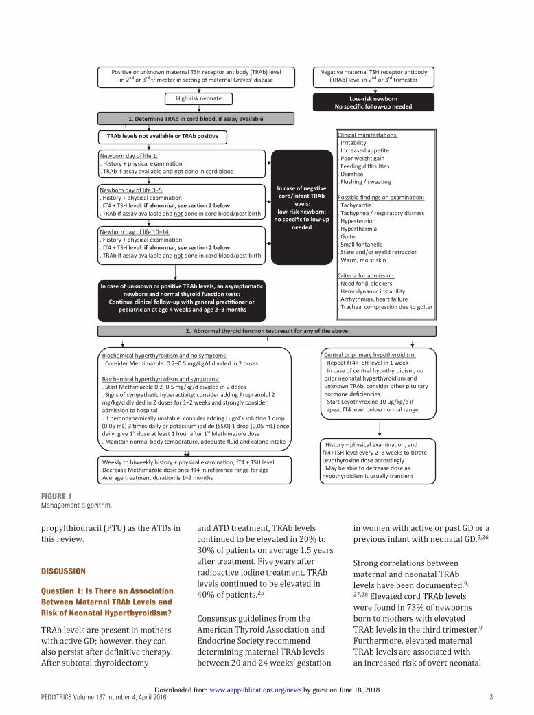
algorithm (Fig 1) that addresses the
following questions:
• Is there an association between
maternal TRAb levels and risk of
neonatal hyperthyroidism?
• Is there utility to determination of
TRAb levels in cord blood?
• Are cord blood TSH and fT4 levels
valuable in predicting neonatal
hyperthyroidism?
• When should TSH and fT4 levels be
measured in the “at-risk” newborn?
• Do maternal ATDs influence the
newborn’s presentation?
• What clinical indicators should
prompt initiation of treatment?
• How long should ATD treatment be
continued?
• Are there other abnormalities of
thyroid function in newborns born
to mothers with GD?
METHODS
Medline, Embase, and Cochrane
databases were searched with the
assistance of a reference librarian
from our hospital. The following
Medline MeSH terms were used:
“Graves disease, ” “hyperthyroidism,
” or “thyrotoxicosis.” Search limits
included publication in the past
15 years (January 1, 2000–May
22, 2015); English language, and
infants (0–23 months). This search
resulted in 283 publications. After
reviewing the abstracts, 179 articles
were not applicable. The remaining
104 articles were read and 68 were
included in this review; the other 36
articles addressed topics beyond the
scope of this review. In addition, we
included 18 pre-2000 original reports
cited as references in the 68 articles.
The literature includes case reports,
case series, and observational
cohort studies; we did not identify
relevant randomized controlled
studies or case-control studies.
Thus, the quality of evidence was
graded as moderate (observational
studies with methodological flaws,
inconsistent or indirect evidence) to
low (case series and nonsystematic
clinical observations). The strength
of recommendation is weak
(benefits and risks or burdens are
closely balanced or uncertain, best
action may differ depending on
circumstances or patients).24 We
therefore used the term “suggestion”
instead of “recommendation.”
In this review, we denote “positive”
TRAb levels as levels that exceeded
the reference range. “Negative”
TRAb levels denote levels within
the reference range or that are
undetectable. Because methimazole
(MMI) is the active metabolite of
carbimazole, we chose MMI and
2 by guest on June 18, 2018www.aappublications.org/newsDownloaded from
PEDIATRICS Volume 137 , number 4 , April 2016
propylthiouracil (PTU) as the ATDs in
this review.
DISCUSSION
Question 1: Is There an Association Between Maternal TRAb Levels and Risk of Neonatal Hyperthyroidism?
TRAb levels are present in mothers
with active GD; however, they can
also persist after definitive therapy.
After subtotal thyroidectomy
and ATD treatment, TRAb levels
continued to be elevated in 20% to
30% of patients on average 1.5 years
after treatment. Five years after
radioactive iodine treatment, TRAb
levels continued to be elevated in
40% of patients.25
Consensus guidelines from the
American Thyroid Association and
Endocrine Society recommend
determining maternal TRAb levels
between 20 and 24 weeks’ gestation
in women with active or past GD or a
previous infant with neonatal GD.5, 26
Strong correlations between
maternal and neonatal TRAb
levels have been documented.9,
27, 28 Elevated cord TRAb levels
were found in 73% of newborns
born to mothers with elevated
TRAb levels in the third trimester.9
Furthermore, elevated maternal
TRAb levels are associated with
an increased risk of overt neonatal
3
FIGURE 1Management algorithm.
by guest on June 18, 2018www.aappublications.org/newsDownloaded from
VAN DER KAAY et al
hyperthyroidism.7–9, 27–33 In a study
that included 35 pregnancies in
29 women with GD, 6 newborns
(17.1%) developed hyperthyroidism.
TRAb levels fourfold above the
reference range predicted neonatal
hyperthyroidism with a positive
predictive value of 40%, whereas
levels less than fourfold above the
reference range were associated
with a negative predictive value of
100%.8 In another study describing
230 pregnancies in 172 women with
GD, 6 newborns (2.6%) developed
overt hyperthyroidism and another
7 (3.0%) developed asymptomatic
biochemical hyperthyroidism.
Maternal TRAb levels were twofold
to fivefold above the reference range
in 8 of these 13 newborns.7 In a
recent report, none of 35 infants
born to mothers with negative TRAb
levels during pregnancy developed
hyperthyroidism.9 The risk of
neonatal hyperthyroidism after
being born to women with negative
TRAb levels is therefore regarded as
negligible.34
There are currently 2 methods
to measure TRAb levels. Second-
generation receptor binding
assays measuring thyroid-binding
inhibitory immunoglobulins
are widely available, but do not
distinguish between stimulating and
nonstimulating immunoglobulins.
Third-generation bioassays
measure thyroid-stimulating or
blocking immunoglobulins through
cyclic adenosine monophosphate
production.35 These bioassays
are less widely available, time-
consuming, and more expensive.
It has been demonstrated that a
maternal thyroid-binding inhibitory
immunoglobulin level of >3.3 times
the upper reference range had a
sensitivity of 100% and specificity
of 43% for identifying affected
newborns. Thyroid-stimulating
antibody activity exceeding 400%
(considered “strong” activity)
increased the specificity to 85%35;
however, generalizing exact numeric
cutoffs is confounded by lack of assay
harmonization. Laboratories involved
in the care of these newborns should
state clearly which assay is used.36, 37
Suggestion:
TRAb levels should be determined
between weeks 20 and 24 of
pregnancy. If maternal TRAb levels
are negative, no specific GD-related
follow-up is necessary. If TRAb
levels are unavailable or positive, the
newborn should be regarded as being
“at risk” for hyperthyroidism.
Question 2: Is Determination of TRAb Levels in Cord Blood Useful?
Skuza et al38 compared TRAb levels
in 14 infants born to mothers
with GD. Cord blood TRAb levels
were normal in 7 infants who
remained euthyroid, whereas levels
were threefold to sixfold above
the reference range in 7 infants
who developed hyperthyroidism.
Similarly, Besançon et al9 described
9 of 9 newborns with negative cord
blood TRAb levels who remained
euthyroid. Several other studies also
have demonstrated that positive
TRAb levels in cord blood correlate
with the likelihood of development of
hyperthyroidism in the first 2 weeks
of life, whereas negative antibodies
are associated with little or no risk
of neonatal hyperthyroidism.9, 30, 38,
39 Positive cord TRAb levels (up to
2.5 times the assay upper reference
limit), however, have been reported
in newborns with normal thyroid
function, 40 demonstrating that low
levels of antibodies can be seen in
euthyroid newborns.
Although TRAb levels provide
important clinical information, the
utility of cord blood TRAb levels can
be limited by the availability of the
test and the turnaround time, which
varies between 1 day and 2 weeks.
Suggestion:
If the assay is available, determine
TRAb levels in cord blood, or as soon
as possible thereafter, as this will
allow those newborns with negative
antibodies to be discharged from
follow-up.
Question 3: Are Cord Blood TSH and fT4 Levels Valuable in Predicting Neonatal Hyperthyroidism?
Several studies have demonstrated
that cord blood TSH and fT4 levels
reflect fetal thyroid function but
do not predict neonatal thyroid
function.30, 38, 39 Among 6 newborns
who developed hyperthyroidism,
Polak et al40 demonstrated that
cord blood levels indicated
hyperthyroidism, hypothyroidism,
and euthyroidism in equal numbers.
A recent observational study included
68 women with GD; all women were
receiving ATD treatment and were
well-controlled. Of 7 newborns
who developed hyperthyroidism, 2
had hypothyroidism in cord blood
tests.9 Collectively these studies
demonstrate that cord blood TSH and
fT4 levels do not reliably predict the
risk of neonatal hyperthyroidism.
Suggestion:
Determination of cord TSH and
fT4 levels is not indicated, because
these levels do not predict neonatal
hyperthyroidism.
Question 4: When Should TSH and fT4 Levels Be Measured in the “At-Risk” Newborn?
Overt neonatal hyperthyroidism can
present at birth; however, the onset
can be delayed due to maternal ATD
treatment (as discussed in question
5) or the coexistence of TSH-receptor
blocking antibodies. Several reports
demonstrate that >95% of newborns
who develop symptoms, do so
between 1 and 29 days of life and
most are diagnosed within the first 2
weeks.9, 38, 40, 41
In 1 study, fT4 and TSH levels were
determined in 96 at-risk newborns
during the first month of life.42 Four
(4%) newborns developed clinical
hyperthyroidism, the ages of onset
were not specified. In the full group,
4 by guest on June 18, 2018www.aappublications.org/newsDownloaded from
PEDIATRICS Volume 137 , number 4 , April 2016
fT4 levels peaked and were above
the 95th percentile in 92.9% of
newborns on day 5 of life, returning
to the reference range at day 15.
More than 60% of this cohort had a
TSH level below the fifth percentile
at day 6 of life. This study indicates
that a significant proportion of
at-risk newborns have abnormal
TFTs without symptoms. Similar
to cord blood, TFTs before 3 days
of life did not predict subsequent
hyperthyroidism; hence, these
authors suggested first assessing
TFTs at day 3 to 5 of life. Because
TFTs normalized by day of life 15 in
most asymptomatic newborns, the
authors also suggested that, when
thyroid function is normal at 2 weeks
of life, no further testing is necessary.
However, infants should continue
to be followed clinically, because
development of hyperthyroidism
as late as day 45 of life has been
described.41–44
These recommendations for the first
2 weeks of life are consistent with
those of others.6, 9, 40 After 2 weeks
of life, Besançon et al9 recommend
weekly clinical and biochemical
evaluation for all newborns with
positive TRAb levels until levels
become negative, although it is not
clear if data support this level of
prolonged and intensive monitoring
in all infants.
The same temporal patterns appear
to be present in preterm infants.
One study described 7 preterm
infants from 5 pregnancies born
after a mean gestational age (GA)
of 30 (range 25–36) weeks.45 Mean
age at diagnosis of hyperthyroidism
was 9 (range 1–16) days. One
infant developed thyroid storm
characterized by tachypnea,
tachycardia, cardiac failure, and
pulmonary edema.
Whether asymptomatic newborns
with biochemical hyperthyroidism
should be treated is perhaps the
greatest area of uncertainty in
this population. Further data on
neurocognitive outcomes (as
discussed in question 6) are needed
to inform this decision. In the
absence of definitive data, it seems
prudent to obtain TFTs even among
asymptomatic infants, as the results
may inform clinical follow-up.
Suggestion:
TFTs should initially be measured
at 3 to 5 days of life unless clinical
signs warrant earlier investigations.
If these data are within age-specific
reference ranges, repeat TFTs at day
10 to 14 of life. If no abnormalities
are identified after 2 weeks of life,
routine testing can be discontinued.
At 4 weeks of life and again at 2 and
3 months of life, infants should be
assessed clinically to identify the
small population of infants with
delayed presentation. Because
TSH and fT4 levels are influenced
by variations in analytical assays,
hospitals should establish age-
specific reference ranges to inform
these decisions.
Question 5: Do Maternal ATDs Infl uence the Newborn’s Presentation?
ATDs can delay presentation of
hyperthyroidism because these
cross the placenta.46 The duration
of action of MMI is 36 to 72 hours
and of PTU is 12 to 24 hours.47 It
has been reported that newborns
born to untreated mothers tended
to be diagnosed at day 1 to 3 of life,
whereas newborns from mothers
treated with ATDs were diagnosed
between days 7 and 17.41 These
variations would be detected by
using the schedule delineated
previously and in Figure 1.
ATDs can reach the newborn through
breast milk, but only in small
quantities.48 PTU in doses <300 mg
per day and MMI <20 to 30 mg per
day do not impair thyroid function in
the newborn and are regarded as safe
during breastfeeding.26, 49–51 Although
there is insufficient literature to
make a definitive statement, it
seems unlikely that this degree of
medication transfer would affect the
presentation of neonatal GD.
As this review focuses on evaluation
and treatment of newborns, it is
worth noting that MMI use during
pregnancy has been associated with
congenital anomalies in some52–54
but not all1, 55, 56 studies, and that
high-dose PTU treatment has been
associated with an increased risk
of low birth weight.3 Because PTU
has (rarely) been associated with
liver failure in pregnant women, 5,
57 current guidelines recommend
switching to MMI after the first
trimester.5, 26
Suggestion:
Although ATDs may delay the
presentation of hyperthyroidism, the
first TFTs should still be performed
on day of life 3 to 5 in neonates born
to mothers on ATD treatment, with
subsequent testing as suggested
previously.
Question 6: What Clinical Indications Should Prompt Initiation of Treatment?
Treatment should be initiated at the
onset of symptoms to avoid short-
term (cardiac failure) and long-
term (craniosynostosis, intellectual
impairment) complications. It is
unclear whether asymptomatic
newborns with biochemical
hyperthyroidism should be treated,
and it is difficult to compare
thresholds used to initiate treatment
in one study versus another, as
different assays and different
reference ranges confound direct
comparison.
Among 7 newborns with clinical
hyperthyroidism, PTU was
initiated at an fT4 level >64 pmol/L
(reference range 10–30 pmol/L) in
1 report.38 In 6 patients who were
asymptomatic, MMI was initiated at
an average fT4 level of 49.6 pmol/
L40 in another report. Besançon et
al, 9 who reported mostly on the
same cohort as previously published
by Polak et al40 and Luton et al, 30
5 by guest on June 18, 2018www.aappublications.org/newsDownloaded from

VAN DER KAAY et al
describe ATD treatment being started
between age 2 and 15 days when
fT4 levels exceeded 35 pmol/L in
7 asymptomatic newborns (mean
fT4 46.5 ± 13.8 pmol/L; reference
range 21.5–27.8 pmol/L at day 7 and
16.9–20.2 pmol/L at day 15 of life).9
The goal of the recommendation
to start treatment when fT4 levels
exceed 35 pmol/L is to prevent
clinical hyperthyroidism with its
potential morbidity and mortality.9,
40 However, data linking the initiation
of therapy in these asymptomatic
newborns with better clinical and
neurocognitive outcomes are lacking.
Related to this uncertainty, other
case reports and series describe
initiating treatment only when
both biochemical hyperthyroidism,
with fT4 levels ranging from 43 to
154 pmol/L, and symptoms were
present.14–18, 42, 58–66 Arguing against
this approach is the small series
reported by Daneman and Howard23
in which untreated neonatal GD
was associated with later-life
cognitive impairment. Overall, the
literature addressing treatment
of asymptomatic newborns is
inconclusive, as it comprises only
a few studies, often with small
numbers, and lacks defined outcomes
and/or untreated control groups for
comparison.
PTU and MMI inhibit thyroid
peroxidase and consequently
synthesis of thyroid hormone. PTU
also inhibits peripheral deiodination
of T4 to T3. In 2010, the US Food
and Drug Administration issued a
warning regarding the association
between PTU and development of
liver failure. Subsequent American
Thyroid Association guidelines
recommend that PTU should be
offered only as a short course in
case of thyroid storm or severe
adverse reactions to MMI treatment,
other than agranulocytosis, when
treatment options such as radioactive
iodine or thyroidectomy are not
available.67, 68
Because a response to ATDs is seen
only once thyroid hormone stores are
depleted, it can take several days to
weeks before clinical and biochemical
effects are noticeable. In symptomatic
patients, nonselective β-adrenergic
blockers such as propranolol can
decrease sympathetic hyperactivity.
In refractory cases, Lugol solution or
potassium iodide (oral solution) can
be added.11 The first dose of iodide
should be given at least 1 hour after
the first dose of MMI to prevent the
initial iodide from being used for
new thyroid hormone synthesis.
Less commonly, hyperthyroidism
is (initially) treated with repeated
doses of iodide instead of ATDs.18,
69–71 In extremely ill newborns
requiring admission to a NICU for
respiratory or cardiac support, a
short course of glucocorticoids,
which inhibit thyroid hormone
secretion and impair peripheral
deiodination of T4 to T3, may be
necessary.
Side effects of MMI occur in up
to 28% of children.72 The most
common side effects are mild, such as
transient elevations of liver enzymes,
mild and transient leukopenia, skin
rashes, gastrointestinal symptoms,
arthralgia, and myalgia.68, 72 Serious
side effects (0.5% of children)
include agranulocytosis, liver injury,
vasculitis and Stevens-Johnson
syndrome.68, 72 Agranulocytosis
most commonly presents with
fever, sore throat, or mouth sores.
Parents should be instructed to stop
ATDs immediately if these occur,
consult a physician, and obtain a
complete blood count. To the best
of our knowledge, only a single case
report described the development
of neutropenia in a preterm (GA 30
weeks) neonate treated with MMI
who recovered after decreasing the
dose.71
Prematurity is not a contraindication
to ATD use. However, in 1 study, 2
extremely preterm newborns (GA 25
weeks) demonstrated an unusually
rapid (within 48 hours) decrease in
fT4 levels after starting carbimazole,
indicating that is important to
monitor TFTs more closely in
preterm newborns.45
Suggestion:
Initiate treatment with MMI with
signs or symptoms of neonatal
hyperthyroidism in the setting
of biochemical hyperthyroidism.
Empiric therapy could be started
after drawing TFTs in emergent
situations. There is a lack of
consensus regarding the starting
dose for infants. A range from 0.2
to 1 mg/kg per day divided in 1
to 3 doses, with a typical dose of
0.2 to 0.5 mg/kg per day, has been
reported.14, 59, 64, 66, 71, 73 For full-term
newborns, we therefore recommend
initiating MMI at 0.625 mg twice
daily (0.4 mg/kg per day for a 3-kg
newborn). The infant should be
assessed clinically and biochemically
on a weekly base until stable, then
every 1 to 2 weeks with titration of
MMI dose as tolerated. Treatment
of asymptomatic neonates remains
controversial.
With sympathetic hyperactivity,
such as tachycardia, hypertension,
and poor feeding, propranolol 2 mg/
kg per day divided in 2 doses for 1
to 2 weeks can be added. Admission
to hospital should be considered
for cardiac monitoring and to
ensure adequate fluid and caloric
intake and temperature control.
Lugol solution 1 drop (0.05 mL) 3
times per day or potassium iodide
(oral solution) 1 drop per day may
be used in conjunction with MMI.
Hemodynamic instability, respiratory
distress or cardiac failure warrants
NICU admission. In these cases,
a short course of treatment with
prednisolone 2 mg/kg per day in 1 to
2 divided doses should be considered
in addition to MMI.
Question 7: How Long Should ATD Treatment Be Continued?
Neonatal hyperthyroidism due to
maternal GD is self-limited, with
6 by guest on June 18, 2018www.aappublications.org/newsDownloaded from
PEDIATRICS Volume 137 , number 4 , April 2016
duration determined by the rate of
disappearance of maternal TRAb
from the infant circulation. TRAb
half-lives have been reported to be
approximately 12 days.74 Depending
on the initial TRAb level, neonatal GD
generally resolves by 6 months after
birth, 35, 38, 41, 75 although 1 instance of
persistence to 12 months has been
reported.12 Treatment duration is
most commonly 1 to 2 months.6, 9,
35, 38 MMI dose should be decreased
and eventually discontinued when
fT4 levels are within the reference
range. Alternatively, the addition of
levothyroxine to MMI treatment has
been practiced, 9, 14, 59 although recent
guidelines recommend against this
“block and replace” practice.73 The
decision to discontinue treatment
should be based on clinical status and
ongoing normal thyroid hormone
levels.
Suggestion:
While on treatment, thyroid
function should be measured
weekly until hormone levels are
stable and subsequently every 2
weeks. Treatment duration is most
commonly 1 to 2 months.
Question 8: Are There Other Abnormalities of Thyroid Function in Neonates Born to Mothers With GD?
In addition to neonatal
hyperthyroidism, transient central
hypothyroidism, transient primary
hypothyroidism, and transient
isolated hyperthyropinemia
(elevated TSH with normal fT4 levels
and no clinical symptoms) have been
described.9, 39, 76–83 One case series
described 18 infants with central
hypothyroidism born to mothers
with GD who were inadequately
treated during pregnancy. Eleven
infants were diagnosed in the
context of a primary T4-based
newborn screening during days 4
and 7 of life. One infant presented
with transient hyperthyroidism
before evolving into central
hypothyroidism. Six others were
euthyroid before developing central
hypothyroidism during the first
month of life. Seventeen infants
started levothyroxine treatment.76
Transient central hypothyroidism, 39, 78, 79 sometimes followed by
hyperthyroidism, 80–82 has been
reported by others. Recovery
from hypothyroidism is usually
seen between 3 and 19 months
of age. Some physicians decrease
levothyroxine supplementation as
the hypothalamic-pituitary-thyroid
axis recovers, but others advise
ongoing treatment until
3 years of age to ensure adequate
thyroid hormone levels during
this important period of brain
development.76, 83 In rare
instances, central hypothyroidism
can be prolonged and may be
permanent.83
The etiology of central
hypothyroidism in these infants
is unknown but may stem from
impaired maturation and/or
regulation of the fetal hypothalamic-
pituitary-thyroid axis. Another
explanation invokes direct binding
of TRAb to the TSH-receptor in the
pituitary gland with suppression of
TSH production independent of T4
production.82, 84
Maternal ATD treatment has been
associated with elevated cord
blood TSH levels in 14% to 21%
and low fT4 levels in 6% to 7% of
newborns.85 No relationship between
TSH and fT4 levels with ATD dose
was found. Other studies have found
transiently elevated TSH levels and
transient primary hypothyroidism
in 7.8% and 2.0% to 9.0% of
newborns, respectively.7, 33 Primary
hypothyroidism can sometimes
precede hyperthyroidism.9 The
interplay between TSH-receptor
stimulating and blocking antibodies
might explain the switch from
hypothyroidism to hyperthyroidism
and vice versa.86, 87
Suggestion:
Be cognizant that central or primary
hypothyroidism can occur in these
newborns. One must be aware of
the clinical signs of hypothyroidism,
including poor feeding, lethargy,
prolonged jaundice, hypotonia, dry
skin, large fontanelle, distended
abdomen, umbilical hernia, and
reduced linear growth, and monitor
TFTs. Levothyroxine 10 μg/kg per
day should be started when the
diagnosis of hypothyroidism has been
established. In the setting of central
hypothyroidism without a previous
diagnosis of hyperthyroidism, it is
important to consider a differential
diagnosis including pituitary
dysfunction.
CONCLUSIONS
Neonatal hyperthyroidism
due to maternal GD requires
early recognition and treatment
to prevent potential morbidity
or mortality. We hope our
literature review and related
algorithm will assist generalists
and subspecialists manage these
patients. Refinement of this
algorithm based on future studies
and feedback on its use will be
important.
ACKNOWLEDGMENT
We thank Dr Guy van Vliet, at the
Centre Hospitalier Universitaire
Sainte-Justine and Department of
Pediatrics, University of Montreal,
Montreal, Canada, for critically
reviewing this manuscript before
submission.
7
ABBREVIATIONS
ATD: antithyroid drug
fT4: free T4
GA: gestational age
GD: Graves’ disease
MMI: methimazole
PTU: propylthiouracil
TFT: thyroid function test
TRAb: TSH-receptor antibodies
TSH: thyroid stimulating
hormone
by guest on June 18, 2018www.aappublications.org/newsDownloaded from

VAN DER KAAY et al
REFERENCES
1. Korelitz JJ, McNally DL, Masters MN,
Li SX, Xu Y, Rivkees SA. Prevalence of
thyrotoxicosis, antithyroid medication
use, and complications among
pregnant women in the United States.
Thyroid. 2013;23(6):758–765
2. Rivkees SA, Mandel SJ. Thyroid disease
in pregnancy. Horm Res Paediatr.
2011;76(suppl 1):91–96
3. Chen CH, Xirasagar S, Lin CC, Wang
LH, Kou YR, Lin HC. Risk of adverse
perinatal outcomes with antithyroid
treatment during pregnancy: a
nationwide population-based study.
BJOG. 2011;118(11):1365–1373
4. Wang W, Teng W, Shan Z, et al. The
prevalence of thyroid disorders
during early pregnancy in China: the
benefi ts of universal screening in the
fi rst trimester of pregnancy. Eur J
Endocrinol. 2011;164(2):263–268
5. De Groot L, Abalovich M, Alexander
EK, et al. Management of thyroid
dysfunction during pregnancy
and postpartum: an Endocrine
Society clinical practice guideline.
J Clin Endocrinol Metab.
2012;97(8):2543–2565
6. Ogilvy-Stuart AL. Neonatal thyroid
disorders. Arch Dis Child Fetal
Neonatal Ed. 2002;87(3):F165–F171
7. Mitsuda N, Tamaki H, Amino N, Hosono
T, Miyai K, Tanizawa O. Risk factors
for developmental disorders in
infants born to women with Graves
disease. Obstet Gynecol. 1992;80(3 pt
1):359–364
8. Peleg D, Cada S, Peleg A, Ben-
Ami M. The relationship between
maternal serum thyroid-stimulating
immunoglobulin and fetal and neonatal
thyrotoxicosis. Obstet Gynecol.
2002;99(6):1040–1043
9. Besançon A, Beltrand J, Le Gac I, Luton
D, Polak M. Management of neonates
born to women with Graves’ disease:
a cohort study. Eur J Endocrinol.
2014;170(6):855–862
10. Pitcher-Wilmott RW, Hindocha P, Wood
CB. The placental transfer of IgG
subclasses in human pregnancy. Clin
Exp Immunol. 1980;41(2):303–308
11. Polak M, Legac I, Vuillard E,
Guibourdenche J, Castanet M, Luton
D. Congenital hyperthyroidism:
the fetus as a patient. Horm Res.
2006;65(5):235–242
12. Zimmerman D. Fetal and neonatal
hyperthyroidism. Thyroid.
1999;9(7):727–733
13. Polak M, Van Vliet G. Therapeutic
approach of fetal thyroid disorders.
Horm Res Paediatr. 2010;74(1):1–5
14. Regelmann MO, Sullivan CK, Rapaport
R. Thyroid “vise” in an infant with
neonatal Graves’ disease. Pediatrics.
2013;132(4). Available at: www.
pediatrics. org/ cgi/ content/ full/ 132/ 4/
e1048
15. Loomba-Albrecht LA, Bremer AA, Wong
A, Philipps AF. Neonatal cholestasis
caused by hyperthyroidism. J Pediatr
Gastroenterol Nutr. 2012;54(3):433–434
16. Oden J, Cheifetz IM. Neonatal
thyrotoxicosis and persistent
pulmonary hypertension necessitating
extracorporeal life support. Pediatrics.
2005;115(1). Available at: www.
pediatrics. org/ cgi/ content/ full/ 115/ 1/
e105
17. Obeid R, Kalra VK, Arora P, Quist F,
Moltz KC, Chouthai NS. Neonatal
thyrotoxicosis presenting as persistent
pulmonary hypertension. BMJ Case
Rep. 2012;2012
18. Carroll DN, Kamath P, Stewart L.
Congenital viral infection? Lancet.
2005;365(9464):1110
19. Mestman JH. Hyperthyroidism in
pregnancy. Clin Obstet Gynecol.
1997;40(1):45–64
20. Eisenstein Z, Weiss M, Katz Y,
Bank H. Intellectual capacity of
subjects exposed to methimazole or
propylthiouracil in utero. Eur J Pediatr.
1992;151(8):558–559
21. Smit BJ, Kok JH, Vulsma T, Briët JM,
Boer K, Wiersinga WM. Neurologic
development of the newborn and
young child in relation to maternal
thyroid function. Acta Paediatr.
2000;89(3):291–295
22. Messer PM, Hauffa BP, Olbricht T,
Benker G, Kotulla P, Reinwein D.
Antithyroid drug treatment of Graves’
disease in pregnancy: long-term
effects on somatic growth, intellectual
development and thyroid function
of the offspring. Acta Endocrinol
(Copenh). 1990;123(3):311–316
23. Daneman D, Howard NJ. Neonatal
thyrotoxicosis: intellectual impairment
and craniosynostosis in later years. J
Pediatr. 1980;97(2):257–259
24. Owens DK, Lohr KN, Atkins D, et al.
AHRQ series paper 5: grading the
strength of a body of evidence when
comparing medical interventions—
Agency for Healthcare Research
and Quality and the effective health-
care program. J Clin Epidemiol.
2010;63(5):513–523
25. Laurberg P, Wallin G, Tallstedt L,
Abraham-Nordling M, Lundell G,
8
DOI: 10.1542/peds.2015-1878
Accepted for publication Aug 31, 2015
Address correspondence to Daniëlle C.M. van der Kaay, MD, PhD, Haga Hospital/Juliana Children’s Hospital, Division of Pediatrics, Leyweg 275, 2545 CH Hague,
Netherlands. E-mail: [email protected]
PEDIATRICS (ISSN Numbers: Print, 0031-4005; Online, 1098-4275).
Copyright © 2016 by the American Academy of Pediatrics
FINANCIAL DISCLOSURE: The authors have indicated they have no fi nancial relationships relevant to this article to disclose.
FUNDING: No external funding.
POTENTIAL CONFLICT OF INTEREST: The authors have indicated they have no potential confl icts of interest to disclose.
by guest on June 18, 2018www.aappublications.org/newsDownloaded from
PEDIATRICS Volume 137 , number 4 , April 2016
Tørring O. TSH-receptor autoimmunity
in Graves’ disease after therapy
with anti-thyroid drugs, surgery, or
radioiodine: a 5-year prospective
randomized study. Eur J Endocrinol.
2008;158(1):69–75
26. Stagnaro-Green A, Abalovich M,
Alexander E, et al; American Thyroid
Association Taskforce on Thyroid
Disease During Pregnancy and
Postpartum. Guidelines of the
American Thyroid Association for
the diagnosis and management of
thyroid disease during pregnancy
and postpartum. Thyroid.
2011;21(10):1081–1125
27. Matsuura N, Konishi J, Fujieda K, et al.
TSH-receptor antibodies in mothers
with Graves' disease and outcome in
their offspring. Lancet. 1988;1(8575-6):
14–17
28. Mortimer RH, Tyack SA, Galligan JP,
Perry-Keene DA, Tan YM. Graves’
disease in pregnancy: TSH receptor
binding inhibiting immunoglobulins
and maternal and neonatal thyroid
function. Clin Endocrinol (Oxf).
1990;32(2):141–152
29. Hamada N, Momotani N, Ishikawa
N, et al. Persistent high TRAb values
during pregnancy predict increased
risk of neonatal hyperthyroidism
following radioiodine therapy for
refractory hyperthyroidism. Endocr J.
2011;58(1):55–58
30. Luton D, Le Gac I, Vuillard E, et al.
Management of Graves’ disease during
pregnancy: the key role of fetal thyroid
gland monitoring. J Clin Endocrinol
Metab. 2005;90(11):6093–6098
31. McKenzie JM, Zakarija M. Fetal
and neonatal hyperthyroidism and
hypothyroidism due to maternal
TSH receptor antibodies. Thyroid.
1992;2(2):155–159
32. Clavel S, Madec AM, Bornet H, Deviller
P, Stefanutti A, Orgiazzi J. Anti TSH-
receptor antibodies in pregnant
patients with autoimmune thyroid
disorder. Br J Obstet Gynaecol.
1990;97(11):1003–1008
33. Uenaka M, Tanimura K, Tairaku
S, Morioka I, Ebina Y, Yamada H.
Risk factors for neonatal thyroid
dysfunction in pregnancies
complicated by Graves’ disease.
Eur J Obstet Gynecol Reprod Biol.
2014;177:89–93
34. Laurberg P, Nygaard B, Glinoer
D, Grussendorf M, Orgiazzi J.
Guidelines for TSH-receptor antibody
measurements in pregnancy: results
of an evidence-based symposium
organized by the European Thyroid
Association. Eur J Endocrinol.
1998;139(6):584–586
35. Abeillon-du Payrat J, Chikh K,
Bossard N, et al. Predictive value of
maternal second-generation thyroid-
binding inhibitory immunoglobulin
assay for neonatal autoimmune
hyperthyroidism. Eur J Endocrinol.
2014;171(4):451–460
36. Barbesino G, Tomer Y. Clinical
review: Clinical utility of TSH receptor
antibodies. J Clin Endocrinol Metab.
2013;98(6):2247–2255
37. Tan K, Loh TP, Sethi S. Lack of
standardized description of TRAb
assays. Endocrine. 2013;43(3):732–733
38. Skuza KA, Sills IN, Stene M, Rapaport R.
Prediction of neonatal hyperthyroidism
in infants born to mothers with Graves
disease. J Pediatr. 1996;128(2):264–268
39. Tamaki H, Amino N, Takeoka K, et
al. Prediction of later development
of thyrotoxicosis or central
hypothyroidism from the cord serum
thyroid-stimulating hormone level in
neonates born to mothers with Graves
disease. J Pediatr. 1989;115(2):318–321
40. Polak M, Le Gac I, Vuillard E, et al.
Fetal and neonatal thyroid function in
relation to maternal Graves’ disease.
Best Pract Res Clin Endocrinol Metab.
2004;18(2):289–302
41. Papendieck P, Chiesa A, Prieto L,
Gruñeiro-Papendieck L. Thyroid
disorders of neonates born to mothers
with Graves’ disease. J Pediatr
Endocrinol Metab. 2009;22(6):547–553
42. Levy-Shraga Y, Tamir-Hostovsky
L, Boyko V, Lerner-Geva L, Pinhas-
Hamiel O. Follow-up of newborns of
mothers with Graves’ disease. Thyroid.
2014;24(6):1032–1039
43. Zakarija M, McKenzie JM, Hoffman
WH. Prediction and therapy of
intrauterine and late-onset neonatal
hyperthyroidism. J Clin Endocrinol
Metab. 1986;62(2):368–371
44. Uçkun Kitapçı A, Çalıkoğlu AS. Neonatal
hyperthyroidism associated with
isolated submandibular sialadenitis: is
it just a coincidence? J Clin Res Pediatr
Endocrinol. 2010;2(1):43–45
45. Smith C, Thomsett M, Choong C,
Rodda C, McIntyre HD, Cotterill AM.
Congenital thyrotoxicosis in premature
infants. Clin Endocrinol (Oxf).
2001;54(3):371–376
46. Mortimer RH, Cannell GR, Addison
RS, Johnson LP, Roberts MS, Bernus
I. Methimazole and propylthiouracil
equally cross the perfused human
term placental lobule. J Clin Endocrinol
Metab. 1997;82(9):3099–3102
47. Clark SM, Saade GR, Snodgrass WR,
Hankins GD. Pharmacokinetics and
pharmacotherapy of thionamides
in pregnancy. Ther Drug Monit.
2006;28(4):477–483
48. Mandel SJ, Cooper DS. The use of
antithyroid drugs in pregnancy and
lactation. J Clin Endocrinol Metab.
2001;86(6):2354–2359
49. Momotani N, Yamashita R, Makino
F, Noh JY, Ishikawa N, Ito K. Thyroid
function in wholly breast-feeding
infants whose mothers take high doses
of propylthiouracil. Clin Endocrinol
(Oxf). 2000;53(2):177–181
50. Speller E, Brodribb W. Breastfeeding
and thyroid disease: a literature
review. Breastfeed Rev.
2012;20(2):41–47
51. Azizi F, Bahrainian M, Khamseh ME,
Khoshniat M. Intellectual development
and thyroid function in children
who were breast-fed by thyrotoxic
mothers taking methimazole.
J Pediatr Endocrinol Metab.
2003;16(9):1239–1243
52. Clementi M, Di Gianantonio E, Cassina
M, Leoncini E, Botto LD, Mastroiacovo
P; SAFE-Med Study Group. Treatment
of hyperthyroidism in pregnancy and
birth defects. J Clin Endocrinol Metab.
2010;95(11):E337–E341
53. Yoshihara A, Noh J, Yamaguchi
T, et al. Treatment of Graves’
disease with antithyroid drugs in
the fi rst trimester of pregnancy
and the prevalence of congenital
malformation. J Clin Endocrinol Metab.
2012;97(7):2396–2403
9 by guest on June 18, 2018www.aappublications.org/newsDownloaded from
VAN DER KAAY et al
54. Andersen SL, Olsen J, Wu CS, Laurberg
P. Birth defects after early pregnancy
use of antithyroid drugs: a Danish
nationwide study. J Clin Endocrinol
Metab. 2013;98(11):4373–4381
55. Rosenfeld H, Ornoy A, Shechtman S,
Diav-Citrin O. Pregnancy outcome,
thyroid dysfunction and fetal
goitre after in utero exposure
to propylthiouracil: a controlled
cohort study. Br J Clin Pharmacol.
2009;68(4):609–617
56. Di Gianantonio E, Schaefer C,
Mastroiacovo PP, et al. Adverse effects
of prenatal methimazole exposure.
Teratology. 2001;64(5):262–266
57. Cooper DS, Rivkees SA. Putting
propylthiouracil in perspective.
J Clin Endocrinol Metab.
2009;94(6):1881–1882
58. Benjamin JS, Chong E, Ramayya MS.
A preterm, female newborn with
tachycardia, hypertension, poor weight
gain, and irritability. Clin Pediatr
(Phila). 2012;51(10):994–997
59. Dierickx I, Decallonne B, Billen J,
et al. Severe fetal and neonatal
hyperthyroidism years after surgical
treatment of maternal Graves’ disease.
J Obstet Gynaecol. 2014;34(2):117–122
60. Bisschop PH, van Trotsenburg AS.
Images in clinical medicine. Neonatal
thyrotoxicosis. N Engl J Med.
2014;370(13):1237
61. Dryden C, Simpson JH, Hunter
LE, Jackson L. An unusual
cause of neonatal coagulopathy
and liver disease. J Perinatol.
2007;27(5):320–322
62. Trapali C, Dellagrammaticas HD,
Nika A, Iacovidou N. A unique case of
reversible myocardial ischemia in a
hyperthyroid neonate. Pediatr Cardiol.
2008;29(1):180–182
63. Aslam M, Inayat M. Fetal and neonatal
Graves disease: a case report and
review of the literature. South Med J.
2008;101(8):840–841
64. Smith CM, Gavranich J, Cotterill
A, Rodda CP. Congenital neonatal
thyrotoxicosis and previous
maternal radioiodine therapy. BMJ.
2000;320(7244):1260–1261
65. Beroukhim RS, Moon TD, Felner
EI. Neonatal thyrotoxicosis and
conjugated hyperbilirubinemia.
J Matern Fetal Neonatal Med.
2003;13(6):426–428
66. Groom K, Snowise S, Wheeler B,
Mekhail A, Farrand S, Parry E.
Maternal thyrotoxicosis and fetal
goitre requiring in utero therapy
for hypothyroidism and subsequent
neonatal therapy for hyperthyroidism.
J Paediatr Child Health.
2013;49(1):E102–E103
67. Bahn RS, Burch HS, Cooper DS, et
al. The role of propylthiouracil in
the management of Graves’ disease
in adults: report of a meeting
jointly sponsored by the American
Thyroid Association and the Food
and Drug Administration. Thyroid.
2009;19(7):673–674
68. Karras S, Tzotzas T, Krassas GE.
Toxicological considerations for
antithyroid drugs in children.
Expert Opin Drug Metab Toxicol.
2011;7(4):399–410
69. Maragliano G, Zuppa AA, Florio MG, et
al. Effi cacy of oral iodide therapy on
neonatal hyperthyroidism caused by
maternal Graves’ disease. Fetal Diagn
Ther. 2000;15(2):122–126
70. Zuppa AA, Sindico P, Savarese I, et al.
Neonatal hyperthyroidism: neonatal
clinical course of two brothers born
to a mother with Graves-Basedow
disease, before and after total
thyroidectomy. J Pediatr Endocrinol
Metab. 2007;20(4):535–539
71. Angelis D, Kubicky RA, Zubrow AB.
Methimazole associated neutropenia
in a preterm neonate treated for
hyperthyroidism. Case Rep Endocrinol.
2015;2015:680191
72. Rivkees SA, Stephenson K, Dinauer
C. Adverse events associated with
methimazole therapy of graves'
disease in children. Int J Pediatr
Endocrinol. 2010;2010:176970
73. Bahn Chair RS, Burch HB, Cooper DS,
et al; American Thyroid Association;
American Association of Clinical
Endocrinologists. Hyperthyroidism
and other causes of thyrotoxicosis:
management guidelines of the
American Thyroid Association
and American Association of
Clinical Endocrinologists. Thyroid.
2011;21(6):593–646
74. Karpman BA, Rapoport B, Filetti S,
Fisher DA. Treatment of neonatal
hyperthyroidism due to Graves’
disease with sodium ipodate. J Clin
Endocrinol Metab. 1987;64(1):119–123
75. Kamishlian A, Matthews N, Gupta A,
et al. Different outcomes of neonatal
thyroid function after Graves’ disease
in pregnancy: patient reports and
literature review. J Pediatr Endocrinol
Metab. 2005;18(12):1357–1363
76. Kempers MJ, van Tijn DA, van
Trotsenburg AS, de Vijlder JJ, Wiedijk
BM, Vulsma T. Central congenital
hypothyroidism due to gestational
hyperthyroidism: detection where
prevention failed. J Clin Endocrinol
Metab. 2003;88(12):5851–5857
77. Brown RS, Bellisario RL, Botero D, et
al. Incidence of transient congenital
hypothyroidism due to maternal
thyrotropin receptor-blocking
antibodies in over one million
babies. J Clin Endocrinol Metab.
1996;81(3):1147–1151
78. Higuchi R, Miyawaki M, Kumagai
T, et al. Central hypothyroidism in
infants who were born to mothers
with thyrotoxicosis before 32 weeks’
gestation: 3 cases. Pediatrics.
2005;115(5). Available at: www.
pediatrics. org/ cgi/ content/ full/ 115/ 3/
e623
79. Lee YS, Loke KY, Ng SC, Joseph R.
Maternal thyrotoxicosis causing
central hypothyroidism in
infants. J Paediatr Child Health.
2002;38(2):206–208
80. Brand F, Liégeois P, Langer B. One
case of fetal and neonatal variable
thyroid dysfunction in the context of
Graves’ disease. Fetal Diagn Ther.
2005;20(1):12–15
81. O’Connor MJ, Paget-Brown AO, Clarke
WL. Premature twins of a mother
with Graves’ disease with discordant
thyroid function: a case report. J
Perinatol. 2007;27(6):388–389
82. Zwaveling-Soonawala N, van
Trotsenburg P, Vulsma T. Central
hypothyroidism in an infant born
to an adequately treated mother
with Graves’ disease: an effect of
maternally derived thyrotrophin
receptor antibodies? Thyroid.
2009;19(6):661–662
10 by guest on June 18, 2018www.aappublications.org/newsDownloaded from
PEDIATRICS Volume 137 , number 4 , April 2016
83. Kempers MJ, van Trotsenburg AS,
van Rijn RR, et al. Loss of integrity
of thyroid morphology and function
in children born to mothers with
inadequately treated Graves’
disease. J Clin Endocrinol Metab.
2007;92(8):2984–2991
84. Prummel MF, Brokken LJ, Wiersinga
WM. Ultra short-loop feedback control
of thyrotropin secretion. Thyroid.
2004;14(10):825–829
85. Momotani N, Noh JY, Ishikawa N,
Ito K. Effects of propylthiouracil
and methimazole on fetal thyroid
status in mothers with Graves’
hyperthyroidism. J Clin Endocrinol
Metab. 1997;82(11):3633–3636
86. Balucan FS, Morshed SA, Davies
TF. Thyroid autoantibodies in
pregnancy: their role, regulation
and clinical relevance. J Thyroid Res.
2013;2013:182472
87. McLachlan SM, Rapoport B.
Thyrotropin-blocking autoantibodies
and thyroid-stimulating autoantibodies:
potential mechanisms involved
in the pendulum swinging from
hypothyroidism to hyperthyroidism or
vice versa. Thyroid. 2013;23(1):14–24
11 by guest on June 18, 2018www.aappublications.org/newsDownloaded from
DOI: 10.1542/peds.2015-1878 originally published online March 15, 2016; 2016;137;Pediatrics
Daniëlle C.M. van der Kaay, Jonathan D. Wasserman and Mark R. PalmertManagement of Neonates Born to Mothers With Graves' Disease
ServicesUpdated Information &
http://pediatrics.aappublications.org/content/137/4/e20151878including high resolution figures, can be found at:
Referenceshttp://pediatrics.aappublications.org/content/137/4/e20151878#BIBLThis article cites 82 articles, 10 of which you can access for free at:
Subspecialty Collections
http://www.aappublications.org/cgi/collection/endocrinology_subEndocrinologyfollowing collection(s): This article, along with others on similar topics, appears in the
Permissions & Licensing
http://www.aappublications.org/site/misc/Permissions.xhtmlin its entirety can be found online at: Information about reproducing this article in parts (figures, tables) or
Reprintshttp://www.aappublications.org/site/misc/reprints.xhtmlInformation about ordering reprints can be found online:
by guest on June 18, 2018www.aappublications.org/newsDownloaded from
DOI: 10.1542/peds.2015-1878 originally published online March 15, 2016; 2016;137;Pediatrics
Daniëlle C.M. van der Kaay, Jonathan D. Wasserman and Mark R. PalmertManagement of Neonates Born to Mothers With Graves' Disease
http://pediatrics.aappublications.org/content/137/4/e20151878located on the World Wide Web at:
The online version of this article, along with updated information and services, is
1073-0397. ISSN:60007. Copyright © 2016 by the American Academy of Pediatrics. All rights reserved. Print
the American Academy of Pediatrics, 141 Northwest Point Boulevard, Elk Grove Village, Illinois,has been published continuously since 1948. Pediatrics is owned, published, and trademarked by Pediatrics is the official journal of the American Academy of Pediatrics. A monthly publication, it
by guest on June 18, 2018www.aappublications.org/newsDownloaded from