Embed Size (px)
Citation preview

1
OEP Copyright (C)1991- 2005
Management of Myopia
Presented by the Optometric Extension Program Foundation
(OEPF)Paul Harris, OD, Robert Hohendorf, OD, Robin Lewis, OD
OEP Copyright (C)1991- 2005
What is myopia?• “The refractive condition of the eye represented
by the location of the conjugate focus of the retina at some finite point in front of the eye, when accommodation is said to be relaxed, or the extent of that condition represented in the number of diopters of concave lens power required to compensate to the optical equivalent of emmetropia.”
Dictionary of Visual Science
OEP Copyright (C)1991- 2005
What is myopia? (2)
• There are then 56 different subtypes of myopia mentioned:
• From: acquired, adventitious, apparent, associated, astigmatic, atypical, axial, benign…..
• To: transitory, traumatic, true, twilight, typical, and finally very high!
Dictionary of Visual Science

2
OEP Copyright (C)1991- 2005
What is nearsightedness?
• Nearsight. Myopia• Nearsighted. Myopic• Nearsightedness. Myopia
Dictionary of Visual Science
OEP Copyright (C)1991- 2005
How about some functional definitions?
• Nearsighted/nearsightedness: a refractive condition where concave lenses are needed to restore standard visual acuity at far.
• Myopia: “the tendency to shrink visual and perceptual space and to restrict the musculature (and often the emotions) while attempting to solve the problem of responding to visual stress.” Gallop, S., Myopia Reduction a View From the Inside, JBO Vol 5/1994 115-120.
OEP Copyright (C)1991- 2005
Other Myopic BehaviorsSteve Gallop
• Keeping to oneself thoughts or emotions• Tends to not plan ahead• Tends towards fatalistic views of things
rather than deterministic• Does not take responsibility for how they
see. It’s up to us to fix things for them.

3
OEP Copyright (C)1991- 2005
Relationship Between Myopia/Nearsightedness
• Usually, myopia and nearsightedness go together. However:
• One can act myopic without being nearsighted.• One can be nearsighted without acting myopic.• Displays of behavioral tendencies of myopia may
come and go and change in degree within a single person.
OEP Copyright (C)1991- 2005
Some Statistics• In Singapore the prevalence of myopia has
been increasing in young adults– 1970’s 26%– 1980’s 43%– 1990’s 66%
• “Does Education Explain Ethnic Differences in Myopia Prevalence? A Population-Based Study of Young Adult Males in Singapore” Wu, et.al. Optometry and Vision Science Vol. 78, No. 4, April 2001
OEP Copyright (C)1991- 2005
Statistics continued
• “In Israel, the prevalence of myopia among Orthodox schoolboys (who spent longer hours reading and writing) was 81.3%, as compared with 27.4% among boys from general schools.”
• “Myopia in Singapore kindergarten children”, Saw, et.al. Optometry, Volume 72/Number 5, May 2001 page 286

4
OEP Copyright (C)1991- 2005
Nature vs. Nurture
• Nature: There must be a genetic determination because so many studies show high correlations to the number of parents that are nearsighted and their degree of nearsightedness.
• Nurture: Environmental demands are the cause.
OEP Copyright (C)1991- 2005
Nature vs. Nurture (2)
• It is always BOTH!• Two conditions must be satisfied:
– 1. There must be an underlying physiological mechanism that is part of the human visual system that can alter the parameters of the physiology itself.
– 2. There must be environmental triggers that activate the above named processes.
OEP Copyright (C)1991- 2005
It’s Both!• “The causes of myopia are not known.
Epidemiological correlation suggest that lengthy periods of close work are probably a contributory factor, and that there is some genetic predisposition to myopia and its severity, with chromosomes 18q, 12p, 7q, and 17q having been implicated. Gene-environment interactions are being defined.” 12

5
OEP Copyright (C)1991- 2005
Some Early Work on Physiology
• Ludlum et al. on the myopia research project done at the Optometric Center of New York.– 1964-69 develop testing devices– 1969-74 testing phase– Over 500 subjects seen every 6-months
OEP Copyright (C)1991- 2005
What was the #1 finding that predicted who would become a
progressive myope?
OEP Copyright (C)1991- 2005
Ludlum et. al. Continued
• Lowered PRA was another major risk factor. With all other findings being equal:– The lower the PRA the higher the risk to
progress.– The higher the PRA the better able to remain
stable.

6
OEP Copyright (C)1991- 2005
More on IOP/Physiology• Phil Kruger SUNY showed IOP changes of from 2-3 mm
HG following 30 minutes of sustained reading.• Ben Lane confirmed Ludlum’s IOP findings in his nutrition
studies on myopia.• Lane also found greatly reduced PRA a major risk factor.• Lane found decreased chromium and increased vanadium,
which cause a decrease in accommodative ability leading to increased accommodative effort, which he felt may cause excess production of aqueous and hence the IOP increase.
• Lane also found changes in scleral cross linking and Ca++ handling.
• Getz reported on Russian use of vitamin C, which lowered IOP 2-3 mm HG.
OEP Copyright (C)1991- 2005
Ludlum Group Comparisons
-0.1749 mm-0.3917 mmAnterior Lens Radius
-0.2923 mm-0.1848 mmAnterior Chamber Depth
-1.5289D0.0960 DRefraction
0.0608 D0.0348 DKeratometer
9.45 years8.99 yearsAge
Sub Group B (N=30)
Sub Group A (N=30)
Net Changes
OEP Copyright (C)1991- 2005
Group Comparisons
0.061 D-0.4657 DTotal Power
0.7426 mm0.242 mmPosterior Segment Length
0.0374 D-0.2823 DLens Power
0.0759 mm-0.366mmPosterior Lens Curvature
0.0032 mm-0.0157 mmLens Thickness
Sub Group B (N=30)
Sub Group A (N=30)
Net Changes

7
OEP Copyright (C)1991- 2005
Group Comparisons
2.2031 D1.4426 DExpected Myopic Shift (3x change in axial length)
-4.782 mm Hg-4.857 mm HgIntra Ocular Pressure
0.7285 mm0.4697 mmAxial Length
Sub Group B (N=30)
Sub Group A (N=30)
Net Changes
OEP Copyright (C)1991- 2005
Newer Concepts of Physiological Mechanisms of Change
• Emmetropization– Selective tuning of the components of the optics
as the system matures. – Many changes occur in infants with shifts towards
a low degree of hyperopia over the first 2 years of life. The further from +0.75 the faster the shifts occur.
• Retinal defocus/local hormone/enzyme release
OEP Copyright (C)1991- 2005
Emmetropization
• Emmetropization is the process occurring during the normal growth period by which the eye changes from a state of ametropia to emmetropia.

8
OEP Copyright (C)1991- 2005
Emmetropization Some Questions
• Active vs. Passive• What structures change?• What effect does deprivation have?• Where is the control? Local or higher?• What do animal models and studies say?• Can we apply this to humans?
OEP Copyright (C)1991- 2005
Active vs. Passive
• Norton, T., Siegwart, J., “These experiments have provided convincing evidence that emmetropization in animals is an active regulatory process.”
• Sorsby, “Ametropia results from failure of coordination of the optical components with the axial components.”
OEP Copyright (C)1991- 2005
What structures change?• It might be easier to ask what doesn’t
change? Nearly everything does. • While the average eye is moving from
moderate hyperopia to low hyperopia the eye is growing in axial length, the cornea and lens powers are decreasing, anterior chamber depth is increasing, etc. all in a controlled symphony of movement!

9
OEP Copyright (C)1991- 2005
What effect does deprivation have?
• “When eyes are deprived of normal form vision they typically become axially elongated and myopic.” 1 (tree shrew)
• There are two periods of growth– Early period of fast change– Juvenile period of slow growth
• “Eyes deprived of form vision early in the “juvenile” period became myopic, and had a thinner sclera and a reduced amount of collagen and proteoglycans than in control eyes.” 3 (tree shrew)
OEP Copyright (C)1991- 2005
Deprivation: Sector/Complete
OEP Copyright (C)1991- 2005
Total deprivation 2 & 6 weeks

10
OEP Copyright (C)1991- 2005
Deprivation nasal: 2 & 6 weeks
OEP Copyright (C)1991- 2005
Deprivation temporal: 2 & 6 weeks
OEP Copyright (C)1991- 2005
Deprivation

11
OEP Copyright (C)1991- 2005
Lenses
• “Altering the location of the focal plane with minus lenses produces a compensatory change in the elongation rate of the eye so as to match the axial length to the altered focal plane.” 1
• “It appears that some blur must be present to produce normal elongation, but too much blur, too much of the time, produces excessive elongation.” 1
See next slide for examples of this.
OEP Copyright (C)1991- 2005
Lenses
OEP Copyright (C)1991- 2005
Local or Higher Control?• “In chickens, rhesus macaque monkeys, and tree
shrews, deprivation-induced myopia has been found to develop even after the central communication from the retina was severed surgically or chemically.” 1
• “The amount of retinal blur may affect the quality or quantity of the retinal neural activity. This, in turn, may alter the release of dopamine, acetylcholine, or other neurotransmitters.” 1

12
OEP Copyright (C)1991- 2005
Local or Higher Control?(Hung and Ciuffreda)
• “Elongation occurs even when the optic nerve is severed.” A4
• “Moreover, these appropriate changes in growth rate occurred even when the optic nerve was severed or the midbrain nuclei for controlling accommodation were lesioned, thus precluding any central or cortical visual feedback mechanism. Hence, the retina is the site for controlling the rate of axial length growth.” A5
OEP Copyright (C)1991- 2005
Local or Higher Control?
• “…emmetropization can take place even in the absence of accommodation just by maximizing the average retinal image contrast by a long-term integrating mechanism in the retina.” 6
• “The biochemical basis of the integrator has yet to be determined.” 6
OEP Copyright (C)1991- 2005
Tree shrew retinal changes
“A” above control left eye. “B” above is the deprived right eye.

13
OEP Copyright (C)1991- 2005
Tree shrew retinal changes, sector deprivation
Sector deprivation caused retinal pigment changes only in area of deprivation.
OEP Copyright (C)1991- 2005
Some Speculation• “Some human myopias might result from a normal
emmetropization mechanism interacting with a visual environment that exceeds the capability of the mechanism.” 1
• “For instance, Wallman et al. have suggested that reading text may be a form of visual form deprivation, with no images outside of the fovea that can stimulate retinal activity sufficient to mediate emmetropization.” 1 (more on this coming)
OEP Copyright (C)1991- 2005
Scleral Thinning: an early or late change?
• “The eye enlargement is accompanied by scleral thinning, which occurs early in the development of myopia and which is due not to passive stretching but to tissue remodeling. A later feature is a reduction in the diameter of the scleral collagen fibrils, which further weakens the sclera.” 12

14
OEP Copyright (C)1991- 2005
Some Cellular Biology
• “The fibroblasts secrete collagen, the main structural component of the sclera. During the development of myopia collagen degradation is accelerated and collagen synthesis is reduced.”13
OEP Copyright (C)1991- 2005
Some Cellular Biology (2)
• “Fibroblasts are the only cell type found in the sclera. The activity of fibroblasts is affected by many factors, including local release of growth-modifying factors, retinal activity, signals from the matrix and iris-ciliary body, and systemic factors such as hormones, steroids, and cytokines. The finding that proliferation of fibroblasts in the sclera declines during development of myopia but increases during recovery raises hopes for an intervention.” 12
OEP Copyright (C)1991- 2005
What Makes it Work?
• “During an increment of normal genetically-driven axial length growth, the magnitude of retinal defocus will have decreased or increased, respectively. It is proposed that feedback regulation provided by the interplexiform neurons from the inner to the outer plexiform layers, which aims to maintain a relatively constant sensitivity to retinal-image contrast, leads to a decrease or increase, respectively in neuromodulators, such as dopamine.” 2

15
OEP Copyright (C)1991- 2005
Retinal Mechanisms• “Transient ganglion cells respond to change in the
surround via amacrine cells in the inner plexiform layer. Thus, these neurons relay information regarding the change in retinal-defocus magnitude.”A1
• “The magnitude of retinal defocus can be represented by the difference in center and surround excitation. A change in this signal, and thus retinal-defocus magnitude, provides the requisite sign for modulating ocular growth. The sensitivity to local retinal-image contrast is maintained at a relatively constant level by means of feedback regulation of horizontal cell gain provided by the interplexiform neurons.” A1
OEP Copyright (C)1991- 2005
Retinal Physiology
• “The retina contains a large array of neurochemicals. These can be broadly divided into two classes: neurotransmitters and neuromodulators. The neurotransmitters, such as glutamate, acetylcholine, and GABA, respond rapidly to retinal stimulation. On the other hand, neuromodulators, such as dopamine, serotonin, and nueropeptides, act over a longer period, and in addition, may cause changes in the neuronal synapses.” A3
OEP Copyright (C)1991- 2005
Retinal Physiology (2)
• “Dopamine serves as a neuromodulator rather than a neurotransmitter because it does not exert its influence by acting directly on the horizontal cell membrane channels. Instead, it acts on enzymes that activate protein kinase A, which adds phosphate groups to specific proteins in the horizontal cell membrane to alter their properties and thereby decrease the flow of current across the membrane.” A3

16
OEP Copyright (C)1991- 2005
Retinal Physiology (3)
• “The result of dopamine administration is a relatively long-term (months to years) reduction in sensitivity to local-image contrast. This mechanism of centripetal feedback via the interplexiform cells can serve to regulate and maintain a relatively constant long-term steady-state operating level and permit relatively normal transient (integrated over hours to days) responsivity to changes in local-image contrast.”A3
OEP Copyright (C)1991- 2005
Retinal Physiology (4)
• “This feedback regulation mechanism helps to shift the steady-state operating level to permit detection of change in magnitude of retinal defocus, even for a large initial magnitude.” A3
• “Thus, after a steady-state level had been attained, a transient increase or decrease in defocus would result in an increase or decrease, respectively, in the rate of release of neuromodulators as well as the rate of proteoglycan synthesis.” A3
OEP Copyright (C)1991- 2005
Retinal Physiology (5)
• “A neuromodulator, such as dopamine, transmits this increase via both volume conduction and a cascade of signals through the choroid to the sclera. This in turn results in an increase in proteoglycan synthesis rate, which increases the structural integrity of the sclera.” A5

17
OEP Copyright (C)1991- 2005
More Gene Stuff
• “Gene expression of matrix metalloproteinases (MMP’s) and their tissue inhibitors are both increased during development of myopia, which indicates tight control of collagen degradation.” 12
OEP Copyright (C)1991- 2005
Muscarinic Receptors
• “Muscarinic receptors are found on virtually every type of fibroblast. In cultured human scleral fibroblasts the muscarinic antagonist atropine, and to a lesser extent pirenzepine, reduce proliferation whereas the agonist carbachol increases it slightly. Yet antagonists can slow the progression of myopia.” 12
OEP Copyright (C)1991- 2005
How Does Atropine Work Then?• “A non-selective antimuscarinic effect on scleral
fibroblasts is a possibility, as are blocking of accommodation, suppression of retinal signals that control eye growth, and suppression of growth hormone secretion. Previous trials, however, have indicated that 1% atropine may be associated with an increased probability of local and systemic effects, and there are concerns about possible long-term side-effects of chronic pupillary dilation, such as retinal damage from ultraviolet light or cataract formation.” 12

18
OEP Copyright (C)1991- 2005
Recent Atropine Study
• 2-year findings by the Atropine in the Treatment of Myopia (ATOM) group reported at ARVO:– 400 Singapore children aged 6-12 with myopia
–1 to –6D got either 1% atropine in one eye or a placebo in one eye.
– Average progression in eye with:• Atropine: 0.25• Placebo: 1.25
OEP Copyright (C)1991- 2005
Pirenzepine Ophthalmic Gel
OEP Copyright (C)1991- 2005
Pirenzepine Ophthalmic Gel• In phase II clinical trials• Specifically for the treatment of myopia• Reduces the progression rate by 50% in the
first 12 months.• Muscarinic antagonist: relatively selective
for M1 receptors: less likely to affect accommodation and pupil size
• Gel instilled BID

19
OEP Copyright (C)1991- 2005
Pirenzepine Ophthalmic Gel• Devadas and Morgan (2001) found that its action
is dose-dependent, working only at very high concentrations, rendering it as “physiologically insignificant”.10
• McBrien, Leech and Cottrial found approximately the same thing, with intra-vitreally-injected Pirenzepine working well, but with sub-conjunctivally placed Pirenzepine not working nearly as well.11
OEP Copyright (C)1991- 2005
Newest data on Pirenzepine
• 1-year data reported at 2003 ARVO: 20 sites; 7 in Asia and 13 in the US.– With the gel applied BID (2 times per day), the
mean progression was 0.26 D compared to 0.53 in the control group in the US and 0.47 as compared to 0.84 in the Asian children.
– The gel was not effective when given once a day!
OEP Copyright (C)1991- 2005
Pirenzepine Words of Caution• “I am very cautious about using drugs long term
with children when we still do not know how and where they work in slowing myopia development.” Neville McBrien, University of Melbourne
• “Until we confirm and understand more about the long-term efficacy, and safety of pharmacological treatments, the mainstay of therapy must remain refractive or optical correction in terms of spectacles or contact lenses, or refractive surgery in adulthood.” Donald Tan, director of SERI (Singapore Eye Research Institute)

20
OEP Copyright (C)1991- 2005
Gene Switching• “The same gene can be reused in different
places and at different times simply by putting a set of different promoters by it.” 14
• “To make grand changes in the body plan of animals, there is no need to invent new genes. All you need to do is to switch the same ones on and off in different patterns. Suddenly, here is a mechanism for creating large and small evolutionary changes from small genetic differences.” 14
OEP Copyright (C)1991- 2005
Gene Switching (2)• Frenchman François Jacob and Jacques
Monod discovered in the 1950’s how this switching occurs. – “Known as promoters and enhancers, these
switches are the key to the development of a body from an embryo. Many genes require several activators to attach to their promoters; activators can work in different combinations; and some genes can be switched on by different sets of activators.” 14 …
OEP Copyright (C)1991- 2005
Gene Switching (3)• … “The result is that the same gene can be used in
different species of in different parts of the body to produce completely different effects depending on which other genes are also active.” 14
• “Suddenly, here is a very different way of viewing genes: as a set of developmental switches. All tissues carry the complete set of genes, but the genes are switched on in different combinations in different tissues. Now forget the sequence of the gene; what counts is where and how the gene is expressed.” 14

21
OEP Copyright (C)1991- 2005
A final thought on genes
• “Genes are devices for extracting information from the environment. Every minute, every second, the pattern of genes being expressed in your brain changes, often in direct or indirect response to events outside the body. Genes are the mechanisms of experience.” 14
OEP Copyright (C)1991- 2005
OK, So I have a different gene, does that mean I’m doomed?
• “Development accommodates to the environment: it is capable of coping with different circumstances and still achieving a result that works. If different developments can result from the same set of genes, then different genes might also be capable of achieving the same outcome. Technically one can say, development is well ‘buffered’against minor genetic changes.” 14
OEP Copyright (C)1991- 2005
Human Refractive Trends5
(Yackle/Fitzgerald)
• Sphere: Born <+2.50 => emmetropia• Sphere: Born >+2.50 => increase hyperopia until age
3.5• Cylinder: at birth average 2.98 diopters! By 2.5-5
years of age decreases to about 1.00D.• Ametropia: Infants lose 1/3 of their spherical
equivalent and 2/3 of their astigmatism by age 2!• Aniso: 3.00 D or more of aniso: 90% chance of
staying aniso by the age of 10. 60% are at risk for amblyopia!

22
OEP Copyright (C)1991- 2005
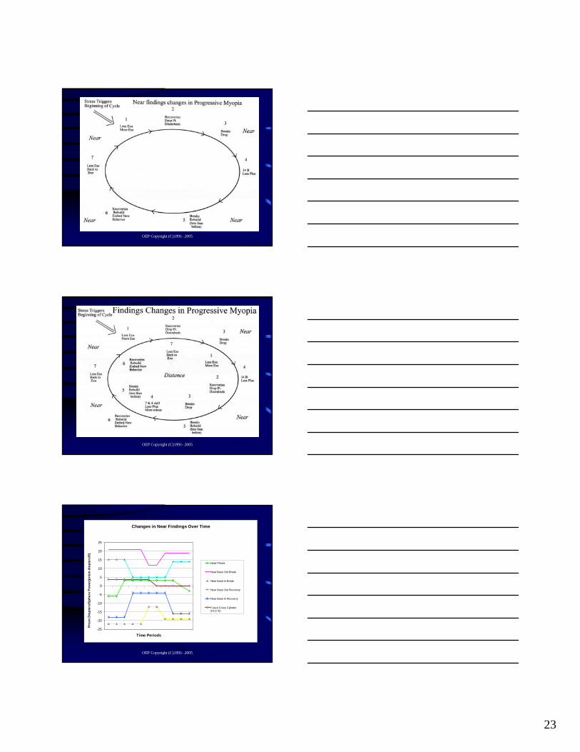
Clinical Signs• The first sign of a person at risk to develop
nearsightedness is the drive to center closer than identification. – This manifests as a closer working distance at near that
typically gets closer and closer as the person sustains working at near.
– Next we see the development of esophoria at near in the analytical testing.
• The problem begins at near and spreads to distance leading to a cascade of changes in the findings over time.
OEP Copyright (C)1991- 2005
The Harmon Distance
OEP Copyright (C)1991- 2005
The Harmon Distance (2)
23
OEP Copyright (C)1991- 2005
OEP Copyright (C)1991- 2005
OEP Copyright (C)1991- 2005
Changes in Near Findings Over Time
-25
-20
-15
-10
-5
0
5
10
15
20
25
Time Periods
Pris
m D
iopt
ers/
Sphe
re P
ower
(pris
m d
iopt
ers/
5)
Near Phoria
Near Base Out Break
Near Base In Break
Near Base Out Recovery
Near Base In Recovery
Fused Cross Cylinder(FCC*5)

24
OEP Copyright (C)1991- 2005
Changes in Distance Findings Over Time
-15
-10
-5
0
5
10
15
20
25
Time Periods
Pris
m D
iopt
ers/
Sphe
re P
ower
(pris
m d
iopt
ers/
5)
Distance Phoria
Distance Base Out Break
Distance Base In Break
Distance Base OutRecovery
Distance Base InRecovery
Subjective 7/7A (7/7A*5)
OEP Copyright (C)1991- 2005
Changes in the Analytical Over Time
-25
-20
-15
-10
-5
0
5
10
15
20
25
Time Periods
Pris
m D
iopt
ers/
Sph
ere
Pow
er(p
rism
dio
pter
s/5)
Distance Phoria
Distance Base Out Break
Distance Base In Break
Distance Base Out Recovery
Distance Base In Recovery
Subjective 7/7A (7/7A*5)
Near Phoria
Near Base Out Break
Near Base In Break
Near Base Out Recovery
Near Base In Recovery
Fused Cross Cylinder (FCC*5)
OEP Copyright (C)1991- 2005
Contact Lenses
• Soft lenses: seem to be about the same as compensatory glasses!
• Rigid lenses: generally provide stability– Old style PMMA: seemed to stop progress dead
in its tracks.– Gas Permeable Rigid Lenses: almost like
PMMA but it depends on fit and lens design

25
OEP Copyright (C)1991- 2005
RGP Lenses and Myopia
• “Morrison in 1956 fit more than 1000 myopia patients between the ages of 7 and 19 years with poly-methyl methacrylate (PMMA) contact lenses and found that none of them progressed in myopia after 2 years.” 7
OEP Copyright (C)1991- 2005
RGP Lenses and Myopia (2)
• “Stone concluded that the effects of the contact lenses on the progression of myopia could not be entirely due to corneal flattening and was the first to suggest that hard contact lenses might influence the axial growth of the eye.” 8
OEP Copyright (C)1991- 2005
RGP Lenses and Myopia (3)• Jeffrey Keller, “Myopia Control with RGP’s in
Children”;– “The mean change in refraction (spherical
equivalent, right eye) during the two-year study period was +0.09 (+/- 0.32D standard deviation), a decrease in myopia of 0.04D per year.” 9
– “I used a 9.5 mm diameter lens whenever possible, but in a few cases with small palpebral apertures, I used a 9.0 mm diameter lens.” 9

26
OEP Copyright (C)1991- 2005
Potential Contact Lens and Myopia Progression (CLAMP) Problems
• The control group is wearing soft contact lenses.
• Fitting parameters: – Diameter: 9.2 mm standard– Optic Zone: 7.8 mm standard– No other specifications such as:
• Large/small lens design – relationship to lids• Center thickness/flex of the contact lens
OEP Copyright (C)1991- 2005
CLAMP Benefit
• “78.4% of 8- to 11-year-old children were able to adapt to rigid gas-permeable contact lens wear.” 8
OEP Copyright (C)1991- 2005
RGP Lens Designs for Stopping Progression
• Look for a large diameter lid control design. We want the lens to stay up under the upper lid. Diameters from 9.6 to 10.4. This may be difficult with an oblate cornea.
• Use an “On-K” fit, not flatter than “K”. This is not “Ortho-K”!
• Thicker than normal. We don’t want the lens to flex. Instead of .12 use .16.
• Plus lenses at near are desirable, but still seem to get stabilization even without it!

27
OEP Copyright (C)1991- 2005
Conjecture on Mechanism
• Wallman et.al. suggest that THE problem is deprivation of form vision.
• They suggest that reading is a type of form deprivation in that it does not provide adequate amounts of differences in the person’s peripheral vision from fixation to fixation.
• This results in a relative global form deprivation, in spite of massive use of the central aspects of vision for decoding.
• The lack of stimulation causes a lack of neuronal activity in the peripheral nets.
OEP Copyright (C)1991- 2005
Conjecture on Mechanism (2)
• The lack of neuronal activity may then be linked to lack of use and transport of essential nutrients and the retina functions “as-if” it is visually deprived.
• Thus, the natural elongation that occurs in the form deprived conditions continues unabated.
• Luminance: paper has only a 10 times light ratio differences between the letters and the background. Normal outdoor scenes have many orders of magnitude differences more.
OEP Copyright (C)1991- 2005
Conjecture on Mechanism (3)
• “Some children may chronically under-accommodate while reading or performing other near work. If so, they may experience a situation similar to tree shrews reared with minus lenses, where an intact emmetropization mechanism produces an elongated eye because the focal plane is shifted posteriorly, increasing the amount of blur and triggering elongation.”

28
OEP Copyright (C)1991- 2005
First Alert
JBO2004
OEP Copyright (C)1991- 2005
Didn’t see in real time
• I tend to not read my journals as they come in.• Several e-mails came in asking my opinion.• I didn’t get to it for a few months.• When I looked I saw only 4 pages.• I started several times but it seemed too dense.
OEP Copyright (C)1991- 2005
O’Leary Article

29
OEP Copyright (C)1991- 2005
O’Leary Summary• 94 subjects watched for 2 years• Full Rx was to 20/20 (6/6)• Under-compensated Rx was to 20/40 (6/12);
generally +0.75 less minus.• Only used Malay and Chinese• At the end of 24 months progression rates:
– -0.77 Diopters in the Full Rx group– -1.00 Diopters in the Under-compensated group
OEP Copyright (C)1991- 2005
O’Leary Data
OEP Copyright (C)1991- 2005
Concern
• O’Leary data was a cause for concern.– When it first came out the cry from some in
Europe was that this would initiate an exodus from the behavioral concept of vision care. Undercompensation was no good. It had been proven!
– So I decided to see how, in 4 pages, Hung and Ciuffreda’s model dealt with O’Leary’s data.

30
OEP Copyright (C)1991- 2005
Dense Writing
• After an hour of intense study I still wasn’t closer to understanding.
• I asked about the review process.• I then asked Greg to read this so he could
set me straight! • Today we hope, by working through this
with you that we all can understand this theory better.
OEP Copyright (C)1991- 2005
Hung and Ciuffreda Statements of Principle
• “Repeated cycles of near-work-induced transient myopia leads to repeated periods of decrease in retinal-image defocus, whose cumulative effect over an extended period of time also results in an increase in axial growth that produces permanent myopia.” 6
• “Manipulations of the optical environment are effective in producing refractive error mainly during the ocular growth and maturational period.” 1
OEP Copyright (C)1991- 2005
Hung and Ciuffreda Statements of Principle (2)
• “It is somewhat ironic that rather than being a failure of the emmetropization process, myopia development is actually a result of the emmetropization process that operates under the constraints of the AS/R (Accommodative Stimulus/Response) function during increments of genetically-programmed ocular growth.” 1

31
OEP Copyright (C)1991- 2005
Hung and Ciuffreda Statements of Principle (3)
• “Recent evidence in animals has shown that during ocular development, the eye grows in the direction of the induced retinal-image defocus. This suggests that the eye may either elongate or decrease in its growth rate in response to the resultant defocused retinal-image as part of the emmetropization process.” 4
OEP Copyright (C)1991- 2005
Hung and Ciuffreda Statements of Principle (4)
• “Elongation occurs even when the optic nerve is severed.” 4
• “Moreover, these appropriate changes in growth rate occurred even when the optic nerve was severed or the midbrain nuclei for controlling accommodation were lesioned, thus precluding any central or cortical visual feedback mechanism. Hence, the retina is the site for controlling the rate of axial length growth.” 5
OEP Copyright (C)1991- 2005
Hung & Ciuffreda
ModelBuilt on AS/AR

32
OEP Copyright (C)1991- 2005
Near Work and Accommodative Lag
• “During Near work, which is represented by a relatively large accommodative stimulus, the accommodative response lags the stimulus, i.e., it exhibits chronic hyperopic defocus.” 3
• “Near work is associated with a larger lag of accommodation, or a greater amount of hyperopic defocus. If the hyperopic defocus is prolonged and sustained, it can result in an increase in the axial growth rate.” 2
OEP Copyright (C)1991- 2005
• “In the case of the development of school myopia relatively small amounts of retinal defocus are present over extended periods of time (i.e., weeks or months). Following an increment of normal genetically-programmed axial length growth, the effective axial length optical power will have decreased, so that the effective accommodative stimulus (or the optical power needed to focus onto the retina of the now incrementally lengthened eyeball) will have also decreased. Thus, less accommodative response would now be necessary for clear retinal imagery. This is equivalent to moving slightly downward and to the left on the AS/R function. The decrease in accommodative error is now associated with a decrease in defocus blur.” 1
OEP Copyright (C)1991- 2005
Accommodation at Distance
• “Although all other refractive groups showed over-accommodation for far targets per the standard hyperfocal refraction procedure, hyperopes under-accommodated at far because of their initial negative refractive error.” 2

33
OEP Copyright (C)1991- 2005
Theories of Myopia Development• Oculomotor Interactive Theory
– “Advocates the use of a near ADD that establishes ocuolmotor balance or equilibrium between the accommodative and vergence systems.” 2
– “Prescription of low-powered near ADD’s in a patient with high AC/A ratio would act to reduce accommodative drive and thus decrease the amount of accommodative convergence, thereby shifting the person’s phoria to a more divergent position. This is consistent with recent evidence demonstrating that near ADD’s are most effective in myopia prevention in children with a high AC/A ratio and near esophoria, with both ocuolmotor findings appearing to be ‘risk factors’ for myopigenesis.” 2
OEP Copyright (C)1991- 2005
Theories of Myopia Development (2)
• Biomechanical Theory– Advocates the use of high powered near ADD’s in the
range of +1.5 to +3.0 D.– The process of accommodation per se produces excessive
biomechanical stress and strain (i.e., abuse) on the sclera and contiguous structures, thus causing small but chronic repeated mechanical stretching which slowly and eventually results in axial elongation and myopia.
• This model is based on motor control of accommodative and vergence components with an indirect impact on retinal defocus.
OEP Copyright (C)1991- 2005
Theories of Myopia Development (3)• Sensory-Based Retinal Defocus Model
– Advocates the use of a near ADD which minimizes the mean level of retinal defocus, especially at near.
– The introduction of a plus ADD is proposed to counteract this process by reducing the amount of hyperopic retinal defocus, which then inhibits the increased growth process, and thereby reduce the axial growth rate.
– This model is applicable under both monocular and binocular conditions.
– This model demonstrates that there is a specific ‘Optimal’ADD, based on the model equations solutions and computer simulations that shift the minimum AErms to occur at IRE equal to zero.
– This model is based on sensory processing with a directimpact on retinal defocus.

34
OEP Copyright (C)1991- 2005
They Build a Model
• Mathematical modeling of the underlying mechanisms of physiological change that are involved with the process of emmetropization and the “tuning” of the refractive components of the eye.
OEP Copyright (C)1991- 2005
Some Characteristics of the Model
• “Local retinal-defocus magnitude is critical in the development of environmentally induced refractive error.” 1
• “The change in magnitude of retinal defocus during an increment of genetically-programmed axial length growth provides the critical information for directional modulation of growth rate.” 1
OEP Copyright (C)1991- 2005
Defocus and Incremental Growth

35
OEP Copyright (C)1991- 2005
Simple Model
OEP Copyright (C)1991- 2005
OEP Copyright (C)1991- 2005
How are Hyperopes Different?
• “With regard to nearwork-induced transient myopia, it was found that young-adult myopes were particularly susceptible, whereas the hyperopes were particularly insusceptible. Thus, further study of hyperopia may lead to a better understanding of myopia, and furthermore yield insight into possible early pre-myopic clinical intervention.” 4

36
OEP Copyright (C)1991- 2005
Some Assumptions
• “Corneal growth is not part of emmetropization process after age 2 years.” 3
• “Added to this basic model is a long-term growth loop that is driven in part by the root-mean square (rms: equal to the average of the absolute value of the instantaneous response over a time interval) of the accommodative error, which simulates retinal defocus that is believed to trigger axial length growth. Due to the squaring process, the directional sign of the blur is potentially lost, although simulations show that during an incremental increase in normal ocular growth or an oculomotor parameter, the blur direction can still be ascertained.” 2
OEP Copyright (C)1991- 2005
Complex Model
OEP Copyright (C)1991- 2005
Accommodative Error / Refractive Error

37
OEP Copyright (C)1991- 2005
AE/IRE• “The sign of the retinal defocus that was preserved in the
V-shaped curve can be extracted by means of an incremental increase in IRE (induced refractive error), which represents the equivalent optical power for an incremental increase in normal axial length growth, and would be manifest as a resultant small change in refractive error that evolved slowly in real life but was still uncorrected optically.” 2
• “Simulation results indicated that refractive error and absolute value of the accommodative error, or retinal defocus (which provides the perception of blur if the defocus is of sufficient magnitude), are involved in a long-term feedback loop in which they interact and modulate each other.” 3
OEP Copyright (C)1991- 2005
Lenses Shift the Curves
OEP Copyright (C)1991- 2005
ADD’s and Accommodative Error
• “Note that in general, infants are hyperopic at birth, and thus this myopic progression would be a natural part of an emmetropization process. However, for ADD = 1 and 2 D, the operational region is on the right, or hyperogenic, branch. Moreover, with an ADD = 0.5 D, the operational region is at a minimum AErms, which represents a relatively optimal condition that would exhibit neither myopic nor hyperopic development since the potential retinal defocus is minimal.” 2

38
OEP Copyright (C)1991- 2005
What Makes it Work?
• “During an increment of normal genetically-driven axial length growth, the magnitude of retinal defocus will have decreased or increased, respectively. It is proposed that feedback regulation provided by the interplexiform neurons from the inner to the outer plexiform layers, which aims to maintain a relatively constant sensitivity to retinal-image contrast, leads to a decrease or increase, respectively in neuromodulators, such as dopamine.” 2
OEP Copyright (C)1991- 2005
Retinal Defocus Pathways & Scleral Growth
OEP Copyright (C)1991- 2005
Retinal Mechanisms• “Transient ganglion cells respond to change in the
surround via amacrine cells in the inner plexiform layer. Thus, these neurons relay information regarding the change in retinal-defocus magnitude.” 1
• “The magnitude of retinal defocus can be represented by the difference in center and surround excitation. A change in this signal, and thus retinal-defocus magnitude, provides the requisite sign for modulating ocular growth. The sensitivity to local retinal-image contrast is maintained at a relatively constant level by means of feedback regulation of horizontal cell gain provided by the interplexiform neurons.” 1

39
OEP Copyright (C)1991- 2005
Retinal Physiology
• “The retina contains a large array of neurochemicals. These can be broadly divided into two classes: neurotransmitters and neuromodulators. The neurotransmitters, such as glutamate, acetylcholine, and GABA, respond rapidly to retinal stimulation. On the other hand, neuromodulators, such as dopamine, serotonin, and nueropeptides, act over a longer period, and in addition, may cause changes in the neuronal synapses.” 3
OEP Copyright (C)1991- 2005
Retinal Physiology (2)
• “Dopamine serves as a neuromodulator rather than a neurotransmitter because it does not exert its influence by acting directly on the horizontal cell membrane channels. Instead, it acts on enzymes that activate protein kinase A, which adds phosphate groups to specific proteins in the horizontal cell membrane to alter their properties and thereby decrease the flow of current across the membrane.” 3
OEP Copyright (C)1991- 2005
Retinal Physiology (3)
• “The result of dopamine administration is a relatively long-term (months to years) reduction in sensitivity to local-image contrast. This mechanism of centripetal feedback via the interplexiform cells can serve to regulate and maintain a relatively constant long-term steady-state operating level and permit relatively normal transient (integrated over hours to days) responsivity to changes in local-image contrast.” 3

40
OEP Copyright (C)1991- 2005
Retinal Physiology (4)
• “This feedback regulation mechanism helps to shift the steady-state operating level to permit detection of change in magnitude of retinal defocus, even for a large initial magnitude.” 3
• “Thus, after a steady-state level had been attained, a transient increase or decrease in defocus would result in an increase or decrease, respectively, in the rate of release of neuromodulators as well as the rate of proteoglycan synthesis.” 3
OEP Copyright (C)1991- 2005
Retinal Physiology (5)
• “A neuromodulator, such as dopamine, transmits this increase via both volume conduction and a cascade of signals through the choroid to the sclera. This in turn results in an increase in proteoglycan synthesis rate, which increases the structural integrity of the sclera.” 5
OEP Copyright (C)1991- 2005
Expanded Retinal Model

41
OEP Copyright (C)1991- 2005
Changes Over Time
OEP Copyright (C)1991- 2005
What Do
Lenses Do?
OEP Copyright (C)1991- 2005
Clinical Implications
• “The ability to control and shift the position of this minimum would provide a potentially powerful means to prescribe specific near bifocal ADD’s based on an individual’s own model parameter values.”2

42
OEP Copyright (C)1991- 2005
Treatment Recommendations
• “The optimum value for the control of myopigenesis would therefore be at the point where the accommodative error as well as the induced refractive error are at their minima. With both AErms and IRE at their minimum values, relative refractive stability should be achieved.” 2
• “Since a multifocal lens would allow for focusing the target at both near and far, the IRDT would predict relatively little effect on refractive change during ocular growth.” 5
OEP Copyright (C)1991- 2005
Treatment Recommendations (2)
• “Given the above we suggest the following: full distance refractive correction in conjunction with a low plus add at near to minimize the level of chronic retinal defocus, and hence myopic progression.” 5
OEP Copyright (C)1991- 2005
Why don’t high plus ADD’s result in hyperogenesis?
• “Thus, although it may seem that a large plus ADD would produce hyperogenesis, or at least a reduction in relative myopic progression, its main effect instead appears to be that of simply introducing larger retinal defocus, with the intended hyperogenesis having relatively little effect in young human subjects.” 2

43
OEP Copyright (C)1991- 2005
Why high ADD’s are bad:
• “For an ADD = 2.0 D, the minimum occurs at IRE = -1.5 D. Under this latter condition, the minimum would be shifter further away from the optimal position. Thus, this would argue against the high ADD’s advocated by the “Biomechanical” Theory.” 2
OEP Copyright (C)1991- 2005
A Paradox?
• “The rationale for this approach is that the ADD reduces the net accommodative stimulus, and thus the accommodative error, and its sensory equivalent, namely retinal defocus/blur. Since retinal defocus/blur has been implicated in the stimulation of axial length growth, its reduction would inhibit axial elongation and the subsequent development of myopia.” 2
OEP Copyright (C)1991- 2005
References1. Norton, T., Siegwart, J., “Animal models of emmetropization: matching axial length to the focal plane.”
JAOA, Vol. 66/No. 7 July 19952. Wu, Hu-Min, et. al. “Does Education Explain Ethnic Differences in Myopia Prevalence? A Population-
Based Study of Young Adult Males in Singapore” Optometry and Vision Science Vol. 78, No. 4, April 2001
3. Kang RN, “Alterations of scleral morphology in tree shrews with induced myopia” invest Oplhthalmol VisSci. 1993;34:1209
4. Wallman, J. et.al., “Local Retinal Regions Control Local Eye Growth and Myopia”, Science, July 1987; 237 (4810) page 73-77
5. Yackle, K, Fitzgerald, D., “Emmetropization: An Overview”, JBO Volume 10/1999/Number 2/ Page 38-43.
6. Bartmann, M, Schaeffel, F., “Letter to the Editor – A Simple Mechanism for Emmetropization Without Cues from Accommodation or Colour”, Vision Res. Vol. 34, No. 7, pp 873-876, 1994
7. Morrison, RJ, “Contact lenses and the progression of myopia”, Optom. Weekly 1956:47:1487-8.8. Walline, J. et.al., “The Contact Lens and Myopia Progression (CLAMP) Study: Design and Baseline
Data”, Optometry and Vision Science, Vol. 78, No.4, April 2001, pp 223-233.9. Keller, J., “Myopia Control with RGP’s in Children”,
www.clspectrum.com/archive/subject/1996/Keller_myopia/10. Devadas M, Morgan I; Retinal Control of Scleral Precursor Synthesis, Aust N Z J Ophthalmol 25 Suppl
1:S-73-75, May 1997.11. Leech EM, Cottrial CL, McBrien NA; Pirenzipine Prevents Form Deprivation Myopia in a Dose-dependent
Manner; Ophthalmic Physiol Opt, 15(5):351-356, Sept 1995.12. Choo, Vivien, “A look at slowing progression of myopia”, The Lancet, Volume 361, Number 9369, May
2003 13. J. Biol. Chem. 2003; 278: 16587-94.14. Ridley, Matt, “Nature via Nutrure – Genes, Experience & What Makes Us Human”, HarperCollins, 2003,
ISBN 0-06-000678-1

44
OEP Copyright (C)1991- 2005
More ReferencesA1. Hung, G., Ciuffreda, K., “A Unifying Theory of Refractive Error
Development”, Bulletin of Mathematical Biology (2000) 62, 1087-1108 A2. Hung, G., Ciuffreda, K., “Quantitative Analysis of the Effect of Near Lens
Addition on Accommodation and Myopigensis.” Current Eye Research, 2000, Vol. 20, No. 4, pp 293-312
A3. Hung, G., Ciuffreda, K., “Differential retinal-defocus magnitude during eye growth provides the appropriate direction signal”, Diagnostics and Medical Technology, Med Sci. Monit, 2000l 6(4): 791-795
A4. Hung, G., Ciuffreda, K., “Model of human refractive error development”, Current Eye Research, 1999, Vol. 19, No. 1, pp 41-52
A5. Hung, G., Ciuffreda, K., “Incremental Retinal-Defocus Theory Predicts Experimental Effect on Under-Correction on Myopic Progression”, JBO, Vol. 15, Number 3, pp 59-63
A6. Hung, G., Ciuffreda, K., “An Incremental Retinal-Defocus Theory of the Development of Myopia”, Comments on Theoretical Biology, 8:511-538, 2003