Embed Size (px)
Citation preview
18
Management of Bone Loss in Primary and Revision Knee Replacement Surgery
Matteo Fosco1, Rida Ben Ayad1, Luca Amendola1,
Dante Dallari1 and Domenico Tigani2 1First Ward of Orthopaedic Surgery, University of Bologna,
Rizzoli Orthopaedic Institute, Bologna 2Department of Orthopaedic Surgery,
Santa Maria alle Scotte Hospital ,Siena Italy
1. Introduction
Total knee arthroplasty (TKA) often deal with bone defect localized in areas corresponding
to tibial and femoral articular surfaces, a condition that is often observed in revision knee
prosthetic surgery but occasionally in primary arthroplasty of the knee too. Such
intraoperative situation, could create a main problem in maintaining proper alignment of
the implant components and in establishing sufficient bone stock to achieve a stable bone-
implant interface. The surgeon must assess the degree of complexity preoperatively and
intraoperatively and have a broad armamentarium available during surgery.
Multiple surgical options are available to repair or reconstruct the loss of bone, these include: bone cement, bone grafts, metal augments and custom-made implants. Principles to consider in bone loss management are knee-related (particularly defect size and location, ligament stability, limb alignment) and patient-related (age, body mass index, activity level, life expectancy).
2. Primary TKA
Bone loss observed during primary arthroplasty of the knee is less frequent than during revision surgery. In primary implants causes of bone defect include:
Osteonecrosis;
Sequelae of fracture of tibial plateau or femoral condyles;
Bone cysts;
Previous tibial osteotomies;
Inflammatory arthropathies. However final cause is always represented by bone erosion, secondary to varus or valgus
deformity of the knee; the consequent overload of medial or lateral compartment bring to
collapse of the subchondral bone (Tigani et al., 2004).
Typically, in varus knee bone defect contribute to collapse of the medial tibial plateau, firstly in the posteromedial site. Instead, in valgus knee, bone defect may involve the tibia (with a
www.intechopen.com
Recent Advances in Arthroplasty
388
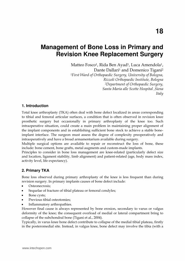
contained central defect) and the external femoral condyle, that is defective in the distal and posterior sites (Insall & Easley, 2001) (Fig. 1).
Fig. 1. Usually varus knee appears with bone defect in posteromedial site of tibial plateau. Instead, in valgus knee bone defect usually involves the central part of lateral tibial hemiplateau and the external femoral condyle.
www.intechopen.com
Management of Bone Loss in Primary and Revision Knee Replacement Surgery
389
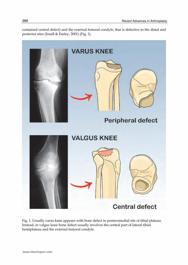
Type Single condylar/hemiplate
involvement (%) Depth(mm)
I (a/b) minimal < 50% < 5
II (a/b) > 50 < 70% 5-10
III (a/b) > 70% < 90% > 10
IV (a/b) > 90% > 10
a) Intact peripheral rim b) Deficient peripheral rim
Table 1. Rand classification of bone loss (modified from Rand JA, 1991).
First classification of bone defects thus consists on distinction between central forms (with
defect confined within the peripheral bone cortex), and peripheral forms (characterized by
involvement of the peripheral cortex). In 1991 Rand proposed a classification that considers
the percentage extent of the defect into the tibial plateau or femoral condyle, distinguishing
between four grades of increasing severity of the lesion (Rand, 1991) (Table 1).
The most common defect is observed in the presence of a varus knee, with defect located in
the posteromedial region of the tibia; the lesion is characterized by the presence of an
important sclerosis of the subchondral bone and its depth usually doesn’t exceed 8 to 10mm.
In such simple cases, resection of the tibial plateau allow to completely remove the defect,
without requiring further procedures.
Instead, in deeper and more severe lesions, tibial resection of more than 12 mm, could lead
to sacrifice important ligamentous structures and has been observed to considerably alters
bone quality, thus requiring other options (Laskin, 1989).
3. Revision TKA
Bone loss in revision TKA could always be considered as a consequence of the previous
arthroplasty. In these cases, on preoperative radiographs bone loss is often
underestimated relative to the true bone loss found during revision surgery. In a
retrospective analysis of 31 patients with symptomatic TKAs who had osteolytic lesions
confirmed by computed tomography, plain radiography detected only 17% of the
osteolytic lesions (Reish et al, 2004). The final evaluation of bone loss is made more accurately during surgery, after component removal; so that various classification systems used are mainly based on the size and type of the defect found intraoperatively. Clatworthy and Gross firstly classified defects as contained central forms, and uncontained
peripheral forms (with involvement of the peripheral cortical rim), then further
distinguishing between intact metaphyseal bone or not (Clatworthy & Gross, 2003). The
most practical system is the Anderson Orthopedic Research Institute (AORI) classification
described by Engh, that considers bone loss from the tibia and femur independently (T and
F) (Engh & Ammeen, 1999); distinction is then made depending on involvement of 1
condyle/plateau (A) or 2 (B) (Table 2,3).
www.intechopen.com
Recent Advances in Arthroplasty
390
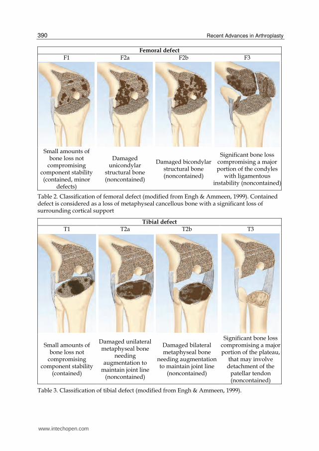
Femoral defectF1 F2a F2b F3
Small amounts of
bone loss not compromising
component stability (contained, minor
defects)
Damaged unicondylar
structural bone (noncontained)
Damaged bicondylar structural bone (noncontained)
Significant bone loss compromising a major portion of the condyles
with ligamentous instability (noncontained)
Table 2. Classification of femoral defect (modified from Engh & Ammeen, 1999). Contained defect is considered as a loss of metaphyseal cancellous bone with a significant loss of surrounding cortical support
Tibial defectT1 T2a T2b T3
Small amounts of bone loss not
compromising component stability
(contained)
Damaged unilateral metaphyseal bone
needing augmentation to
maintain joint line (noncontained)
Damaged bilateral metaphyseal bone
needing augmentation to maintain joint line
(noncontained)
Significant bone loss compromising a major portion of the plateau,
that may involve detachment of the
patellar tendon (noncontained)
Table 3. Classification of tibial defect (modified from Engh & Ammeen, 1999).
www.intechopen.com
Management of Bone Loss in Primary and Revision Knee Replacement Surgery
391
Main causes of bone loss during revision arthroplasty are stress shielding, wear debris and
implant loosening; these factors may be interrelated (Lombardi et al, 2010):
Stress shielding causes an “osteopenic” type of bone loss behind the prosthetic
components, due to pressure shielded by the implant and redistributed to the bone-
cement-implant interface;
Wear debris of polyethylene, cement and metal particles; in contrast to the less common
osteopenic type of bone loss seen in stress shielding, wear causes an “osteolytic” type of
bone loss, around apparently stable implants (Van Loon et al, 1999). Osteolysis is
defined as the periprosthetic replacement of bone by chronic inflammatory tissue
without evidence of loosening. This type of osteolysis is more common in young, male,
overweight patients with osteoarthritis;
Implant aseptic loosening resulting in direct mechanical bone loss at the cement-bone
interface;
Implant septic loosening;
Iatrogenic damage resulting directly from implant removal, thus representing an
important factor in preserving bone stock during revision TKA. Removal of
components with no loosening or with an intercondylar box or stems will create large
bone defects. Cement should be removed using small sagittal saw and flexible
osteotomes, avoiding levering which could cause a fracture.
The objectives of revision surgery include reestablishment of the anatomic joint line, long-
term joint stability, and restoration of bone stock with a fast return to full weight-bearing
and function.
So that, implant selection should be based not only on the status of the ligamentous and soft
tissue stabilizing structures, but also on the severity and type of bone loss.
4. Management of bone defects
Various options exist to manage bone defects, available in both primary and revision surgery. Indication to whether option to use, depends on knee-related and patient-related factors.
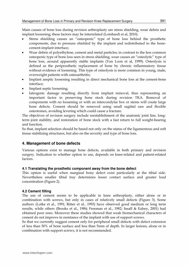
4.1 Translating the prosthetic component away from the bone defect This option is useful when marginal bony defect exist particularly at the tibial side. Nevertheless smaller tibial tray determines lesser contact surface and greater load concentration (Figure 2).
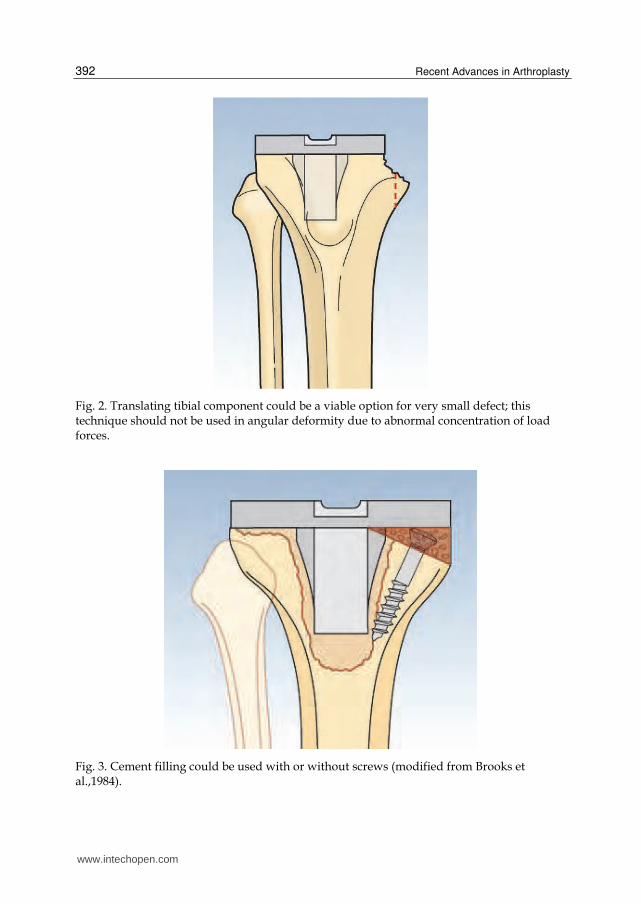
4.2 Cement filling The use of cement seems to be applicable in knee arthroplasty, either alone or in combination with screws, but only in cases of relatively small defects (Figure 3). Some authors (Lotke et al., 1991; Ritter et al., 1993) have observed good medium or long term results, while others (Brooks et al., 1984; Freeman et al., 1982; Insall & Ealsey, 2001) had obtained poor ones. Moreover these studies showed that weak biomechanical characters of cement do not improve in resistance of the implant with use of support screws. So that we currently suggest cement only for peripheral small defects with defect extension of less than 50% of bone surface and less than 5mm of depth. In larger lesions, alone or in combination with support screws, it is not recommended.
www.intechopen.com
Recent Advances in Arthroplasty
392
Fig. 2. Translating tibial component could be a viable option for very small defect; this technique should not be used in angular deformity due to abnormal concentration of load forces.
Fig. 3. Cement filling could be used with or without screws (modified from Brooks et al.,1984).
www.intechopen.com
Management of Bone Loss in Primary and Revision Knee Replacement Surgery
393
4.3 Bone grafting Recently, grafting constitutes a frequent option used for the treatment of bone defect in knee arthroplasty. The rationale in using bone grafts consisted in the possibility of a new formation of vital bone, through a process of osteoinduction and/or osteoconduction. Autoplastic bone grafts are likely to be used for limited defects, while structural allograft could be necessary in cases of larger lesions. Bone grafts, both homoplastic and autoplastic, are to be preferred in younger patients because they allow for bone regeneration that represents an essential condition in case of reintervention.
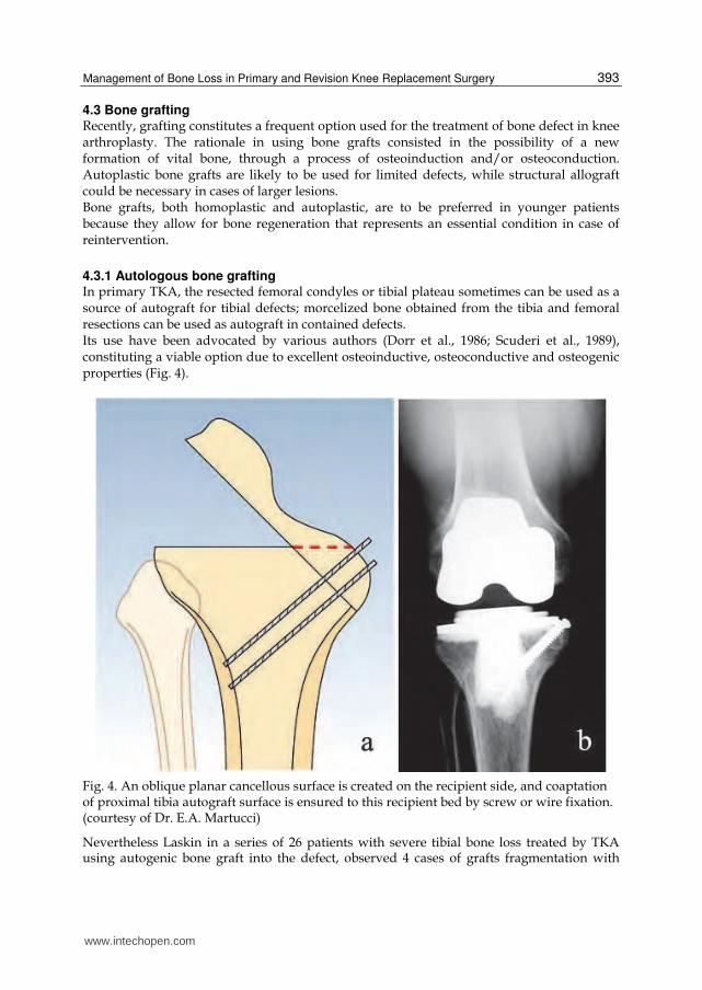
4.3.1 Autologous bone grafting In primary TKA, the resected femoral condyles or tibial plateau sometimes can be used as a source of autograft for tibial defects; morcelized bone obtained from the tibia and femoral resections can be used as autograft in contained defects. Its use have been advocated by various authors (Dorr et al., 1986; Scuderi et al., 1989), constituting a viable option due to excellent osteoinductive, osteoconductive and osteogenic properties (Fig. 4).
Fig. 4. An oblique planar cancellous surface is created on the recipient side, and coaptation of proximal tibia autograft surface is ensured to this recipient bed by screw or wire fixation. (courtesy of Dr. E.A. Martucci)
Nevertheless Laskin in a series of 26 patients with severe tibial bone loss treated by TKA using autogenic bone graft into the defect, observed 4 cases of grafts fragmentation with
www.intechopen.com

Recent Advances in Arthroplasty
394
implant subsidence within the first year (Laskin, 1989). Moreover needle biopsy in 9 cases in which the graft had not fragmented, revealed osteocytes in the lacunae in only 4 grafts. In each of four knees, there was a complete radiolucency between the graft and the tibial host bone. The final author’s conclusion is to reevaluate the use of modular prostheses in large fragment defects but to continue using bone graft for smaller, circumscribed defects. Possible reason of autoplastic graft failure have been ipothetised: Varus alignment of the leg. Avascular host bed for the graft. Insufficient graft press-fit. Incomplete coverage of the graft by the tibial component. Extravasation of the cement between graft and host. Currently there are insufficient clinical data to state with certainty that bone stock restoration with autogenic bone graft will in fact aid future revisions when necessary.
4.3.2 Allogeneic bone grafting The limited availability of autologous graft may not be enough to compensate an extensive bone defect and use of homoplastic bone could be indicated (Fig. 5). Cancellous or structured bone allograft could be used but always have to be considered some rules: The graft have to be modeled, thus to precisely adapt it to the defect; The graft have to be perfectly stabilized; The graft have to be “protected” by use of an intramedullary stems, able to reduce
mechanical stress forces.
Fig. 5. (a) Cancellous bone allograft, added with antibiotic. (b) Structural bone allograft molded intraoperatively before implanting.
www.intechopen.com
Management of Bone Loss in Primary and Revision Knee Replacement Surgery
395
Nevertheless difficulties using bone homologous grafts consisted on:
Need of a bone bank support;
Grafts resorbtion;
Increased risk of infection;
Disease transmission.
4.3.3 Structural massive bone allograft Structural bone allograft offers numerous advantages, including biocompatibility, bone
stock restoration and potential for ligaments reattachment. Regarding their versatility it is
possible to treat a wide range of bone deficiency allowing the surgeon to shape the allograft
to fit the bone defect and avoid unnecessary removal of host bone. Finally allograft is
relatively cost-effective if compared to the high cost of custom-made implants.
The goals of structural allograft reconstruction are to maximize the stability of the graft-
host bone contact and provide a stable platform for fixation of the implant. The first step
is to remove all the nonviable bone and soft tissue from in and around the defect, the
presence of viable bone is absolutely necessary to maximize the likelihood of graft
incorporation. Conversion of the oblique peripheral defects into rectangular space with
vertical and horizontal surfaces has been demonstrate to improve stability for
components fixation. The angular patterns have also a biological advantage since it
allows improving the contact area of the host-graft construct maximizing the probability
of graft incorporation. Regarding the choice of the graft it is important to shape the
allograft similarly in order to fit the defect precisely. Graft fixation too is an important
step to be taken in consideration: mainly used are partially or fully threaded cancellous
screws.
In the literature there are various papers reporting about the use of allograft to restore
bone defect during revision knee arthroplasty, especially for uncontained defects. In some
cases with circumferential segmental bone defect of tibial plateau, have been
demonstrated about 25% of allograft failure (De Long et al., 2007; Engh & Ammeen, 2007).
Engh & Ammeen, whose have reviewed the results of 49 knees with severe tibial bone
loss, found only four cases of failure for reasons not-directly related to collapse or
resorption of the graft; most of patients had contained defect and ten presented an
uncontained deficiency of which only four cases were restored with full segment allograft.
Recently Backstein D, et al have reported 85.2% of success rate at an average follow-up of
5.4 years in a series of 68 revision that required a structural allograft for the treatment of
uncontained defect (Backstein et al., 2006).
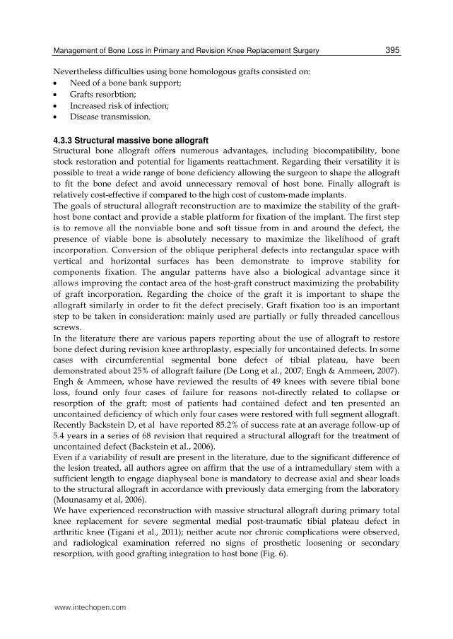
Even if a variability of result are present in the literature, due to the significant difference of the lesion treated, all authors agree on affirm that the use of a intramedullary stem with a sufficient length to engage diaphyseal bone is mandatory to decrease axial and shear loads to the structural allograft in accordance with previously data emerging from the laboratory (Mounasamy et al, 2006). We have experienced reconstruction with massive structural allograft during primary total
knee replacement for severe segmental medial post-traumatic tibial plateau defect in
arthritic knee (Tigani et al., 2011); neither acute nor chronic complications were observed,
and radiological examination referred no signs of prosthetic loosening or secondary
resorption, with good grafting integration to host bone (Fig. 6).
www.intechopen.com
Recent Advances in Arthroplasty
396
Fig. 6. (a) Pre-operative plain radiographs showing post-traumatic severe depressed medial bone stock loss; (b) preparation of tibial plateau, in a way to convert oblique defect to rectangular stepped one; (c) Fitting the allograft into the defect; (d) last follow-up plain radiographs, 12 months after the surgery.
4.4 Modular components (metal augments) The use of metal augments for bone deficiencies has become quite popular since mid
Eighties, after the work of Brooks et al which indicated that biomechanically the modular
augments are equivalent to a custom implant (Brooks et al., 1984). In last decades a new
biomaterial is largely used in knee prosthetic surgery: porous tantalum, in its trabecular
form (trabecular metal, TM; Zimmer, Warsaw, Ind, US). TM show excellent mechanical
characteristics compared to conventional implant materials (i.e. titanium and cobalt
chromium): good biocompatibility, high porosity, low modulus of elasticity (Levine et al.,
2007). Moreover biological advantages of TM include its negative charge and
interconnective pores, which form a scaffolding and surface for osteoblast-mediated bone
www.intechopen.com
Management of Bone Loss in Primary and Revision Knee Replacement Surgery
397
ingrowth (Bobyn et al., 1999). These unique material properties of porous trabecular metal
allow it to achieve immediate structural support, together with early bony ingrowth and late
restoration of bone stock.
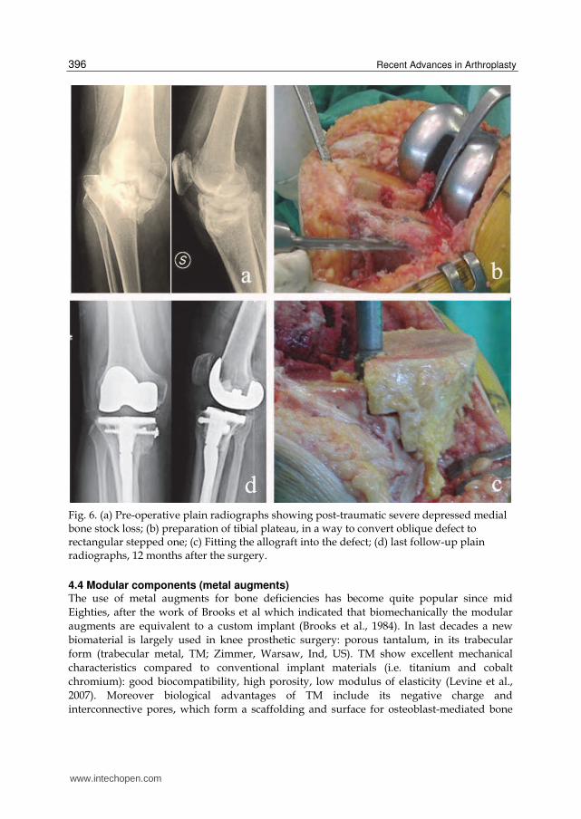
TM is available in many shapes and forms (Fig. 7): porous or solid, rectangular or wedge shaped, and can be attached with the use of cement or screws. It can be applied quickly, allow intraoperative custom fabrication, supply excellent biomechanical properties, and require minimal bone resection as the augments attach on the residual bone.
Fig. 7. TM modular augments (courtesy of Zimmer, Warsaw, Ind, US).
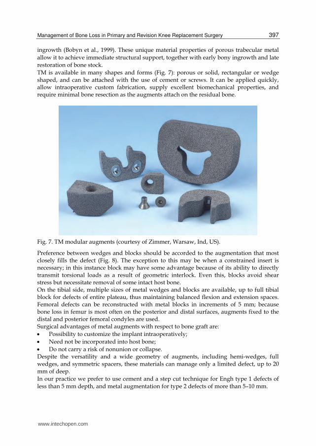
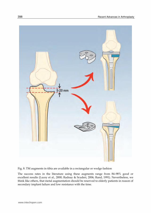
Preference between wedges and blocks should be accorded to the augmentation that most closely fills the defect (Fig. 8). The exception to this may be when a constrained insert is necessary; in this instance block may have some advantage because of its ability to directly transmit torsional loads as a result of geometric interlock. Even this, blocks avoid shear stress but necessitate removal of some intact host bone. On the tibial side, multiple sizes of metal wedges and blocks are available, up to full tibial block for defects of entire plateau, thus maintaining balanced flexion and extension spaces. Femoral defects can be reconstructed with metal blocks in increments of 5 mm; because bone loss in femur is most often on the posterior and distal surfaces, augments fixed to the distal and posterior femoral condyles are used. Surgical advantages of metal augments with respect to bone graft are:
Possibility to customize the implant intraoperatively;
Need not be incorporated into host bone;
Do not carry a risk of nonunion or collapse. Despite the versatility and a wide geometry of augments, including hemi-wedges, full wedges, and symmetric spacers, these materials can manage only a limited defect, up to 20 mm of deep. In our practice we prefer to use cement and a step cut technique for Engh type 1 defects of
less than 5 mm depth, and metal augmentation for type 2 defects of more than 5–10 mm.
www.intechopen.com
Recent Advances in Arthroplasty
398
Fig. 8. TM augments in tibia are available in a rectangular or wedge fashion
The success rates in the literature using these augments range from 84–98% good or excellent results (Lucey et al., 2000; Radnay & Scuderi, 2006; Rand, 1991). Nevertheless, we think like others, that metal augmentation should be reserved to elderly patients in reason of secondary implant failure and low resistance with the time.
www.intechopen.com
Management of Bone Loss in Primary and Revision Knee Replacement Surgery
399
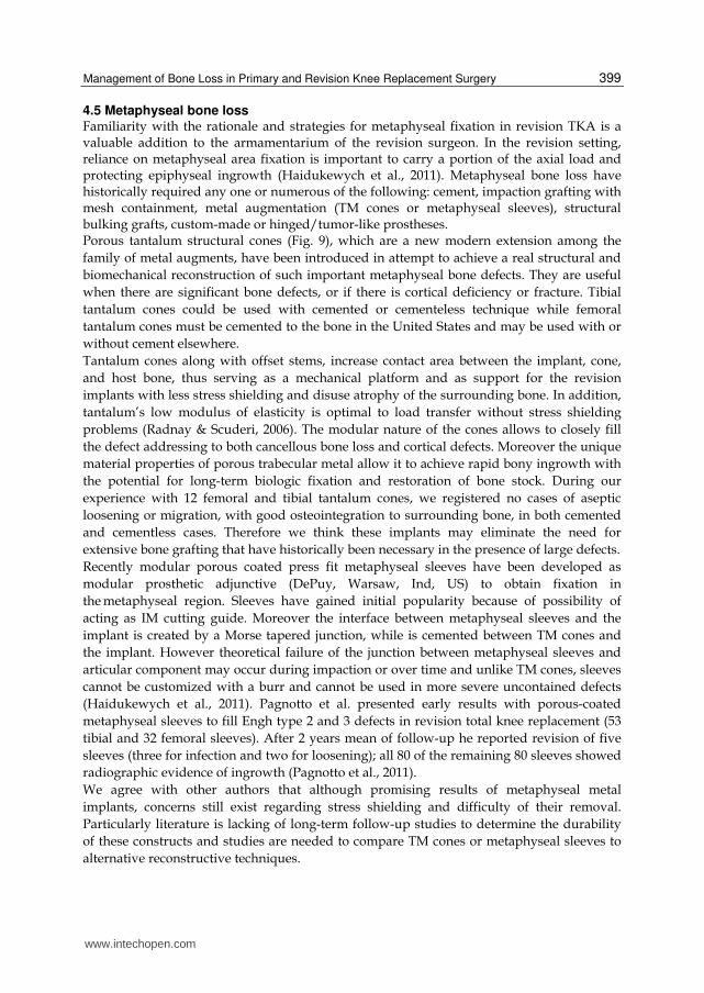
4.5 Metaphyseal bone loss Familiarity with the rationale and strategies for metaphyseal fixation in revision TKA is a valuable addition to the armamentarium of the revision surgeon. In the revision setting, reliance on metaphyseal area fixation is important to carry a portion of the axial load and protecting epiphyseal ingrowth (Haidukewych et al., 2011). Metaphyseal bone loss have historically required any one or numerous of the following: cement, impaction grafting with mesh containment, metal augmentation (TM cones or metaphyseal sleeves), structural bulking grafts, custom-made or hinged/tumor-like prostheses. Porous tantalum structural cones (Fig. 9), which are a new modern extension among the
family of metal augments, have been introduced in attempt to achieve a real structural and
biomechanical reconstruction of such important metaphyseal bone defects. They are useful
when there are significant bone defects, or if there is cortical deficiency or fracture. Tibial
tantalum cones could be used with cemented or cementeless technique while femoral
tantalum cones must be cemented to the bone in the United States and may be used with or
without cement elsewhere.
Tantalum cones along with offset stems, increase contact area between the implant, cone,
and host bone, thus serving as a mechanical platform and as support for the revision
implants with less stress shielding and disuse atrophy of the surrounding bone. In addition,
tantalum’s low modulus of elasticity is optimal to load transfer without stress shielding
problems (Radnay & Scuderi, 2006). The modular nature of the cones allows to closely fill
the defect addressing to both cancellous bone loss and cortical defects. Moreover the unique
material properties of porous trabecular metal allow it to achieve rapid bony ingrowth with
the potential for long-term biologic fixation and restoration of bone stock. During our
experience with 12 femoral and tibial tantalum cones, we registered no cases of aseptic
loosening or migration, with good osteointegration to surrounding bone, in both cemented
and cementless cases. Therefore we think these implants may eliminate the need for
extensive bone grafting that have historically been necessary in the presence of large defects.
Recently modular porous coated press fit metaphyseal sleeves have been developed as
modular prosthetic adjunctive (DePuy, Warsaw, Ind, US) to obtain fixation in
the metaphyseal region. Sleeves have gained initial popularity because of possibility of
acting as IM cutting guide. Moreover the interface between metaphyseal sleeves and the
implant is created by a Morse tapered junction, while is cemented between TM cones and
the implant. However theoretical failure of the junction between metaphyseal sleeves and
articular component may occur during impaction or over time and unlike TM cones, sleeves
cannot be customized with a burr and cannot be used in more severe uncontained defects
(Haidukewych et al., 2011). Pagnotto et al. presented early results with porous-coated
metaphyseal sleeves to fill Engh type 2 and 3 defects in revision total knee replacement (53
tibial and 32 femoral sleeves). After 2 years mean of follow-up he reported revision of five
sleeves (three for infection and two for loosening); all 80 of the remaining 80 sleeves showed
radiographic evidence of ingrowth (Pagnotto et al., 2011).
We agree with other authors that although promising results of metaphyseal metal
implants, concerns still exist regarding stress shielding and difficulty of their removal.
Particularly literature is lacking of long-term follow-up studies to determine the durability
of these constructs and studies are needed to compare TM cones or metaphyseal sleeves to
alternative reconstructive techniques.
www.intechopen.com
Recent Advances in Arthroplasty
400
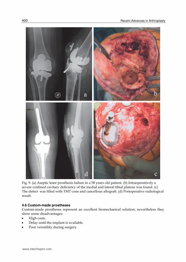
Fig. 9. (a) Aseptic knee prosthesis failure in a 58 years old patient. (b) Intraoperatively a severe confined cavitary deficiency of the medial and lateral tibial plateau was found. (c) The defect was filled with TMT cone and cancellous allograft. (d) Postoperative radiological result.
4.6 Custom-made prostheses Custom-made prostheses represent an excellent biomechanical solution; nevertheless they show some disadvantages:
High costs.
Delay until the implant is available.
Poor versatility during surgery.
www.intechopen.com
Management of Bone Loss in Primary and Revision Knee Replacement Surgery
401
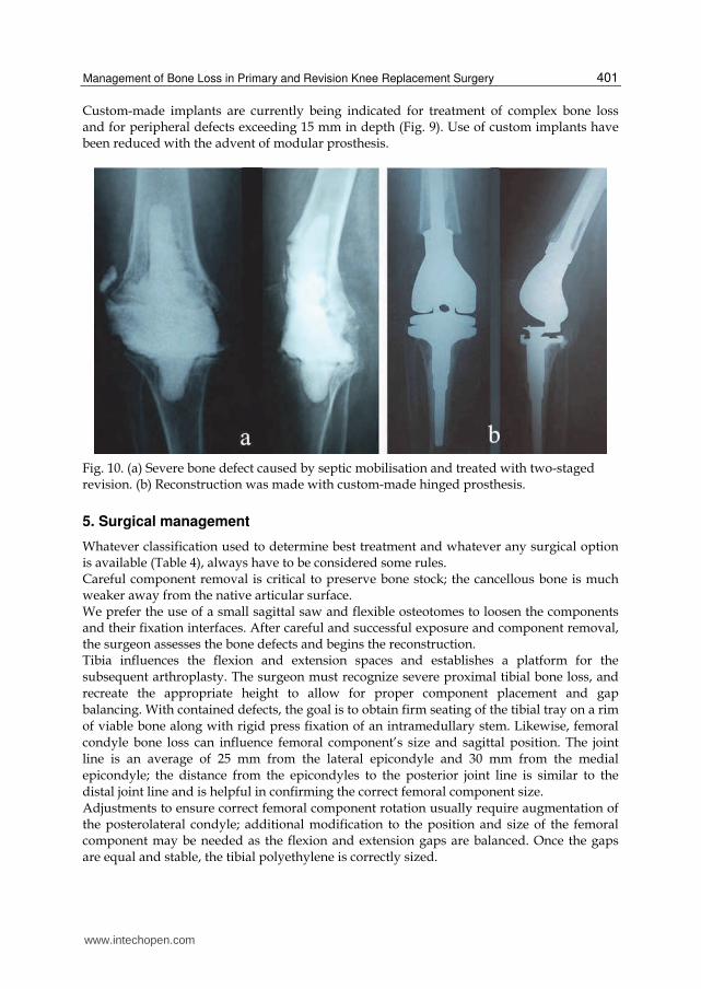
Custom-made implants are currently being indicated for treatment of complex bone loss and for peripheral defects exceeding 15 mm in depth (Fig. 9). Use of custom implants have been reduced with the advent of modular prosthesis.
Fig. 10. (a) Severe bone defect caused by septic mobilisation and treated with two-staged revision. (b) Reconstruction was made with custom-made hinged prosthesis.
5. Surgical management
Whatever classification used to determine best treatment and whatever any surgical option is available (Table 4), always have to be considered some rules. Careful component removal is critical to preserve bone stock; the cancellous bone is much weaker away from the native articular surface. We prefer the use of a small sagittal saw and flexible osteotomes to loosen the components and their fixation interfaces. After careful and successful exposure and component removal, the surgeon assesses the bone defects and begins the reconstruction. Tibia influences the flexion and extension spaces and establishes a platform for the subsequent arthroplasty. The surgeon must recognize severe proximal tibial bone loss, and recreate the appropriate height to allow for proper component placement and gap balancing. With contained defects, the goal is to obtain firm seating of the tibial tray on a rim of viable bone along with rigid press fixation of an intramedullary stem. Likewise, femoral condyle bone loss can influence femoral component’s size and sagittal position. The joint line is an average of 25 mm from the lateral epicondyle and 30 mm from the medial epicondyle; the distance from the epicondyles to the posterior joint line is similar to the distal joint line and is helpful in confirming the correct femoral component size. Adjustments to ensure correct femoral component rotation usually require augmentation of the posterolateral condyle; additional modification to the position and size of the femoral component may be needed as the flexion and extension gaps are balanced. Once the gaps are equal and stable, the tibial polyethylene is correctly sized.
www.intechopen.com
Recent Advances in Arthroplasty
402
If there is functional loss of the medial or lateral collateral ligaments, soft tissue instability, inability to balance the flexion and extension spaces, or a severe valgus deformity, then a constrained condylar prosthesis is necessary.
Defect Type
Treatment Options Clatworthy
classification
Engh
classificationRand classification
Contained,
undamaged
metaphysis
1
<5 mm depth, <50%
unicondylar/plateau
involvement
Prosthetic component
translation, PMMA fill,
morcelized allograft,
autograft
Contained
undamaged
metaphysis
2a
5-10 mm depth, >50%
unilateral condylar/plateau
involvement
Morcelized allograft or
metal augments
Contained
undamaged
metaphysis
2a
10-20 mm depth,
unicondylar/plateau
involvement
Metal augments,
metaphyseal sleeves,
structural allografts
Contained
undamaged
metaphysis
2b
<20mm depth,
bicondylar/plateau
involvement
Metal augments (full
block in tibia),
metaphyseal sleeves,
structural allografts
Uncontained
damaged
metaphysis
2b
<20mm depth,
bicondylar/plateau
involvement
TM cones, structural
allografts, custom-
made prostheses
Uncontained
damaged
metaphysis
3 >20 mm depth
Structural allografts,
megaprostheses,
custom-made
prostheses, TM cones
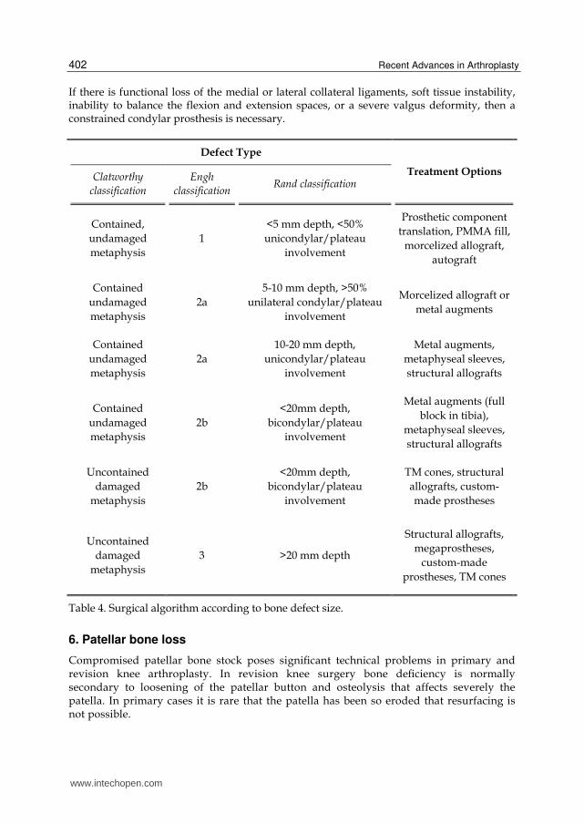
Table 4. Surgical algorithm according to bone defect size.
6. Patellar bone loss
Compromised patellar bone stock poses significant technical problems in primary and revision knee arthroplasty. In revision knee surgery bone deficiency is normally secondary to loosening of the patellar button and osteolysis that affects severely the patella. In primary cases it is rare that the patella has been so eroded that resurfacing is not possible.
www.intechopen.com
Management of Bone Loss in Primary and Revision Knee Replacement Surgery
403
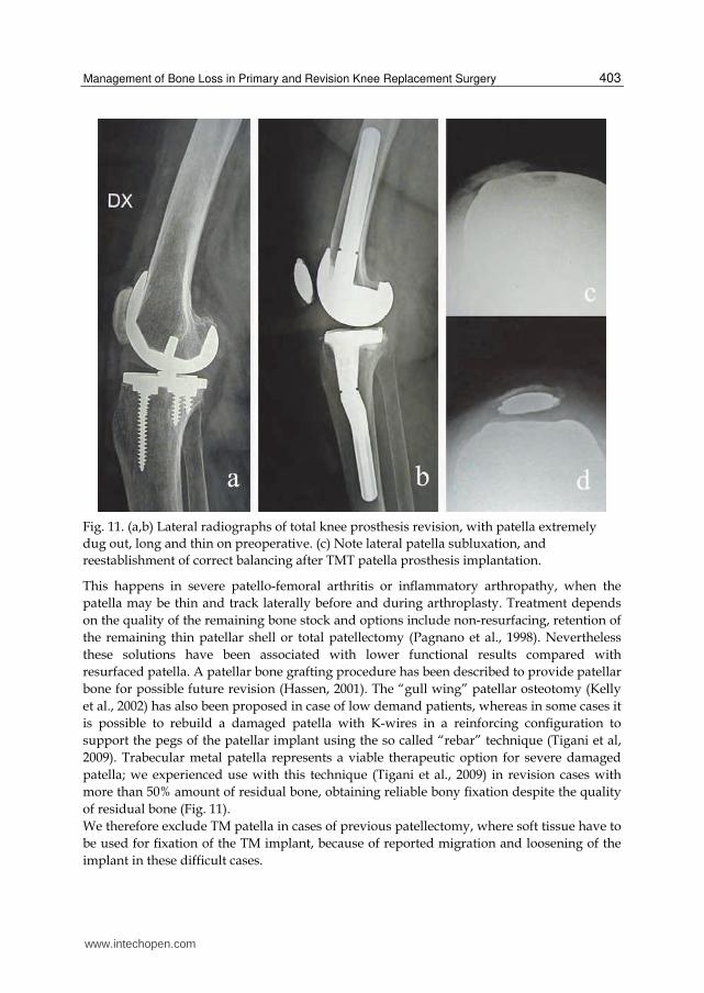
Fig. 11. (a,b) Lateral radiographs of total knee prosthesis revision, with patella extremely
dug out, long and thin on preoperative. (c) Note lateral patella subluxation, and
reestablishment of correct balancing after TMT patella prosthesis implantation.
This happens in severe patello-femoral arthritis or inflammatory arthropathy, when the
patella may be thin and track laterally before and during arthroplasty. Treatment depends
on the quality of the remaining bone stock and options include non-resurfacing, retention of
the remaining thin patellar shell or total patellectomy (Pagnano et al., 1998). Nevertheless
these solutions have been associated with lower functional results compared with
resurfaced patella. A patellar bone grafting procedure has been described to provide patellar
bone for possible future revision (Hassen, 2001). The “gull wing” patellar osteotomy (Kelly
et al., 2002) has also been proposed in case of low demand patients, whereas in some cases it
is possible to rebuild a damaged patella with K-wires in a reinforcing configuration to
support the pegs of the patellar implant using the so called “rebar” technique (Tigani et al,
2009). Trabecular metal patella represents a viable therapeutic option for severe damaged
patella; we experienced use with this technique (Tigani et al., 2009) in revision cases with
more than 50% amount of residual bone, obtaining reliable bony fixation despite the quality
of residual bone (Fig. 11).
We therefore exclude TM patella in cases of previous patellectomy, where soft tissue have to
be used for fixation of the TM implant, because of reported migration and loosening of the
implant in these difficult cases.
www.intechopen.com
Recent Advances in Arthroplasty
404
7. Intramedullary stems
In revision total knee arthroplasty, the mechanical stability of the femoral and tibial components is increased by the addition of intramedullary stems. Whatever solution is indicating to compensate bone loss, the use of an extended femoral and tibial stems reduce the stress forces on the metaphyseal region and bone-implant interface. Stem extensions, with or without offset, can supplement fixation, decrease stress at the bone-implant interface, and help address asymmetric bone defects. Offset stems can assist with implant alignment on the metaphysis, reducing the incidence of coronal or sagittal components malalignment and helping balance the flexion and extension spaces by effectively translating the components. The stem ability to protect the proximal tibia or distal femur has been demonstrated in a laboratory setting using finite element model and cadaver models. Brooks et al used stems in conjunction with various bone augmentation techniques for defect in the proximal tibia: they suggested that a 70-mm stem carried from 23% to 38% of the axial load (Brooks et al., 1984). A finite element analysis has revealed that the predicted bone loss is even greater in
stemmed components compared to stemless ones; this may have consequences to
discouraged routine use of stems in revision TKA (van Lenthe et al., 2002). Nevertheless
Stern and Insall (Stern & Insall, 1992) advocates routine use of stemmed components in
revision TKA. Engh et al used femoral stems mainly to protect large structural grafts in
revision TKA (Engh et al., 1997). Meneghini et al. advise the use of cemented stemmed
extension to maximize early implant fixation and allow for successful biologic ingrowth of
the TM cones into the remaining part of the bone (Meneghini et al., 2008). We recommend to
add stem extensions when using metal augments to decrease stress at the bone-implant
interface.
Cemented stems allow for intraoperative adjustment with unusual anatomy and achieve fixation in large canals and osteopenic bone (Murray et al., 1994). The main disadvantages are that they are difficult to remove if revision is necessary and since they are not canal filling, they do not guarantee alignment (Parsley et al., 2003). Cementless press-fit stem extensions are easy to use and facilitate component alignment, and diaphyseal engaging stems ensure fixation (Radnay & Scuderi, 2006). In our practice with stemmed components of TKA, we prefer to use hybrid fixation in both femur and tibia, with proximal cementation just in the metaphyseal area as usual. In fact, Jazrawi et al. demonstrated in a cadaveric study that a press-fit modular cementless stem could achieve equivalent stability than a somewhat shorter fully cemented stem (Jazrawi et al., 2001), whereas Albrektsson et al. showed with radiostereogrammetic analysis that a long cementless stem provide optional stability (Albrektsson et al., 1990).
8. Future perspectives
We verified that treatment of bone defects associated with knee prosthetic surgery include
many different surgical techniques and options, each with specific disadvantages and
complications. Generally, in the elderly bone loss should be accomplished using artificial
materials, while in younger patients, therapy should be addressed to regeneration of new
bone as a foundation for future revision procedures. So that, an ideal bone substitute should
possesses osteogenic, osteoinductive and osteoconductive potential.
www.intechopen.com
Management of Bone Loss in Primary and Revision Knee Replacement Surgery
405
The ‘‘osteogenic’’ potential of the graft corresponds to capacity of cells living within the donor graft to survive during transplantation, then proliferate and differentiate to osteblasts and eventually to osteocytes. ‘‘Osteoinduction’’ on the other hand is the stimulation and activation of host mesenchymal stem cells from the surrounding tissue, which differentiate into bone-forming osteoblasts. This process is mediated by a cascade of signals and the activations of several extra and intracellular receptors the most important of which belong to the TGF-beta family (Cypher & Grossman, 1996). “Osteoconduction” describes the facilitation and orientation of blood-vessel and the creation of the new Haversian systems into the bone scaffold. Finally these three properties together allow ‘‘osteointegration’’ between the host bone and the grafting material surfaces (Giannoudis et al., 2005). The gold standard for regeneration of new bone is autologous bone graft, which contains a scaffold, osteoblasts, and the necessary signalling proteins and molecules. However, autograft is of limited availability and may be insufficient due to poor quality (eg, osteoporosis). Furthermore, it may fail in clinical practice as most of the cellular (osteogenic) elements do not survive transplantion (Sandhu et al., 1999). Thus alternatives to autograft bone have emerged. Perhaps the most common bone substitute is cancellous allograft, which is osteoconductive only, and rely on a viable vascularized bone bed for incorporation. Moreover bone graft provided by musculoskeletal tissue bank could provoke immune response and transmission of viral disease; the processing of allograft tissue lowers this risk but can significantly weaken the biologic and mechanical properties initially present in the bone tissue (Giannoudis et al., 2005). Bone substitutes could be used to replenish lost bone stock during total knee arthroplasty. A bone-graft substitute to be useful should be: osteoconductive, osteoinductive, biocompatible, bio-resorbable, structurally similar to bone, easy to use, and cost-effective (Giannoudis et al., 2005). Recombinant growth factors such as bone morphogenetic proteins (BMPs) have
osteoinductive capacity (Greenwald et al., 2001). Nevertheless because they are powerful in
small amounts, and they are expensive, their indication in knee prosthetic surgery is still
limited. Contrary platelet rich plasma (PRP) is largely available as it is prepared from
centrifugation of autologous blood; it is an osteopromotive adjunct with the ability to
enhance natural bone formation by stimulatory signals. Both BMPs and PRP need a scaffold
to support their bone regeneration properties.
Demineralized bone matrix (DBM) corresponds to portion of bone without the mineral
phases, extracted by strong acids (Peterson et al., 2004). The demineralization process leaves
behind the growth factors, the noncollagenous proteins and collagen, and therefore DBM is
osteoconductive and osteoinductive. However, few prospective, randomized clinical studies
delineate the efficacy of DBM, and the material can be costly.
Nowadays many synthetic substitutes are available, used either alone or in combination
with other biologic adjuncts. Synthetic bone graft materials available include calcium
sulphates, special glass ceramics (bioactive glasses) and calcium phosphates. Calcium
phosphate-based implants have the most similar composition to human bone, in particular
those made of hydroxyapatite (HA) (Paderni et al., 2009). HA-based substitutes provide an
osteoconductive scaffold to which mesenchymal stem cells and osteoinductive growth
factors can migrate and differentiate into functioning osteoblasts (Fujishiro et al., 2005).
These materials can be sterilized and are moldable, but generally do not have sufficient
mechanical properties to support full immediate weight bearing.
www.intechopen.com
Recent Advances in Arthroplasty
406
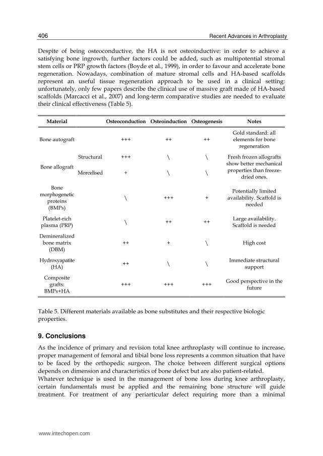
Despite of being osteoconductive, the HA is not osteoinductive: in order to achieve a satisfying bone ingrowth, further factors could be added, such as multipotential stromal stem cells or PRP growth factors (Boyde et al., 1999), in order to favour and accelerate bone regeneration. Nowadays, combination of mature stromal cells and HA-based scaffolds represent an useful tissue regeneration approach to be used in a clinical setting: unfortunately, only few papers describe the clinical use of massive graft made of HA-based scaffolds (Marcacci et al., 2007) and long-term comparative studies are needed to evaluate their clinical effectiveness (Table 5).
Material Osteoconduction Osteoinduction Osteogenesis Notes
Bone autograft +++ ++ ++ Gold standard: all elements for bone
regeneration
Bone allograft
Structural +++ \ \ Fresh frozen allografts show better mechanical properties than freeze-
dried ones. Morcelised + \ \
Bone morphogenetic
proteins (BMPs)
\ +++ + Potentially limited
availability. Scaffold is needed
Platelet-rich plasma (PRP)
\ ++ ++ Large availability. Scaffold is needed
Demineralized bone matrix
(DBM) ++ + \ High cost
Hydroxyapatite (HA)
++ \ \ Immediate structural
support
Composite grafts:
BMPs+HA +++ +++ +++
Good perspective in the future
Table 5. Different materials available as bone substitutes and their respective biologic properties.
9. Conclusions
As the incidence of primary and revision total knee arthroplasty will continue to increase,
proper management of femoral and tibial bone loss represents a common situation that have
to be faced by the orthopedic surgeon. The choice between different surgical options
depends on dimension and characteristics of bone defect but are also patient-related.
Whatever technique is used in the management of bone loss during knee arthroplasty,
certain fundamentals must be applied and the remaining bone structure will guide
treatment. For treatment of any periarticular defect requiring more than a minimal
www.intechopen.com
Management of Bone Loss in Primary and Revision Knee Replacement Surgery
407
prosthetic augment, it is imperative to use stemmed components to transfer stress away
from the joint line. Reestablishment of well-aligned and stable implants is necessary for
successful reconstruction, but this can’t be accomplished without a sufficient restoration of
an eventual bone loss.
10. Acknowledgment
We thank Mrs Mariapia Cumani for graphical support. The authors did not receive any outside funding or grants in support of their research or for preparation of this work.
11. References
Albrektsson, B.E.; Ryd, L.; Carlsson, L.V.; Freeman, M.A.; Herberts, P.; Regnér, L. & Selvik, G. (1990). The effect of a stem on the tibial component of knee arthroplasty. A roentgen stereophotogrammetric study of uncemented tibial components in the Freeman-Samuelson knee arthroplasty. J Bone Joint Surg Br, Vol.72, No.2, (March 1990), pp. 252-8, ISSN 0301-620X
Backstein, D.; Safir, O. & Gross, A. (2006). Management of bone loss: structural grafts in revision total knee arthroplasty. Clin Orthop Relat Res, Vol.446, (May 2006), pp.104-12, ISSN 1528-1132
Bobyn, J.D.; Stackpool, G.J.; Hacking, S.A.; Tanzer, M. & Krygier, J.J. (1999). Characteristics of bone ingrowth and interface mechanics of a new porous tantalum biomaterial. J Bone Joint Surg Br, Vol.81, No.5, (September 1999), pp.907-14, ISSN 0301-620X
Boyde, A.; Corsi, A.; Quarto, R.; Cancedda, R. & Bianco, P. (1999). Osteoconduction in large macroporous hydroxyapatite ceramic implants: evidence for a complementary integration and disintegration mechanism. Bone, Vol.24, No.6, (June 1999), pp.579–89, ISSN 1873-2763
Brooks, P.J.; Walker, P.S. & Scott, R.D. (1984). Tibial component fixation in deficient tibial bone stock. Clin Orthop Relat Res, Vol.184, (April 1984), pp.302-8, ISSN 1528-1132
Clatworthy, M. & Gross, A.E. (2003). Management of bony defects in revision total knee arthroplasty. In: The Adult Knee, JJ Callaghan, AG Rosenberg, HE Rubash, PT Simonian, TL Wickiewicz, (Eds.), 1455-64, Lippincott Williams & Wilkins, ISBN 0-7817-3247-6, Philadelphia, PA
Cypher, T.J. & Grossman, J.P. (1996). Biological principles of bone graft healing. J Foot Ankle Surg, Vol.35, No.5, (September-October 1996), pp.413-7, ISSN 0449-2544
De Long, W.G. Jr; Einhorn, T.A.; Koval, K.; McKee, M.; Smith, W.; Sanders, R. & Watson, T. (2007). Bone grafts and bone graft substitutes in orthopaedic trauma surgery. A critical analysis. J Bone Joint Surg Am, Vol.89, No.3, (March 2007), pp.649-58, ISSN 1535-1386
Dorr, L.D.; Ranawat, C.S.; Sculco, T.A.; McKaskill, B. & Orisek, B.S. (1986). Bone graft for tibial defects in total knee arthroplasty. Clin Orthop Relat Res Vol.205, (April 1986), pp.153-65, ISSN 1528-1132
Engh, G.A. & Ammeen, D.J. (1999). Bone loss with revision total knee arthroplasty: defect classification and alternatives for reconstruction. Instr Course Lect, Vol.48, (1999), pp.167-75, ISSN 0065-6895
www.intechopen.com
Recent Advances in Arthroplasty
408
Engh, G.A. & Ammeen, D.J. (2007). Uses of structural allograft in revision total knee arthroplasty in knees with sever tibial bone loss. J Bone Joint Surg Am, Vol.89, No.12, (December 2007), pp.2640-7, ISSN 1535-1386
Engh, G.A.; Herzwurm, P.J. & Parks, N.L. (1997).Treatment of major defects of bone with bulk allografts and stemmed components during total knee arthroplasty.JBone JointSurg Am,Vol.79,No.7,(July 1997),pp.1030-9,ISSN 1535-1386
Freeman, M.A.; Bradley, G.W. & Revell, P.A. (1982). Observations upon the interface between bone and polymethylmethacrylate cement. J Bone Joint Surg Br, Vol.64, No.4, (1982), pp.489-93, ISSN 0301-620X
Fujishiro, T.; Nishikawa, T.; Niikura, T.; Takikawa, S.; Nishiyama, T.; Mizuno, K.; Yoshiya, S. & Kurosaka, M. (2005). Impaction bone grafting with hydroxyapatite: increased femoral component stability in experiments using Sawbones. Acta Orthop, Vol.76, No.4, (August 2005), pp.550–4, ISSN 1745-3682
Giannoudis, P.V.; Dinopoulos, H. & Tsiridis, E. (2005). Bone substitutes: an update. Injury, Vol.36, Suppl 3, (November 2005), pp.20-7, ISSN 1879-0267
Greenwald, A.S.; Boden, S.D.; Goldberg, V.M.; Khan, Y.; Laurencin, C.T. & Rosier, R.N. (2001). Bone-graft substitutes: facts, fictions and application. J Bone Joint Surg Am. Vol.83, Suppl 2 Pt 2, (2001), pp.98-103, ISSN 1535-1386
Haidukewych, G.J.; Hanssen, A. & Jones, R.D. (2011). Metaphyseal fixation in revision total knee arthroplasty: indications and techniques. J Am Acad Orthop Surg. Vol.19, No.6, (June 2011), pp.311-8, ISSN 1067-151X
Hassen, A. (2001). Bone-grafting for severe patellar bone loss during revision knee arthroplasty. J Bone J Surg Am, Vol.83, No.2, (February 2001), pp.171–6, ISSN 1535-1386
Insall, J.N. & Easley, M.E. (2001). Surgical techniques and instrumentation in total knee arthroplasty. In: Surgery of the knee, JN Insall, WN Scott (Eds.), ISBN 978-0-443-06682-5, Churchill-Livingstone, New York
Jazrawi, L.M.; Bai, B.; Kummer, F.J.; Hiebert, R. & Stuchin, S.A. (2001). The effect of stem modularity and mode of fixation on tibial component stability in revision total knee arthroplasty. J Arthroplasty, Vol.16, No.6, (September 2001), pp.759-67, ISSN 1532-8406
Kelly, M.A. (2004). Extensor mechanism complications in total knee arthroplasty. Instr Course Lect, Vol.53, (2004), pp.193-9, ISSN 0065-6895
Laskin, R.S. (1989). Total knee arthroplasty in the presence of bony defects of the tibia and marked knee instability. Clin Orthop Relat Res, Vol.248, (November 1989), pp.66-70, ISSN 1528-1132
Levine, B.; Sporer, S.; Della Valle, C.J.; Jacobs, J.J. & Paprosky, W. (2007). Porous tantalum in reconstructive surgery of the knee: a review. J Knee Surg, Vol.20, No.3, (July 2007), pp.185-94, ISSN 1938-2480
Lombardi, A.V.; Berend, K.R. & Adams, J.B. (2010). Management of bone loss in revision TKA: it's a changing world. Orthopedics, Vol.33, No.9, (September 2010), pp.662, ISSN 1938-2367
Lotke, P.A.; Wong, R.Y.& Ecker, M.L. (1991). The use of methylmethacrylate in primary total knee replacements with large tibial defects. Clin Orthop Relat Res, Vol.270, (September 2001), pp.288-94, ISSN 1528-1132
www.intechopen.com
Management of Bone Loss in Primary and Revision Knee Replacement Surgery
409
Lucey, S.D.; Scuderi, G.R.; Kelly, M.A. & Insall, J.N. (2000). A practical approach to dealing with bone loss in revision total knee arthroplasty. Orthopedics, Vol.23, No.10, (October 2000), pp.1036–41, ISSN 1938-2367
Marcacci, M.; Kon, E.; Mukhacev, V.; Lavroukov, A.; Kutepov, S.; Quarto, R.; Mastrogiacomo, M. & Cancedda, R. (2007). Stem cells associated with macroporous bioceramics for long bone repair: 6 to 7-year outcome of a pilot clinical study. Tissue Eng. Vol.13, No.5, (May 2007), pp.947–955, ISSN 1557-8690
Meneghini, R.M.; Lewallen, D.G.& Hanssen, A.D. (2008). Use of porous tantalum metaphyseal cones for severe tibial bone loss during revision total knee replacement. J Bone Joint Surg Am, Vol.9, No. 1, Januay 2008, pp.78-84, ISSN 1535-1386
Mounasamy, V.; Ma, S.Y.; Schoderbek, R.J.; Mihalko, W.M.; Saleh, K.J. & Brown, T.E. (2006). Primary total knee arthroplasty with condylar allograft and MCL reconstruction for a comminuted medial condyle fracture in an arthritic knee-a case report. Knee, Vol.13, No.5, (October 2006), pp.400-3, ISSN 1873-5800
Murray, P.B.; Rand, J.A. & Hanssen, A.D. (1994). Cemented long-stem revision total knee arthroplasty. Clin Orthop Relat Res, Vol.309, (December 1994), pp.116-23, ISSN 1528-1132
Paderni, S.; Terzi, S. & Amendola, L. (2009). Major bone defect treatment with an osteoconductive bone substitute. Chir Organi Mov, Vol.93, No.2, (September 2009), pp.89-96, ISSN 1973-2538
Pagnano, M.W.; Scuderi, G.R. & Insall, J.N. (1998). Patellar component resection in revision and reimplantation TKA. Clin Orthop Relat Res, Vol.356, (November 1998), pp.134–8, ISSN 1528-1132
Pagnotto, M.; Fedorka, C.J.; McGough, R.L.; Crossett, L.S. & Klatt, B.A. (2011). Revision total knee replacement with porous-coated metaphyseal sleeves. Paper. In: AAOS 2011 Annual Meeting, San Diego, February 2011
Parsley, B.S.; Sugano, N.; Bertolusso, R. & Conditt, M.A. (2003). Mechanical alignment of tibial stems in revision total knee arthroplasty. J Arthroplasty. Vol.18, Suppl 1, (October 2003), pp.33–6, ISSN 1532-8406
Peterson, B.; Whang, P.G.; Iglesias, R.; Wang, J.C. & Lieberman, J.R. (2004). Osteoconductivity of commercially available demineralized bone matrix. J Bone Joint Surg Am. Vol.86, No.10, (October 2004), pp.2243-50, ISSN 1535-1386
Radnay, C.S. & Scuderi, G.R. (2006). Management of bone loss: augments, cones, offset stems. Clin Orthop Relat Res, Vol.446, (May 2006), pp.83-92, ISSN 1528-1132
Rand, J.A. (1991). Bone deficiency in total knee arthroplasty. Use of metal wedge augmentation. Clin Orthop Relat Res, Vol.271, (October 1991), pp.63-71, ISSN 1528-1132
Rand, J.A. (1998). Modular augments in revision total knee arthroplasty. Orthop Clin North Am, Vol. 29, No.2, (April 1998), pp.347–53, ISSN 1558-1373
Reish, T.G.; Clarke, H.D.; Scuderi, G.R.; Math, K.R. & Scott, W.N. (2006). Use of multi-detector computed tomography for the detection of periprosthetic osteolysis in total knee arthroplasty. J Knee Surg, Vol.19, No.4, (October 2006), pp.259-64, ISSN 1938-2480
www.intechopen.com
Recent Advances in Arthroplasty
410
Ritter, M.A.; Keating, E.M.& Faris, P.M. (1993). Screw and cement fixation of large defects in total knee arthroplasty. A sequel. J Arthroplasty, Vol.8, No.1, (February 1993), pp.63-5, ISSN 1532-8406
Sandhu, H.S.; Grewal, H.S. & Parvataneni, H. (1999). Bone grafting for spinal fusion. Orthop Clin North Am, Vol.30, No.4, (October 1999), pp.685-98, ISSN 1558-1373
Scuderi, G.R.; Insall, J.N. & HaasSB. (1989). Inlay autogenic bone grafting of tibial defects in primary total knee arthroplasty Clin Orthop Relat Res, Vol.248, (November 1989), pp.93-7
Stern, S.H. & Insall, J.N. (1992). Posterior stabilized prosthesis. Results after follow-up of nine to twelve years. J Bone Joint Surg Am, Vol.74, No.7, (August 1992), pp.980-6, ISSN 1535-1386
Tigani, D.; Dallari, D.; Coppola, C.; Ben Ayad, R.; Sabbioni, G. & Fosco, M. (2011). Total knee arthroplasty for post-traumatic proximal tibial bone defect: three cases report. Open Orthop J, Vol.14,No.5,(April 2011),pp.143-50,ISSN 1874-3250
Tigani, D.; Trentani, P.; Trentani, F.; Andreoli, I.; Sabbioni, G. & Del Piccolo, N. (2009). Trabecular metal patella in total knee arthroplasty with patella bone deficiency. Knee, Vol.16, No.1, (January 2009), pp.46-9, ISSN 1873-5800
Tigani, D.; Trentani, P.; Trentani, F.; Marinelli, A.; Bianchi, G. & Fravisini, M. (2004). The treatment of bone defects in primary arthroplasty of the knee. Chir Organi Mov, Vol.89, No.1, (January-March 2004),pp.29-33, ISSN 1973-2538
van Lenthe, G.H.; Willems, M.M.; Verdonschot, N.; de Waal Malefijt, M.C. & Huiskes, R. (2002). Stemmed femoral knee prostheses: effects of prosthetic design and fixation on bone loss. Acta Orthop Scand, Vol.73, No.6, (December 2002), pp.630-7, ISSN 0001-6470
Van Loon, C.J.; de Waal Malefijt, M.C.; Buma, P.; Verdonschot, N. & Veth, R.P. (1999). Femoral bone loss in total knee arthroplasty. A review. Acta Orthop Belg, Vol.65, No.2, (June 1999), pp.154-63, ISSN 0001-6462
www.intechopen.com
Recent Advances in ArthroplastyEdited by Dr. Samo Fokter
ISBN 978-953-307-990-5Hard cover, 614 pagesPublisher InTechPublished online 27, January, 2012Published in print edition January, 2012
InTech EuropeUniversity Campus STeP Ri Slavka Krautzeka 83/A 51000 Rijeka, Croatia Phone: +385 (51) 770 447 Fax: +385 (51) 686 166www.intechopen.com
InTech ChinaUnit 405, Office Block, Hotel Equatorial Shanghai No.65, Yan An Road (West), Shanghai, 200040, China
Phone: +86-21-62489820 Fax: +86-21-62489821
The purpose of this book was to offer an overview of recent insights into the current state of arthroplasty. Thetremendous long term success of Sir Charnley's total hip arthroplasty has encouraged many researchers totreat pain, improve function and create solutions for higher quality of life. Indeed and as described in a specialchapter of this book, arthroplasty is an emerging field in the joints of upper extremity and spine. However,there are inborn complications in any foreign design brought to the human body. First, in the chapter oninfections we endeavor to provide a comprehensive, up-to-date analysis and description of the management ofthis difficult problem. Second, the immune system is faced with a strange material coming in huge amounts ofmicro-particles from the tribology code. Therefore, great attention to the problem of aseptic loosening hasbeen addressed in special chapters on loosening and on materials currently available for arthroplasty.
How to referenceIn order to correctly reference this scholarly work, feel free to copy and paste the following:
Matteo Fosco, Rida Ben Ayad, Luca Amendola, Dante Dallari and Domenico Tigani (2012). Management ofBone Loss in Primary and Revision Knee Replacement Surgery, Recent Advances in Arthroplasty, Dr. SamoFokter (Ed.), ISBN: 978-953-307-990-5, InTech, Available from: http://www.intechopen.com/books/recent-advances-in-arthroplasty/management-of-bone-loss-in-primary-and-revision-knee-replacement-surgery

© 2012 The Author(s). Licensee IntechOpen. This is an open access articledistributed under the terms of the Creative Commons Attribution 3.0License, which permits unrestricted use, distribution, and reproduction inany medium, provided the original work is properly cited.