Embed Size (px)
Citation preview
Management in Acute Pancreatitis
Ioana Grintescu
Anesthesia and Intensive Care ClinicClinical Emergency Hospital of Bucharest
Carol Davila University of General Medicine, Bucharest

Acute pancreatitis
Definition

Acute pancreatitis - definition
• Inflammatory disease caused by activation, interstitial liberation and autodigestion of the pancreas by its own enzymes
• A group of reversible lesions characterised by inflammation of the pancreas
Bradley EL III. Atlanta, Ga, September 11-13, 1992. Arch Surg 1993,128, 586-590
Cavallini G, Uomo G, Pezilli R et al – Pancreatology 2001, 1: 129-199
Acute pancreatitis
Anatomy
Physiology

Normal Anatomy & Physiology
• neutralize chyme
• digestive enzymes
• hormones
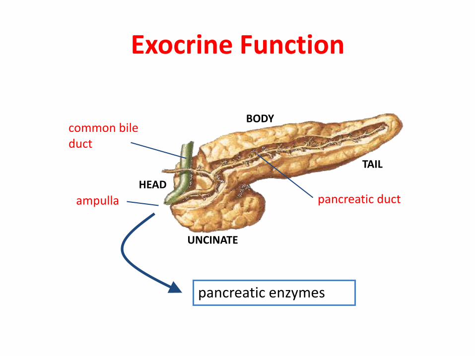
Exocrine Function
pancreatic duct
common bileduct
ampulla
pancreatic enzymes
TAIL
BODY
HEAD
UNCINATE

Enzyme Secretion
pancreatic duct
duodenum
acinus
microscopic viewof pancreatic acini

Enzyme Secretion
HormonalCCKgastrin
Neuralacetylcholine
VIPGRP
Secretin (hormonal)
H2Obicarbonate

Digestive Enzymes in the Pancreatic Acinar Cell
PROTEOLYTIC LIPOLYTIC ENZYMES ENZYMES Lipase
Trypsinogen Prophospholipase A2Chymotrypsinogen Carboxylesterase lipaseProelastaseProcarboxypeptidase A NUCLEASESProcarboxypeptidase B Deoxyribonuclease (DNAse)
Ribonuclease (RNAse)AMYOLYTIC ENZYMESAmylase OTHERS
ProcolipaseTrypsin inhibitor
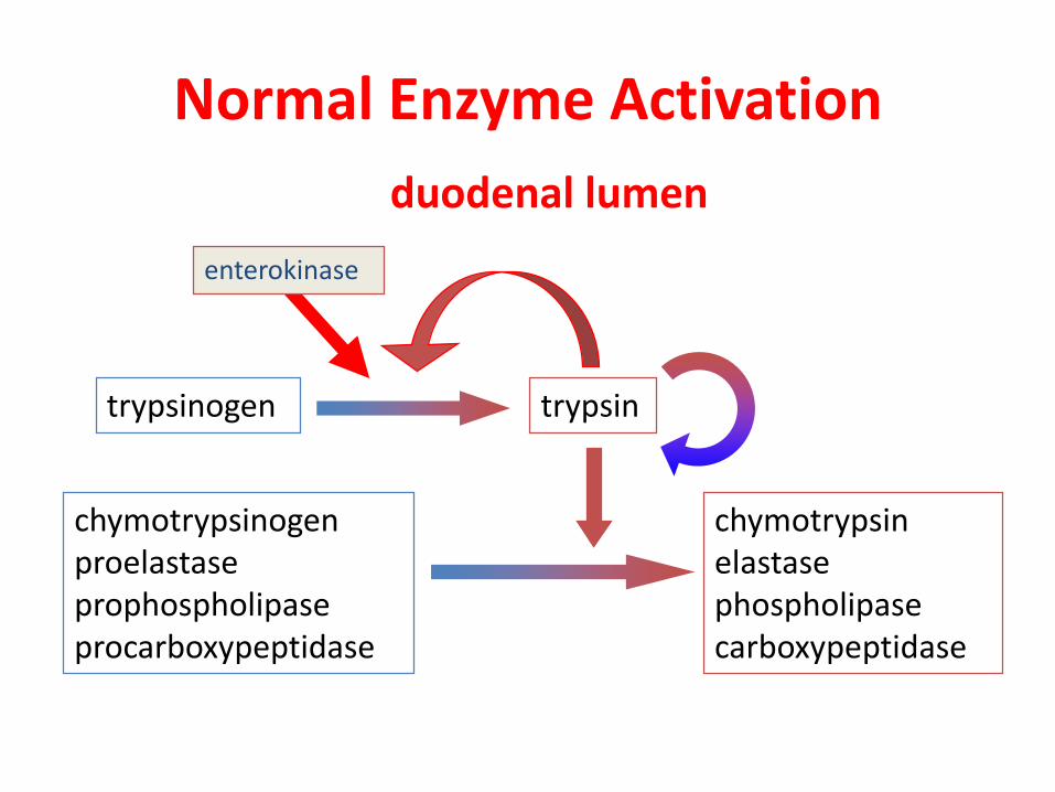
Normal Enzyme Activation
trypsinogen trypsin
chymotrypsinelastasephospholipasecarboxypeptidase
enterokinase
chymotrypsinogenproelastaseprophospholipaseprocarboxypeptidase
duodenal lumen
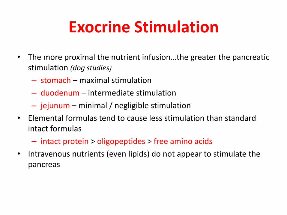
Exocrine Stimulation
• The more proximal the nutrient infusion…the greater the pancreatic stimulation (dog studies)
– stomach – maximal stimulation
– duodenum – intermediate stimulation
– jejunum – minimal / negligible stimulation
• Elemental formulas tend to cause less stimulation than standard intact formulas
– intact protein > oligopeptides > free amino acids
• Intravenous nutrients (even lipids) do not appear to stimulate the pancreas

Protective Measures
• COMPARTMENTALIZATION - digestive enzymes are contained within zymogen granules in acinar cells
• REMOTE ACTIVATION - digestive enzymes are secreted as inactive proenzymes within the pancreas
• PROTEASE INHIBITORS – trypsin inhibitor is secreted along with the proenzymes to suppress any premature enzyme activation
• AUTO “SHUT-OFF” – trypsin destroys trypsin in high concentrations

Acute pancreatitis
Pathogenesis

Pathogenesis
Colipase
Elastase
Chymotrypsin
Phospholipase A2
Xanthynedehydrogenase
Kallycrein
C3a
C5a
Plasminogen
XIIa Factor
Systemic circulation
Alfa2 + Trypsin
Alfa2-M
RESLiverSpleenBone marrowNodes
Clearance
Procolipase
Proelastase
Chymotrypsinogen
Prophospholipase A2
Xanthynedehydrogenase
Prokallycrein
C3
C5
Plasminogen
XII Factor
Kininogens
Kinins
Trypsinogen
Trypsin
Trypsin
PSTI + Trypsin
PSTI
Alfa1-AT + Trypsin
Alfa1-AT
MesotrypsinEnzyme Y
Norman J. Am J Surg 1998; 175:76-83. (modified)

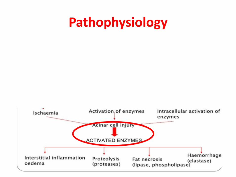
autodigestion of pancreatic tissue
release ofenzymes intothe circulation
activationof whiteblood cells
local
complications
localvascularinsufficiency
premature enzyme activation
distant
organ failure
Acute PancreatitisPathogenesis

Oxidated
Phospholipids
Pathophysiology
TNF
PAF IL-8
IL-6
Activated Macrophages
TNFPAF
Tissue injury
IL-8
Link between pankreatic inflammation and sytemic tissue damage
Systemic Circulation
(from R.Stocker – “Acute pancreatitis nutritional simposium” 2000
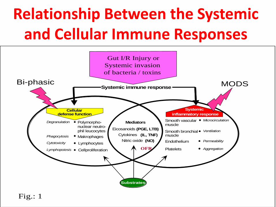
Relationship Between the Systemic and Cellular Immune Responses
Mediators
inflammatory response
Substrates
Systemic immune response
Cellular
Degranulation
Phagocytosis
Cytotoxicity
Lymphopoiesis
Smooth bronchialmuscle
Endothelium
Platelets
Smooth vascularmuscle
Microcirculation
Ventilation
Permeability
Aggregation
Systemic invasion of
bacteria
toxins
Makrophages
Lymphocytes
Polymorpho-
nuclear neutro-
phil leucocytes
Cellproliferation
Eicosanoids (PGE, LTB)
Cytokines (IL, TNF)
Nitric oxide (NO)
Systemic
defense function
Bi-phasic MODS
Gut I/R Injury or
Systemic invasion
of bacteria / toxins
Fig.: 1
OFR

1st – 2nd week 3rd – 4rd week
MOF
infection
Mo
rta
lity
Two mortality peaks of severe acute pancreatitis
Pathophysiology

Pathophysiology

Pathophysiology

Acute pancreatitis
Etiology

Etiology
• Non-traumatic (75%)• Biliary tract disease• Alcohol• Viral infection (EBV, CMV, mumps)• Drugs (steroid, thiazide, furosemide)• Scorpion bites• Hyperlipidemia• Hyperparathyroidism
• Traumatic (5%)• Operative trauma• Blunt/penetrating trauma• Lab test ERCP/angiography
• Idiopathic (20%)

Etiology
Gallstones (35%-60%)– Gallstone pancreatitis risk is
highest among patients with small GS < 5mm and with microlithiasis
– GS pancreatitis risk is also increased in women > 60 yrs

Etiology
Alcohol (30-40%)
– Mechanism not fully understood
– Not all alcoholics get pancreatitis (only about 15%)
– This suggests a subset of the population predisposed to
pancreatitis, with alcohol acting more as a co-precipitant

Etiology – Trauma
Blunt Trauma
– Automobile
– Bicycle handlebar injuries
– Abuse
Iatrogenic – ERCP (1-7%)
– Likely secondary to contrast but also very operator dependant
– Risk is also increased with Sphincter of Oddi manometry

Etiology - 1
Transabdominal ultrasound should be performed in all patients with acute pancreatitis(strong recommendation, low quality of evidence)
In the absence of gallstones and/or history of alcohol use serum triglyceride should be obtained and considered the etiology if > 1000 mg/dL (conditional recommendation,moderate quality of evidence)
In a patient older than 40 years, a pancreatic tumor should be considered as a possible cause of AP (conditional recommendation, low quality of evidence)

Etiology - 2
Endoscopic investigation in patients with acute idiopathic pancreatitis should be limited, asthe risks and benefits of investigation in these patients are unclear(conditional recommendation, low quality of evidence)

Acute PancreatitisEtiology
EtOH
35%
Idiopathic
10%
Other
10%Gallstones
45%
Clinical Emergency Hospital of Bucharest, 2009Fagenholz H et al. AEP 2007;17: 491-49

Incidence
23 female
53 male
0
5
10
15
20
25
30 21-30
31-40
41-50
51-60
61-70
71-80
Male:female ratio is• 1:3– in those with gallstone and• 6:1 in those with alcoholism
Sex and age distribution of AP
Clinical Emergency Hospital

Famous people who have had pancreatitis
Alexander the Great
Ludwig von Beethoven
Dizzie Gillespie
Maximilian Schell
John Ashcroft

Acute pancreatitis
Terminology
Classification
Epidemiology
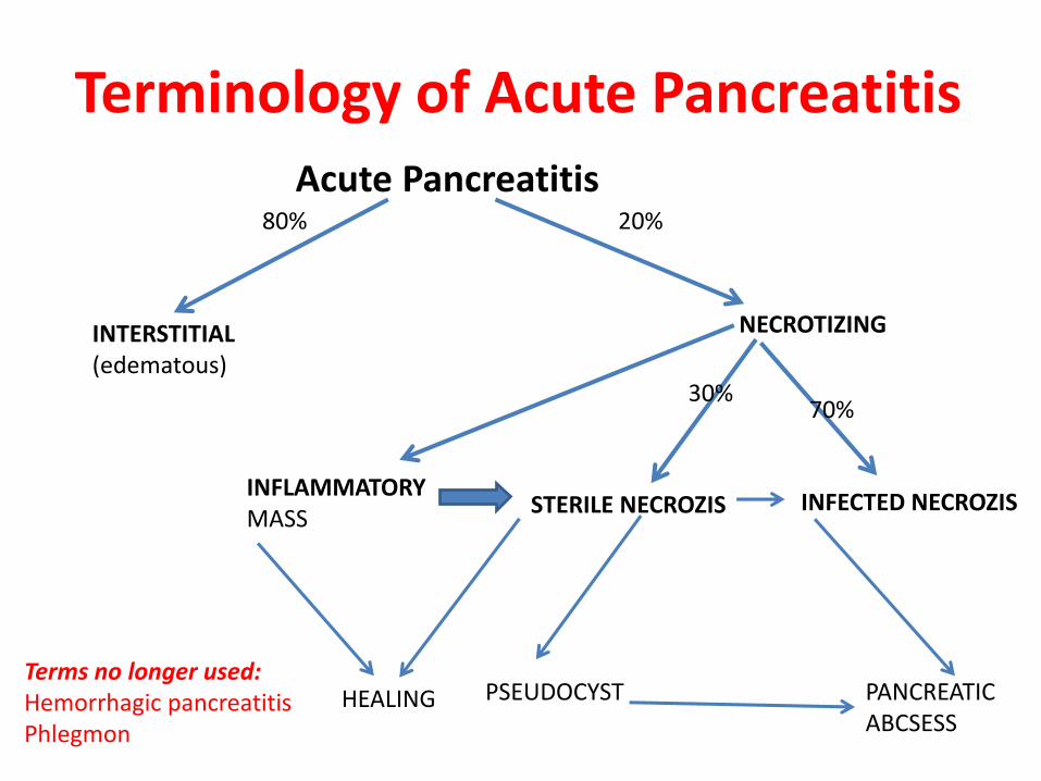
Acute Pancreatitis
INTERSTITIAL(edematous)
NECROTIZING
INFLAMMATORYMASS
STERILE NECROZIS INFECTED NECROZIS
PSEUDOCYST PANCREATICABCSESS
80% 20%
30%70%
Terminology of Acute Pancreatitis
HEALINGTerms no longer used:Hemorrhagic pancreatitisPhlegmon

Definitions of severity in acute pancreatitis: comparison of Atlanta and recent revision

Determinat- based ClassificationBased on the factors that are causally associated with severity of acute pancreatitis
Mild AP Moderate AP Severe AP Critical AP
(Peri)pancreatic necrosis
NO Sterile Infected Infected
AND AND/OR OR AND
Organ failure NO Transient Persistent Persistent

Determinat- based ClassificationBased on the factors that are causally associated with severity of acute pancreatitis
Infected (peri)pancreatic necrosis• Gas bubbles within (CT)• Positive culture obtained
• by image guiidet fine needle aspiration• During the first drainage or necrosectomy
Sterile (peri)pancreatic necrosis • Absence of proven infection in necresis
(Peri)pancreatic necrosis• Pancreatic, peiripancreatic, both• Solid, semisolid, without a radiologically wall
Local determinanat Systemic determinanatDefinition
Organ failure• Cardiovascular:
need for inotropic agent• Renal: creatinine≥ 171mol/L(≥2.0 mg/dL)
• Respuratory:
PaO2/ FiO 2 ≤300 mmHg (≤40kPa)
Persistent organ failureFor 48 hours or more
Transient organ failureFor less than 48 hours

Acute Pancreatitis – Epidemiology
Fagenholz PJ1, Castillo CF, Harris NS, Pelletier AJ, Camargo CA Jr Increasing United States hospital admissions for acute pancreatitis,
1988-2003.Ann Epidemiol. 2007 Jul;17(7):491-7. Epub 2007 apr 19.
Increases in total hospitalisation for acute pancreatitis and in the population rate of hospitalisation
for acute pancreatitis during the study period ( p for trend = 0.001 for both)

Acute pancreatitis
Diagnosis

Clinical Presentation
Pain (95%)
– Acute onset
• Mid-abdominal or mid-epigastric
• Radiates to the back (50%)
– Peak intensity in 30 minutes
• Lasts for several hours
Differential Diagnosis
• Choleledocholithoasis
• Perforated ulcer
• Mesenteric ischemia
• Intestinal obstructuion
• Ectopic pregnancy
Nausea and vomiting (80%)
Abdominal distention (75%)
Abdominal guarding and tenderness (50%)
Restlessness and agitation
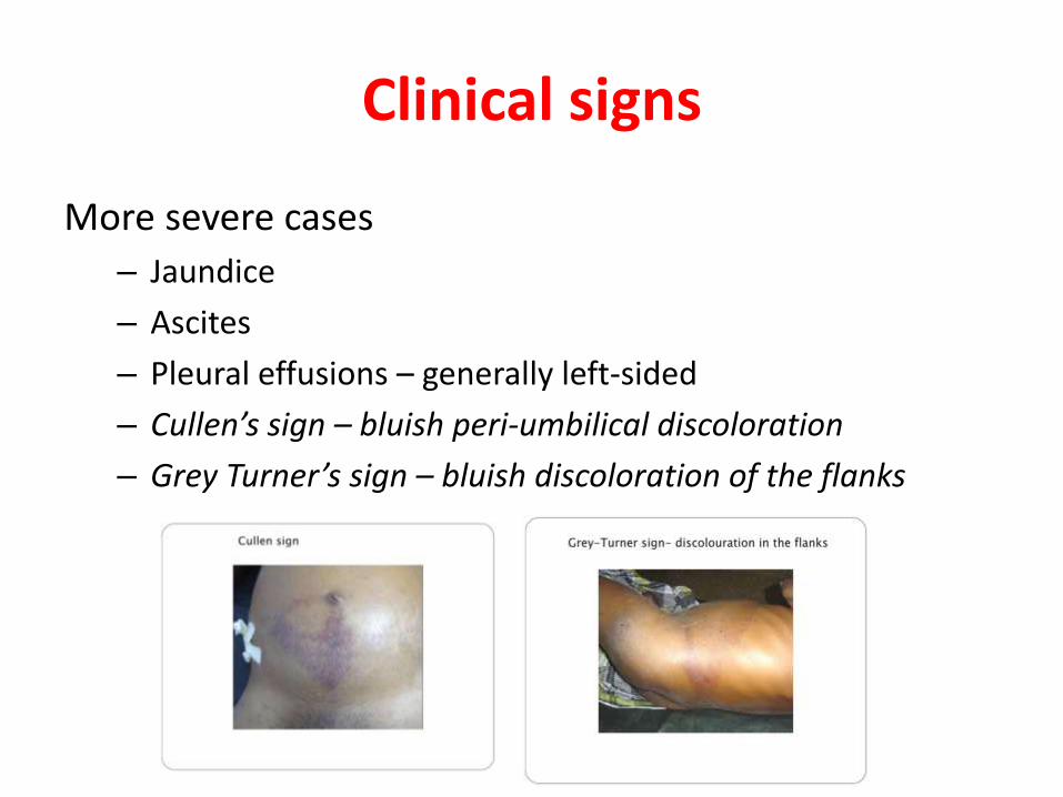
Clinical signs
More severe cases
– Jaundice
– Ascites
– Pleural effusions – generally left-sided
– Cullen’s sign – bluish peri-umbilical discoloration
– Grey Turner’s sign – bluish discoloration of the flanks
Labs
Amylase
• Elevates within HOURS and can remain elevated for 4-5 days
• High specificity when using levels >3x normal
• Most specific = pancreatic isoamylase (fractionated amylase)
• Many false positives
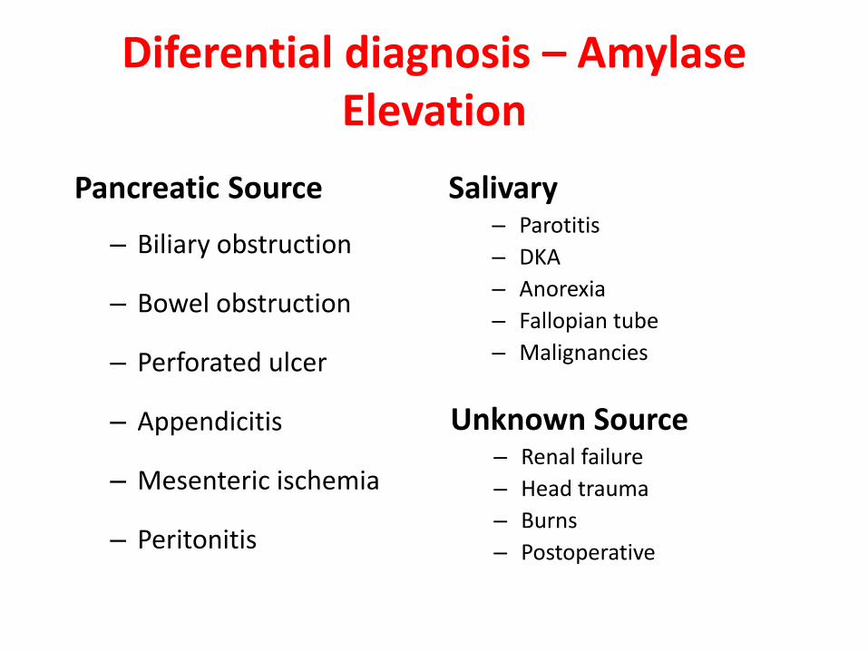
Diferential diagnosis – Amylase Elevation
Salivary– Parotitis
– DKA
– Anorexia
– Fallopian tube
– Malignancies
Unknown Source– Renal failure
– Head trauma
– Burns
– Postoperative
Pancreatic Source
– Biliary obstruction
– Bowel obstruction
– Perforated ulcer
– Appendicitis
– Mesenteric ischemia
– Peritonitis
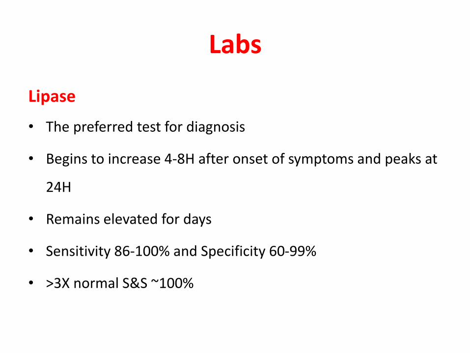
Labs
Lipase
• The preferred test for diagnosis
• Begins to increase 4-8H after onset of symptoms and peaks at
24H
• Remains elevated for days
• Sensitivity 86-100% and Specificity 60-99%
• >3X normal S&S ~100%

Lab Investigations
• Full blood count:neutrophil, leucocytosis
• Electrolyte abnormalities include hypokaemia,hipocalcemia
• Elevated LDH in biliary desease
• Glycosuria (10% of cases)
• Blood sugar: hyperglycaemiain severe cases
• Ultrasound look for stones diseases

Diagnosis
The diagnosis of AP is most often established by the presence of two of the three followingcriteria: (I) abdominal pain; (II) serum amylase and/or lipase greater than three times theupper limit of normal, and/or (III) characteristic findings from abdominal imaging(strong recommendation, moderate quality of evidence)
Contrast-enhanced CT and/or MRI of the pancreas should be reserved for patients in whomthe diagnosis is unclear or who fail to improve clinically within the first 48-72 h after hospitaladmission (strong recommendation, low quality of evidence)

Diagnosis – Imaging
CT– Excellent pancreas imaging
– Recommended in all patients with persisting organ failure, sepsis or deterioration in clinical status (6-10 days after admission)
– Search for necrosis – will be present at least 4 days after onset of symptoms; if ordered too early it will underestimate severity
– Follow-up months after presentation as clinically warranted for CT severity index of >3

CT FindingsSevere Pancreatitis
Tail Indistinct
Intraperitoneal fluid
UnenhancingNecrosis
Peripancreatic edema and inflammation
72 H
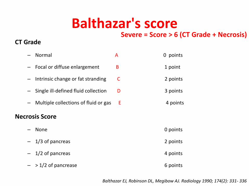
Balthazar's scoreCT Grade
– Normal A 0 points
– Focal or diffuse enlargement B 1 point
– Intrinsic change or fat stranding C 2 points
– Single ill-defined fluid collection D 3 points
– Multiple collections of fluid or gas E 4 points
Necrosis Score
– None 0 points
– 1/3 of pancreas 2 points
– 1/2 of pancreas 4 points
– > 1/2 of pancrease 6 points
Balthazar EJ, Robinson DL, Megibow AJ. Radiology 1990; 174(2): 331- 336
Severe = Score > 6 (CT Grade + Necrosis)

CT Severity Index
necrosis none < 33% 33-50% > 50%
score 0 2 4 6
score morbidity mortality
1-2 4% 0%
7-10 92% 17%
Balthazar et al. Radiology 1990.

Acute pancreatitis
ER presentation
cytokine releaseorgan failure
Predictors of Severity

Predictors of Severity
Why are they needed?
– appropriate patient triage & therapy
– compare results of studies of the impact of therapy
When are they needed?
– optimally, within first 24 hours (damage control must begin early)
Which is best?

Determining severity
• Clinical criteria • Early development/persistence of organ dysfunction
– Ranson criteria – Atlanta criteria – POP score– BISAP
• Clinical assessment• Frequent VS, fluid status/UOP, pulse oximetry
• Radiographic criteria– CT severity index
• Necrosis may not be evident until 48-72h
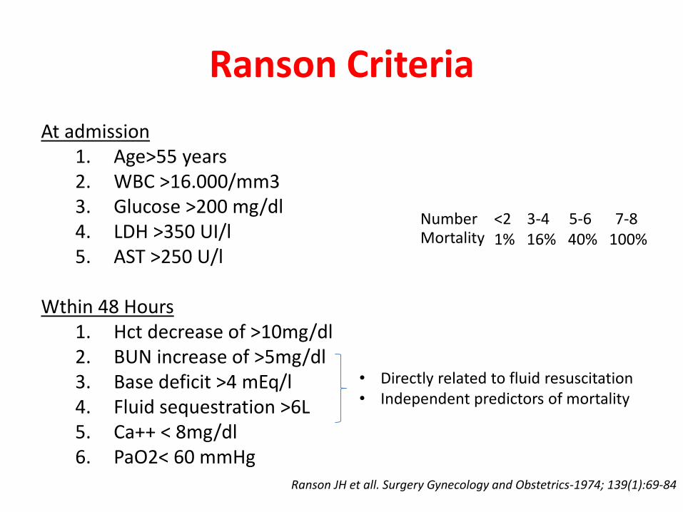
Ranson Criteria
At admission1. Age>55 years2. WBC >16.000/mm33. Glucose >200 mg/dl4. LDH >350 UI/l5. AST >250 U/l
Wthin 48 Hours1. Hct decrease of >10mg/dl2. BUN increase of >5mg/dl3. Base deficit >4 mEq/l4. Fluid sequestration >6L5. Ca++ < 8mg/dl6. PaO2< 60 mmHg
• Directly related to fluid resuscitation• Independent predictors of mortality
Ranson JH et all. Surgery Gynecology and Obstetrics-1974; 139(1):69-84
Number Mortality
<2 3-4 5-6 7-81% 16% 40% 100%

Clinical findings associated with a severe course for initial risk assessment –Intrinsic patient-related risk factors for developing of severe disease

Acute pancreatitis
Treatment
Initial assessment and risk stratification
Risk assessment should be performed to stratify patients in to higher and lower risk categories to assist triage, such as admission to ICU (conditional recommendation, moderate quality of evidence)
Patients with organ failure should be admitted to ICU or intermediary care setting whenever possible (strong recommendation, low quality of evidence)

When Do I Transfer to the Intensive Care?
• Severe pancreatitis
• Multi-organ failure – Pulmonary
– Renal
• Consider it if you are placing the patient on antibiotics and/or ordering a CT to evaluate non-improvement

When Do I Transfer to the Intensive Care?
• Cardiovascular
– Hypotension
– HR, CO and SVR
• Respiratory
– Hypoxemia
– Pleural effusion
• Renal
– ATN
– Oliguria
• Haematologic
– DIC
– Thrombocytosis
• Hepatic
– Encephalopathy
– T bili (3mg/dl)
– AST/ALT 2x nl
• GI
– Stress ulcer
– Acalculous cholecystitis
Therapeutical goals
•Ventilatory support
•Fluid resuscitation
•Haemodynamic support (vasopresors, inotropes)
•Antibiotherapy
•Sedation
•Analgesia
•Early enteral nutrition
•Glycemic and triglycerides control
•Prokinetics
•Stress ulcer prophylaxis
•Thromboprophylaxis

Fluids initial management
Aggressive hydration, defined as 250-500 ml/h of isotonic crystalloid solution should be provided to all patients, unless cardiovascular and/or renal comorbidites exist. Early aggressive iv hydration is most beneficial the first 12-24 h, and may have little benefit beyond (strong recommendation, moderate quality of evidence)
In patients with severe volume depletion (hypotension and tachycardia), more rapid repletion (bolus) may be needed (conditional recommendation, moderate quality of evidence)
Lactated Ringer’s solution may be preferred (conditional recommendation,moderate quality of evidence)
Fluid requirements should be reassessed every 6h for the next 24-48 h. The goal of aggressive hydration should be to decrease the blood urea nitrogen (strong recommendation, moderate quality of evidence)

Fluid Resuscitation
• Fluid resuscitation and “early goal-directed therapy” are cornerstones of critical care management
• Excessive fluid resuscitation is an independent predictor of IAH/ACS and should be avoided
• The use of goal-directed hemodynamic monitoring should be considered to achieve appropriate fluid resuscitation
• Fluid resuscitation volume should be carefully monitored to avoid over-resuscitation in patients at risk for IAH/ACS (Grade 1B)
• Hypertonic crystalloid and colloid-based resuscitation should be considered in patients with IAH to decrease the progression to secondary ACS (Grade 1C)
Balogh Z et al.(2003) Arch Surg 138:637–642; McNelis J et al (2002) Arch Surg 137:133–136; Oda J et al (2006) J Trauma 60:64–71; O’Mara MS et al. (2005) J Trauma 58:1011–1018

Graphic Display Of IAP, APP, UOP

How IAP Should Be Measured?
• Physical examination is insensitive in detecting IAH
• IAP monitoring is a cost-effective, safe, and accurate tool for identifying the presence of IAH and guiding resuscitative therapy for ACS
• Serial IAP measurements are necessary to guide resuscitation of patients with IAH / ACS
Malbrain ML et al. (2005) Incidence and prognosis of intraabdominal hypertension in a mixed population of critically ill patients: a multiple-center epidemiological study. • Crit Care Med 33:315–322 Ivatury RR et al (1998) Intra-abdominal hypertension after life-threatening penetrating abdominal trauma: prophylaxis, incidence, and clinical relevance to gastric mucosal pH and abdominal compartment syndrome. • J Trauma 44:1016–1021. Balogh Z et al. (2003) Both primary and secondary abdominal compartment syndrome can be predicted early and are harbingers of multiple organ failure. J Trauma 54:848–859
• If two or more risk factors for IAH / ACS are present, a baseline IAP measurement should be obtained (Grade 1B)
• If IAH is present, serial IAP measurements should be performed throughout the patient’s critical illness (Grade1C)
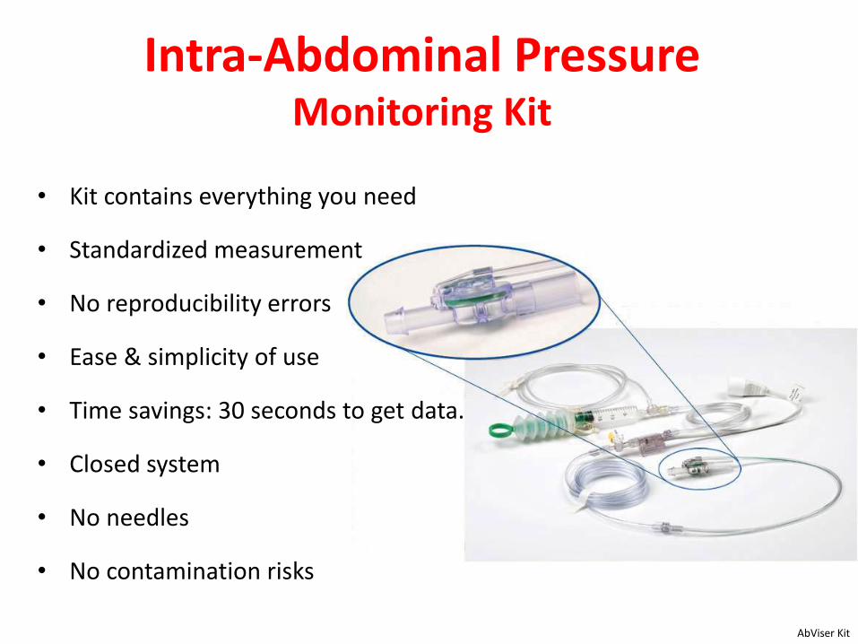
Intra-Abdominal Pressure Monitoring Kit
• Kit contains everything you need
• Standardized measurement
• No reproducibility errors
• Ease & simplicity of use
• Time savings: 30 seconds to get data.
• Closed system
• No needles
• No contamination risks
AbViser Kit

When Do I Start Antibiotics?
• Acute pancreatitis - infection ~10%– 30-40% of those with necrosis get infection
• Prophylactic antibiotics
– Controversial
• No benefit in mild EtOH pancreatitis
• Imipenem or meropenem in necrotizing pancreatitis
• Selective gut decontamination may be beneficial?
• Abx do not appear to promote fungal infection
• General recommendation for use:
– Biliary pancreatitis with signs of cholangitis
– >30% necrosis on CT scan

The role of antibiotics in acute pancreatitis - 1
Routine use of prophylactic antibiotics in patients with severe AP is not recommended(strong recommendation, moderate quality of evidence)
The use of antibiotics in patients with sterile necrosis to prevent the development ofinfected necrosis is not recommended (strong recommendation, moderate quality of evidence)
Infected necrosis should be considered in patients with pancreatic or extrapancreaticnecrosis who deteriorate or fail to improve after 7-10 days of hospitalization. In these patients, either (I) initial CT-guided fine needle aspiration (FNA) for Gram stain and culture to guide use of appropriate antibiotics or (II) empiric use of antibiotics without CT FNA should be given (strong recommendation, low quality of evidence)

The role of antibiotics in acute pancreatitis - 2
In patients with infected necrosis, antibiotics known to penetrate pancreatic necrosis, such as carbapenems, quinolones, and metronidazole, may be useful(conditional recommendation, low quality of evidence)
Antibiotics should be given for an extrapancreatic infection, such as cholangitis,catheter-aquired infections, bacteremia, urinary tract infections, pneumonia(strong recommendation, high quality of evidence)
Routine administration of antifungal agents is not recommended (conditional recommendation, low quality of evidence)

Guidelines for managing pain

Epidural analgesia
• Thoracic trauma(Bulger EM et al. Surgery 2004; 136:426-430)
• Cardiac surgery(Liu SS et al. Anesthesiology 2004, 101:153-161)
• Acute pancreatitis
• The effectiveness and safety of epidural analgesia has also been demonstrated in critically ill patients with severe acute pancreatitis
(Bernhardt A et al. Anaesthesiol Reanim 2002, 27:16-22)

Epidural analgesia
• time to extubation
• ICU stay
• incidence of renal failure
• morphine consumption during the first 24 hours
• maximal glucose and cortisol blood concentrations
• improves forced vital capacity(Guay J. J Anesth 2006, 20:335-340)
• Gold standard - thoracic epidural analgesia (TEA) with a local anaesthetic/opioid infusion

Thoracic epidural analgesia
• sympathetic activity and the stress response(A segmental temporary sympathetic block)
• Improved mucosal capillary perfusionDaudel F, Freise H, Westphal M, et al. Shock 2007; 28: 610–4
Freise H, Lauer S, Anthonsen S, et al. Anesthesiology 2006; 105: 354–9
• Accelerated recovery of intestinal function(Jorgensen H, Wetterslev J, Moiniche S, Dahl JB. Cochrane Database Syst Rev 2000; CD001893)
• The faster resolution of postoperative ileus after major open surgery has been attributed to superior pain therapy, reduced opioid consumption, and sympathetic block

Epidural analgesia – adverse effectsHypotension• 3.0% to 10.2%
• Corelate with hypovolemia(Wheatley RG, Schug SA & Watson D Br J Anaesth 2001;87(1): 47–61)
Treatment failure
• 22% premature termination of postoperative epidural infusions
– dislodgement (10%)
– inadequate analgesia (3.5%)
– sensory or motor deficit (2.2%)(Ballantyne JC, McKenna JM & Ryder E .Acute Pain 2003;4: 89–97)
Neurological injury
Epidural abscess

Nutrition
Mild - moderate pancreatitis
– Calories from IVF (D5W) are sufficient
– No benefit from additional nutritional support
– Oral intake advancing to low fat diet once pain/anorexia resolve
– NGT decompression
• If frequent emesis or evidence of ileus on plain films
• Tube feed if anticipate NPO > 1 week
DO NOT follow amylase and lipase levels

Nutrition
Severe AP
Enteral nutrition is preferred
– Begin nutritional support as early as possible
• NJ tube preferred
– however nasogastric feeds have been shown to be effective in 80% of cases
– NGTs should be used with caution in patients with ACS
TPN only if
– Can’t maintain adequate jejunal access
– Unable to meet caloric demands enterally for > 5 days

TPN + Glutamine in severe acute pancreatitis
double-blind study
Gln reduces the severityof acute-phase response
Gln supports lymphocyteproliferation

Glutamine-TPN in acute pancreatitis
• reduced acute-phase response and better lymphocyte proliferation
De Beaux, Nutrition 1998
• reduced length of TPN (10 vs 16 days, p< 0.05)• reduced length of hospital stay (21 vs 25 days)
Ockenga et al, Clin Nutr 2002

Glutamine-TPN in acute pancreatitis :other RCTs
• less infections and reinterventionsFuentes-Orozco et al, JPEN 2008
• less patients with complicationsSahin et al , Eur J Clin Nutr 2007
• lower incidence of complications, prevention ofpancreatic infections
He et al , Clin Nutr Suppl 2004

Nutrition in acute pancreatitis
In mild AP oral feeding can be started immediately if there is no nausea and vomiting and no abdominal pain (conditional recommendation, moderate quality of evidence)In mild AP initiation of feeding with a low-fat solid diet appears as safe as clear liquid diet(conditional recommendation, moderate quality of evidence)
In severe AP enteral nutrition is recommended to prevent infectious complications .Parenteral nutrition should be avoided unless the enteral route is not available, not toleratedor not meeting caloric requirement (strong recommendation, high quality of evidence)
Nasogastric and nasojejunal delivery of enteral feeding appear comparable (strong recommendation, moderate quality of evidence)

ERCP in acute pancreatitis
Patients with acute pancreatitis and concurrent acute cholangitis should undergo ERCP within 24 h of admission (strong recommendation, moderate quality of evidence).
ERCP is not needed in most patients with gallstone pancreatitis who lack laboratory or clinical evidence of ongoing biliary obstruction (strong recommendation, low quality of evidence).
In the absence of cholangitis and / or jaundice, MRCP or endoscopic ultrasound (EUS) rather than diagnostic ERCP should be used to screen for choledocholithiasis if highly suspected (conditional recommendation, low quality of evidence).
Pancreatic duct stents and / or postprocedure rectal nonsteroidal anti-inflammatory drug (NSAID) suppositories should be utilized to prevent severe post-ERCP pancreatitis in high-risk patients (conditional recommendation, moderate quality of evidence).

The role of surgery in acute pancreatitis - 1
In patients with mild AP, found to have gallstones in the gallbladder, a cholecystectomy should be performed before discharge to prevent a recurrence of AP (strong recommendation, moderate quality of evidence).
In a patient with necrotizing biliary AP, in order to prevent infection, cholecystectomy is to be deferred until active inflammation subsides and fluid collections resolve or stabilize (strong recommendation, moderate quality of evidence).
The presence of asymptomatic pseudocysts and pancreatic and / or extrapancreatic necrosis do not warrant intervention, regardless of size, location, and / or extension (strong recommendation, moderate quality of evidence).
The role of surgery in acute pancreatitis - 2
In stable patients with infected necrosis, surgical, radiologic, and / or endoscopic drainage should be delayed preferably for more than 4 weeks to allow liquefication of the contents and the development of a fibrous wall around the necrosis (walled-off necrosis) (strong recommendation, low quality ofevidence).
In symptomatic patients with infected necrosis, minimally invasive methods of necrosectomy are preferred to open necrosectomy (strong recommendation, low quality of evidence).

Conclusions
• Severe acute pancreatitis should be managed in ICU by a
multidisciplinary team (surgeon, intensive care,
gastroenterology, radiologist, nutritionist etc.)
• Infected necrosis carries a high mortality
• Antibiotics for suspected infected necrosis
• Tube feedings preferred, post ligament of Treiz
• Always look for the myriad of complications
• Guidelines are useful but not enough

studiu de caz
Admision
Day 4
Day 24