Embed Size (px)
Citation preview
Article
Mammalian Diaphanous 1
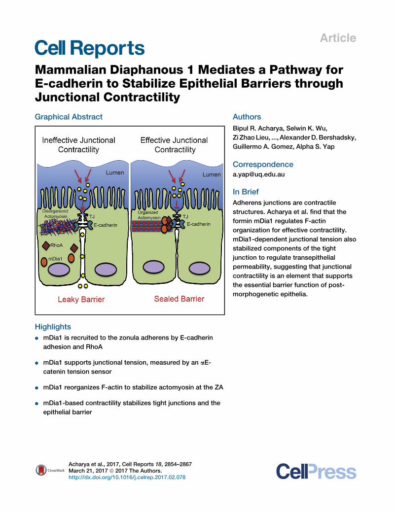
Mediates a Pathway forE-cadherin to Stabilize Epithelial Barriers throughJunctional ContractilityGraphical Abstract
Highlights
d mDia1 is recruited to the zonula adherens by E-cadherin
adhesion and RhoA
d mDia1 supports junctional tension, measured by an aE-
catenin tension sensor
d mDia1 reorganizes F-actin to stabilize actomyosin at the ZA
d mDia1-based contractility stabilizes tight junctions and the
epithelial barrier
Acharya et al., 2017, Cell Reports 18, 2854–2867March 21, 2017 ª 2017 The Authors.http://dx.doi.org/10.1016/j.celrep.2017.02.078
Authors
Bipul R. Acharya, Selwin K. Wu,
Zi ZhaoLieu, ..., AlexanderD.Bershadsky,
Guillermo A. Gomez, Alpha S. Yap
In Brief
Adherens junctions are contractile
structures. Acharya et al. find that the
formin mDia1 regulates F-actin
organization for effective contractility.
mDia1-dependent junctional tension also
stabilized components of the tight
junction to regulate transepithelial
permeability, suggesting that junctional
contractility is an element that supports
the essential barrier function of post-
morphogenetic epithelia.
Cell Reports
Article
Mammalian Diaphanous 1 Mediates a Pathwayfor E-cadherin to Stabilize Epithelial Barriersthrough Junctional ContractilityBipul R. Acharya,1 Selwin K. Wu,1 Zi Zhao Lieu,3 Robert G. Parton,1,2 Stephan W. Grill,4,5 Alexander D. Bershadsky,3
Guillermo A. Gomez,1 and Alpha S. Yap1,6,*1Division of Cell Biology and Molecular Medicine, Institute for Molecular Bioscience, The University of Queensland St. Lucia, Brisbane,
QLD 4072, Australia2Centre for Microscopy and Microanalysis, The University of Queensland St. Lucia, Brisbane, QLD 4072, Australia3Mechanobiology Institute of Singapore, National University of Singapore, Singapore 117411, Singapore4Biotechnology Center, Technical University Dresden, Tatzberg 47/49, 01307 Dresden, Germany5Max Planck Institute of Molecular Cell Biology and Genetics, Pfotenhauerstrasse 108, 01307 Dresden, Germany6Lead Contact*Correspondence: [email protected]
http://dx.doi.org/10.1016/j.celrep.2017.02.078
SUMMARY
Formins are a diverse class of actin regulators that in-fluence filament dynamics and organization. Severalformins have been identified at epithelial adherensjunctions, but their functional impact remains incom-pletely understood. Here, we tested the hypothesisthat formins might affect epithelial interactionsthrough junctional contractility. We focused onmDia1, which was recruited to the zonula adherens(ZA) of established Caco-2 monolayers in responseto E-cadherin and RhoA. mDia1 was necessary forcontractility at the ZA, measured by assays thatinclude a FRET-based sensor that reports molecu-lar-level tension across aE-catenin. This reflected arole in reorganizing F-actin networks to form stablebundles that resistedmyosin-induced stress. Finally,we found that the impact of mDia1 ramified beyondadherens junctions to stabilize tight junctions andmaintain the epithelial permeability barrier. There-fore, control of tissue barrier function constitutesa pathway for cadherin-based contractility tocontribute to the physiology of established epithelia.
INTRODUCTION
Cell-cell junctions couple epithelial individual cells together to
form coherent populations. They are necessary for morphogen-
esis and allow epithelia to form effective tissue barriers. Special-
ized epithelial junctions serve distinct functions: E-cadherin-
based adherens junctions (AJs) promote cell-cell adhesion
(Takeichi, 2014), whereas tight junctions (TJs) seal the intercel-
lular space (Marchiando et al., 2010; Anderson and Van Itallie,
2009). AJs and TJs are also dynamic structures, whosemorphol-
ogies alter, and molecular constituents turn over, in response to
physiological and pathological stimuli (Takeichi, 2014; March-
2854 Cell Reports 18, 2854–2867, March 21, 2017 ª 2017 The AuthoThis is an open access article under the CC BY-NC-ND license (http://
iando et al., 2010; Shen et al., 2008). Recent advances have
identified cellular contractility as an important determinant of
junctional function (Lecuit and Yap, 2015; Priya et al., 2013;
Marchiando et al., 2010). AJs, in particular, are active mechani-
cal structures that display contractile tension (Lecuit and Yap,
2015). This is best understood to function during morphogenesis
(Mason et al., 2016; Martin and Goldstein, 2014), but tension is
also found at AJs in stable monolayers that are not undergoing
morphogenetic movements (Kannan and Tang, 2015; Ratheesh
et al., 2012). What role this post-morphogenetic contractility
plays in epithelial biology, however, is less well understood.
Contractile tension at AJs is generated by an actomyosin cor-
tex that couples to E-cadherin adhesions (Lecuit and Yap, 2015).
This is especially evident in cells that form a zonula adherens
(ZA), a site of high junctional tension in polarized simple epithelia
(Wu et al., 2014), where prominent actomyosin bundles run adja-
cent to a ring of stabilized E-cadherin (Wu et al., 2014; Priya et al.,
2013; Kovacs et al., 2011; Smutny et al., 2010). These actomy-
osin bundles are dynamic structures (Michael et al., 2016; Ko-
vacs et al., 2011; Yamada et al., 2005) sustained by an ensemble
of molecular processes, many of which are recruited to the junc-
tional cortex in response to E-cadherin (Lecuit and Yap, 2015).
These include RhoA signaling, which activates non-muscle
myosin (NMII) (Priya and Yap, 2015; Ratheesh et al., 2012). In
addition, molecules that control the assembly and organization
of junctional F-actin networks are important for effective con-
tractile tension (Jodoin et al., 2015; Kovacs et al., 2011). Not
only is junctional actin assembly necessary to sustain F-actin
content (Verma et al., 2012), but it also incorporates a process
of architectural reorganization that converts disorganized
nascent networks into the bundles seen at the mature ZA
(Michael et al., 2016). We found that architectural reorganization
was necessary to generate effective contractility and identified
Coronin 1B as one element of its cellular apparatus (Michael
et al., 2016), although it is unlikely to be the only one.
Formins are a large class of actin-binding proteins that
are implicated in generating parallel F-actin networks (Goode
and Eck, 2007). Biochemically, formins nucleate and elongate
rs.creativecommons.org/licenses/by-nc-nd/4.0/).
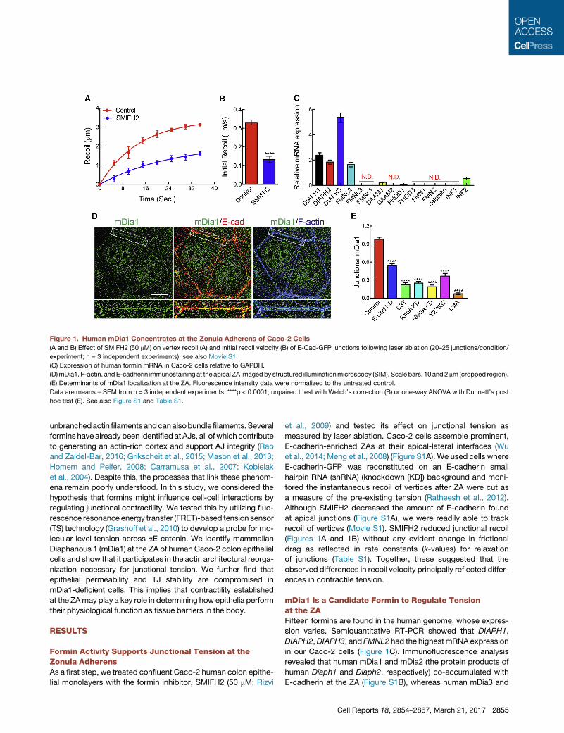
Figure 1. Human mDia1 Concentrates at the Zonula Adherens of Caco-2 Cells
(A and B) Effect of SMIFH2 (50 mM) on vertex recoil (A) and initial recoil velocity (B) of E-Cad-GFP junctions following laser ablation (20–25 junctions/condition/
experiment; n = 3 independent experiments); see also Movie S1.
(C) Expression of human formin mRNA in Caco-2 cells relative to GAPDH.
(D)mDia1, F-actin, and E-cadherin immunostaining at the apical ZA imaged by structured illuminationmicroscopy (SIM). Scale bars, 10 and2 mm (cropped region).
(E) Determinants of mDia1 localization at the ZA. Fluorescence intensity data were normalized to the untreated control.
Data are means ± SEM from n = 3 independent experiments. ****p < 0.0001; unpaired t test with Welch’s correction (B) or one-way ANOVA with Dunnett’s post
hoc test (E). See also Figure S1 and Table S1.
unbranchedactin filamentsandcanalsobundlefilaments.Several
formins have already been identifiedat AJs, all of which contribute
to generating an actin-rich cortex and support AJ integrity (Rao
and Zaidel-Bar, 2016; Grikscheit et al., 2015; Mason et al., 2013;
Homem and Peifer, 2008; Carramusa et al., 2007; Kobielak
et al., 2004). Despite this, the processes that link these phenom-
ena remain poorly understood. In this study, we considered the
hypothesis that formins might influence cell-cell interactions by
regulating junctional contractility. We tested this by utilizing fluo-
rescence resonance energy transfer (FRET)-based tension sensor
(TS) technology (Grashoff et al., 2010) to develop a probe for mo-
lecular-level tension across aE-catenin. We identify mammalian
Diaphanous 1 (mDia1) at the ZA of human Caco-2 colon epithelial
cells and show that it participates in the actin architectural reorga-
nization necessary for junctional tension. We further find that
epithelial permeability and TJ stability are compromised in
mDia1-deficient cells. This implies that contractility established
at the ZAmay play a key role in determining how epithelia perform
their physiological function as tissue barriers in the body.
RESULTS
Formin Activity Supports Junctional Tension at theZonula AdherensAs a first step, we treated confluent Caco-2 human colon epithe-
lial monolayers with the formin inhibitor, SMIFH2 (50 mM; Rizvi
et al., 2009) and tested its effect on junctional tension as
measured by laser ablation. Caco-2 cells assemble prominent,
E-cadherin-enriched ZAs at their apical-lateral interfaces (Wu
et al., 2014; Meng et al., 2008) (Figure S1A). We used cells where
E-cadherin-GFP was reconstituted on an E-cadherin small
hairpin RNA (shRNA) (knockdown [KD]) background and moni-
tored the instantaneous recoil of vertices after ZA were cut as
a measure of the pre-existing tension (Ratheesh et al., 2012).
Although SMIFH2 decreased the amount of E-cadherin found
at apical junctions (Figure S1A), we were readily able to track
recoil of vertices (Movie S1). SMIFH2 reduced junctional recoil
(Figures 1A and 1B) without any evident change in frictional
drag as reflected in rate constants (k-values) for relaxation
of junctions (Table S1). Together, these suggested that the
observed differences in recoil velocity principally reflected differ-
ences in contractile tension.
mDia1 Is a Candidate Formin to Regulate Tensionat the ZAFifteen formins are found in the human genome, whose expres-
sion varies. Semiquantitative RT-PCR showed that DIAPH1,
DIAPH2,DIAPH3, and FMNL2 had the highestmRNA expression
in our Caco-2 cells (Figure 1C). Immunofluorescence analysis
revealed that human mDia1 and mDia2 (the protein products of
human Diaph1 and Diaph2, respectively) co-accumulated with
E-cadherin at the ZA (Figure S1B), whereas human mDia3 and
Cell Reports 18, 2854–2867, March 21, 2017 2855

Figure 2. Construction and Validation of an aE-Catenin Tension Sensor
(A) Schematic of the aE-catenin tension sensor constructs (see Results for details).
(B) Apical junctional localization of aE-catenin-GFP (aCat-WT) and aE-catenin tension sensor (aCat TS) expressed in aE-catenin siRNA cells (see also
Figure S2A).
(C) FRAP of junctional aCat-WT and aCat-TS expressed in aE-Catenin.
(legend continued on next page)
2856 Cell Reports 18, 2854–2867, March 21, 2017

FMNL2 were principally nuclear (Figure S1B). To assess whether
their junctional localization might show interdependence (Peng
et al., 2003), we depleted either mDia1 or mDia2 with RNAi (Fig-
ures S1C and S1D) and tested whether localization of the other
protein was altered (Figure S1E). Small interfering RNA (siRNA)
selectively reduced either total mDia1 (Figure S1C) or mDia2
(Figure S1D) by �95% and substantially reduced their junctional
immunostaining (Figure S1E). Whereas mDia2 was reduced at
junctions in mDia1 KD cells, junctional mDia1 was unaffected
by mDia2 KD (Figure S1E), implying that mDia1 exerts a domi-
nant influence on the junctional localization of mDia2.
We then used structured illumination microscopy (SIM) to bet-
ter define the localization ofmDia1 at apical junctions (Figure 1D).
Polarized Caco-2 cells display actomyosin bundles that lie adja-
cent to E-cadherin in the apical ZA (Kovacs et al., 2011). mDia1
decorated the junctional membrane between the F-actin bun-
dles (Figure 1D), and this staining pattern was substantially
reduced by E-cadherin RNAi (Figure 1E). This suggested that
mDia1 was recruited in response to E-cadherin. The ZA is also
a prominent site for GTP-RhoA (Priya et al., 2015; Ratheesh
et al., 2012), a known activator of mDia1 (Li and Higgs, 2003).
Indeed, junctional mDia1 was reduced when RhoA was inacti-
vatedwith C3-transferase or RNAi (Figure 1E). mDia1 localization
was also compromised when we blocked a recently reported
feedback network that allows NMII to stabilize the junctional
GTP-RhoA zone (Priya et al., 2015), either by NMIIA RNAi or by
blocking ROCK with Y-27632 (Figure 1E), keys to the feedback
network. Overall, these findings suggest that E-cadherin adhe-
sions may define cortical sites for mDia to accumulate through
their ability to establish a stable zone of RhoA signaling at the
ZA (Priya et al., 2013). Junctional mDia1 was also blocked by la-
trunculin A, suggesting that a pre-existing F-actin scaffold might
be needed for its recruitment (Figure 1E). As a junctional formin
that responded to AJ and RhoA signaling, mDia1 then consti-
tuted an interesting candidate to potentially contribute to junc-
tional contractility.
Development of an aE-catenin Tension SensorIn order to measure molecular-level forces at the interface be-
tween E-cadherin adhesions and the actomyosin cytoskeleton,
we employed FRET-based TS technology (Grashoff et al.,
2010) to develop a probe for tension across aE-catenin (Shapiro
andWeis, 2009). We generated a putative force-sensor (aE-Cat-
TS) by inserting the TSmodule between the N-terminal b-catenin
binding domain of aE-catenin and its F-actin binding domain
(Figure 2A). We postulated that here the TS module would be
sensitive to forces exerted from the actomyosin cytoskeleton
onto cadherin-bound aE-catenin (Figure S2A). As a negative
(D and E) Representative images (D) and quantitation (E) of E-cadherin, F-actin, NM
with GFP (left), aE-Cat-WT (center), or aE-Cat-TS (right).
(F) Representative FRET index images of aE-catenin TS constructs.
(G) Effect of manipulating contractility on FRET index for aCat-TS and aE-cateni
(H) Comparison of FRET index results and junctional recoil when contractility was
(I) Contractile sensitivity of a18mAb (left) and vinculin (right) staining (normalized to
TS or aCatD-TS.
Data are means ± SEM (n = 3 independent experiments). For FRAP analysis, 20
*p < 0.05, **p < 0.01,***p < 0.001, and ****p < 0.0001; one-way ANOVA with Dunn
10 mm (B, D, and F) and 2 mm (F, cropped region). See also Figure S2 and Table
control, we inserted the TS module at the very C terminus
of aE-catenin (aE-Cat-TL, ‘‘tension-less’’) where, being down-
stream of the actin-binding domain, it should not experience
contractile force (Figures 2A and S2A).
We then expressed aE-Cat-TS in aE-catenin siRNA cells
where endogenous protein was reduced by >90% (Figure S2B)
and compared its function with that of a GFP-tagged aE-catenin
transgene (aE-Cat-WT) expressed at similar levels (Figure S2B).
Both constructs localized to cell-cell junctions (Figure 2B) and
co-immunoprecipitated E-cadherin (Figure S2C). Further, aE-
Cat-TS showed comparable steady-state dynamics to aE-Cat-
WT when measured by fluorescence recovery after photo-
bleaching (FRAP) (Figure 2C; Table S2). Importantly, whereas
aE-catenin RNAi decreased the amounts of E-cadherin, actomy-
osin, and vinculin found at apical junctions, these were all
restored by aE-Cat-TS as effectively as by aE-Cat-WT (Figures
2D, 2E, and S2D). Overall, these findings established the aE-
Cat-TS fusion protein as a functional aE-catenin.
We then tested whether aE-Cat-TS was sensitive to contrac-
tile forces. First, we compared FRET across junctional aE-Cat-
TS with that across the negative control, aE-Cat-TL (Figures 2F
and 2G), which also localized effectively to the ZA. We used a
FRET index (described in detail in Experimental Procedures)
to correct for variation in transgene expression. aE-Cat-TS dis-
played a lower FRET index than did aE-Cat-TL, implying that
the FRET acceptor/donor pair was further separated in aE-
Cat-TS and consistent with the notion that it might be subject
to tensile force (Figure 2G). This was supported by the observa-
tion that energy exchange across aE-Cat-TS was increased by
Y-27632 or NMIIA siRNA (Figures 2G, S2E, and S2F), maneuvers
that decrease junctional contractility (Leerberg et al., 2014;
Ratheesh et al., 2012). Conversely, Calyculin A, which can stim-
ulate NMII (Cai et al., 2010) and increased junctional recoil in
Caco-2 cells (Figures 2H, S2H, and S2I), decreased aCat-TS
FRET (Figures 2G and S2F), consistent with an increase in force
upon aE-Cat-TS. None of these manipulations affected FRET
from aE-Cat-TL (Figure S2G).
To correlate these responses with an independent measure of
junctional tension, we performed laser ablation experiments in
cells expressing either aE-Cat-TS or aE-Cat-TL (Figures S2H–
S2K) and plotted these results against the FRET index data (Fig-
ure 2H). These showed a close negative relationship between
initial recoil and FRET index for aE-Cat-TS, but not for aE-Cat-
TL (Figure 2H), further supporting the notion that aE-Cat-TS
was reporting changes in contractile force.
Formally, however, force-induced changes in the proximity of
the fluorophores of the TS module could reflect conformational
changes in the overall molecule that condition their proximity
IIA, and junctional Vinculin/aE-catenin in aE-catenin siRNA cells reconstituted
n mutant tension sensor (aCatD-TS).
altered in aE-catenin siRNA cells reconstituted with either aCat-TS or aCat-TL.
aE-catenin) at the ZA in aE-catenin siRNA cells reconstituted with either aCat-
–25 junctions were analyzed per condition per experiment. ns, not significant,
ett’s post hoc test (E and G) and Sidak’s post hoc test (I). Scale bars represent
s S1 and S2.
Cell Reports 18, 2854–2867, March 21, 2017 2857
rather than tensile stretching of the protein linker that separates
them. This alternative possibility is pertinent in the case of aE-
catenin, which increasingly appears to be able to exist in an auto-
inhibited conformation (Choi et al., 2012), where vinculin binding
and the epitope recognized by the a-18mAb aremasked, as well
as in an ‘‘open’’ configuration that can be promoted by contrac-
tile force (Yonemura et al., 2010). Consistent with this notion,
vinculin recruitment to the ZA is force sensitive in Caco-2
cells (Leerberg et al., 2014), as is a-18 mAb staining, which
was decreased by Y-27632 or NMIIA KD and increased by
Calyculin A (Figures S2M and S2N).
To discriminate between these possibilities, we designed a
further deletion construct (aE-CatD-TS, Figure 2A) lacking resi-
dues 510–670 of aE-catenin, which are reported to be necessary
for the vinculin and a-18 mAb binding sites to be masked by the
‘‘closed’’ conformation (Yonemura et al., 2010). This deletion
mutant localized effectively to AJs (Figure 2F). We then immuno-
labeled cells expressing aE-CatD-TS with both anti-vinculin and
a18 mAbs to test the prediction that this mutant exists in the
‘‘open’’ conformation. aE-catenin KD cells reconstituted with
aE-CatD-TS displayed higher baseline levels of junctional vincu-
lin and a18 mAb than did cells expressing aE-Cat-TS (Figure 2I).
Furthermore, labeling for both markers was decreased by
Y-27632 in aE-Cat-TS cells, consistent with these being force
dependent, but not in aE-CatD-TS cells. Overall, these support
the conclusion that the aE-CatD-TS mutant exists in an open
configuration that is independent of force, as previously pro-
posed (Yonemura et al., 2010).
We then compared the FRET response of aE-CatD-TS with
that of aE-Cat-TS (Figure 2G). If the FRET changes in the
reporters were dominated by conformational changes, rather
than stretch of the reporter, we predicted that energy exchange
across aE-Cat-TS would be lower at baseline than that of the
constitutively ‘‘open’’ aE-CatD-TS. Further, when contractility
was inhibited, FRET would increase across aE-Cat-TS but
not across aE-CatD-TS. Instead, we found that both reporters
showed similar degrees of energy transfer at baseline, which
also increased comparably when contractility was inhibited
with Y-27632. Finally, Calyculin A decreased energy exchange
across aE-CatD-TS as it did for aE-Cat-TS (Figures 2G and
S2L). The indistinguishable behaviors of the wild-type (WT) and
constitutively ‘‘open’’ reporters implied that in both contexts
the TS module was principally responding to changes in tension
rather than changes in conformation.
mDia1 Supports Junctional Tension at the ZAWe then applied these assays tomDia1 KD cells (Figure 3). Using
laser ablation, we found that instantaneous recoil of E-cadherin-
tagRFP-T at vertices was substantially reduced in mDia1 KD
cells (Figures 3A and 3B), without significant change in k-values
(Table S1). Recoil was restored by expression of exogenous
RNAi-resistant GFP-mDia1 (Figures 3A, 3B, and S1C), confirm-
ing a specific effect. In contrast, FMNL2 siRNA did not alter junc-
tional recoil (Figures S3A–S3C).
We then used aE-Cat-TS reconstituted into aE-catenin KD
cells to test whether mDia1 affected molecular-level forces at
the cadherin adhesion complex. Energy transfer was increased
significantly across aE-Cat-TS when mDia1 was depleted by
2858 Cell Reports 18, 2854–2867, March 21, 2017
siRNA (Figure 3D), correlating negatively with the change in initial
recoil (Figure 3E), and this was corrected by expression of
mCherry-mDia1 (Figures 3C–3E). Furthermore, the constitutively
‘‘open’’ aE-CatD-TS responded identically (Figures S3D and
S3E), reinforcing the conclusion that mDia1 KD was altering ten-
sion across aE-catenin. In support, a18mAb (Figures 3F and 3G)
and vinculin (Figures 3F and 3H) at the ZA were also reduced
upon mDia1 depletion and restored by expression of GFP-
mDia1. Overall, these findings implicate mDia1 in generating
contractile force at the ZA. They further imply that, while other
formins may localize to junctions, they did not compensate to
support contractility when mDia1 was depleted.
mDia1 KD Disturbs the Stability and Organization ofJunctional NMIITo begin to understand how mDia1 supported junctional
contractility, we first examined junctional NMII. Junctional NMIIA
(Figures 4A and 4B) and, to a lesser extent, NMIIB (Figures 4A
and 4C) were reduced in mDia1 KD cells. Further, GFP-NMIIA
turnover was increased in mDia1 KD cells, as reflected in an
increased mobile fraction by FRAP (Figure 4D; Table S2). This
suggested that mDia1 might stabilize NMII at the junctions.
Then we examined the organization of NMIIA at the ZA by SIM.
In control cells, NMIIA localized in puncta that aligned with the
perijunctional actin bundles (Figure S3F), whereas NMIIA puncta
appeared more dispersed at mDia1 KD junctions. We then ex-
pressedGFP-NMIIA in Caco-2 cells and used its nativeGFP fluo-
rescence to visualize the N termini of the molecules along with
indirect immunofluorescence to identify antibodies directed
against the C termini of NMIIA (Beach et al., 2014; Ebrahim
et al., 2013). As shown previously (Michael et al., 2016), this
revealed alternating patterns (green-red-green) consistent with
the head-tail-head disposition of NMII minifilaments (Figures
4F and S4A). We then quantitated the orientation of NMIIA mini-
filaments by measuring the angle between the axis of a minifila-
ment and the junction (Figure S4B). In control cells, the majority
of NMIIA minifilaments aligned parallel to junctions (Figure 4G)
with a limited dispersion (SD) of minifilament angles (Figure 4H).
In contrast, in mDia1 KD cells, myosin minifilaments were
located further away from the junctions (Figure 4I) and displayed
a much greater dispersion of angles (Figures 4G and 4H). Minifi-
lament length, however, was unaltered (Figure 4J). Overall, these
observations suggest that mDia1 KD did not affect the ability of
cells to form NMIIA minifilaments but profoundly disturbed their
distribution and stability at the junctions.
mDia1 Is Necessary for Dynamic Stability of theJunctional Actin CytoskeletonAs F-actin conditions the recruitment of NMII to the ZA (Verma
et al., 2012), we then asked how the junctional actin cytoskeleton
was affected in mDia1 KD cells. The junctional cortex is distin-
guished by populations of dynamic actin filaments, which are
nucleated at the junctional membrane and then become reor-
ganized and incorporated into the prominent perijunctional bun-
dles that run adjacent to the ZA (Michael et al., 2016; Kovacs
et al., 2011).
We found that mDia1 KD profoundly disturbed the junctional
cytoskeleton at the ZA (Figure 5A). Steady-state F-actin levels

Figure 3. mDia1 Supports Contractile Tension at the Zonula Adherens
(A and B) Effect of mDia1 KD or reconstitution with RNAi-resistant GFP-mDia1 on vertex recoil (A) and initial recoil velocities (B) at the ZA after laser ablation of
E-cadherin-TagRFP-T junctions (25–30 junctions/condition/n; n = 3).
(C and D) Effect of mDia1 KD and its reconstitution on FRET index across aCat-TS at the ZA. Representative images (C) and quantitation (D) are shown.
(E) Effect of mDia1 KD (and reconstitution) on aCat-TS FRET index compared to laser ablation (initial recoil velocity) assays.
(F–H) Representative images (F) and fluorescence quantitation (normalized to aE-catenin) for a18 mAb (G) and vinculin (H) staining at the ZA in mDia1 KD and
reconstituted cells.
Data are means ± SEM from n = 3 independent experiments. ns, not significant, *p < 0.05,***p < 0.001, ****p < 0.0001, one-way ANOVA with Dunnett’s post hoc
test. Scale bars represent 10 mm (C and F) and 2 mm (cropped region on C). See also Figure S3 and Table S1.
were reduced (Figures 5A and 5B), and, more strikingly, SIM im-
aging revealed qualitative changes in actin network organization
(Figure 5A). The prominent F-actin bundles found at the ZA in
control cells were replaced with a more dispersed network that
appeared to extend from the membrane into the cytoplasm.
To quantitate this change in organization, we used a recently
described approach to skeletonize F-actin patterns and
measured the number of overlapping intersects that were found
in image fields of defined size (Michael et al., 2016). Indeed, the
density of intersects was significantly increased by mDia1 KD
and restored by expression of RNAi-resistant GFP-mDia1
(Figure 5C).
mDia1 KD also affected the dynamics of the junctional cyto-
skeleton. Levels of fluorescent G-actin incorporation at junctions
were reduced in mDia1 KD cells (Figure 5D), suggesting that
mDia1 might contribute to nucleation or protect barbed ends
from capping at these sites (Goode and Eck, 2007). In FRAP
studies, mDia1 KD significantly increased the mobile fraction
Cell Reports 18, 2854–2867, March 21, 2017 2859

Figure 4. mDia1 Regulates Myosin II Organization and Stability at the ZA
(A–C) Impact of mDia1 KD or reconstitution on junctional NMII. Representative confocal images (A) and quantitation for NMIIA (B) and NMIIB (C) are shown.
(D) FRAP of transiently expressed GFP-NMIIA.
(E) Effect of Y-27632 on FRAP of GFP-NMIIA in mDia1 KD cells.
(F–J) Effect ofmDia1 KD on junctional NMIIAminifilament organization. Representative SIM images (F), polar histogram plots ofminifilament orientation respect to
the junction (G), dispersion (H), distance from junction (I), and minifilament length (J) are shown.
Data are means ± SEM from n = 3 independent experiments. For FRAP, 20–25 junctions were analyzed/condition/experiment. ns, not significant, *p < 0.05,
**p < 0.01,***p < 0.001, and ****p < 0.0001; unpaired t-Test with Welch’s correction (H–J) or one-way ANOVA with Dunnett’s post hoc test (B and C). Scale bars
represent 10 mm (A), 5 mm (F), and 2 mm (F, cropped region). See also Figure S4 and Table S2.
of GFP-actin at the ZA, accompanied by a decrease in the half-
life (T1/2) of recovery (Figure 5E; Table S2), suggesting that junc-
tional F-actin had become significantly more dynamic. To pursue
2860 Cell Reports 18, 2854–2867, March 21, 2017
this, we then measured fluorescence decay after activation of
actin tagged with photoactivatable GFP (PAGFP-actin). Fluores-
cence decay was accelerated in the mDia1 KD cells compared
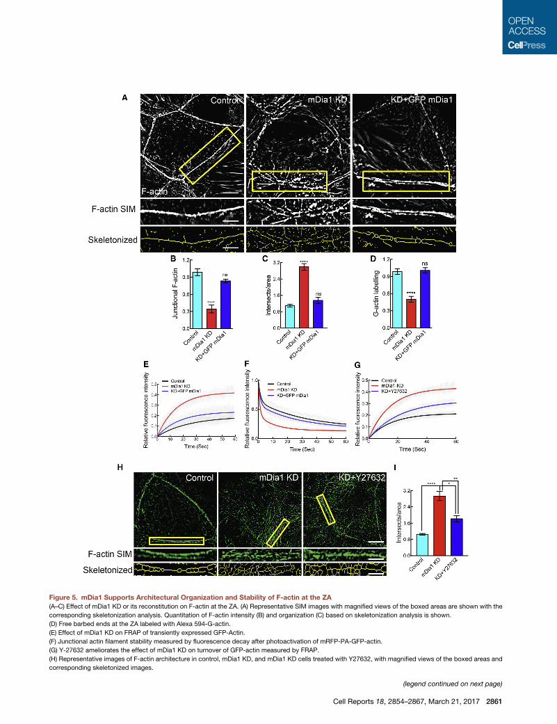
Figure 5. mDia1 Supports Architectural Organization and Stability of F-actin at the ZA
(A–C) Effect of mDia1 KD or its reconstitution on F-actin at the ZA. (A) Representative SIM images with magnified views of the boxed areas are shown with the
corresponding skeletonization analysis. Quantitation of F-actin intensity (B) and organization (C) based on skeletonization analysis is shown.
(D) Free barbed ends at the ZA labeled with Alexa 594-G-actin.
(E) Effect of mDia1 KD on FRAP of transiently expressed GFP-Actin.
(F) Junctional actin filament stability measured by fluorescence decay after photoactivation of mRFP-PA-GFP-actin.
(G) Y-27632 ameliorates the effect of mDia1 KD on turnover of GFP-actin measured by FRAP.
(H) Representative images of F-actin architecture in control, mDia1 KD, and mDia1 KD cells treated with Y27632, with magnified views of the boxed areas and
corresponding skeletonized images.
(legend continued on next page)
Cell Reports 18, 2854–2867, March 21, 2017 2861
with controls (Figure 5F; Table S3), reinforcing the concept that
mDia1 promotes actin filament stability at the ZA. Overall, this
suggested that mDia1 played a necessary role in supporting
dynamic stability and organization of the junctional actin
cytoskeleton.
Junctional Actomyosin Is More Sensitive toStress-Induced Destabilization in mDia1 KD CellsTogether, these observations implied that in mDia1 KD cells
NMIIA minifilaments associate with F-actin networks that are
more disorganized than those found in control cells. Disorgani-
zation can predispose F-actin networks to stress-induced frag-
mentation and severing (Murrell et al., 2015; Reymann et al.,
2012; Haviv et al., 2008). We therefore wondered whether the
destabilization of junctional actomyosin that we found in mDia1
KD cells might reflect the impact of NMII acting on a more disor-
ganized F-actin network. Indeed, we found that the enhanced
turnovers of both GFP-actin (Figure 5G) and GFP-NMIIA (Fig-
ure 4E) seen in mDia1 KD cells were ameliorated by Y-27632.
Furthermore, Y-27632 caused the polydispersed junctional
F-actin networks of mDia1 KD cells to become more compact
and better organized (Figures 5H and 5I). Together, these obser-
vations suggested that architectural reorganization by mDia1
helps the junctional F-actin network resist stress-induced turn-
over and thereby stabilizes actomyosin.
mDia1 Promotes Network Reorganization duringBiogenesis of the Junctional CortexTo pursue this possibility, we then analyzed how the junctional
actin cytoskeleton of mDia1 KD cells reassembled after it had
been disrupted by latrunculin A. We recently found that network
reorganization is a prominent feature of this process, where
initially polydisperse F-actin networks are converted into co-
linear bundles (Michael et al., 2016).
To test this, we used skeletonization analysis to quantitate
F-actin network organization. As previously observed (Michael
et al., 2016; Tang and Brieher, 2012), junctional F-actin networks
recover from residual, latrunculin-resistant puncta that are found
at cell-cell junctions (Figures 6A and S4C). The initial phase
(0–30 min) is insensitive to SMIFH2 (Michael et al., 2016) and
yields a dispersed network with a relatively large number of
F-actin intersects that progressively decrease in number over
60–90 min, as the co-linear bundles form (Figures 6A and 6C).
In contrast, mDia1 KD cells displayed a higher number of inter-
sects at 30 min, and this did not decrease with time (Figures
6A and 6C). This was accompanied by defects in NMIIA minifila-
ment organization (Figure 6B). Control cells initially recruited
NMIIA minifilaments that were relatively dispersed (Figures 6B
and 6D). But as actin reorganization occurred, the dispersion
of NMIIA minifilaments decreased, and these came progres-
sively closer to the junctional membrane (Figures 6B, 6D, and
6E). In mDia1 KD cells, however, although their length was not
(I) Skeletonization analysis of F-actin organization.
Data are means ±SEM (n = 3 independent experiments). For FRAP and photoactiv
*p < 0.05, **p < 0.01, ****p < 0.0001; one-way ANOVAwith Dunnett’s post hoc test (
(cropped, H). See also Tables S2 and S3.
2862 Cell Reports 18, 2854–2867, March 21, 2017
altered (Figure 6F), NMIIAminifilaments retained a dispersed dis-
tribution (Figure 6D), located further away from the junction (Fig-
ure 6E), throughout the assay. Overall, these findings support the
concept that mDia1 contributes to the architectural reorganiza-
tion of both F-actin and NMII that is needed to build an effective
contractile apparatus.
Functional Impact of mDia1 in Established EpithelialMonolayersAs its junctional recruitment depended on E-cadherin, manipu-
lating mDia1 provided an opportunity for us to test how the gen-
eration of contractility at the ZA might affect epithelial integrity in
established Caco-2 monolayers (Figures 7 and S5). Although
AJs formed between mDia1 KD cells (Figure S5A), the amount
of E-cadherin that concentrated to form an apical ZA was signif-
icantly reduced (Figure S5A), without change in the total or sur-
face levels of E-cadherin (Figure S5B). Together, these findings
suggested that mDia1 promotes the concentrated localization
of E-cadherin to form the ZA, consistent with earlier evidence
that implicated contractility in stabilizing E-cadherin to form the
ZA (Wu et al., 2014; Priya et al., 2013; Ratheesh et al., 2012;
Smutny et al., 2010; Homem and Peifer, 2008).
Further, we found that although TJs formed in mDia1 KD cells,
the junctional levels of their constituent proteins, ZO-1, occludin,
and Par3, were all significantly reduced compared with controls
(Figures 7A and 7B). As contractility stabilizes E-cadherin at the
ZA (Priya et al., 2013), we wondered whether the ability of mDia1
to support junctional contractility might also affect the stability of
TJ components. Indeed, FRAP revealed that themobile fractions
of both ZO-1-Emerald (Figures 7C and 7D) and occludin-
Emerald (Figures 7E and 7F) were increased in mDia1 KD cells.
The notion that contractility might stabilize these TJ components
was further supported by finding that Y-27632 alone decreased
the steady-state junctional levels of ZO-1 and occludin (Figures
7B and S5C) and also increased their mobile fractions (Figures
7C–7E).
As the TJ constitutes a key barrier to the transepithelial flux of
solutes and macromolecules (Marchiando et al., 2010), we then
tested howmDia1 KD affected epithelial permeability. Measured
by transepithelial resistance (TER) to ionic flux, barrier integrity
increased progressively during culture to reach a plateau at
�16 days (Figure 7G). In contrast, TER remained significantly
lower in mDia1 KD cells (Figures 7G and 7H). Furthermore, trans-
epithelial flux of 4 kDa dextran was significantly higher in mDia1
KD cells than in controls (Figure 7I). Therefore, although mDia1
KD cells were able to assemble TJs, their ability to seal the para-
cellular pathway was compromised. Furthermore, the epithelial
barrier of Caco-2 monolayers was also significantly reduced
when contractility was inhibited with Y-27632 alone (Figures
7G–7I).
Collectively, these data suggested that basal TJ function in
established monolayers might be conditioned by the ability of
ation, 20–25 junctions were analyzed/condition/experiment. ns, not significant,
B–D) and Sidak’s post hoc test (I). Scale bars represent 5 mm (A andH) and 2 mm

Figure 6. mDia1 Mediates Network Architectural Reorganization during Cytoskeletal Biogenesis at the ZA
Effect of mDia1 KD on F-actin and NMIIA minifilament organization as the junctional actomyosin cytoskeleton reassembles following Latrunculin A wash out is
shown.
(A) Representative SIM and skeletonization images (cropped regions from images shown in Figure S4C).
(B) Representative SIM and polar histogram plots of NMIIA minifilament distribution relative to the junctional membrane.
(C) Skeletonization analysis of F-actin organization.
(D–F) NMIIA minifilament dispersion (D), distance (E), and length (F).
Data are means ± SEM from n = 3 independent experiments. ns, not significant, *p < 0.05, **p < 0.01 and ***p < 0.001; one-way ANOVA with Dunnett’s post hoc
test (C–F). Scale bars represent 2 mm (A and B). See also Figure S4.
mDia1 to support junctional contractility at the TJs, as well as
at the ZA. Consistent with this, mDia1 co-accumulated with
ZO-1 at TJs (Figure S5D), proximate to the actomyosin (Fig-
ure S5E) that is found in this region (Fanning et al., 2012). More-
over, actomyosin at the TJs was disrupted in mDia1 KD cells
(Figure S5E). However, the level of mDia1 observed with
ZO-1 at the TJs was reduced by E-cadherin KD (Figure S5F),
suggesting that it might reflect initial cadherin-dependent
recruitment to the ZA.
Finally, since increased sensitivity to contractile stress contrib-
uted to destabilizing junctional actomyosin in mDia1 KD cells
(Figures 4E and 5G–5I), we wondered whether a similar sensiti-
zation to NMII also affected the TJs. To test this, we asked
whether Y-27632 could ameliorate the TJ defects in mDia1 KD
Cell Reports 18, 2854–2867, March 21, 2017 2863

Figure 7. mDia1 Supports Tight Junction Stability and Barrier Integrity
Effects of mDia1 KD, Y-27632 alone, or in combination on TJs.
(A and B) Representative confocal images (A) and quantitation (B) of ZO-1, occludin, and Par3.
(legend continued on next page)
2864 Cell Reports 18, 2854–2867, March 21, 2017
cells, as it did for actomyosin at the ZA (Figures 4E and 5G–5I).
Indeed, we found that Y-27632 partially rescued the junctional
level of TJ proteins (Figure 7B) and also stabilized ZO-1-Emerald
and occludin-Emerald in mDia1 KD cells (Figures 7C–7F).
Further, Y-27632 restored the TER in mDia1 KD cells to levels
approaching those of controls and higher than seen with either
mDia1 KD or Y-27632 alone (Figures 7G and 7H). Y-27632 also
partially restored dextran permeability in mDia1 KD cells (Fig-
ure 7I). Together, these results suggest that the contribution of
mDia1 to epithelial barrier function reflects its ability to generate
actin networks that are optimized to resist stress and can
thereby effectively generate contractility to stabilize TJs as well
as AJs.
DISCUSSION
Our present study yields three major conclusions. First, mDia1 is
recruited to the junctional cortex in response to E-cadherin
adhesion and RhoA, where it plays a major role in generating
junctional contractility. For these experiments, we comple-
mented a suite of assays for junctional tension with an aE-cate-
nin tension sensor. Although it was possible that conformational
change in the aE-catenin backbone might influence energy ex-
change across the sensor, this seems unlikely, as aE-Cat-TS
behaved identically to a mutant aE-Cat-TS reporter that was de-
signed to be constitutively open. Together, our results support
evidence that Diaphanous regulates junctional myosin in the fly
embryo (Homem and Peifer, 2008) and reinforce the concept
that mechanical force is an important regulator of aE-catenin
(Buckley et al., 2014; Yao et al., 2014; Yonemura et al., 2010)
that can affect processes such as junctional actin dynamics
(Leerberg et al., 2014).
Second, mDia1 facilitates the cellular process of network
architectural reorganization that allows the dynamic cytoskel-
eton to generate actomyosin bundles at the ZA (Michael et al.,
2016). Thus, mDia1-deficient cells failed to assemble perijunc-
tional bundles, displaying instead a disorganized pattern akin
to that seen before nascent networks undergo architectural reor-
ganization (Michael et al., 2016). Indeed, in latrunculin wash-out
experiments, junctional F-actin reassembled but failed to reor-
ganize into bundles when mDia1 was depleted. A key effect of
this architectural regulation is to enhance the resistance of F-net-
works to the stress of NMII binding. Disorganized actin networks
are both inefficient scaffolds for force generation and also more
sensitive to stress-induced severing (Murrell et al., 2015; Rey-
mann et al., 2012; Haviv et al., 2008). Consistent with this, we
found that the distribution of NMIIAminifilaments was also disor-
ganized at the junctional cortex of mDia1 KD cells. Further, the
destabilization of both junctional F-actin and NMIIA seen in
mDia1 KD cells was largely corrected when contractility was
blocked with Y-27632.
(C–F) FRAP of ZO-1-Emerald (C and D) and Occludin-Emerald (E and F). Recove
(G–I) Effect on development of transepithelial electrical resistance (TER) (G), norm
(J) Model of mDia1 as a necessary facilitator of E-cadherin-based contractility
epithelial barriers, respectively. See also Figure S5 and Table S2.
Data are means ± SEM from n = 3 independent experiments. ns, not significant, F
*p < 0.05, **p < 0.01, ***p < 0.001, and ****p < 0.0001; one-way ANOVA with Sid
Altogether, these suggest that many abnormal features of the
mDia1-deficient junctional cytoskeleton reflect howmDia1 regu-
lates actin network architecture to allow it to withstand contrac-
tile stress. Whether mDia1 nucleates non-branched filaments de
novo (Bovellan et al., 2014) or works on filaments that are initi-
ated by another nucleator (Breitsprecher et al., 2012), such as
Arp2/3, remains to be elucidated. We favor the second hypoth-
esis, as Arp2/3 inhibition compromises the formation of contrac-
tile bundles (Verma et al., 2012), and SMIFH2 did not affect the
initial phase of actin reassembly in latrunculin wash-out experi-
ments (Michael et al., 2016; Tang and Brieher, 2012). If so,
then mDia1 might cooperate with a mechanism to debranch
Arp2/3-nucleated networks, such asmight be provided by Coro-
nin 1B (Cai et al., 2008), which also participates in architectural
reorganization (Michael et al., 2016).
Third, mDia1 stabilized proteins of both the AJs and TJs to
control epithelial permeability. This impact on E-cadherin is
consistent with earlier evidence that RhoA-dependent contrac-
tility stabilizes E-cadherin (Smutny et al., 2010; Priya et al.,
2013). As junctional requires cadherin adhesion to couple the
contractile cortices of cells together (Priya and Yap, 2015), stabi-
lization of E-cadherin may also have contributed to the ability of
mDia1 to support tension at the ZA. However, the functional
impact of mDia1-dependent contractility was not confined to
the ZA. Although mDia1 KD cells formed TJs, their protein con-
tent was reduced, and their epithelial barrier to ions and small
macromolecules was compromised. Whereas earlier studies re-
ported that epithelial permeability was increased when cellular
contractility is stimulated (Marchiando et al., 2010; Blair et al.,
2006; Turner et al., 1997), our present results reinforce the
complementary notion that junctional contractility also supports
steady-state TJs (Kannan and Tang, 2015; Nusrat et al., 1995).
Physical connections between TJ plaques and perijunctional
bundles (Madara, 1987) might allow tension to be transmitted
to stabilize dynamic TJ components (Shen et al., 2008), akin to
what is seen for E-cadherin at the ZA (Priya et al., 2013). Indeed,
we found that the turnover of both ZO-1 and occludin was
increased by mDia1 KD and also by Y-27632. How molecular
stability of TJ components may be influenced by contractile
force will be an interesting issue for future research.
We therefore propose that E-cadherin-dependent RhoA
signaling recruits mDia1 to establish effective contractility at
the ZA, which ramifies to affect other components of the apical
junction complex (Figure 7J). Together, these findings provide
new insights into how AJ contractility contributes to epithelial
biology. First, they imply that the junctional contractility evident
even for morphogenetically stable monolayers supports their
function as tissue barriers. Second, our findings provide insight
on the functional interrelationship between cadherin adhesion
and TJs. Earlier studies reported that cadherin adhesion may
support TJ biogenesis in some (Tunggal et al., 2005; Wheelock
ry curves (C and E) and mobile fractions (D and F) are shown.
alized TER (H), and transepithelial dextran flux (I) after 20-day culture.
that stabilizes AJs and TJs to promote cell-cell adhesion and the integrity of
or FRAP analysis, 20-25 junctions were analyzed per condition per experiment;
ak’s post hoc test (B, D, F, H, and I). Scale bars represent 10 mm (A and B).
Cell Reports 18, 2854–2867, March 21, 2017 2865

and Jensen, 1992; Gumbiner et al., 1988), but not all (Capaldo
and Macara, 2007; Ohsugi et al., 1997), circumstances. We
suggest that the impact of E-cadherin on TJs may reflect not
just adhesion but also an impact of the contractile cytoskeleton
that cadherin adhesion establishes. Finally, previous efforts
to understand how contractility can regulate epithelial barrier
function have focused on elucidating the role of myosin regula-
tion (Marchiando et al., 2010). Our data highlight the importance
of mechanisms, such as mDia1, that allow actin networks to
resist stress for effective contractility. More broadly, our find-
ings, taken with their role in the biogenesis of junctional acto-
myosin (Michael et al., 2016; Jodoin et al., 2015; Homem and
Peifer, 2008), suggest that actin regulators can provide comple-
mentary targets to control barrier function both in health and
disease.
EXPERIMENTAL PROCEDURES
Construction of TS Plasmids
The TSmodule withmTFP1-F40-venusA206Kwas adapted from Vinculin TS (a
kind gift from Martin Schwartz [Grashoff et al., 2010]). The TSmod was cloned
in to pEGFP-N1 vector at Xho1 and Not1 restriction sites. For aE-Cat-TS, the
mouse N-terminal (1–697 aa) of aE-catenin was inserted before mTFP1 by in-
fusion cloning (Takara Bioscience, Clontech) at the Xho1 restriction site. The
C-terminal fragment (698–906 aa) of aE-catenin was similarly cloned after
VenusA206K at the Not1 restriction site. For aE-Cat-TL, the full-length aE-cat-
enin (1–906) was inserted at the Xho1 site, and a stop codon is inserted after
VenusA206K in the plasmid. For aE-CatD-TS, the aE-Cat-TS was digested
with NheI and XmaI to cut out a fragment corresponding to 1–670 aa, and a
PCR-amplified fragment corresponding to amino acids 1–509 from mouse
aE-catenin was ligated back at the same restriction enzyme sites. All clones
were confirmed by sequencing.
SUPPLEMENTAL INFORMATION
Supplemental Information includes Supplemental Experimental Procedures,
five figures, four tables, and onemovie and can be found with this article online
at http://dx.doi.org/10.1016/j.celrep.2017.02.078.
AUTHOR CONTRIBUTIONS
The project was conceived by B.R.A., S.K.W., and A.S.Y.; experiments were
performed by B.R.A., S.K.W., and Z.Z.L.; G.A.G. developed FRET, ablation,
and actomyosin minifilament analysis tools and the design of the aE-Cat TS
together with B.R.A.; R.G.P., S.W.G., and A.D.B. contributed expertise; the pa-
per was written by B.R.A., G.A.G., and A.S.Y. and approved by all the authors.
ACKNOWLEDGMENTS
We thank Zev Bryant (Stanford) for many stimulating discussions, all our col-
leagues who generously provided reagents, and our lab colleagues for their
support, advice, and good cheer. This work was supported by the Human
Frontiers Science Program (RGP0023/2014 to S.W.G., Zev Bryant, and
A.S.Y.), National Health and Medical Research Council of Australia (1037320
and 1067405), Cancer Council Queensland (1086857), and the Australian
Research Council (DP120104667, 150101367). A.S.Y. and R.G.P. are
Research Fellows of the NHMRC (1044041, 1058565). G.A.G. is a Research
Fellow of the ARC (FT160100366). S.W.G. acknowledges additional support
from the DFG and the European Research Council (grant no. 281903). Optical
imaging was performed at the ACRF/IMB Cancer Biology Imaging Facility, es-
tablished with the generous support of the Australian Cancer Research Foun-
dation, and the Queensland Brain Institute microscopy facility supported by
ARC LIEF grant (LE130100078).
2866 Cell Reports 18, 2854–2867, March 21, 2017
Received: October 2, 2016
Revised: January 19, 2017
Accepted: February 27, 2017
Published: March 21, 2017
REFERENCES
Anderson, J.M., and Van Itallie, C.M. (2009). Physiology and function of the
tight junction. Cold Spring Harb. Perspect. Biol. 1, a002584.
Beach, J.R., Shao, L., Remmert, K., Li, D., Betzig, E., and Hammer, J.A., 3rd.
(2014). Nonmuscle myosin II isoforms coassemble in living cells. Curr. Biol. 24,
1160–1166.
Blair, S.A., Kane, S.V., Clayburgh, D.R., and Turner, J.R. (2006). Epithelial
myosin light chain kinase expression and activity are upregulated in inflamma-
tory bowel disease. Lab. Invest. 86, 191–201.
Bovellan, M., Romeo, Y., Biro, M., Boden, A., Chugh, P., Yonis, A., Vaghela,
M., Fritzsche, M., Moulding, D., Thorogate, R., et al. (2014). Cellular control
of cortical actin nucleation. Curr. Biol. 24, 1628–1635.
Breitsprecher, D., Jaiswal, R., Bombardier, J.P., Gould, C.J., Gelles, J., and
Goode, B.L. (2012). Rocket launcher mechanism of collaborative actin assem-
bly defined by single-molecule imaging. Science 336, 1164–1168.
Buckley, C.D., Tan, J.L., Anderson, K.L., Hanein, D., Volkmann, N., Weis, W.I.,
Nelson, W.J., and Dunn, A.R. (2014). Cell adhesion. The minimal cadherin-cat-
enin complex binds to actin filaments under force. Science 346. http://dx.doi.
org/10.1126/science.1254211.
Cai, L., Makhov, A.M., Schafer, D.A., and Bear, J.E. (2008). Coronin 1B antag-
onizes cortactin and remodels Arp2/3-containing actin branches in lamellipo-
dia. Cell 134, 828–842.
Cai, Y., Rossier, O., Gauthier, N.C., Biais, N., Fardin, M.A., Zhang, X., Miller,
L.W., Ladoux, B., Cornish, V.W., and Sheetz, M.P. (2010). Cytoskeletal coher-
ence requires myosin-IIA contractility. J. Cell Sci. 123, 413–423.
Capaldo, C.T., and Macara, I.G. (2007). Depletion of E-cadherin disrupts
establishment but not maintenance of cell junctions in Madin-Darby canine
kidney epithelial cells. Mol. Biol. Cell 18, 189–200.
Carramusa, L., Ballestrem, C., Zilberman, Y., and Bershadsky, A.D. (2007).
Mammalian diaphanous-related formin Dia1 controls the organization of
E-cadherin-mediated cell-cell junctions. J. Cell Sci. 120, 3870–3882.
Choi, H.J., Pokutta, S., Cadwell, G.W., Bobkov, A.A., Bankston, L.A., Lidding-
ton, R.C., and Weis, W.I. (2012). aE-catenin is an autoinhibited molecule that
coactivates vinculin. Proc. Natl. Acad. Sci. USA 109, 8576–8581.
Ebrahim, S., Fujita, T., Millis, B.A., Kozin, E., Ma, X., Kawamoto, S., Baird,M.A.,
Davidson, M., Yonemura, S., Hisa, Y., et al. (2013). NMII forms a contractile
transcellular sarcomeric network to regulate apical cell junctions and tissue
geometry. Curr. Biol. 23, 731–736.
Fanning, A.S., Van Itallie, C.M., and Anderson, J.M. (2012). Zonula occludens-
1 and -2 regulate apical cell structure and the zonula adherens cytoskeleton in
polarized epithelia. Mol. Biol. Cell 23, 577–590.
Goode, B.L., and Eck, M.J. (2007). Mechanism and function of formins in the
control of actin assembly. Annu. Rev. Biochem. 76, 593–627.
Grashoff, C., Hoffman, B.D., Brenner, M.D., Zhou, R., Parsons, M., Yang, M.T.,
McLean, M.A., Sligar, S.G., Chen, C.S., Ha, T., and Schwartz, M.A. (2010).
Measuring mechanical tension across vinculin reveals regulation of focal
adhesion dynamics. Nature 466, 263–266.
Grikscheit, K., Frank, T., Wang, Y., and Grosse, R. (2015). Junctional actin as-
sembly is mediated by Formin-like 2 downstream of Rac1. J. Cell Biol. 209,
367–376.
Gumbiner, B., Stevenson, B., and Grimaldi, A. (1988). The role of the cell adhe-
sion molecule uvomorulin in the formation and maintenance of the epithelial
junctional complex. J. Cell Biol. 107, 1575–1587.
Haviv, L., Gillo, D., Backouche, F., and Bernheim-Groswasser, A. (2008). A
cytoskeletal demolition worker: Myosin II acts as an actin depolymerization
agent. J. Mol. Biol. 375, 325–330.
Homem, C.C., and Peifer, M. (2008). Diaphanous regulates myosin and adhe-
rens junctions to control cell contractility and protrusive behavior during
morphogenesis. Development 135, 1005–1018.
Jodoin, J.N., Coravos, J.S., Chanet, S., Vasquez, C.G., Tworoger, M., King-
ston, E.R., Perkins, L.A., Perrimon, N., and Martin, A.C. (2015). Stable Force
Balance between Epithelial Cells Arises from F-Actin Turnover. Dev. Cell 35,
685–697.
Kannan, N., and Tang, V.W. (2015). Synaptopodin couples epithelial contrac-
tility to a-actinin-4-dependent junction maturation. J. Cell Biol. 211, 407–434.
Kobielak, A., Pasolli, H.A., and Fuchs, E. (2004). Mammalian formin-1 partici-
pates in adherens junctions and polymerization of linear actin cables. Nat. Cell
Biol. 6, 21–30.
Kovacs, E.M., Verma, S., Ali, R.G., Ratheesh, A., Hamilton, N.A., Akhmanova,
A., and Yap, A.S. (2011). N-WASP regulates the epithelial junctional actin cyto-
skeleton through a non-canonical post-nucleation pathway. Nat. Cell Biol. 13,
934–943.
Lecuit, T., and Yap, A.S. (2015). E-cadherin junctions as active mechanical in-
tegrators in tissue dynamics. Nat. Cell Biol. 17, 533–539.
Leerberg, J.M., Gomez, G.A., Verma, S., Moussa, E.J., Wu, S.K., Priya, R.,
Hoffman, B.D., Grashoff, C., Schwartz, M.A., and Yap, A.S. (2014). Tension-
sensitive actin assembly supports contractility at the epithelial zonula adhe-
rens. Curr. Biol. 24, 1689–1699.
Li, F., and Higgs, H.N. (2003). Themouse ForminmDia1 is a potent actin nucle-
ation factor regulated by autoinhibition. Curr. Biol. 13, 1335–1340.
Madara, J.L. (1987). Intestinal absorptive cell tight junctions are linked to the
cytoskeleton. Am. J. Physiol. 253, C171–C175.
Marchiando, A.M., Graham, W.V., and Turner, J.R. (2010). Epithelial barriers in
homeostasis and disease. Annu. Rev. Pathol. 5, 119–144.
Martin, A.C., and Goldstein, B. (2014). Apical constriction: Themes and varia-
tions on a cellular mechanism driving morphogenesis. Development 141,
1987–1998.
Mason, F.M., Tworoger, M., and Martin, A.C. (2013). Apical domain polariza-
tion localizes actin-myosin activity to drive ratchet-like apical constriction.
Nat. Cell Biol. 15, 926–936.
Mason, F.M., Xie, S., Vasquez, C.G., Tworoger, M., and Martin, A.C. (2016).
RhoA GTPase inhibition organizes contraction during epithelial morphogen-
esis. J. Cell Biol. 214, 603–617.
Meng, W., Mushika, Y., Ichii, T., and Takeichi, M. (2008). Anchorage of micro-
tubule minus ends to adherens junctions regulates epithelial cell-cell contacts.
Cell 135, 948–959.
Michael, M., Meiring, J.C., Acharya, B.R., Matthews, D.R., Verma, S., Han,
S.P., Hill, M.M., Parton, R.G., Gomez, G.A., and Yap, A.S. (2016). Coronin
1B Reorganizes the Architecture of F-Actin Networks for Contractility at
Steady-State and Apoptotic Adherens Junctions. Dev. Cell 37, 58–71.
Murrell, M., Oakes, P.W., Lenz, M., and Gardel, M.L. (2015). Forcing cells into
shape: The mechanics of actomyosin contractility. Nat. Rev. Mol. Cell Biol. 16,
486–498.
Nusrat, A., Giry, M., Turner, J.R., Colgan, S.P., Parkos, C.A., Carnes, D.,
Lemichez, E., Boquet, P., and Madara, J.L. (1995). Rho protein regulates tight
junctions and perijunctional actin organization in polarized epithelia. Proc.
Natl. Acad. Sci. USA 92, 10629–10633.
Ohsugi, M., Larue, L., Schwarz, H., and Kemler, R. (1997). Cell-junctional and
cytoskeletal organization in mouse blastocysts lacking E-cadherin. Dev. Biol.
185, 261–271.
Peng, J., Wallar, B.J., Flanders, A., Swiatek, P.J., and Alberts, A.S. (2003).
Disruption of the Diaphanous-related formin Drf1 gene encoding mDia1 re-
veals a role for Drf3 as an effector for Cdc42. Curr. Biol. 13, 534–545.
Priya, R., and Yap, A.S. (2015). Active tension: The role of cadherin adhesion
and signaling in generating junctional contractility. Curr. Top. Dev. Biol. 112,
65–102.
Priya, R., Yap, A.S., and Gomez, G.A. (2013). E-cadherin supports steady-
state Rho signaling at the epithelial zonula adherens. Differentiation 86,
133–140.
Priya, R., Gomez, G.A., Budnar, S., Verma, S., Cox, H.L., Hamilton, N.A., and
Yap, A.S. (2015). Feedback regulation throughmyosin II confers robustness on
RhoA signalling at E-cadherin junctions. Nat. Cell Biol. 17, 1282–1293.
Rao, M.V., and Zaidel-Bar, R. (2016). Formin-mediated actin polymerization at
cell-cell junctions stabilizes E-cadherin and maintains monolayer integrity dur-
ing wound repair. Mol. Biol. Cell 27, 2844–2856.
Ratheesh, A., Gomez, G.A., Priya, R., Verma, S., Kovacs, E.M., Jiang, K.,
Brown, N.H., Akhmanova, A., Stehbens, S.J., and Yap, A.S. (2012). Central-
spindlin and a-catenin regulate Rho signalling at the epithelial zonula adhe-
rens. Nat. Cell Biol. 14, 818–828.
Reymann, A.C., Boujemaa-Paterski, R., Martiel, J.L., Guerin, C., Cao, W.,
Chin, H.F., De LaCruz, E.M., Thery, M., andBlanchoin, L. (2012). Actin network
architecture can determine myosin motor activity. Science 336, 1310–1314.
Rizvi, S.A., Neidt, E.M., Cui, J., Feiger, Z., Skau, C.T., Gardel, M.L., Kozmin,
S.A., and Kovar, D.R. (2009). Identification and characterization of a small
molecule inhibitor of formin-mediated actin assembly. Chem. Biol. 16, 1158–
1168.
Shapiro, L., andWeis,W.I. (2009). Structure and biochemistry of cadherins and
catenins. Cold Spring Harb. Perspect. Biol. 1, a003053.
Shen, L., Weber, C.R., and Turner, J.R. (2008). The tight junction protein com-
plex undergoes rapid and continuous molecular remodeling at steady state.
J. Cell Biol. 181, 683–695.
Smutny, M., Cox, H.L., Leerberg, J.M., Kovacs, E.M., Conti, M.A., Ferguson,
C., Hamilton, N.A., Parton, R.G., Adelstein, R.S., and Yap, A.S. (2010). Myosin
II isoforms identify distinct functional modules that support integrity of the
epithelial zonula adherens. Nat. Cell Biol. 12, 696–702.
Takeichi, M. (2014). Dynamic contacts: Rearranging adherens junctions to
drive epithelial remodelling. Nat. Rev. Mol. Cell Biol. 15, 397–410.
Tang, V.W., and Brieher, W.M. (2012). a-Actinin-4/FSGS1 is required for
Arp2/3-dependent actin assembly at the adherens junction. J. Cell Biol. 196,
115–130.
Tunggal, J.A., Helfrich, I., Schmitz, A., Schwarz, H., G€unzel, D., Fromm, M.,
Kemler, R., Krieg, T., and Niessen, C.M. (2005). E-cadherin is essential for
in vivo epidermal barrier function by regulating tight junctions. EMBO J. 24,
1146–1156.
Turner, J.R., Rill, B.K., Carlson, S.L., Carnes, D., Kerner, R., Mrsny, R.J., and
Madara, J.L. (1997). Physiological regulation of epithelial tight junctions is
associated with myosin light-chain phosphorylation. Am. J. Physiol. 273,
C1378–C1385.
Verma, S., Han, S.P., Michael, M., Gomez, G.A., Yang, Z., Teasdale, R.D.,
Ratheesh, A., Kovacs, E.M., Ali, R.G., and Yap, A.S. (2012). A WAVE2-Arp2/
3 actin nucleator apparatus supports junctional tension at the epithelial zonula
adherens. Mol. Biol. Cell 23, 4601–4610.
Wheelock, M.J., and Jensen, P.J. (1992). Regulation of keratinocyte intercel-
lular junction organization and epidermal morphogenesis by E-cadherin.
J. Cell Biol. 117, 415–425.
Wu, S.K., Gomez, G.A., Michael, M., Verma, S., Cox, H.L., Lefevre, J.G.,
Parton, R.G., Hamilton, N.A., Neufeld, Z., and Yap, A.S. (2014). Cortical F-actin
stabilization generates apical-lateral patterns of junctional contractility that
integrate cells into epithelia. Nat. Cell Biol. 16, 167–178.
Yamada, S., Pokutta, S., Drees, F., Weis, W.I., and Nelson, W.J. (2005).
Deconstructing the cadherin-catenin-actin complex. Cell 123, 889–901.
Yao, M., Qiu, W., Liu, R., Efremov, A.K., Cong, P., Seddiki, R., Payre, M., Lim,
C.T., Ladoux, B., Mege, R.M., and Yan, J. (2014). Force-dependent conforma-
tional switch of a-catenin controls vinculin binding. Nat. Commun. 5, 4525.
Yonemura, S., Wada, Y., Watanabe, T., Nagafuchi, A., and Shibata, M. (2010).
alpha-Catenin as a tension transducer that induces adherens junction devel-
opment. Nat. Cell Biol. 12, 533–542.
Cell Reports 18, 2854–2867, March 21, 2017 2867


![CD146 mediates an E-cadherin-to-N-cadherin switch during TGF-β … · 2018. 7. 10. · expression [16–19]. N-cadherin is reported to be upregulated by TGF-β signaling [20,21]](https://img.dokumen.tips/doc/110x75/6126bb2c5b910b6f974c32bd/cd146-mediates-an-e-cadherin-to-n-cadherin-switch-during-tgf-2018-7-10-expression.jpg)