Embed Size (px)
Citation preview

Pathology International 1994; 44: 865-873
Case Report Malignant peripheral nerve sheath tumor with prominent intracytoplasmic vacuolation: Report of a case
Yasushi Horie,’ Hajime Fujita,2 Kohichi Mi~obuchi ,~ Yoshihiko H ~ s h i d a , ~ lchiro M ~ r a k a m i , ~ Kohji TaguchP and Tadaatsu Akagi’ ’Department of Clinical Laboratory Medicine, Tottori University, Faculty of Medicine, Yonago, 2Department of Pathology, Kagawa Prefectural Central Hospital, Takamatsu, 3Department of Anatomical Pathology, Kaga wa Rosai Hospital, Marugame, ‘Department of Pathology, Kobe West City Hospital, Kobe, SDepartment of Pathology, lwakuni National Hospital, Iwakuni, 6Department of Pathology, Okayama University Hospital, Okayarna and ’Second Department of Pathology, Okayama University Medical School, Okayama, Japan
A case of malignant peripheral nerve sheath tumor with uncommon features is reported. A mass was noted in the left thigh of a 16 year old man. Histologically, most areas of the tumor exhibited the typical appearance of malignant periph- eral nerve sheath tumor, but some tumor cells had rounded nuclei and cytoplasm, resembling an epithelioid pattern. It was noted that some rounded tumor cells showed prominent intracytoplasmic vacuolation. lmmunohistochemically, almost all of the tumor cells, including the rounded and vacuolated ones, were positive for S-100 protein and vimentin. Electron microscopic study revealed well-developed cytoplasmic processes, intracytoplasmic intermediate-sized filaments, basement lamina formation and extracellular long-spacing collagens. These findings were compatible with those of Schwann cell differentiation. Moreover, ultrastructurally, the vacuolated spaces contained a few granular materials and were derived from the dilatation of the rough endoplasmic reticulum. It is speculated that intracytoplasmic vacuolation in malignant peripheral nerve sheath tumor would be caused by degeneration of the tumor cells.
Key words: electron microscopy, endoplasmic reticulum, imrnunohistochernistry, malignant peripheral nerve sheath tumor, malignant schwannoma, S-100 protein, vacuolation
Malignant peripheral nerve sheath tumor has also been referred to as malignant schwannoma, neurogenic sarcoma or neurofibro~arcorna.~-~ Most of these tumors arise from nerves involved by neurofibromas as part of von Reckling- hausen’s disease, but some of these tumors may arise de
~~~~~~ ~
Correspondence: Yasushi Horie, MD, Department of Clinical Labora- tory Medicine, Tottori University, Faculty of Medicine, 36-1 Nishi- rnachi, Yonago 683, Japan.
Received 19 April 1994. Accepted for publication 26 July 1994.
novo. The epithelioid subtype is a rare histologic variant comprising approximately 5-1 7% of malignant peripheral nerve sheath DiCarlo et al. described three forms of epithelial or epithelial-like tissues that may be seen in the epithelioid subtype:’ (i) glands are seen within the so-called glandular malignant peripheral nerve sheath tumor; (ii) rosettes or primitive neuroepithelia may be seen within a malignant peripheral nerve sheath tumor; and (iii) the cells in a malignant peripheral nerve sheath tumor can assume either a rounded or polygonal shape and grow in nests or cords, resembling a carcinoma or malignant melanoma. When these areas predominate within a malignant peripheral nerve sheath tumor, the tumors are designated the epithelioid ~ubtype.‘ ,~
lntracytoplasmic vacuoles may appear not only in the epithelial tumors, but also in the non-epithelial tumors. These changes have been documented in some cases of liposar- coma,9 angiosarcoma,’o smooth muscle tumor,” and malig- nant l ymph~ma. ’~ - ’~ To our knowledge, only limited attention has been directed to the intracytoplasmic vacuoles in malig- nant peripheral nerve sheath tumors.
In this report, we present an uncommon case of the malignant peripheral nerve sheath tumor with prominent intracytoplasmic vacuolation and discuss its morphogenesis.
CLINICAL SUMMARY
A 16 year old man noticed a lesion in his left thigh for 3 years before visiting the Department of Orthopedic Surgery, Kagawa Prefectural Central Hospital. Computerized tomography and angiography revealed a huge soft tissue tumor in the proxi- mal thigh with the extension to the lower buttock. Laboratory
866 Y. Horie eta/.
data were non-contributory to the diagnosis. Neither the patient nor his family had any history of von Reckling- hausen’s disease. There was no pre-operative chemotherapy or irradiation. After incisional biopsy, the tumor was widely resected in February 1986. In April 1990, a local recurrence was noted in the left thigh. Then, multiple pulmonary metas- tases developed in April 1993.
PATHOLOGICAL FINDINGS
Histological findings
The tumor was 14 x 10 x 5 cm in size. Macroscopically, the tumor was located from the deep subcutaneous tissue to the muscle, and there was no connection between the tumor and branches of the peripheral nerves. The cut surface showed a multinodular solid pattern with a swollen appear- ance, and was white-tan to yellow with several reddish hem- orrhagic foci (Fig. 1). There were no cystic changes. The mass was relatively well demarcated in most areas, but the border was partially unclear, suggesting an infiltrative nature.
Microscopically, spindled tumor cells were arranged in fasci- cles in most areas (Fig. 2). The spindled cytoplasm was eosinophilic on hematoxylin-eosin (HE) stain. The elongated nuclei of the tumor cells were hyperchromatic and pleo- morphism was noted (Fig. 3). The cellularity of the tumor was increased in most areas, and mitotic figures were easily identified, counting about 3 per 10 per high-power fields. In the peripheral part, the tumor cells had directly invaded the muscular tissue. In some areas, the cytoplasm of the tumor cells became richer and rounded, resembling an epithelioid pattern (Fig. 4). It was noted that some rounded tumor cells showed prominent vacuolated changes in the cytoplasm (Fig. 5). The large-sized single vacuoles were predominant, but small-sized multiple vacuoles were also seen. Transitional features were found among these different areas. There were no glandular or heterogeneous components in the tumor. There were no mucinous materials in the cytoplasm of the vacuolated tumor cells on periodic acid-Schiff reaction or alcian blue stain. Oil-red 0 and Sudan 111 stain revealed that the vacuolated spaces contained no lipid materials. In the stroma, there were relatively abundant reticulin and collagen fibers among the tumor cells on reticulin staining.
Figure 1 Macroscopic appearance of the tumor from the left thigh. Multinodular and hemorrhagic changes are seen on the cut surface.
Figure 2 Low-powerview of the tumor. Spindle-shaped tumor cells proliferate in a fascicular pattern. Note the increased cellularity of the tumor cells.

Malignant peripheral nerve sheath tumor 867
Figure 3 High-power view of the spindle-shaped tumor cells. The elongated nuclei of the tumor cells show hyperchromatic changes, suggesting malignancy. A mitotic figure is seen (arrow) .
Figure 4 In this area, the tumor cells have a rounded nuclei and richer eosinophilic cytoplasm, resembling an epithelioid pattern. There are small-sized vacuoles in the cytoplasm of some tumor cells.
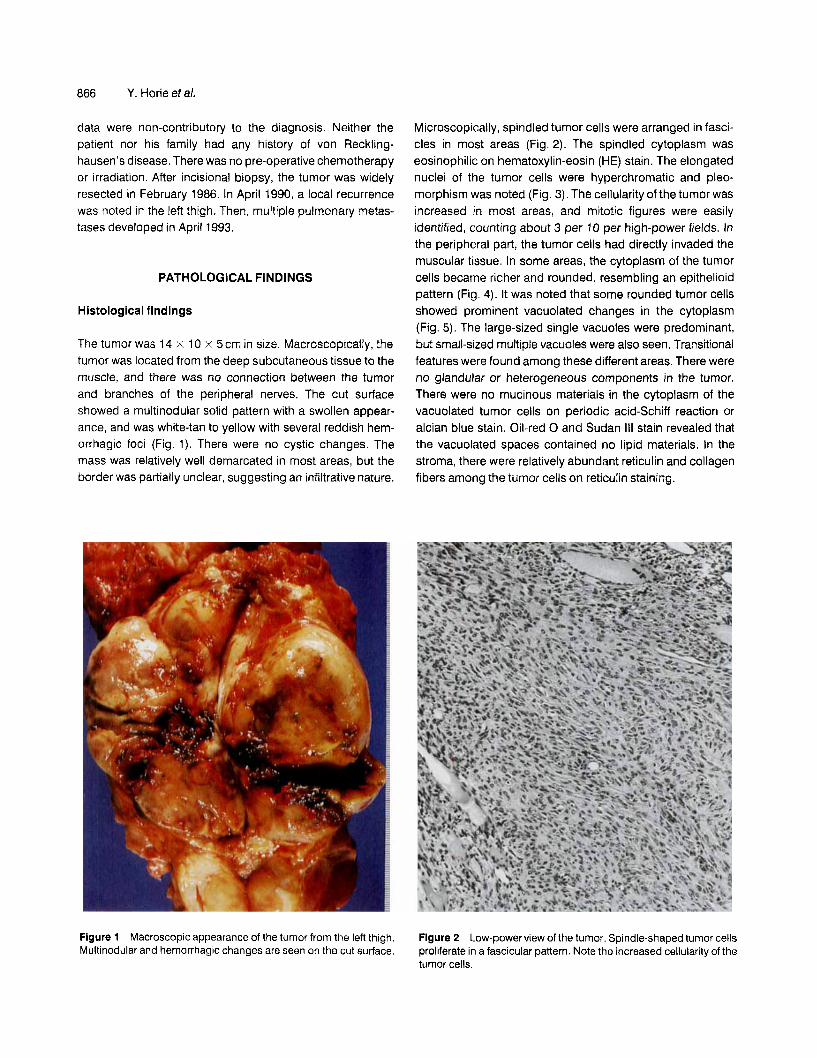
lmmunohistochemical findings
The 10% formalin-fixed, paraffin-embedded materials were conventionally processed and examined by peroxidase anti- peroxidase or avidin-biotin complex methods. The nuclei and cytoplasm of the tumor cells, including not only the spindled type but also the rounded and vacuolated ones, were immu- noreactive for S-100 protein (polyclonal, diluted 1 :loo, Dako Japan Co. Ltd, Kyoto, Japan; Fig. 6a,b) and vimentin (mono- clonal, prediluted, Seikagaku Co., Tokyo, Japan). Tumor cells were negative for the following antibodies: carcino- embryonic antigen (polyclonal, diluted 1 : 50, Dako Japan); epithelial membrane antigen (monoclonal, diluted 1 : 20, Dako Japan); cytokeratin (monoclonal, prediluted, Dako Japan); factor VIII-related antigen (monoclonal, diluted 1 : 50, Dako Japan); myoglobin (monoclonal, diluted 1 : 50, Dako Japan); desmin (monoclonal, prediluted, Seikagaku Co.); or alpha- smooth muscle actin (monoclonal, diluted 1 :50, Dako Japan).
e Figure 5 Large intracytoplasmic vacuoles are seen in the rounded tumor cells.
868 Y. Horie et a/.
Figure 6 The nuclei and cytoplasm of the tumor cells are immunoreactive for S-100 protein. (a) Spindle-shaped tumor cells. (b) Rounded and vacuolated tumor cells (Immunostain).
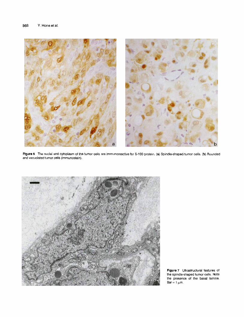
Figure 7 Ultrastructural features of the spindle-shaped tumor cells. Note the presence of the basal lamina. Bar = 1 Wm.
Malignant peripheral nerve sheath tumor 869
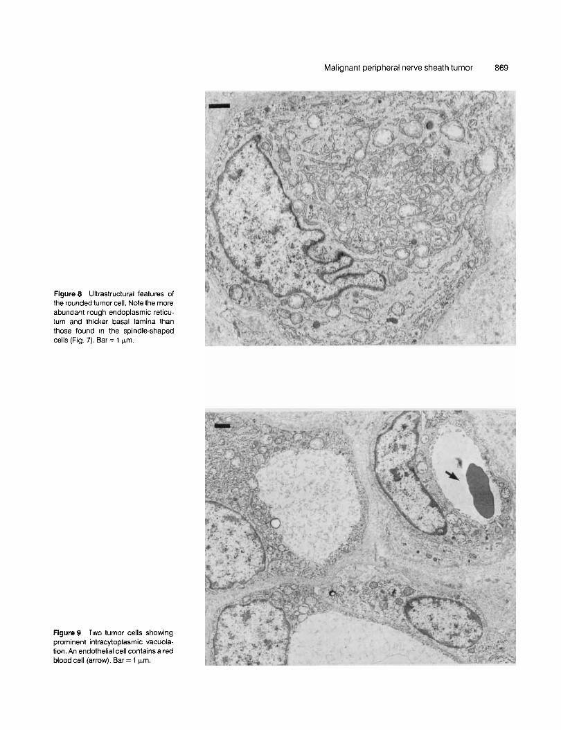
Figure 8 Ultrastructural features of the rounded tumor cell. Note the more abundant rough endoplasmic reticu- lum and thicker basal lamina than those found in the spindle-shaped cells (Fig. 7). Bar = 1 p,m.
Figure 9 Two tumor cells showing prominent intracytoplasmic vacuola- tion. An endothelial cell contains a red blood cell (arrow). Bar = 1 pm.
870 Y. Horie et a/.
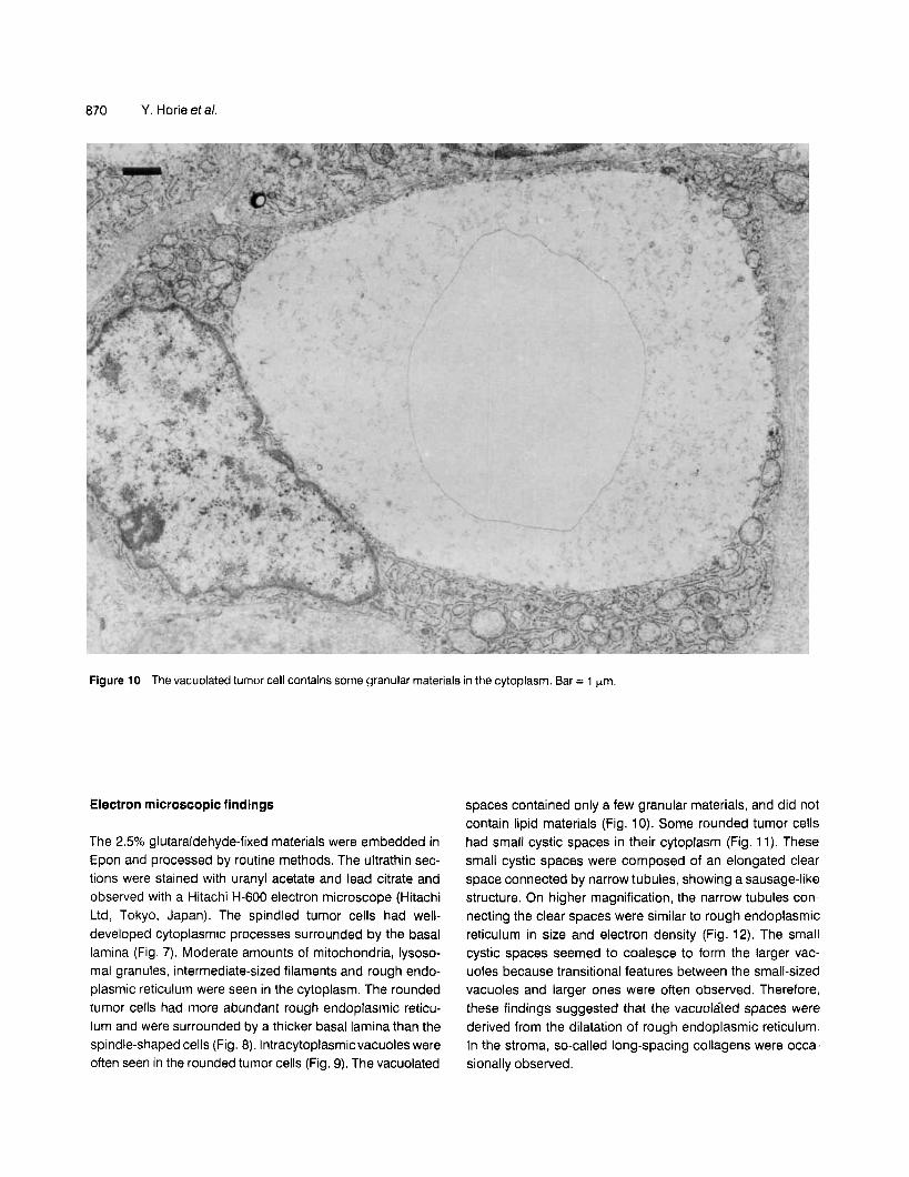
Figure 10 Thevacuolated tumor cell contains some granular materials in the cytoplasm. Bar = 1 pm.
Electron microscopic findings
The 2.5% glutaraldehyde-fixed materials were embedded in Epon and processed by routine methods. The ultrathin sec- tions were stained with uranyl acetate and lead citrate and observed with a Hitachi H-600 electron microscope (Hitachi Ltd, Tokyo, Japan). The spindled tumor cells had well- developed cytoplasmic processes surrounded by the basal lamina (Fig. 7) . Moderate amounts of mitochondria, lysoso- ma1 granules, intermediate-sized filaments and rough endo- plasmic reticulum were seen in the cytoplasm. The rounded tumor cells had more abundant rough endoplasmic reticu- lum and were surrounded by a thicker basal lamina than the spindle-shaped cells (Fig. 8). lntracytoplasmicvacuoles were often seen in the rounded tumor cells (Fig. 9). The vacuolated
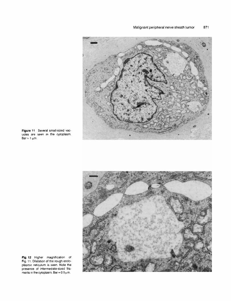
spaces contained only a few granular materials, and did not contain lipid materials (Fig. 10). Some rounded tumor cells had small cystic spaces in their cytoplasm (Fig. 11). These small cystic spaces were composed of an elongated clear space connected by narrow tubules, showing a sausage-like structure. On higher magnification, the narrow tubules con- necting the clear spaces were similar to rough endoplasmic reticulum in size and electron density (Fig. 12). The small cystic spaces seemed to coalesce to form the larger vac- uoles because transitional features between the small-sized vacuoles and larger ones were often observed. Therefore, these findings suggested that the vacuolated spaces were derived from the dilatation of rough endoplasmic reticulum. In the stroma, so-called long-spacing collagens were occa- sionally observed.
Malignant peripheral nerve sheath tumor 871
Figure 11 Several small-sized vac- uoles are seen in the cytoplasm. Bar = 1 Fm.
Fig. 12 Higher magnification of Fig. 11. Dilatation of the rough endo- plasmic reticulum is seen. Note the presence of intermediate-sized fila- ments in the cytoplasm. Bar = 0.5 Fm.
872 Y. Horie eta/.
DISCUSSION
In the present case, most areas of the tumor showed the conventional features of malignant peripheral nerve sheath tumors. However, because uncommon features including the rounded tumor cells and prominent intracytoplasmic vacuolation were also observed, it was necessary to dif- ferentiate this tumor from several other types, including liposarcoma, angiosarcoma, smooth muscle tumor, chon- drosarcoma, synovial sarcoma and malignant mesenchy- moma. In particular, we thought that the vacuolated tumor cells simulated signet-ring cells, and that they had morpho- logical similarities to those of liposarcoma on HE stain alone. However, detailed histologic examinations revealed that the tumor cells showed only Schwann cell differentiation. Although some tumor cells simulated an epithelioid pattern, the present case did not belong to the epithelioid subtype due to the following reasons. First, although the aggregates of the rounded tumor cells resembled an epithelioid pattern, the tumor cells did not form a typical cord or nest-like structure as seen in carcinoma or malignant melanoma. Second, these epithelioid-like areas did not occupy the pre- dominant area of the tumor, so it did not fulfill the criterion of the epithelioid subtype of malignant peripheral nerve sheath t ~ m 0 r . l . ~
The immunohistochemical and ultrastructural studies are useful in the differential diagnosis of malignant peripheral nerve sheath tumor. lmmunohistochemically, the tumor cells are positive for S-1 00 protein in most cases, although positiv- ity for S-100 protein is reported in approximately 50-70% of malignant peripheral nerve sheath tumors.16-18 The electron microscopical findings suggestive of malignant peripheral nerve sheath tumor are as follows: (i) prominent cytoplasmic processes; (ii) basal lamina formation; (iii) intracytoplasmic intermediate-sized filaments; (iv) extracellular long-spacing collagens; and (v) intercellular primitive In the present case, both the immunohistochemical and ultrastruc- tural findings were compatible with those of malignant peripheral nerve sheath tumor.
The intracytoplasmic vacuolation may appear not only in mucus-producing epithelial tumors, but also in non-epithelial tumors. These changes have been reported in some cases of liposar~oma,~ angiosarcoma,lo smooth muscle tumor,” and malignant lymph~ma,~*- ’~ although the mechanisms forming the intracytoplasmic vacuoles differ respectively. In liposarcoma, the vacuoles are thought to be derived from the accurnulation of fat.g The lipoblastic tumor cells contain lipid droplets of various shapes and sizes in the cytoplasm, and the lipid droplets coalesce during development. In angiosar- coma, a number of membrane-bound vacuoles may develop in the cytoplasm of autolytic endothelial cells, although the
origin of the vacuoles has not been clearly elucidated.1° In smooth muscle tumor, most investigators have considered the vacuoles to be an artifact due to formalin fixation.’ However, Hyde et a/. reported that numerous vacuolated mitochondria produced a cytoplasmic clearing on HE stain in a case of clear-cell variant of uterine epithelioid leio- myoma.” In malignant lymphoma, a vacuolated subtype is designated signet-ring cell lymphoma, in which three variants have been First, in the Russell body type, the vacuolated appearance is caused by the accumulation of immunoglobulin-containing cisternae of rough endoplasmic reti~ulum.’~ In the second vacuolar type, the cytoplasm is dominated by a large, membrane-bounded vac~ole. ’~ l4 In the third type, the cytoplasm is occupied by granular and/or fibrillar material, but lacks an enclosing membrane.”
However, only limited attention has been given to the intracytoplasmic vacuoles in malignant peripheral nerve sheath tumor. There are no descriptions in several repre- sentative surgical pathology books or monograph^.'^^^*^ To our knowledge, the vacuolated changes of the tumor cells in malignant peripheral nerve sheath tumor have been described in only two reports. In 1991, Yoshitomi and Boorman report- ed on the vacuolated changes in experimentally induced intraocular malignant schwannomas of rats.z4 These vac- uoles were ultrastructurally non-membrane-bound, electron- lucent spaces that occasionally contained flocculent material and were infrequently found together with dilated, membrane- bound structures. In the same year, Laskin eta/. described uncommon cytoplasmic changes resembling signet-ring cells in a case of the epithelioid variant of malignant peripheral nerve sheath tumor.5 However, they considered these changes to be the accumulation of fat droplets, although a detailed demonstration by special stains or ultrastructural method was not presented in their report.
Finally, electron microscopic study in the present case revealed that the vacuolated spaces were derived from dilata- tion of the rough endoplasmic reticulum during development. We speculate that these changes were caused by degenera- tion of the tumor cells. Yoshitomi and Boorman also sug- gested that cytoplasmic vacuolation was an indication of degeneration of the tumor cells in experimentally induced malignant schwannomas in rats.24 Further clinicopathological consideration of this phenomenon in peripheral nerve sheath tumors is necessary.
1
2
REFERENCES Enzinger FM, Weiss SW. Soft Tissue Tumors, 2nd edn. CV Mosby, St Louis, 1988. Harkin JC, Reed RJ. Tumors of the Peripheral Nervous System. Atlas of Tumor Pathology. 2nd series Fascicle 3. Armed Forces Institute of Pathology, Washington, DC, 1969.

Malignant peripheral nerve sheath tumor 873
3 Wanebo JE, Malik JM, VandenBerg SR, Wanebo HJ, Driesen N, Persing JA. Malignant peripheral nerve sheath tumors. A clinico- pathologic study of 28 cases. Cancer 1993; 71 : 1247-1 253.
4 Hruban RH, Shiu MH, Senie RT, Woodruff JM. Malignant periph- eral nerve sheath tumors of the buttock and lower extremity. A study of 43 cases. Cancer 1990; 66: 1253-1265.
5 Laskin WB, Weiss SW, Bratthauer GL. Epithelioid variant of malignant peripheral nerve sheath tumor (malignant epithelioid schwannoma).Am. J. Surg. Pathol. 1991; 15: 1136-1145.
6 Honma K, Watanabe H, Ohnishi Y, Tachikawa S, Tachikawa K. Epithelioid malignant schwannoma. A case report. Acta Pathol. Jpn. 1989; 39: 195-202.
7 DiCarlo EF, Woodruff JM, Bansal M, Erlandson RA. The purely epithelioid malignant nerve sheath tumor. Am. J. Surg. Parhol.
8 Lodding P, Kindblom L-G, Angervall L. Epithelioid malignant schwannoma. A study of 14 cases. Virchows Arch. A. Pathol. Anat. Histopathol. 1986; 409: 433-451.
9 Rossouw DJ, Cinti S, Dickersin,GR. Liposarcorna. An ultrastruc- tural study of 15 cases.Am. J. Clin. Pafhol. 1986; 85: 649-667.
10 Shimizu K, Kishikawa M, Nishimori I. Endothelial intracellular vacuoles in angiosarcoma of the scalp. Virchows. Arch. 6. Cell. Patbol. 1986; 52: 291 -298. Hyde KE, Geisinger KR, Marshall RB, Jones TL. The clear-cell variant of uterine epithelioid leiomyoma. An immunohistologic and ultrastructural study. Arch. Pathol. Lab. Med. 1989; 113:
12 Lertprasertsuke N, Tsutsumi Y, Maruyama T. B-cell lymphoma with vimentin-positive cytoplasmic inclusions. Acta Pathol. Jpn.
13 Eyden BP, Cross PA, Harris M. The ultrastructure of signet-ring cell non-Hodgkin’s lymphoma. Virchows Arch. A. Patbol. Anat. Histopathol.1990; 417: 395-404.
1986; 10: 478-490.
11
551 -553.
1991 ; 41 473-479.
14
15
16
17
18
19
20
21
22
23
24
Cross PA, Eyden BP, Harris M. Signet ring cell lymphoma of T cell type. J. Clin. Pathol. 1989; 42: 239-245. Uccini S, Pescarmona E, Ruco LP, Baroni CD, Morarca B, Modesti A. lmmunohistochernical characterization of a B-cell signet ring cell lymphoma. Report of a case. Pafhol. Res. Pract.
Wick MR, Swanson PE, Scheithauer BW, Manivel JC. Malignant peripheral nerve sheath tumor. An immunohistochemical study of 62 cases. Am. J. Clin. Patbol. 1987; 87: 425-433. Matsunou H, Shimoda T, Kakimoto S, Yamashita H, lshikawa E, Mukai M. Histopathologic and immunohistochemical study of malignant tumors of peripheral nerve sheath (malignant schwannoma). Cancer 1985; 56: 2269-2279. Daimaru Y, Hashimoto H, Enjoji M. Malignant peripheral nerve- sheath tumors (malignant schwannomas). An immunohisto- chemical study of 29 cases. Am. J. Surg. Patbol. 1985; 9: 434-444. Chitale AR, Murthy AK, Desai AP, Lalitha VS. Peripheral nerve sheath tumors: An ultrastructural study of 30 cases. tndian J. Cancer 1991 ; 28: 1-8. Dickersin GR. The electron microscopic spectrum of nerve sheath tumors. Ultrasfrucr. Pathol. 1987; 11 : 103-146. Erlandson RA, Woodruff JM. Peripheral nerve sheath tumors: An electron microscopic study of 43 cases. Cancer 1982; 49:
Taxy JB, Battifora H, Trujillo Y, Dorfman HD. Electron microscopy in the diagnosis of malignant schwannoma. Cancer 1981 ; 48:
Hajdu SI. Differential Diagnosis of Soft Tissue and Bone Tumors. Lea & Febiger, Philadelphia, 1986; 214-218. Yoshitomi K, Boorman GA. lntraocular and orbital malignant schwannomas in F344 rats. Vet. Pathol. 1991 ; 28: 457-466.
1988; 183: 497-501.
273-287.
1381-1391.