Embed Size (px)
Citation preview

Title<Case Report>Malignant Lymphoma Arising in the DuodenumCombined with Gastric Lymphoma and Early Gastric Cancer :A Case Report
Author(s)
UYAMA, NAOKI; KAN, NORIMICHI; INOUE,KAZUTOMO; TORII, AYAO; KAJIYAMA, TORU; UEDA,SHUNJI; SAKAI, MASAHIKO; TAJIMA, MASAROU;IMAMURA, MASAYUKI
Citation 日本外科宝函 (1995), 64(5): 115-122
Issue Date 1995-09-01
URL http://hdl.handle.net/2433/203595
Right
Type Departmental Bulletin Paper
Textversion publisher
Kyoto University

Arch Jpn Chir 64(5), 115~122, Sept., 1995
症例
Malignant Lymphoma Arising in the Duodenum Combined with Gastric
Lymphoma and Early Gastric Cancer: A Case Report
NAOKI UYAMA, NoRIMICHI KAN, KAzuTOMO lNOUE1l, AvAo ToRn, ToRu KAJIYAMA,
SHUNJI UEDA, MASAHIKO SAKAI, MASAROU TAJIMA2) AND MASAYUKI lMAMURA1)
IJfirst Department of Surgery, Faculty Medicine, Kyoto Univers町, Kyoto,Japan 暗 irstDepartment of Internal Medicine, Faculty Medicine, Kyoto University, Kyoto, Japan
Received for Publication, Dec, 27., 1994
Abstract
We report here a 65-year old man with primary duodenal malignant lymphoma combined with
gastric lymphoma and early gastric cancer. Malignant lymphoma in the bulbus of the duodenum
was suspected of by endoscopic biopsy during follow up of duodenal ulcer. Preoperative examina-
tion revealed an extension of malignant lymphoma from the bulbus to the stomach in combination
with early gastric cancer. We perfomed a pancreaticoduodenectomy because the tumor invaded to
the second portion of the duodenum. The postoperative course was uneventful and he received adju-
vant chemotherapy following surgery. To our knowledge, this case is the first report of primary duo-
denal malignant lymphoma combined with gastric lymphoma and early gastric cancer.
Introduction
The incidence of primary gastrointestinal malignant lymphoma was reported to be 4.5% of all
lymphomas1l. Especially, lymphoma arising from the duocenum is rare, constituting 1.6-12% of
primary gastrointestinal lymphoma2・3・4l. Moreover, duodenal malignant lymphoma combined with
early gastric cancer seems to be extremely rare. 、/\/ereport here a patient with duodenal malignant
lymphoma associated with gastric lymphoma and early gastric cancer who underwent successful sur-
gical resection.
On November 24, 1993, a-65-year-old man was admitted to the first department of surgery,
Kyoto University, with suspicion of duodenal malignant lymphoma. Three years earlier, he con-
sulted a doctor for upper abdominal pain and was diagnosed with duodenal ulcer stage Al by en-
doscope and received anti-ulcer drugs. There after, he had undertaken endoscopic examination
twice a year. In September, 1994, an abnormality of duodenal bulbus and pylorus of the stomach
was noted on endoscopy and malignant lymphoma was suspected of by endoscopic biopsy. There-
索引用語:十二指腸,悪性リンバ腫,早期胃癌,超音波内視鏡
Key words: malignant lymphoma, gastric cancer, EUS Present address: Department of Surgery, Toyooka Hospital, 6-35, Tachino Toyooka, Hyougo, 668, Japan.

116 日外宝第64巻第5号(平成 7年9月)
fore, he was referred to our hospital. His history included subarachnoidal hemorrhage which was
treated conservatively five years ago.
Physical examination was as follows. The patient was a well-developed, well-nourished man.
The abdomen was soft and fiat. There was no hepatomegaly, splenomegaly or lymphadenopathy.
Laboratory findings were as follows. Hematological examination was within normal limits.
Biochemical test was normal. Tumor markers were normal except for serum CEA level which was
slightly elevated.
On abdominal ultrasonography, no abnormal findings were noted except hyperechoic mass in
the gallbladder suggesting cholelithiasis.
An upper gastrointestinal series revealed a slight deformity in the angle portion, a filling defect
on the greater curvature of the antrum, and deformity and stenosis in the first portion of the duode-
num (Fig. 1-A, B)ー
Endoscopy showed a JI c like lesion, 20 mm in diameter in the posterior wall of the midcorpus of
the stomach, a shallow and irregular ulcer, 25 mm in diameter in the anterior wall of the angle por-
tion, a small erosion in the antrum, and multiple reddish erosions in the first portion of the duode-
num. Endoscopic biopsy showed well-di仔erentiatedtubular adenocarcinoma in the posterior wall of
the midcorpus and non-Hodgkin lymphoma in the first portion of the duodenum (Fig. 2).
Endoscopic Ultrasonography demonstrated four findings; (1) thickening of the second layer in
the cardia and on the lesser curvature in the upper part of the corpus. (2) the existance of a
hypoechoic mass, 2-3 mm in diameter in the third layer in the posterior wall of the midcorpus阻 don
the greater curvature of the lower part of the corpus. (3) the existance of an homogeneous
hypoechoic mass extending in the angle portion from the anterior wall to the posterior wall. It was
A B
Fig. 1・A,B A; An upper gastrointestinal series in the stomach showing a slight deformity in angle portion and a創I・ing defect on great curvature of the antrum. B; An upper gastrointestinal series in the duodenum show・ ing deformity and stenosis in the first part of the duodenum

MALIGNANT LYMPHOMA ARISING 117
A B
Fig. 2・A,B Endoscopic pictures of the posterior wall in the midcorpus and the duodenum. In the posterior wall of
the midcorpus, II c like leison is seen. In the duodenum, multiple reddish erosions are seen.
A B
Fig. 3・A,B A; In the angle portion of the stomach, EUS showed a hypoechoic mass from the anterior wall to the pos-
terior wall. B; In the duodenum, EUS showed a homogeneoushypoechoic mass in a half circle.

118 日外宝第64巻第5号(平成7年 9月)
derived from the second layer, partially infiltrating to the fourth layer of the anterior wall. The le-
sions extended horizontally rather than vertically. Therefore, we considered this lesion malignant
lymphoma. (4) the existance of a homogeneous hypo巴choicmass, similar to that was found in angle
portion, in the posterior wall of the antrum and in the first portion of the duodenum. It extended in
a half circle in the duodenum (Fig. 3-A, B, C). Computed tomography showed a calcified leison in the gallbladder, which was thought to be
cholelithiasis. There were no other abnormal findings including hepato-splenomegaly or enlarged
abdominal lymph nodes. Surgical findings were as follows: Exploration of the peritoneal cavity revealed that (1) the
spleen and liver were normal. (2) in the gallbladder, stones were palpable (3) in the stomach, the car-
dia and E・cjunction were normal on palpation. A tumor approximately 3 cm in diameter, which
A
B
』 l lymphoma Jづ Ec le1son
制セ Carcinoma
Fig. 4・A,B Macroscopic findings of the resected specimen. A II c like leison was found in the posterior wall of the midcorpus and three submucosal tumor lesions were found in the antrum of the stomach. In duode-num, diffuse erosions and a circular thicker】edwall was found from pyrolus ring to the upper part of the 2nd portion.
恥1ALIGNANTLYMPHOMA ARISING 119
did not infiltrate to the serosa, was palpable on the lesser curvature of the antrum. (4) In the duode-
num, we palpated a circular tumor mainly in the first portion, but partially extending to the second
portion of the duodenum. (5) Lymph nodes were not enlarged on palpation and we did not touch
the thickened mesentery and mesocolon. Based on the above findings, we performed a cholecystec-
tomy and pancreaticoduodenectomy with three-fourths resection of distal stomach and we anasto-
mosed the stomach, pancreas, and common bile duct to the jejunum in order, respectively, from the
oral side.
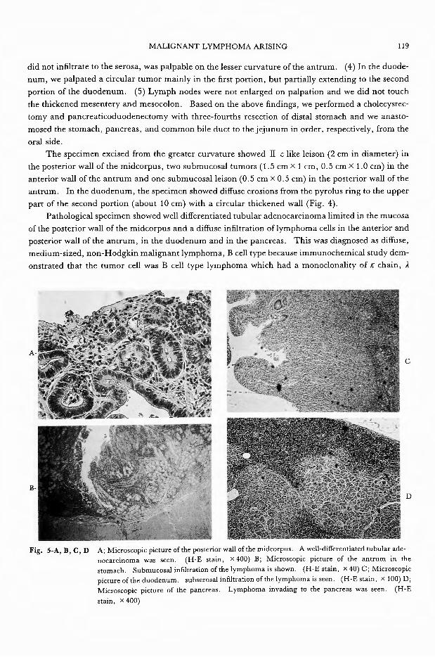
The specimen excised from the greater curvature showed II c like leison (2 cm in diameter) in
the posterior wall of the midcorpus, two submucosal tumors (1.5 cm×1 cm, 0.5 cm×1.0 cm) in the
anterior wall of the antrum and one submucosal leison (0.5 cm×0.5 cm) in the posterior wall of the
antrum. In the duodenum, the specimen showed diffuse erosions from the pyrolus ring to the upper
part of the second portion (about 10 cm) with a circular thickened wall (Fig. 4).
Pathological specimen showed well differentiated tubular adenocarcinoma limited in the mucosa
of the posterior wall of the midcorpus and a diffuse infiltration of lymphoma cells in the anterior and
posterior wall of the antrum, in the duodenum and in the pancreas. This was diagnosed as di釘use,
medium-sized, non-Hodgkin malignant lymphoma, B cell type because immunochemical study dem-
onstrated that the tumor cell was B cell type lymphoma which had a monoclonality of K chain, ).
c
B D
Fig. 5司A,B, C, D A; Microscopic picture of the posterior wall of the midcorpus. A well-differentiated tubular ade-nocarcinoma was seen. (H-E stain, X 400) B; Microscopic picture of the antrum in the stomach. Submucosal infiltration of the lymphoma is shown. (HE stain, X 40) C; Microscopic p凶 ureof the duodenum. subserosal infiltration of the lymphoma is seen田(H-Estain,× 100) D; Microscopic picture of the pancreas. Lymphoma invading to the pancreas was seen. (H-E
stain,× 400)

120 日外宝第64巻第5号(平成7年9月)
chain, μ chain and was positive for surface markers of HLA『 DR,Bl, B2 and B4 (Fig. 5・ABCD).
Postoperative course was satisfactory. On the 18th POD, 67Ga-citrate was performed. It
showed no abnormal accumulation. On the 33rd POD, postoperative chemotherapy (Etoposide,
Methotrexate, Vindesine, Prednisolone) was begun.
Discussion
Sixty percent of primary gastrointestinal lymphoma is reported to arise in the stomach and 20%
in the small intestine9l. The incidence oflymphoma arising in the duodenum is 1.6-12% of primary
small intestinal malignant lymphoma. To our knowledge, 58 reports oflymphoma arising in the du-
odenum were published and there is only one report of primary duodenal malignant lymphoma com-
bined with an early gastric cancer in Japan till 19935). Our case is extremely rare, since histological
examination revealed a primary duodenal malignant lymphoma combined with gastric lymphoma as
well as early gastric cancer.
The patient reported here apparently satisfied Dawson’s criteria6l for primary gastrointestinal
mali伊 antlymphoma. These criteria cons凶 offive points: (1) absence of palpable superficial lym-
phadenopathy; (2) normal leukocytes and di征erentialcounts; (3) absence of enlarged mediastinal
lymph nodes on chest X-ray; (4) no grossly demonstrable involvement beyond the affected segment
of the intestine and its regional mesentric lymph nodes at the time of diagnos町(5)absence of tumor
involvement of the liver and the spleen.
The pathological specimen revealed that the tumor cells in the duodenum infiltrated to the
subserosal layer and as the lesion neared the antrum, infiltration was more superficial. In the an-
trum, the tumor was located in the submucosal layer. In general, malignant lymphoma grows hori-
zontally rather than vertically7l. Therefore, we considered that malignant lymphoma in this patient
arose in the duodenum and extended to the antrum.
Concerning preoperative and postoperative diagnosis, preoperative biopsy and EUS diagnosis
were nearly consistent with the postoperative diagnosis except for the lesion in the angle of the
stomach, which was pseudonegative on preoperative biopsy. Biopsies sometimes result in
pseudonegative findings for malignant lymphoma. We think that it is because biopsies can not cl-
early show the atypism of the cell, which is di侃cultto distinguish from the mesenchymal cell in
mucosal layer, and the tissue of malignant lymphoma is so soft that it tends to be destroyed during
biopsy. However, there are three points which are characteristic of EUS images showing malignant
lymphoma7l ( 1) The tumor infiltrates horizontally than vertically. (2) The tumor is often located
in the 2nd and 3rd layer. (3) The tumor shows a homogeneous low density area. The EUS images
of the angle and the antrum in this case fulfiled the above three points, demonstrating that EUS was useful in the diagnosis of malignant lymphoma.
Finally, we tried to determine the prognosis of this case. Accardi時 toKlaus]. Lewin8l, the pro-
gnosis of the gastrointestinal lymphoma appears to correlate best with the stage of the disease rather
than the histologic type. In this case, lymphoma had spread from the antrum to the 2nd portion
with invasion to the pancreas, but there was no involvement of regional lymph nodes.
Using Ann Arbor classification for malignant lymphoma, this case was in stage lI E and the pro-
gnosis for this stage is not poor. (stage I E; the extent of disease is confined to the viscus/stage lh;
the extent of disease is confined to the viscus and involvement of regional lymph nodes/stage N; wide-spread) On the other hand, using the Contreary classification9l, this case is class ]]] and the prognosis

MALIGNANT LYMPHOMA ARISING
Table 1-A A; Two-year survival rate according to Ann Arbor classifica-
tion shown by Lewin. Stage I E; The extent of disease is
confined to the viscus/Stage Il E; The extent is confined to
the viscus and involvement of regional lymph nodes/Stage
N; widespread.
Stage
IE(17)
IlE(12)
N ( 6)
) ; N
Two-year survival rate
82%
71%
0%
Table 1-B B; Five-year survival rate according to Contrearγs classification. CLASS
I ; lymphoma confined to the bowel without extra intestinal involve-
ment/CLASS Il ; mesentric nodes only/CLASS ill; paraao口icnode involve-ment or extension by contiguity to adjacent viscera.
CLASS I
Overall (102) 59% (41)
Stomach (67) 60% (32)
Small intestine (24) 40% ( 6)
% ; 5 year survival rate ( ) ; N
CLASS Il
72% (15)
80% ( 5)
75%〔6)
CLASS ill
24% (56)
39% (30)
12% (12)
Table 1・C C; Five year survival rate after curative and palliative resection shown by
Contreary.
Total Curative Palliative resect10n resect10n
Over all (102) 38% 64% 17%
Stomach (67) 42% 62% 23%
Small intestine (24) 27% 62% 。%
% ; 5 year-survival rate ( ) ; N
121
is rather poor. (class I; lymphoma confined to the bowel without extraintestinal involvement/class IT; mesentric nodes only/class III; paraaortic node involvement or extension by contiguity to adja-
cent viscera.) However, according to Contreary, the prognosis of the case that undergoes curative
resection is much better than the case without curative resection (Table 1-A, B, C).
Duodenal lesions are reported to carry a worse prognosis than other gastrointestinal lesions1・6・10)
because the extension length of the lesion was already 5 cm or more when clinical symptoms ap-
peared, and duodenal malignant lymphoma tends to infiltrate to other organs (especially, the colon
and pancreas). Neverthless, it seems likely that the prognosis of this patient may not be worse, since
postoperative adjuvant chemotherapy as well as curative resection was performed. Careful follow-
up of this patient must be done.
References
1) Ahmad z. N勾em,John L. Polcano, Benjamin F. Rush Jr. et al: Primary non-Hodgikin’s Lymphoma of the
Duodenum. Cancer 54: 895-898, 1984.

122 日外宝第64巻第5号 (平成 7年 9月)
2) Redentor JG. Pagtalunan, Charles W ‘Mayo, Malcolm B. Dockerty, et al: Primary malignant tumor of the small
intestine. Am. Jour. Surg. 108: 13-18, 1964
3) Marcuse PM, Stout AP, et al: Primary lymphosarcoma of the small intestine. Cancer, 3: 459-474, 1950
4) Fu YS, Perizin: Lymphosarcoma of the small intestine. Cancer, 29: 645-659, 1972
5) Ueno J, Yoshida A, Uyama Y, et al: Malignant Lymphoma of the Duodenum with Early Gastric Carcinoma.
Stomach and Intestine. 17: 1269-1272, 1982
6) IMP. Dawson JS. Cornes, BC. Morsons et al: Primary malignant Lymphoid tumors of the intestinal tract. Br,
Jour. Surg. 49: 80-89, 1961
7) Luigi Bolondi, Paolo Casanova, Gian Carlo Caletti, et al: Primary Gastric Lymphoma versus Gastric Carcinoma;
Endoscopic US Evaluation. Radiology. 165: 821-826, 1987.
8) Klaus J. Lewin, Mahendra Ranchod, Ronald F. Dorfman, et al: Lymphoma of Gastrointestinal Tract, Cancer.
42: 693-707' 1987
9) Kelvin Contreary, Francis C. Nance, Walter F. Becker, et al: Primary Lymphoma of the Gastrointestinal Tract,
Ann, Surg. 191: 593-598, 1980.
10) Herbsman H, Hassan A, Gardner B, et al: Tumor of the small intestine. Curr, Prob, Surg. 17: 127, 1980
和文抄録
十二指腸原発の悪性リンパ腫と早期胃癌を合併した一例
京都大学第一外科
宇山直樹,菅 典道,井上一知, 今村正之
京都大学第一内科
鳥居恵男,梶山 徹,上田俊二,酒井正彦,田罵政朗
消化管原発の悪性りンパ躍のうち十二指腸原発悪性 科を紹介された.術前検査にて病巣は,十二指腸球部
リンパ腫は,比較的稀である 早期胃癌を併発した症 を中心に胃粘膜下に浸潤しており,さらに早期胃癌の
例は更に少なし 本邦では文献上二例目である.今回, 合併が疑われ開腹,弊頭十二指腸切除術にて上記が確
我々は更に胃リンパ腫も合併した非常に稀な症例を経 認された 今回術前の生検で胃悪性リンパ腫は偽陰性
験したのでここに報告する であったが, EUS では悪性リンパ腫に典型的な所見
症例は65高誌の男性.主訴は上腹部痛.現病歴は,三 を呈した部位が有 り,病巣の診断に EUSが有効であ
年前に十二指腸潰蕩 stageAl と診断されその後内視 った また予後については治癒切除し得たが, Con・
鏡にて経過観察していたが,今回十二指腸球部に異常 trearyの分類では CLASSEに相当するため術後に化
を認め,更に生検にて悪性リンパ腫が疑われたので当 学療法 (VEMP療法)を施行した