Embed Size (px)
Citation preview

Life Sciences Learning Center i Copyright © 2013, University of Rochester May be copied for classroom use
i
Male versus Female Brain
Teacher Instructions
Core Concepts:
Overall male and female brain form and function is very similar, however, on average
there are some differences that may explain differences in the rates of some
neurological diseases in male and females.
Endocrine glands secrete hormones that act throughout the body including the brain
Class time required:
Approximately 2 X 40 minute class periods if part 1 is done as a pre-lab, parts 2, 3 and 5 are
done in class and parts 4 and 6 are done as homework.
Teacher Provides:
For ALL PARTS each student will need
Copy of student handout
For Part 3 each team of 2-4 students:
Copy of Brain Map (black and white printed on paper)
Copy of Female Brain Map Overlay (printed in color on transparency)
Copy of Male Brain Map Overlay (printed in color on transparency)
For Part 5 each team of 2-4 students:
Bag or bin containing:
o 1.5 ml microtubes or other small tubes prepared as shown in the chart below:
Label on Tube Contents of Tube – 1 mL of
Jack 3 month old male pH 9 buffer
Jill 3 month old female pH 6 buffer
Testosterone Test Solution 0.01% Bromothymol blue solution
o Droppers labeled:
Jack 3 mo old male
Jill 3 mo old female
Testosterone Test Solution
o Hormone Test Sheet (page 5)printed on plastic transparency sheets
o Small bag labeled “Estrogen Test Paper” containing at least 5 small pieces (cut paper into pieces that are about ½ inch long) of pH 1-12 test paper.

Life Sciences Learning Center ii Copyright © 2013, University of Rochester May be copied for classroom use
ii
o Testosterone and Estrogen Color Charts (page 6).Consider laminating for use by multiple classes.
For Part 6 each team of 2-4 students:
Copy of Hippocampus Neuron (printed in color on paper)
Copy of Amygdala Neuron (printed in color on paper)
Copy of Testosterone Signal Pathway Overlay (printed in color on transparency)
Suggested Class Procedure
1. Assign Part 1 “Don’t get Testy”… handout for homework or discuss as a group as a pre-lab
activity
2. Do Part 2 in class with each student completing the tests on their own
In this activity students will take one or two short memory tests that have been reported
to show male/female differences.
The first test is the California Verbal Learning Test (CVLT) a verbal memory test in
which a list of 16 words (a grocery list) is read aloud to the class. After a pause of 1
minute (or longer) of time students are asked to write down as many of the words as
they can remember. The total number of correct words is then tallied.
http://www.memorylossonline.com/glossary/californiaverballearningtest.html
The second spatial memory test is a that tests spatial memory through a spatial
rotation test. Students are provided with a complex 2 dimensional image and then must
determine which of three possible choices represents the same object rotated in any of
three dimensions. This is considered a memory task as one must keep the original
image in their mind as they compare it to the other available options.
3. Perform Part 3 as students work in small groups. Students will learn some brain anatomy
as they determine which regions of the brain differ in size on average between males and
females.
4. Have students complete Part 4 “Neurological Diseases in Males and Females” as
homework or in class. This section provides students with a table of data on the rates of
neurological diseases in males and females. Alzheimer’s disease is not included as the
difference in Alzheimer’s incidence in males and females (higher in females) is due to the
greater lifespan of females. Males and females of the same age have the same rate of
Alzheimer’s incidence.

Life Sciences Learning Center iii Copyright © 2013, University of Rochester May be copied for classroom use
iii
Important Note: The fact that there are NO neurological diseases which ONLY affect
males or ONLY affect females indicates that there is considerable overlap in the form and
function of male and female brains.
5. Complete Parts 5 “Hormones and the Brain”
Distribute the following materials to each team when students begin Part 5.
Droppers and tubes labeled:
o Jack 3 mo old male
o Jill 3 mo old female
o Testosterone Test Solution
Hormone Test Sheet
Small bag of “Estrogen Test Paper”
Testosterone and Estrogen Color Chart
6. Part 6 “Sorting Out The Signals” as students work in small groups.
In this part the students will use short reading passage and a simple model to develop their
understanding of how hormones can affect neurons. Specifically they will examine the steps
that occur in two different brain neuron types from testosterone binding, to transcription, to
translation, and cellular effects.
7. Provide Part 7 “Bias-ology” as homework or discuss in class.
Additional information/resources Here is a link to a 2012 article by McCarthy et al entitled, “Sex Differences In The Brain: The Not So Inconvenient Truth” http://www.jneurosci.org/content/32/7/2241.full This is an article from 2005 in Scientific American written by Larry Cahill, Professor of Neurobiology at the University of California of http://www.bio.uci.edu/public/press/2005/hisherbrain.pdf

Life Sciences Learning Center iv
Copyright © 2013, University of Rochester May be copied for classroom use
iv
Part 2 Teacher Instructions for memory tests
Test #1 Verbal memory (Females on average tend to perform better at this memory task)
1. Read the list of words below entitled, “Monday’s Shopping List” to the students at a
normal pace.
2. Have students wait one minute after the last word is read.
3. Allow students 1 minute to write down all the words they remember in the student data
table.
4. After 1 minute of writing, read the list aloud again and have students mark all the correct
words that they listed.
5. Have students calculate the total number of words they correctly remembered in the
space provided on the table.
6. Class averages for males and females can be tallied and, if desired, graphed.
Monday’s Shopping list
1. Apples
2. Bananas
3. Grapes
4. Oranges
5. Pepper
6. Salt
7. Sugar
8. Cinnamon
9. Cheese
10. Milk
11. Yogurt
12. Butter
13. Ham
14. Turkey
15. Ground beef
16. Chicken
Test #2 Spatial Memory Test (Males on average tend to perform better on this test)
1. Hand out the spatial rotation tests to the students face down.
2. Students should be instructed that for each image on the left side of the page there are
two images on the right side which represent the SAME object rotated in some direction.
3. Students must correctly identify BOTH of the correct answers for the question to get
credit for that question.
4. Allow 5 minutes for the students to answer the questions.
5. Review the correct answers with the students and have them record the total number
correct.
6. Class averages for males and females can be calculated and if desired, graphed.
Life Sciences Learning Center v
Copyright © 2013, University of Rochester May be copied for classroom use
v
Hormone Test Sheet (print on transparency sheet)
Hormone Test Sheet Testosterone Test Estrogen Test
Jack 3-month old male
Jill 3-month old female
Hormone Test Sheet Testosterone Test Estrogen Test
Jack 3-month old male
Jill 3-month old female
Hormone Test Sheet Testosterone Test Estrogen Test
Jack 3-month old male
Jill 3-month old female
Hormone Test Sheet Testosterone Test Estrogen Test
Jack 3-month old male
Jill 3-month old female
For Part 5

Life Sciences Learning Center vi Copyright © 2013, University of Rochester May be copied for classroom use
vi
Testosterone Test Color Chart
Estrogen Test Color Chart
Testosterone Level
Color Estrogen
Level Color
0-5
0
6-10
1
11-50
2
51-100
3
101-200
4
201-300 5
Testosterone Test Color Chart
Estrogen Test Color Chart
Testosterone Level
Color Estrogen
Level Color
0-5
0
6-10
1
11-50
2
51-100
3
101-200
4
201-300 5
Testosterone Test
Color Chart Estrogen Test
Color Chart
Testosterone Level
Color Estrogen
Level Color
0-5
0
6-10
1
11-50
2
51-100
3
101-200
4
201-300 5
Testosterone Test Color Chart
Estrogen Test Color Chart
Testosterone Level
Color Estrogen
Level Color
0-5
0
6-10
1
11-50
2
51-100
3
101-200
4
201-300 5

Life Sciences Learning Center vii Copyright © 2013, University of Rochester May be copied for classroom use
Brain Map (
For Part 3 (print on regular paper)

Life Sciences Learning Center viii Copyright © 2013, University of Rochester May be copied for classroom use
For Part 6 (print on regular paper)

Life Sciences Learning Center ix
Copyright © 2013, University of Rochester May be copied for classroom use
For Part 6 (print on regular paper)

Life Sciences Learning Center x
Copyright © 2013, University of Rochester May be copied for classroom use
Male--Brain Map Overlay
For Part 3 (print on transparency)

Life Sciences Learning Center xi Copyright © 2013, University of Rochester May be copied for classroom use
Female--Brain Map Overlay (
For Part 3 (print on transparency)

Life Sciences Learning Center xii Copyright © 2013, University of Rochester May be copied for classroom use
For Part 6 (print on transparency)
Life Sciences Learning Center 1
Copyright © 2013, University of Rochester May be copied for classroom use
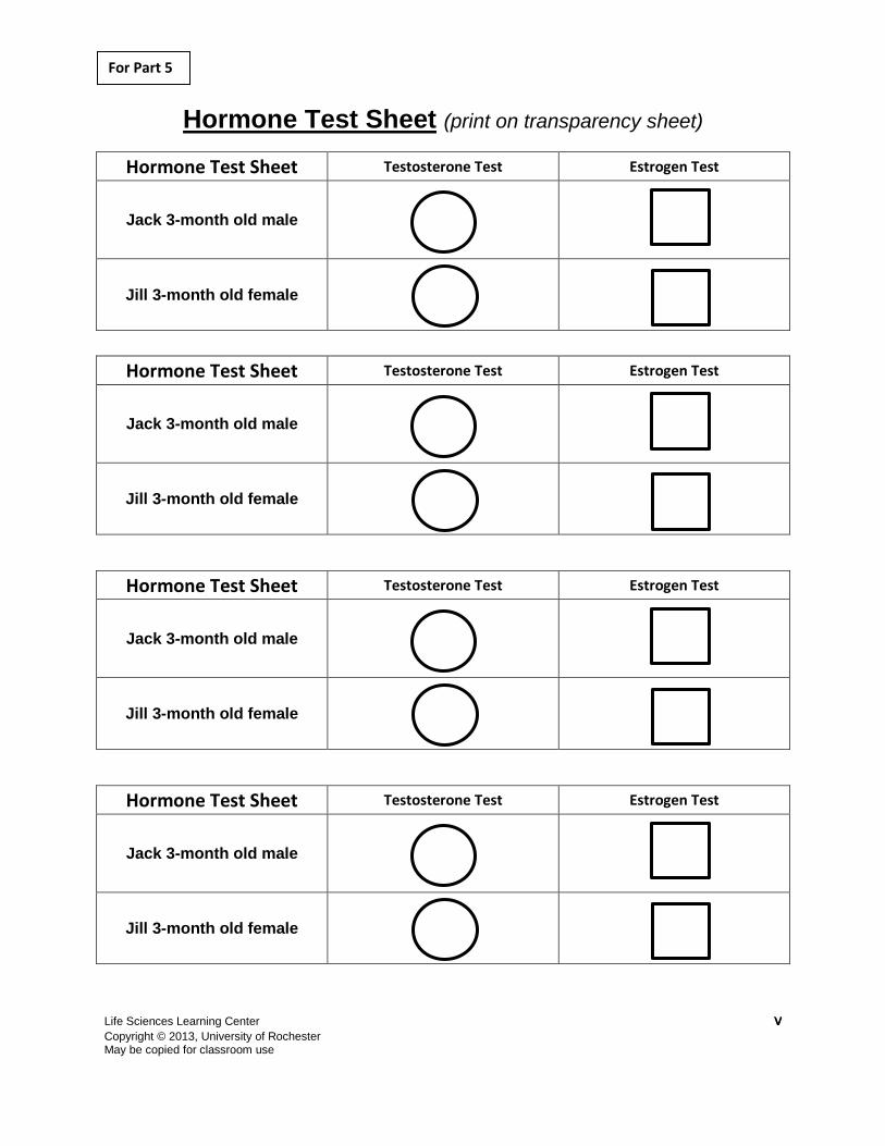
Part 1: Don’t get testy!
Introduction:
Ms. Smith, the high school English Language Arts teacher, made a graph of the exam scores
from a recent test she gave her 27 students as she always did. This time, however, she
decided to plot the results for the 13 males and 14 females separately. While there was
considerable overlap in performance between male and female students, on average there was
a slight difference between the groups.
1. As a group, who performed better on the exam, males or females?
Females
2. What is one possible reason that could explain the different performance of males and females on this English test?
Random chance due to small sample size (27 students)
Different cognitive abilities between males and females
Other social or environmental factors that could differently affect males and females
3. Think of Ms. Smith’s English test as an experiment.
a. What is the independent variable? ______The sex of the students__________
b. What is the dependent variable? _______The test score___________________
0
1
2
3
4
5
6
7
0-10 11-20 21-30 31-40 41-50 51-60 61-70 71-80 81-90 91-100
Nu
mb
er
of
Stu
den
ts
Test score (range)
Males
Females

Life Sciences Learning Center 2
Copyright © 2013, University of Rochester May be copied for classroom use
Part 2: Do Male and Female Brains Work Differently?
Overview:
The following two neurological tests have been developed to test different types of memory.
After taking one or both of the tests, compare the results for males and females in the class to
determine if the results support the idea that male and female brains work differently.
Student Instructions
Memory Test #1 Instructions
1. Listen to the list of words read by your teacher.
2. Wait one minute after the last word is read.
3. Write as many of the words that you just heard as you remember in the table provided.
Scoring the test
4. After you finish writing the list of words, the teacher will read all of the words again.
Place a mark next to all of the words that you remembered correctly.
5. Record your total # correct in the space provided.
YOUR RESULTS
Monday’s Grocery List Correct (√)
Your Total Number Correct

Life Sciences Learning Center 3
Copyright © 2013, University of Rochester May be copied for classroom use
Memory Test #2
1. The shape on the left is your model.
2. Of the 4 shapes on the right, 2 are the same as the model but have been rotated at a
different angle.
3. Mark with an X below the 2 shapes that match the target. An example is given below.
X X
4. There are 8 total problems and you will have 4 minutes to complete them.
Don’t be upset if you don’t finish as this is common.
Scoring the test
5. To score the test, you receive 1 point for each question in which you correctly identified
BOTH of the matching models.
6. No points are awarded if one or more choices are incorrect.

Life Sciences Learning Center 4
Copyright © 2013, University of Rochester May be copied for classroom use
Mark with an X below the two figures on the right that match the one on the left.
There are 2 pages and 8 total questions.
1.
2.
3.
4.

Life Sciences Learning Center 5
Copyright © 2013, University of Rochester May be copied for classroom use
5.
6.
7.
8.
Your Total Correct ____________________

Life Sciences Learning Center 6
Copyright © 2013, University of Rochester May be copied for classroom use
VERBAL MEMORY--CLASS DATA
Total Correct
Student # Females Males
1
2
3
4
5
6
7
8
9
10
11
12
13
14
15
AVERAGE
Based on the class data, does it appear that males or females are better at either the verbal memory task or spatial rotation memory
test?
Answers will vary—opportunity to talk about sample size and the need for more careful analysis.
Teachers should emphasize that any differences observed between males and females on the two tests are most likely not enough
to conclude that there are significant differences between the two groups. However, these tests when given to hundreds or
thousands of males and females under controlled conditions (same lighting, same time of day) reveal average differences.
SPATIAL ROTATION TEST--CLASS DATA
Total Correct
Student # Females Males
1
2
3
4
5
6
7
8
9
10
11
12
13
14
15
AVERAGE

Life Sciences Learning Center 7
Copyright © 2013, University of Rochester May be copied for classroom use
Part 3-Are There Physical Differences in Girls and Boys Brains?
There have been a number of studies comparing the size of brain regions in males and females.
More work still needs to be done, but there appear to be some differences in the sizes of some
brain regions between male and female brains. THESE REPRESENT AVERAGES OF MANY
MALE AND FEMALE BRIANS. INDIVIDUAL MALES AND FEMALES MAY BE MORE
SIMILAR TO EACH OTHER.
Examine the Brain Map and answer the questions below.
1. Which of the brain region(s) shown is/are involved in memory?
___ Hippocampus, Cingulate Gyrus____________________________________
2. What is/are the function(s) of the amygdala?
____Basic Emotion___________________________________________________
3. Place the Male and Female Brain Map Overlays over the Brain Map to identify regions of the brain where the average male and female brain may be different.
If the region appears only purple it indicates that region is similar in size between average male and female brains
Where the pink overlay extends beyond the purple indicates regions which tend to be larger in female brains
Where the blue overlay extends beyond the purple indicates regions which tend to be larger in male brains
Use the Brain Map and Male and Female Brain Map Overlay to complete the table below.
Brain Region Function(s)
No difference or Larger in Males or
Larger in Females
Amygdala Basic emotion Larger in males
Cingulate gyrus Emotion, learning and memory Larger in females
Hippocampus Learning and memory Larger in females
Hypothalamus Hunger, thirst, Sex Larger in males
Inferior temporal gyrus
Form and color perception Larger in males
Lingual gyrus Vision and dreaming Larger in females
Prefrontal cortex
Problem solving complex movements No difference
Thalamus Connection between brain regions No difference

Life Sciences Learning Center 8
Copyright © 2013, University of Rochester May be copied for classroom use
Part 4-Neurological Disease in Males and Females
Biology Brief: Understanding Human Neurological Disease
Biology and math are combined in the field of epidemiology (EPPY-DEE-ME-AH-LO-GEE).
Epidemiologists study disease rates among the population. These types of studies have
revealed that there are some neurological diseases that are more common in males and others
that are more common in females (Table 1).
Table 1: Percentage of Males and Females in U.S. Population with Neurological Conditions
Neurological Condition Females Males
Parkinson’s—Loss of motor control 0.01% 0.02%
Multiple Sclerosis 0.1% 0.03%
Mood Disorders (Depression) 5.9% 3.9%
Tourette’s Syndrome 0.08% 0.23%
Autism 0.2% 0.8%
Questions:
1. Which neurological conditions affect more males than females?
Parkinson’s, Autism, Tourette’s
2. Which neurological conditions are more frequent in females?
Multiple Sclerosis, Mood Disorders
3. Provide two possible explanations for the differences in neurological disease rates
between males and females? Explanations can include information presented in this
lesson or other sources.
Differences in the brain/nervous system between males and females.
Different hormones produced by males and females.
Genes located on the X or Y chromosomes which differ between males and
females
Environmental factors like peer pressure may affect males and females differently
Differences in the rates that males and females seek medical treatment. Maybe
more males do suffer from depression but are undiagnosed because of
unwillingness to seek help.
4. Provide an explanation for why there are no neurological diseases that affect only males
or females?
Male and female brains have much in common and individual males and females
can be similar.

Life Sciences Learning Center 9
Copyright © 2013, University of Rochester May be copied for classroom use
Part 5-Hormones and the Brain Biology Brief: Sex Hormones and the Brain
Hormones, chemical messengers released by specialized organs called endocrine glands, can
travel throughout the body through the circulatory system. Hormones affect the development
and function of other organs including the brain. Humans have about 50 different hormones
including some that are produced by the sex organs (testes and ovaries) called sex hormones.
Testosterone and estrogen are two sex hormones that play important roles during development
from embryonic stages through puberty and into adulthood. These hormones are best known for
inducing the formation of secondary sex characteristics but testosterone and estrogen can also
affect brain development. The level of sex hormones changes throughout development and can
affect brain cells in different ways including:
Promote cell division (mitosis)
Increase cell growth (change in cell size and shape)
Induce cell death (apoptosis)
Increase cell communication (nerve firing)
Direct cell differentiation (development of specific types of neurons)
There are many different types of nerve cells in the brain (like cortex nerve cells, hippocampus
nerve cells, amygdala nerve cells). Different nerve cells may respond in different ways to the
same hormone.
Questions:
1. Hormones are released by _____endocrine glands____.
2. Answer the true false questions below.
Sex hormone levels do not change throughout development
TRUE FALSE
Sex hormones are active only during puberty TRUE FALSE
Sex hormones can affect cell division in the brain TRUE FALSE
3. List three ways that sex hormones can affect neurons.
Induce cell growth
Induce cell death
Promote cell division
Affect cell differentiation
Affect nerve cell
firing/signaling

Life Sciences Learning Center 10
Copyright © 2013, University of Rochester May be copied for classroom use
Overview:
The hormones testosterone and estrogen can be detected in the blood plasma (liquid part of the blood). Your lab kit contains samples of blood plasma collected from Jack and Jill, 3-month old male and female fraternal twins. Your goal is to determine if there are any differences in testosterone or estrogen at this critical stage of brain development. Follow these instructions to determine the levels of testosterone and estrogen in each of the blood plasma samples.
1. Place 1 drop of Jack’s plasma sample in the top circle and the top square.
2. Using a clean dropper place 1 drop of Jill’s plasma sample to the bottom circle and bottom square.
3. Using a clean dropper add 1 drop of Testosterone Test Solution to both of the circles. Use the COLOR CHART to record your results below.
4. Carefully place a piece of the Estrogen Test Paper onto both of the squares. Use the COLOR CHART to record your results below.
Patient Testosterone Level (nanograms/100 ml)
Estrogen Level (nanograms/100 ml)
Jack, 3-month old male
Jill, 3-month old female
4. Based on the information in the data table:
Which hormone (testosterone or estrogen) is most different between males and females at this stage?
Testosterone
Are either testosterone or estrogen secreted in only males or only females?
No

Life Sciences Learning Center 11
Copyright © 2013, University of Rochester May be copied for classroom use
Figure 1: Testosterone signaling may contribute to differences
between male and female brains.
Part 6-Sorting out the Signals
Biology Brief: The Steps of Hormone Signaling
Scientists are still trying to understand how exactly testosterone signaling can influence cells in
the brain and perhaps lead to differences between male and female brains (Figure 1).
Some of the major steps have been worked out. They include the following:
1. In the brain, testosterone can bind to proteins called testosterone receptors within neurons.
Unlike many receptors, testosterone receptors are found in the cytoplasm not at the plasma
membrane.
2. After binding to the testosterone, testosterone receptors enter the nucleus. In the nucleus,
testosterone receptors bind to DNA and other specialized proteins to turn on or off
transcription of other genes.
3. Genes activated by testosterone signaling are then translated into proteins in the cytoplasm
by ribosomes.
4. Proteins turned on or off by testosterone signaling can then affect neuron functions such as:
cell division (mitosis)
cell growth (change in cell size and shape)
cell death (apoptosis)
cell communication (nerve firing)
cell differentiation (development of specific types of neurons)
The effects of testosterone can be different in different nerve cells because these cells may
already have different proteins present before receiving the testosterone signal. These other
proteins may then change how the cell responds to the testosterone signal.
Life Sciences Learning Center 12
Copyright © 2013, University of Rochester May be copied for classroom use
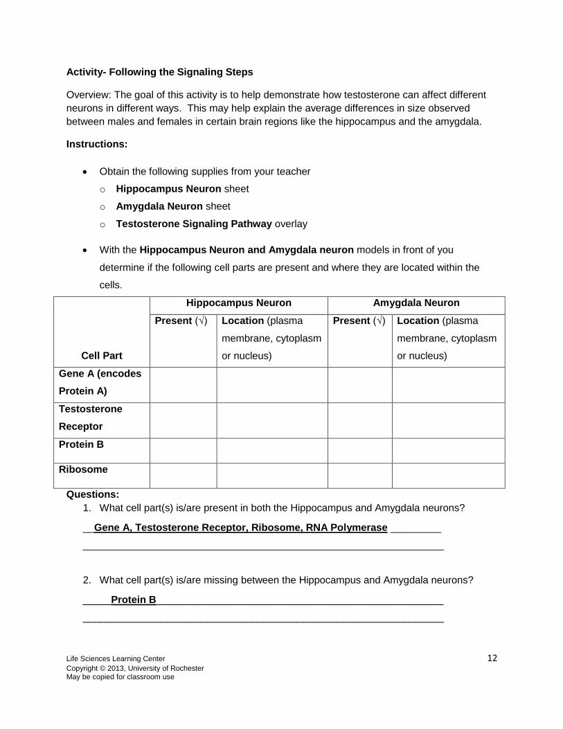
Activity- Following the Signaling Steps
Overview: The goal of this activity is to help demonstrate how testosterone can affect different
neurons in different ways. This may help explain the average differences in size observed
between males and females in certain brain regions like the hippocampus and the amygdala.
Instructions:
Obtain the following supplies from your teacher
o Hippocampus Neuron sheet
o Amygdala Neuron sheet
o Testosterone Signaling Pathway overlay
With the Hippocampus Neuron and Amygdala neuron models in front of you
determine if the following cell parts are present and where they are located within the
cells.
Cell Part
Hippocampus Neuron Amygdala Neuron
Present (√) Location (plasma
membrane, cytoplasm
or nucleus)
Present (√) Location (plasma
membrane, cytoplasm
or nucleus)
Gene A (encodes
Protein A)
Testosterone
Receptor
Protein B
Ribosome
Questions:
1. What cell part(s) is/are present in both the Hippocampus and Amygdala neurons?
__Gene A, Testosterone Receptor, Ribosome, RNA Polymerase _________
________________________________________________________________
2. What cell part(s) is/are missing between the Hippocampus and Amygdala neurons?
_____Protein B___________________________________________________
________________________________________________________________

Life Sciences Learning Center 13
Copyright © 2013, University of Rochester May be copied for classroom use
Signaling Step Tags
Instructions (continued)
Model the effect of hormone signaling by positioning the Testosterone Signaling
Pathway Overlay onto the Hippocampus Neuron so that the testosterone molecules
(resembling a T) are fit into the Testosterone Receptors.
Cut out the Signaling Step Tags below (BOTTTOM RIGHT) and place them in the
spaces near the numbers on the Testosterone Signaling Pathway Overlay to identify
what is happening at each step.
Complete the table below when you are finished.
Step Label used
1 Receptor Binding
2 Transcription
3 Translation
4 Cell Effect
Questions:
3. What may happen to the Hippocampus Neuron in response to testosterone signaling?
(Hint: refer to step 4)
_____The cell may die___________________________________
Note that we can only what MAY happen to the cell because the result will depend on the exact
timing and amount of testosterone signal received by the neuron.
Instructions (continued)
Repeat this process with the Amygdala Neuron model and the testosterone.
Answer the questions below using the model and the information in Part 6-- Biology
Brief: The Steps of Hormone Signaling
Questions
1. Describe what happens in Step 2 of the model?
In the nucleus, testosterone receptors pair with other testosterone
receptors and bind to DNA to turn on transcription of another gene.
2. What may happen to the amygdala neuron exposed to testosterone?
The cell may grow
Extension Questions

Life Sciences Learning Center 14
Copyright © 2013, University of Rochester May be copied for classroom use
3. How might testosterone’s effects on these nerve cells contribute to the average
differences between male and female brains presented in PART 2?
The amygdala is larger in males—maybe due to testosterone promoting growth
The hippocampus is smaller in males. This might be due to cells dying in this part
of the male brain.
4. Given what we know about the brain and male and female differences do you think that
male and female students should be treated differently? Graded separately?
Answers will vary. Students in favor of different treatment should cite the
differences observed in the brain regions associated with learning, memory, and
vision. They may also note any differences between the males and females of the
class performance on the memory tests.
Students in favor of equal treatment should include the fact that there is great
overlap between males and females in tests of knowledge and memory. Also they
may note similarity between male and female brain anatomy in the prefrontal
cortex

Life Sciences Learning Center 15
Copyright © 2013, University of Rochester May be copied for classroom use
Figure 1: Percentage of male and female animals included in published
neuroscience research papers in 2009.
(Beery and Zucker, 2010, Neroscience Behavior Research)
0
10
20
30
40
50
60
70
80
90
100
Animal Studies Human Studies
% Unspecified
% Both
% Female
% Male
Part 7: Bias-ology
Animal models, like mice and rats, are one of the most critical ways that scientists can
understand the causes and potential cures of human diseases. Though there are clearly
diseases that are more common in females than males, most animal research is performed with
male animals. A recent examination of published biological research revealed that in the field of
neuroscience males were
used 5 times more often than
females in single sex animal
studies.
Some scientists say using
males is less expensive and
easier than using female
animals. For one, the
hormonal cycle of female
animals requires that
hormone levels be measured
and matched among
experimental groups. This
requires more time, animals,
and money, which
researchers argue is in short
supply.
Others believe that animal studies must include male and female animals and compare them
separately to determine if there are differences between the sexes. One possible negative
consequence of not studying females is that new drugs that work in males may have
unanticipated side effects in females. Supporters of female research also argue that studying
females may benefit both males and females. In one case, researchers found that pregnancy
decreased symptoms of multiple sclerosis in female mice. This has led to studies exploring
whether female sex hormones can help treat this disease in males.
1. Approximately what percentage of non-human animal studies included females? ~30%
(%female + %both)
2. What are the arguments against using female animals to study disease?
Higher cost, More animals used. More difficult to control for hormonal
differences between individuals
3. What are the arguments for using females in animal research studies?
Better understanding of the causes of disease in females. Development of
treatments for females and males.