Embed Size (px)
Citation preview
UC DavisUC Davis Previously Published Works
TitleMagnetization and stability study of a cobalt-ferrite-based ferrofluid
Permalinkhttps://escholarship.org/uc/item/4j92p6vw
AuthorsKamali, SPouryazdan, MGhafari, Met al.
Publication Date2016-04-15
DOI10.1016/j.jmmm.2015.12.007 Peer reviewed
eScholarship.org Powered by the California Digital LibraryUniversity of California
Journal of Magnetism and Magnetic Materials 404 (2016) 143–147
Contents lists available at ScienceDirect
Journal of Magnetism and Magnetic Materials
http://d0304-88
n CorrE-mURL
journal homepage: www.elsevier.com/locate/jmmm
Magnetization and stability study of a cobalt-ferrite-based ferrofluid
Saeed Kamali a,n, Mohsen Pouryazdan b,c, Mohammad Ghafari b,d, Masayoshi Itou e,Masoud Rahman f, Pieter Stroeve f, Horst Hahn b,c,d, Yoshiharu Sakurai e
a Department of Mechanical, Aerospace and Biomedical Engineering, University of Tennessee Space Institute, Tullahoma, TN 37388, USAb Institute for Nanotechnology, Karlsruhe Institute of Technology, Karlsruhe, Germanyc Joint Research Laboratory Nanomaterials (KIT and TUD) at Technische Universität Darmstadt (TUD), 64287 Darmstadt, Germanyd Herbert Gleiter Institute of Nanoscience, Nanjing University of Science and Technology, Nanjing 210094, People's Republic of Chinae Japan Synchrotron Radiation Research Institute, SPring-8, 1-1-1 Kouto, Sayo-cho, Sayo-gun, Hyogo 679-5198 Japanf Department of Chemical Engineering and Materials Science, University of California, Davis, CA 95616, USA
a r t i c l e i n f o
Article history:Received 3 September 2015Received in revised form9 November 2015Accepted 1 December 2015Available online 8 December 2015
Keywords:FerrofluidCobalt-ferriteNanoparticlesMössbauer spectroscopyMagnetic Compton scatteringNano-magnetism
x.doi.org/10.1016/j.jmmm.2015.12.00753/& 2015 Elsevier B.V. All rights reserved.
esponding author.ail address: [email protected] (S. Kamali).: http://www.utsi.edu (S. Kamali).
a b s t r a c t
In this study the structural and magnetization properties of a CoFe2O4-based ferrofluid was investigatedusing x-ray diffraction (XRD), transmission electron microscopy (TEM), energy dispersive x-ray spec-troscopy (EDS), Mössbauer spectroscopy, and magnetic Compton scattering (MCS) measurements. TheXRD diagram indicates that the nanoparticles in the ferrofluid are inverse spinel and TEM graph showsthat the ferrofluid consists of spherical nanoparticles with an average diameter of 187 1 nm, in goodagreement with the size, 19.4 nm, extracted from line broadening of the XRD peaks. According to EDSmeasurements the composition of the nanoparticles is CoFe2O4. Mössbauer spectroscopy shows that thecation distributions are (Co0.38Fe0.62)[Co0.62Fe1.38]O4. The MCS measurement, performed at 10 K, indicatesthat the magnetization of the nanoparticles is similar to magnetization of maghemite and magnetite.While the magnetization of the inverse spinels are in [111] direction, interestingly, the magnetizationdeduced from MCS is in [100] direction. The CoFe2O4-based ferrofluid is found to be stable at ambientconditions, which is important for applications.
& 2015 Elsevier B.V. All rights reserved.
1. Introduction
Ferrofluids, which are defined as stable colloidal magneticnano-sized particles dispersed in a carrier liquid, have many in-dustrial applications [1,2]. It is already well-known that the ap-plication of magnetic nanoparticles in general and iron-oxide andferrite nanoparticles in particular is very wide due to their novelproperties. They have various applications in different fields, ran-ging from new functional materials [3] such as magnetic recordingmedia [4] to biomedical diagnostics and therapy [5] such as cancertreatment by hyperthermia [6]. Sufficiently small nanoparticles aremagnetic single domain with a magnetic anisotropy energy of thesame order as the thermal energy [7], and the atomic magneticmoments in these nanoparticles fluctuate between the easy axesdirections giving rise to superparamagnetism [8–10]. Suspensionof such nanoparticles in liquids, resulting in ferrofluids, makesthem even more interesting and opens new routes of applications.This class of material was initially developed by NASA to be used in
space to control fluids [11]. Besides their primary application asrotating shaft seals in satellites, they have found a lot of applica-tions from being used in mechanical machines due to their inter-esting mechanical properties [12] to computer hard drives [1,2].Ferrofluids have also many applications in medicine such as forsustaining drug attached to the surface of magnetic nanoparticles,at specific sites in the body by using external field [13] and forimplantable artificial hearing aids in the form of ferrofluidic ac-tuators [1]. It is worth mentioning that the magnetism in ferro-fluids is more complicated compared to nano-powders owing tothe existence of additional parameters such as existence of theBrownian motion, which is simultaneously a mechanism for ro-tation of magnetization direction in nanoparticles due to theparticle rotation. Other parameters, such as interaction betweenmagnetic nanoparticles and the surfactant, which is used based onthe electric polarization of the surfactant to prevent agglomerationof the nanoparticles, together with tensional as well as gravita-tional forces in the ferrofluids make them extremely complexcompounds. Superlattices and self-assembled iron-oxides nano-crystals have also many industrial applications especially in mag-netic recording media and hence recently attracted considerableamount of attention [14–19].
Another interesting class of materials is ferrites. The parent

S. Kamali et al. / Journal of Magnetism and Magnetic Materials 404 (2016) 143–147144
compound, i.e., magnetite (Fe3O4), is a ferrimagnetic compoundwith two different cation sites in a unit cell: 8 tetrahedral A sites,and 16 octahedral B sites. Magnetite is an inverse spinel, in otherwords, (Feþ3)[Feþ2Feþ3]O4, where the parentheses stand for thetetrahedral sites and the square parentheses stand for the octa-hedral sites. Furthermore, the Feþ3 ion has a magnetic moment of5 μB and the Feþ2 ion has 4 μB resulting in total magnetic momentof 4 μB. Above the Verwey [20] transition temperature there is afree electron hopping between Feþ2 and Feþ3 in the B sites,causing magnetite to be a conductor. Below the Verwey transitiontemperature this electron is frozen and the magnetite will be aninsulator. Concerning the above-mentioned magnetic and elec-trical properties of magnetite, it is possible to introduce 3d tran-sition metals in magnetite, hence manipulate and control theseproperties.
It is well-known that in contrast to metallic alloys where Ni andCo, in relatively low concentrations, increase the Fe magneticmoment according to the Slater–Pauling curve [21], the magneti-zation is decreased when such elements are incorporated intomagnetite compared to the pure magnetite [22,23]. On the otherhand, the Fe magnetic hyperfine field is increased both in metallicalloys [24–26] and in such ferrites [23,27].
Mössbauer spectroscopy has been proven to be a powerfultechnique for characterization of different iron-oxide nano-particles [28–30], identifying their coexistence in a nanoparticle[31], or in solid solutions [32]. Furthermore, it is a suitable tech-nique to gain insight into the distribution of cations in ferrites[23,27]. Accordingly, this technique is suitable for characterizationof iron-oxide-based ferrofluids.
Furthermore, as orbital magnetic moment in most iron-oxidesis quenched, the dominant magnetic moment in this class ofmagnetic material is the spin magnetic moment. Spin-polarizedelectron momentum densities (EMDs) can be studied by magneticCompton scattering (MCS) [33]. It is called magnetic Comptonprofile (MCP) [34] of magnetic atoms:
∫ ∫( ) = ( ) ( )−∞
+∞
−∞
+∞J p n dp dpp . 1mag z mag x y
= ( )p p pp , ,x y z is the electron momentum and ( )n pmag is the spin-polarized EMD,
( ) = ( ) − ( ) ( )n n np p p , 2mag up down
where ( )n pup and ( )n pdown are up-spin and down-spin EMDs re-spectively, defined as,
∫∑ ψ( ) = | ( ) ( − · ) |( )
n i dp r p r rexp ,3
up downi
i up down/ , /2
as a function of the up-spin and down-spin electron wave func-tions ψ ( )ri up down, / . The summation in Eq. (3) extends over all oc-cupied states. The pz-axes is taken along the scattering vector. Theshape of MCP depends on the spin-polarized atomic species, theirlocal structures and the direction of scattering vector. In general,Compton scattering refers to x-ray scattering in a deeply inelasticregime, because x-rays interact not only with the electron chargebut also with the electron spin. Therefore, MCP, which is the dif-ference between the majority- and the minority-spin Comptonprofiles, can be extracted by measuring two Compton scatteringcross-sections: one in the direction of magnetization of the spe-cimen parallel to the scattering vector, and the other in the anti-parallel direction of magnetization. MCS has proved to be a pow-erful technique for characterization of different nanostructures[14,35–38].
This study has a two-fold motivation. The first is to investigatethe stability of a Co-ferrite, CoFe2O4, ferrofluid after more than twodecades of its preparation. The second is to characterize it with
powerful techniques to gain insight into its magnetic properties aswell as the electron polarization and atomic arrangement. Thestructure and morphology of the nanoparticles were investigatedby transmission electron microscopy (TEM), high-resolution TEM(HR-TEM), and x-ray diffraction (XRD), while energy-dispersivex-ray spectroscopy (EDS) provides information about the ele-mental distribution.
2. Experimental details
The synthesis of the ferrofluid was based on the wet-grindingapproach the details of which can be found elsewhere [39]. Thecompound was prepared more than two decades ago and has beenkept in the ambient atmosphere. The structure of the nano-particles in the ferrofluid was investigated by XRD using a PhilipsX'Pert diffractometer equipped with a copper tube in Bragg–Brentano geometry operating at 45 kV/40 mA, together with aLynx-eye energy dispersive position sensitive detector (3° open-ing). The radiation used was λ( = ˚ )αCuK 1.5418 A . The crystalstructures were refined using the Rietveld method [40] im-plemented in the FullProf software [41]. Zero point and displace-ment error were refined using an internal Si standard(a¼5.430922 Å, NIST 640b). The instrumental resolution was de-termined by measurements of a LaB6-standard (NIST 660).
The morphology and dimensions of the synthesized nano-particles in the ferrofluid were determined by HR-TEM using aPhilips CM12 transmission electron microscope, and the averagediameter of the Co-ferrite nanoparticles was determined bymeasuring the diameters of 200 particles and fitting the histo-grams to Gaussian distribution function. The EDS measurementswere performed in conjunction with FEI XL30-SFEG scanningelectron microscopy operating at a voltage of 10 kV.
A conventional transmission 57Fe Mössbauer spectrometer,working in constant acceleration mode, was used for this experi-ment. The movement of the radioactive source, 57Co in Rh matrixheld at room temperature (RT), was controlled by a computerizedinterface. The sample was mounted in a He-flow cryostat andspectra were recorded at 10, 50, 80, and 300 K. The recordedspectra were analyzed using the software Recoil [42]. All centroidshifts, δ, are given with respect to metallic α-iron at RT.
The MCS experiment was carried out at the BL08W beamline ofSPring-8. The incident x-rays, with 175 keV energy, were ellipti-cally polarized and perpendicular to the sample surface. Energyspectra of Compton-scattered x-rays were measured at 10 K, in amagnetic field of þ2.5 T (parallel to the scattering vector) and�2.5 T (anti-parallel to the scattering vector). The MCS contribu-tions were separated by taking the difference between the energyspectra at þ2.5 T and �2.5 T. The measurement was repeatedusing the sequence of magnetic fields, ABBABAAB, where A isþ2.5 T and B is �2.5 T, with a data-accumulation duration of 60 seach. The energy spectra of the MCS x-rays were converted to MCPusing a standard data-processing method [43].
3. Results and discussion
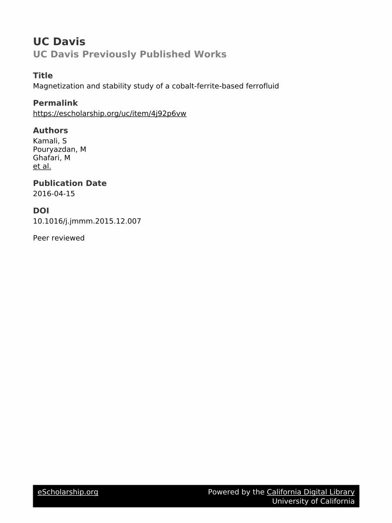
A typical XRD pattern for the sample is shown in Fig. 1. TheXRD diffractogram has relatively sharp peaks, indicating an ex-cellent crystallinity, as it is evident from (220), (311), (222), (400),(422), (511), and (440) peaks, which are typical for inverse spinelstructure nanoparticles [44]. The broadening of the peaks is anindication of the finite size in accordance with the Scherrerequation [45]:
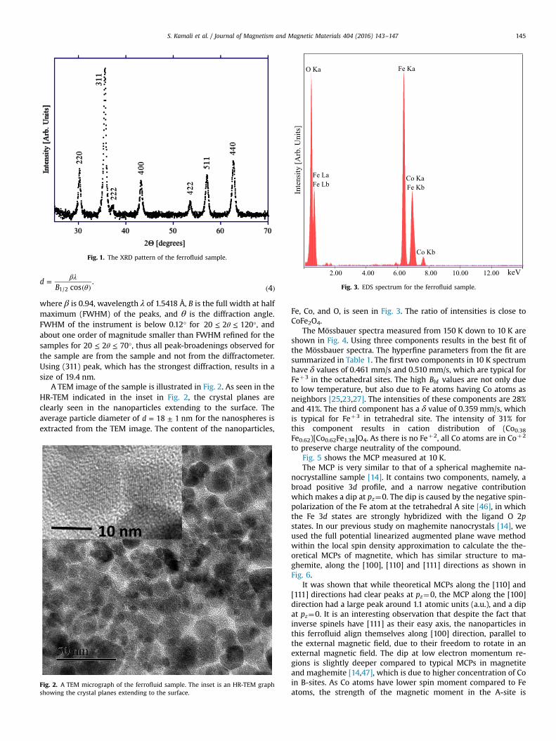
Fig. 1. The XRD pattern of the ferrofluid sample.
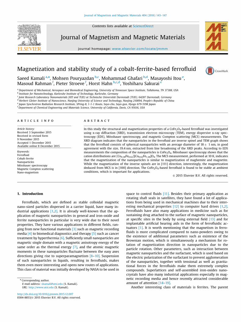
O Ka
Fe LaFe Lb
Fe Ka
Fe KbCo Ka
Co Kb
2.00 4.00 6.00 8.00 10.00 12.00 keV
Inte
nsity
[Arb
. Uni
ts]
Fig. 3. EDS spectrum for the ferrofluid sample.
S. Kamali et al. / Journal of Magnetism and Magnetic Materials 404 (2016) 143–147 145
βλθ
=( ) ( )
dB cos
.41/2
where β is 0.94, wavelength λ of 1.5418 Å, B is the full width at halfmaximum (FWHM) of the peaks, and θ is the diffraction angle.FWHM of the instrument is below 0.12° for θ≤ ≤ °20 2 120 , andabout one order of magnitude smaller than FWHM refined for thesamples for θ≤ ≤ °20 2 70 , thus all peak-broadenings observed forthe sample are from the sample and not from the diffractometer.Using (311) peak, which has the strongest diffraction, results in asize of 19.4 nm.
A TEM image of the sample is illustrated in Fig. 2. As seen in theHR-TEM indicated in the inset in Fig. 2, the crystal planes areclearly seen in the nanoparticles extending to the surface. Theaverage particle diameter of = ±d 18 1 nm for the nanospheres isextracted from the TEM image. The content of the nanoparticles,
Fig. 2. A TEM micrograph of the ferrofluid sample. The inset is an HR-TEM graphshowing the crystal planes extending to the surface.
Fe, Co, and O, is seen in Fig. 3. The ratio of intensities is close toCoFe2O4.
The Mössbauer spectra measured from 150 K down to 10 K areshown in Fig. 4. Using three components results in the best fit ofthe Mössbauer spectra. The hyperfine parameters from the fit aresummarized in Table 1. The first two components in 10 K spectrumhave δ values of 0.461 mm/s and 0.510 mm/s, which are typical forFeþ3 in the octahedral sites. The high Bhf values are not only dueto low temperature, but also due to Fe atoms having Co atoms asneighbors [25,23,27]. The intensities of these components are 28%and 41%. The third component has a δ value of 0.359 mm/s, whichis typical for Feþ3 in tetrahedral site. The intensity of 31% forthis component results in cation distribution of (Co0.38Fe0.62)[Co0.62Fe1.38]O4. As there is no Feþ2, all Co atoms are in Coþ2
to preserve charge neutrality of the compound.Fig. 5 shows the MCP measured at 10 K.The MCP is very similar to that of a spherical maghemite na-
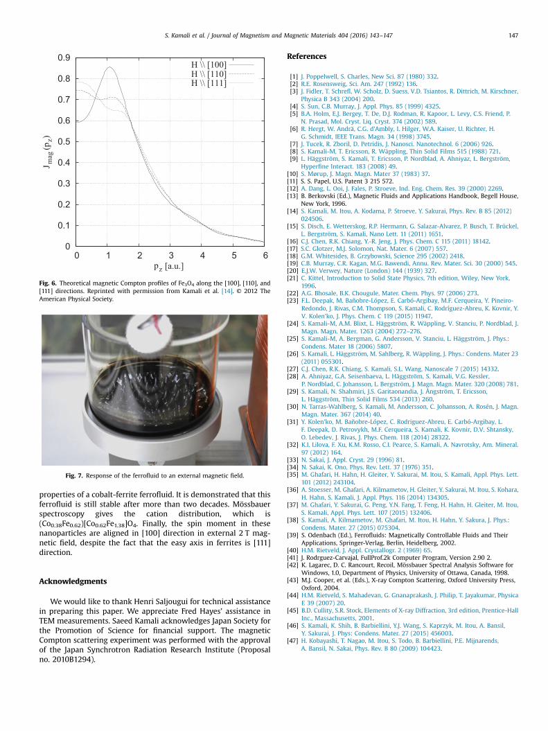
nocrystalline sample [14]. It contains two components, namely, abroad positive 3d profile, and a narrow negative contributionwhich makes a dip at pz¼0. The dip is caused by the negative spin-polarization of the Fe atom at the tetrahedral A site [46], in whichthe Fe 3d states are strongly hybridized with the ligand O 2pstates. In our previous study on maghemite nanocrystals [14], weused the full potential linearized augmented plane wave methodwithin the local spin density approximation to calculate the the-oretical MCPs of magnetite, which has similar structure to ma-ghemite, along the [100], [110] and [111] directions as shown inFig. 6.
It was shown that while theoretical MCPs along the [110] and[111] directions had clear peaks at pz¼0, the MCP along the [100]direction had a large peak around 1.1 atomic units (a.u.), and a dipat pz¼0. It is an interesting observation that despite the fact thatinverse spinels have [111] as their easy axis, the nanoparticles inthis ferrofluid align themselves along [100] direction, parallel tothe external magnetic field, due to their freedom to rotate in anexternal magnetic field. The dip at low electron momentum re-gions is slightly deeper compared to typical MCPs in magnetiteand maghemite [14,47], which is due to higher concentration of Coin B-sites. As Co atoms have lower spin moment compared to Featoms, the strength of the magnetic moment in the A-site is

Fig. 4. Mössbauer spectra of the ferrofluid sample recorded at differenttemperatures.
Table 1Mössbauer parameters of the sample measured at different temperatures: themagnetic hyperfine fields (Bhf ), magnetic hyperfine field distributions (s), centroidshift (δ), quadrupole shift (ϵ), and intensities (I) of the different components. Esti-mated errors in Bhf are 7 0.2 T, in s, 7 0.2 T, in δ and ϵ, 7 0.01 mm/s, and in I, 73%.
Subspectra 10 K 50 K 80 K 180 K
Bhf,1 (T) 53.1 52.9 52.7 51.5s1 (T) 1.2 1.3 1.3 1.4δ1 (mm/s) 0.461 0.464 0.448 0.423ϵ1 (mm/s) 0.079 0.070 0.058 0.042I1 (%) 28 30 29 29
Bhf,2 (T) 51.9 51.5 51.2 49.6s2 (T) 2.0 2.1 2.1 2.8δ2 (mm/s) 0.510 0.517 0.524 0.511ϵ2 (mm/s) �0.064 �0.054 �0.037 �0.026I2 (%) 41 40 41 41
Bhf,3 (T) 50.6 50.5 50.3 49.4s3 (T) 1.2 1.1 1.2 1.4δ3 (mm/s) 0.359 0.343 0.326 0.300ϵ3 (mm/s) 0.024 0.020 0.014 0.012I3 (%) 31 30 30 30
pz[a.u.]
J mag
(pz) [
Arb
. Uni
ts]
0 5-5 10-10
Fig. 5. MCP of the ferrofluid sample measured at 10 K.
S. Kamali et al. / Journal of Magnetism and Magnetic Materials 404 (2016) 143–147146
increased relative to the magnetic moment in the B-sites accordingto the model presented in our previous work [46].
The stability of a ferrofluid is shown in its response to an ex-ternal magnetic field. In case of magnetic nanoparticles suspendedin a solution, only the nanoparticles are attracted to an externalmagnetic field. On the other hand, when a ferrofluid is exposed toan external magnetic field, the whole sample will be attracted tothe external field, and spikes are formed, as shown in Fig. 7 for thisferrofluid. The response is identical to the ferrofluid's response toan external magnetic field when it was synthesized more than twodecades ago (not shown). Besides the results from the character-ization techniques used in this study, i.e., Mössbauer spectroscopy,which shows the existence of Co and Fe in the nanoferrite, andMCP showing the existence of a Co-based ferrite, the visible re-sponse of the ferrofluid to an external magnetic field shows thatthe ferrofluid is still stable after more than two decades.
4. Conclusions
In this work XRD, HR-TEM, EDS, Mössbauer spectroscopy, andMCS have been used to characterize the structural and magnetic
0
0.1
0.2
0.3
0.4
0.5
0.6
0.7
0.8
0.9
0 1 2 3 4 5 6
J mag
(pz)
pz [a.u.]
H \\ [100]H \\ [110]H \\ [111]
Fig. 6. Theoretical magnetic Compton profiles of Fe3O4 along the [100], [110], and[111] directions. Reprinted with permission from Kamali et al. [14]. © 2012 TheAmerican Physical Society.
Fig. 7. Response of the ferrofluid to an external magnetic field.
S. Kamali et al. / Journal of Magnetism and Magnetic Materials 404 (2016) 143–147 147
properties of a cobalt-ferrite ferrofluid. It is demonstrated that thisferrofluid is still stable after more than two decades. Mössbauerspectroscopy gives the cation distribution, which is(Co0.38Fe0.62)[Co0.62Fe1.38]O4. Finally, the spin moment in thesenanoparticles are aligned in [100] direction in external 2 T mag-netic field, despite the fact that the easy axis in ferrites is [111]direction.
Acknowledgments
We would like to thank Henri Saljougui for technical assistancein preparing this paper. We appreciate Fred Hayes' assistance inTEM measurements. Saeed Kamali acknowledges Japan Society forthe Promotion of Science for financial support. The magneticCompton scattering experiment was performed with the approvalof the Japan Synchrotron Radiation Research Institute (Proposalno. 2010B1294).
References
[1] J. Poppelwell, S. Charles, New Sci. 87 (1980) 332.[2] R.E. Rosensweig, Sci. Am. 247 (1992) 136.[3] J. Fidler, T. Schrefl, W. Scholz, D. Suess, V.D. Tsiantos, R. Dittrich, M. Kirschner,
Physica B 343 (2004) 200.[4] S. Sun, C.B. Murray, J. Appl. Phys. 85 (1999) 4325.[5] B.A. Holm, E.J. Bergey, T. De, D.J. Rodman, R. Kapoor, L. Levy, C.S. Friend, P.
N. Prasad, Mol. Cryst. Liq. Cryst. 374 (2002) 589.[6] R. Hergt, W. Andrä, C.G. d'Ambly, I. Hilger, W.A. Kaiser, U. Richter, H.
G. Schmidt, IEEE Trans. Magn. 34 (1998) 3745.[7] J. Tucek, R. Zboril, D. Petridis, J. Nanosci. Nanotechnol. 6 (2006) 926.[8] S. Kamali-M, T. Ericsson, R. Wäppling, Thin Solid Films 515 (1988) 721.[9] L. Häggström, S. Kamali, T. Ericsson, P. Nordblad, A. Ahniyaz, L. Bergström,
Hyperfine Interact. 183 (2008) 49.[10] S. Mørup, J. Magn. Magn. Mater 37 (1983) 37.[11] S. S. Papel, U.S. Patent 3 215 572.[12] A. Dang, L. Ooi, J. Fales, P. Stroeve, Ind. Eng. Chem. Res. 39 (2000) 2269.[13] B. Berkovski (Ed.), Magnetic Fluids and Applications Handbook, Begell House,
New York, 1996.[14] S. Kamali, M. Itou, A. Kodama, P. Stroeve, Y. Sakurai, Phys. Rev. B 85 (2012)
024506.[15] S. Disch, E. Wetterskog, R.P. Hermann, G. Salazar-Alvarez, P. Busch, T. Brückel,
L. Bergström, S. Kamali, Nano Lett. 11 (2011) 1651.[16] C.J. Chen, R.K. Chiang, Y.-R. Jeng, J. Phys. Chem. C 115 (2011) 18142.[17] S.C. Glotzer, M.J. Solomon, Nat. Mater. 6 (2007) 557.[18] G.M. Whitesides, B. Grzybowski, Science 295 (2002) 2418.[19] C.B. Murray, C.R. Kagan, M.G. Bawendi, Annu. Rev. Mater. Sci. 30 (2000) 545.[20] E.J.W. Verwey, Nature (London) 144 (1939) 327.[21] C. Kittel, Introduction to Solid State Physics, 7th edition, Wiley, New York,
1996.[22] A.G. Bhosale, B.K. Chougule, Mater. Chem. Phys. 97 (2006) 273.[23] F.L. Deepak, M. Bañobre-López, E. Carbó-Argibay, M.F. Cerqueira, Y. Pineiro-
Redondo, J. Rivas, C.M. Thompson, S. Kamali, C. Rodríguez-Abreu, K. Kovnir, Y.V. Kolen'ko, J. Phys. Chem. C 119 (2015) 11947.
[24] S. Kamali-M, A.M. Blixt, L. Häggström, R. Wäppling, V. Stanciu, P. Nordblad, J.Magn. Magn. Mater. 1263 (2004) 272–276.
[25] S. Kamali-M, A. Bergman, G. Andersson, V. Stanciu, L. Häggström, J. Phys.:Condens. Mater 18 (2006) 5807.
[26] S. Kamali, L. Häggström, M. Sahlberg, R. Wäppling, J. Phys.: Condens. Mater 23(2011) 055301.
[27] C.J. Chen, R.K. Chiang, S. Kamali, S.L. Wang, Nanoscale 7 (2015) 14332.[28] A. Ahniyaz, G.A. Seisenbaeva, L. Häggström, S. Kamali, V.G. Kessler,
P. Nordblad, C. Johansson, L. Bergström, J. Magn. Magn. Mater. 320 (2008) 781.[29] S. Kamali, N. Shahmiri, J.S. Garitaonandia, J. Ångström, T. Ericsson,
L. Häggström, Thin Solid Films 534 (2013) 260.[30] N. Tarras-Wahlberg, S. Kamali, M. Andersson, C. Johansson, A. Rosén, J. Magn.
Magn. Mater. 367 (2014) 40.[31] Y. Kolen'ko, M. Bañobre-López, C. Rodriguez-Abreu, E. Carbó-Argibay, L.
F. Deepak, D. Petrovykh, M.F. Cerqueira, S. Kamali, K. Kovnir, D.V. Shtansky,O. Lebedev, J. Rivas, J. Phys. Chem. 118 (2014) 28322.
[32] K.I. Lilova, F. Xu, K.M. Rosso, C.I. Pearce, S. Kamali, A. Navrotsky, Am. Mineral.97 (2012) 164.
[33] N. Sakai, J. Appl. Cryst. 29 (1996) 81.[34] N. Sakai, K. Ono, Phys. Rev. Lett. 37 (1976) 351.[35] M. Ghafari, H. Hahn, H. Gleiter, Y. Sakurai, M. Itou, S. Kamali, Appl. Phys. Lett.
101 (2012) 243104.[36] A. Stoesser, M. Ghafari, A. Kilmametov, H. Gleiter, Y. Sakurai, M. Itou, S. Kohara,
H. Hahn, S. Kamali, J. Appl. Phys. 116 (2014) 134305.[37] M. Ghafari, Y. Sakurai, G. Peng, Y.N. Fang, T. Feng, H. Hahn, H. Gleiter, M. Itou,
S. Kamali, Appl. Phys. Lett. 107 (2015) 132406.[38] S. Kamali, A. Kilmametov, M. Ghafari, M. Itou, H. Hahn, Y. Sakura, J. Phys.:
Condens. Mater. 27 (2015) 075304.[39] S. Odenbach (Ed.), Ferrofluids: Magnetically Controllable Fluids and Their
Applications, Springer-Verlag, Berlin, Heidelberg, 2002.[40] H.M. Rietveld, J. Appl. Crystallogr. 2 (1969) 65.[41] J. Rodrguez-Carvajal, FullProf.2k Computer Program, Version 2.90 2.[42] K. Lagarec, D. C. Rancourt, Recoil, Mössbauer Spectral Analysis Software for
Windows, 1.0, Department of Physics, University of Ottawa, Canada, 1998.[43] M.J. Cooper, et al. (Eds.), X-ray Compton Scattering, Oxford University Press,
Oxford, 2004.[44] H.M. Rietveld, S. Mahadevan, G. Gnanaprakash, J. Philip, T. Jayakumar, Physica
E 39 (2007) 20.[45] B.D. Cullity, S.R. Stock, Elements of X-ray Diffraction, 3rd edition, Prentice-Hall
Inc., Massachusetts, 2001.[46] S. Kamali, K. Shih, B. Barbiellini, Y.J. Wang, S. Kaprzyk, M. Itou, A. Bansil,
Y. Sakurai, J. Phys: Condens. Mater. 27 (2015) 456003.[47] H. Kobayashi, T. Nagao, M. Itou, S. Todo, B. Barbiellini, P.E. Mijnarends,
A. Bansil, N. Sakai, Phys. Rev. B 80 (2009) 104423.