Embed Size (px)
Citation preview
Bull. Mater. Sci., Vol. 32, No. 3, June 2009, pp. 231–237. © Indian Academy of Sciences.
231
Magnetic and photocatalytic properties of nanocrystalline ZnMn2O4
#
MENAKA, MOHAMMED QAMAR, SAMUEL E LOFLAND†,
KANDALAM V RAMANUJACHARY††
and ASHOK K GANGULI*
Department of Chemistry, Indian Institute of Technology, New Delhi 110 016, India †Department of Physics and Astronomy, ††Department of Chemistry and Biochemistry, Rowan University, Glassboro, NJ-08028, USA
Abstract. The present study describes the synthesis of ZnMn2O4 nanoparticles with the spinel structure. These
oxide nanoparticles are obtained from the decomposition of metal oxalate precursors synthesized by (a) the
reverse micellar and (b) the coprecipitation methods. Our studies reveal that the shape, size and morphology
of precursors and oxides vary significantly with the method of synthesis. The oxalate precursors prepared
from the reverse micellar synthesis method were in the form of rods (micron size), whereas the coprecipitation
method led to spherical nanoparticles of size, 40–50 nm. Decomposition of oxalate precursors at low tempera-
ture (~ 450°C) yielded phase pure ZnMn2O4 nanoparticles. The size of the nanoparticles of ZnMn2O4 obtained
from reverse micellar method is relatively much smaller (20–30 nm) as compared to those made by the
co-precipitation (40–50 nm) method. Magnetic studies of nanocrystalline ZnMn2O4 confirm antiferro-
magnetic ordering in the broad range of ~ 150 K. The photocatalytic activity of ZnMn2O4 nanoparticles was
evaluated using photo-oxidation of methyl orange dye under UV illumination and compared with nanocrystal-
line TiO2.
Keywords. Nanostructures; chemical synthesis; electron microscopy.
1. Introduction
Zinc manganese oxide, with spinel-like structure has attracted much attention because of its tremendous techno-logical importance as catalysts (Bessekhaud et al 2005), solid electrolytes (Yang et al 1996), negative temperature coefficient (NTC) thermistor (Fritsch et al 2000) and as sensor materials (Sorita and Kawano 1996). ZnMn2O4 has a normal spinel structure with a tetragonal distortion (c/a = 1⋅14) of the face centred pseudocubic cell having cell parameters of a = 8⋅087 and c = 9⋅245 Å (Asbrink et al 1999). The distortion (Jahn–Teller type) causes instability of the Mn+3 ion located at the octahedral site within the oxygen sublattice. Micron-sized particles of ZnMn2O4 have been prepared earlier by various methods like sol–gel (Monros et al 1995), solid-state reaction (Chhor et al 1986; Feltz and Jager 1988; Peiteado et al 2007; El-Aiashy et al 1995) and coprecipitation methods (Rosenberg et al 1963). Zhang et al (2007) synthesized ZnMn2O4 nanoparticles of size 20–50 nm by a hydrothermal method, which requires 118 h (long time) for its comple-tion. However, the above synthetic approaches do not have sufficient control on the size and morphology of the nanoparticles.
The reverse micellar route is known to facilitate the control of size and morphology of various nanoparticles (Ahmad et al 2004, 2005; Ganguli et al 2007). The present study describes the synthesis of nanocrystalline ZnMn2O4 by two methodologies, (i) reverse micellar and (ii) copre-cipitation method. In the reverse micellar method, a micro-emulsion system consisting of an oil phase, a surfactant and an aqueous phase are mixed appropriately to give a thermodynamically stable isotropic dispersion of aqueous phase in a continuous oil phase (Luisi and Straub 1984; Fang et al 1997). The size of the aqueous droplets (reverse micelles) is in the range of 5–20 nm and hence lead to an optically transparent microemulsion (Kuiry and Seal 2004). On mixing the two microemulsions, precipitation takes place due to the collision of the droplets containing the reactants. The reverse micellar route is of importance since it provides a convenient way to synthesize nano-sized and nearly mono-dispersed nanoparticles. Various authors have reported the magnetic behaviour of ZnMn2O4 which shows the presence of antiferromagnetism in ZnMn2O4 with a broad variation in the Neel temperature (Aiyama 1966; Chhor et al 1986; Asbrink et al 1999). The origin of such variation is, however, not very clear. Asbrink et al (1999) reported a Neel temperature of 21⋅5 K (magnetic moment = 4⋅9 μB/Mn) while Aiyama (1966) claimed the Neel temperature to be around 250 K. The unusually high Neel temperature of ZnMn2O4 could not be determined accu-rately using the susceptibility (χ – T) curve but was obtai-ned using specific heat measurement (Aiyama 1966).
*Author for correspondence ([email protected]) #Dedicated to Prof. C N R Rao on his 75th birthday
Menaka et al
232
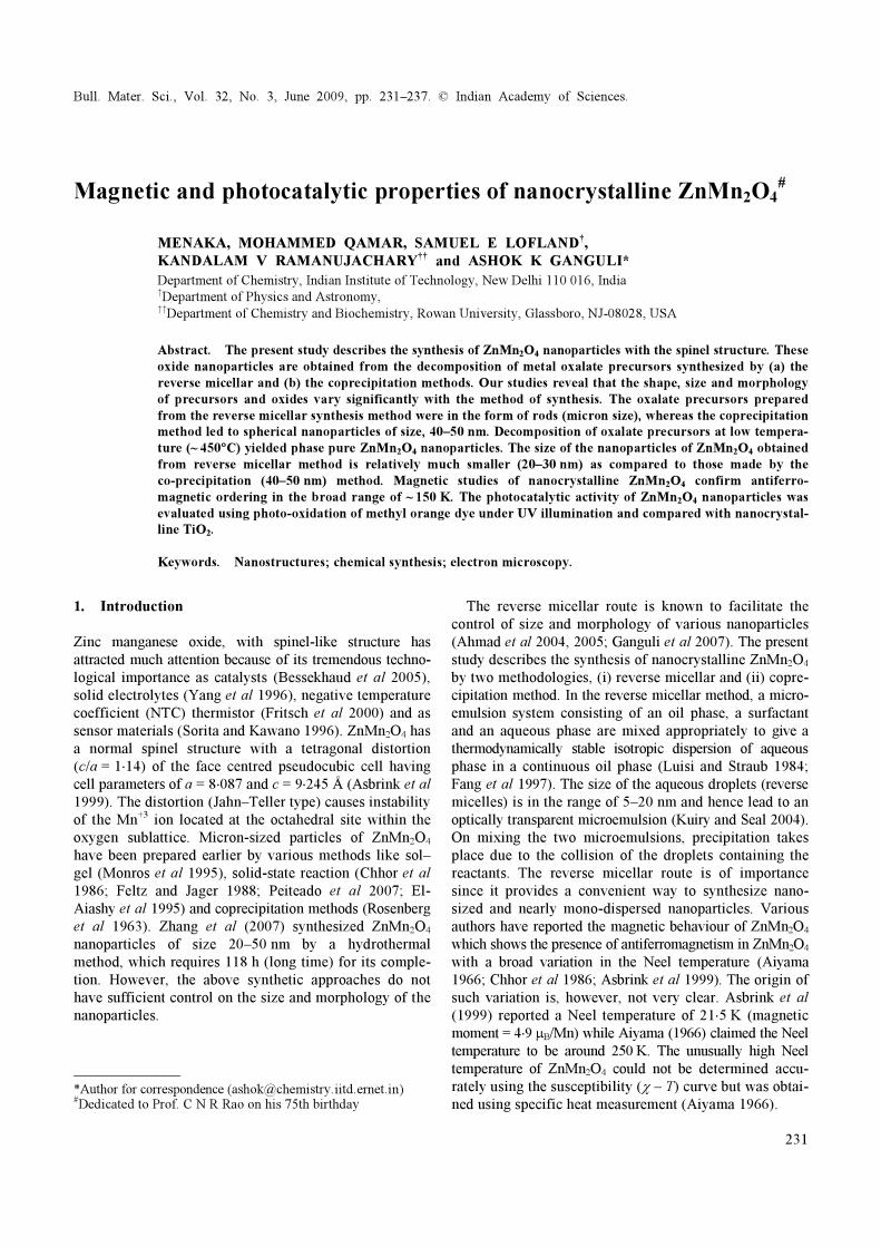
Figure 1. Flow chart indicating the method of synthesis of ZnMn2O4 using (a) reverse micellar method and (b) coprecipitation method.
Here we report the synthesis of rod-shaped zinc and manganese oxalate dihydrates (reverse micellar method) as well as spherical particles (coprecipitation method). Their decomposition yields pure ZnMn2O4 nanoparticles. The rods and nanoparticles have been characterized by powder X-ray diffraction (PXRD), thermogravimetric analysis (TGA), differential thermal analysis (DTA) and transmission electron microscopy (TEM). The magnetic behaviour of ZnMn2O4 nanoparticles was investigated using a Quantum Design Physical Properties Measure-ment system. The photocatalytic activity of ZnMn2O4 was evaluated spectrophotometrically (Bessekhaud et al 2005; Qamar et al 2008).
2. Experimental
We attempted to synthesize the mixed metal oxalate pre-cursor, ZnMn2(C2O4)3⋅xH2O, by the reverse micellar method following the procedure developed earlier for other oxalates (Ahmad et al 2004, 2005). Three micro-
emulsions were prepared using cetyltrimethyl ammonium bromide (CTAB) as the surfactant, 1-butanol as the co-surfactant, isooctane as the non-polar phase and the aqueous solutions were made up of 0⋅1 M of (i) zinc nitrate, (ii) manganese acetate and (iii) ammonium oxalate. The Zn+2 : Mn+2 molar ratio of 1 : 2 was maintained in the starting reagents. The weight fraction of various constitu-ents in the microemulsions was 16⋅86% of CTAB, 14⋅1% of n-butanol, 58⋅91% of isooctane and 10⋅11% of aqueous phase. The synthetic procedure (reverse micellar method) for the synthesis of oxalate precursor as well as oxide nanoparticles is summarized in a flow chart (figure 1a). We have also synthesized the mixture of oxalates of zinc and manganese by the coprecipitation method using ammonium oxalate and mixture of manganese acetate and zinc nitrate. 10 ml of 0⋅1 N aq. zinc nitrate solution was slowly mixed with 20 ml of 0⋅1 N aq. manganese acetate. To this solution 30 ml of 0⋅1 N ammonium oxalate was slowly added and then stirred overnight. The oxide nanoparticles were obtained after decomposition of the
Magnetic and photocatalytic properties of nanocrystalline ZnMn2O4
233
precursors of zinc and manganese oxalate at a tempera-ture of 450°C in air and to improve the crystallinity, further heated the sample at 600°C in air for 12 h. Powder X-ray diffraction studies were carried out on a Bruker D8 Advance diffractometer with Ni-filtered Cu-Kα radiation using a step size of 0⋅02° and a step time of 1 s. Raw data were subjected to background correction and Kα2 lines were removed. The crystallite size was calculated from Scherrer’s formula (Ahmad et al 2004). Thermogravimetric (TGA) and differential thermal analy-sis (DTA) experiments were carried out on a Perkin Elmer TGA and DTA system on well ground samples in a flowing nitrogen atmosphere with a heating rate of 5°C/min. Transmission electron microscopy (TEM) studies were carried out using a Tecnai G2 20 electron micro-scope operated at 200 kV. TEM specimens were prepared by dispersing the oxide powder in ethanol by ultrasonic
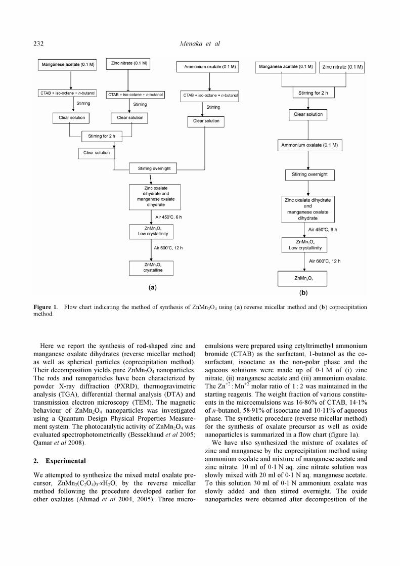
Figure 2. PXRD pattern of the metal oxalates obtained using a. reverse micellar and b. coprecipitation route.
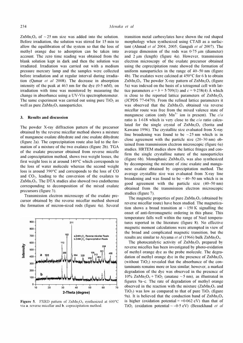
Figure 3. TGA/DTA plot for the decomposition of mixture of zinc oxalate and manganese oxalate prepared using reverse micellar route.
treatment. A few drops were poured onto a porous carbon film supported on a copper grid and then dried in air. Temperature and field dependent magnetization mea-surements were carried out at temperatures ranging from 5–300 K in an applied field of 1 kOe with a Quantum Design Physical Properties Measurement system. The photocatalytic activity of ZnMn2O4 was evaluated with a photo-reactor made up of Pyrex glass equipped with a magnetic stirring bar. For irradiation experiments, 200 ml of methyl orange dye of 0⋅5 mM concentration was taken into the vessel and appropriate amount of the catalyst, ca 180 mg, of commercially available TiO2 nano-powder (Aldrich, 99⋅7%) of ~ 5 nm size and 20 mg of
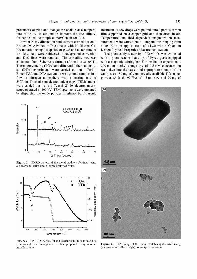
Figure 4. TEM image of the metal oxalates synthesized using (a) reverse micellar and (b) coprecipitation route.
Menaka et al
234
ZnMn2O4 of ~ 25 nm size was added into the solution. Before irradiation, the solution was stirred for 15 min to allow the equilibration of the system so that the loss of methyl orange due to adsorption can be taken into account. The zero time reading was obtained from the blank solution kept in dark and then the solution was irradiated. Irradiation was carried out with a medium pressure mercury lamp and the samples were collected before irradiation and at regular interval during irradia-tion (Qamar et al 2008). The decrease in absorption intensity of the peak at 463 nm for the dye (0⋅5 mM), on irradiation with time was monitored by measuring the change in absorbance using a UV-Vis spectrophotometer. The same experiment was carried out using pure TiO2 as well as pure ZnMn2O4 nanoparticles.
3. Results and discussion
The powder X-ray diffraction pattern of the precursor obtained by the reverse micellar method shows a mixture of manganese oxalate dihydrate and zinc oxalate dihydrate (figure 2a). The coprecipitation route also led to the for-mation of a mixture of the two oxalates (figure 2b). TGA of the oxalate precursor obtained from reverse micelle and coprecipitation method, shows two weight losses, the first weight loss is at around 140°C which corresponds to the loss of water molecule whereas the second weight loss is around 390°C and corresponds to the loss of CO and CO2, leading to the conversion of the oxalates to ZnMn2O4. The DTA studies also showed two endotherms corresponding to decomposition of the mixed oxalate precursors (figure 3). Transmission electron microscopy of the oxalate pre-cursor obtained by the reverse micellar method showed the formation of micron-sized rods (figure 4a). Several
Figure 5. PXRD pattern of ZnMn2O4 synthesized at 600°C via a. reverse micellar and b. coprecipitation method.
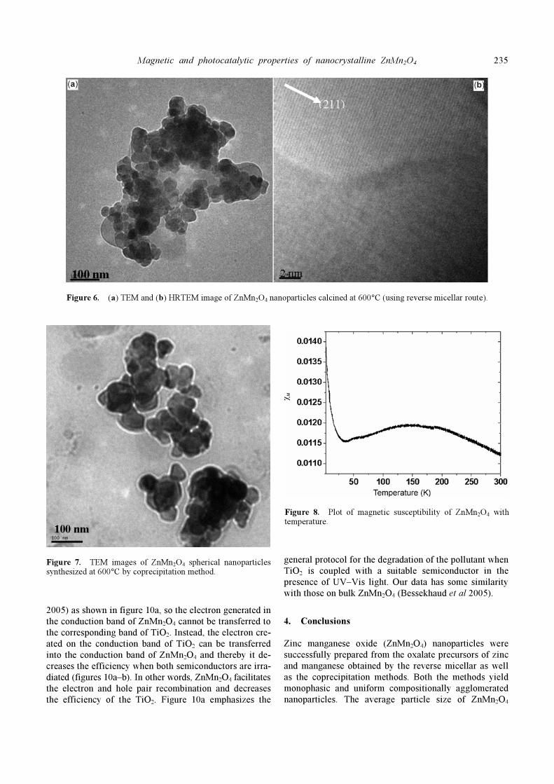
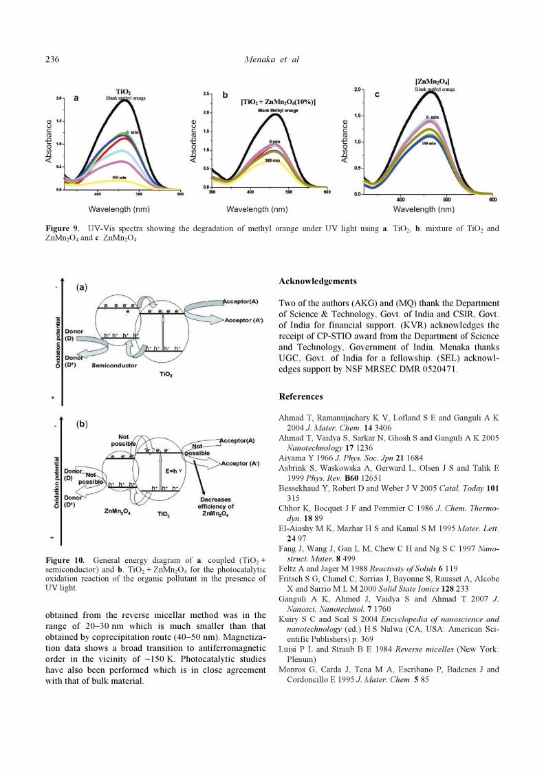
transition metal carboxylates have shown the rod shaped morphology when synthesized using CTAB as a surfac-tant (Ahmad et al 2004, 2005; Ganguli et al 2007). The average dimension of the rods was 0⋅75 μm (diameter) and 2 μm (length) (figure 4a). However, transmission electron microscopy of the oxalate precursor obtained using the coprecipitation route showed the formation of uniform nanoparticles in the range of 40–50 nm (figure 4b). The oxalates were calcined at 450°C for 6 h to obtain ZnMn2O4. The powder X-ray pattern of ZnMn2O4 (figure 5a) was indexed on the basis of a tetragonal cell with lat-tice parameters a = b = 5⋅709(1) and c = 9⋅238(4) Å which is close to the reported lattice parameters of ZnMn2O4 (JCPDS 77-0470). From the refined lattice parameters it was observed that the ZnMn2O4 obtained via reverse micellar route was free from the mixed valence state of manganese cation (only Mn3+ ion is present). The c/a ratio is 1⋅618 which is very close to the c/a ratio calcu-lated for the single crystal of ZnMn2O4 (Sorita and Kawano 1996). The crystallite size evaluated from X-ray line broadening was found to be ~ 25 nm which is in close agreement with the particle size (20–30 nm) ob-tained from transmission electron microscopic (figure 6a) studies. HRTEM studies show the lattice fringes and con-firm the single crystalline nature of the nanoparticles (figure 6b). Monophasic ZnMn2O4 was also synthesized by decomposing the mixture of zinc oxalate and manga-nese oxalate obtained by coprecipitation method. The average crystallite size was evaluated from X-ray line broadening and was found to be ~ 40–50 nm which is in good agreement with the particle size (40–50 nm) obtained from the transmission electron microscopic studies (figure 7). The magnetic properties of pure ZnMn2O4 (obtained by reverse micellar route) have been studied. The magnetiza-tion shows a broad transition at ~ 150 K signalling the onset of anti-ferromagnetic ordering in this phase. This temperature falls well within the range of Neel tempera-tures reported in the literature (figure 8). No effective magnetic moment calculations were attempted in view of the broad and complicated magnetic transition, but the results are similar to Aiyama et al (1966) bulk ZnMn2O4. The photocatalytic activity of ZnMn2O4 prepared by reverse micelles has been investigated by photo-oxidation of methyl orange dye as the probe molecule. The degra-dation of methyl orange dye in the presence of ZnMn2O4 (without TiO2) revealed that the absorbance of the con-taminants remains more or less similar; however, a marked degradation of the dye was observed in the presence of 10% ZnMn2O4 + TiO2 (anatase ~ 5 nm), as illustrated in figures 9a–c. The rate of degradation of methyl orange observed in the reaction with the mixture (ZnMn2O4 and TiO2) was low as compared to that of pure TiO2 (figure 9a). It is believed that the conduction band of ZnMn2O4 is higher (oxidation potential = +0⋅062 eV) than that of TiO2 (oxidation potential ~ –0⋅5 eV) (Bessekhaud et al
Magnetic and photocatalytic properties of nanocrystalline ZnMn2O4
235
Figure 6. (a) TEM and (b) HRTEM image of ZnMn2O4 nanoparticles calcined at 600°C (using reverse micellar route).
Figure 7. TEM images of ZnMn2O4 spherical nanoparticles synthesized at 600°C by coprecipitation method.
2005) as shown in figure 10a, so the electron generated in the conduction band of ZnMn2O4 cannot be transferred to the corresponding band of TiO2. Instead, the electron cre-ated on the conduction band of TiO2 can be transferred into the conduction band of ZnMn2O4 and thereby it de-creases the efficiency when both semiconductors are irra-diated (figures 10a–b). In other words, ZnMn2O4 facilitates the electron and hole pair recombination and decreases the efficiency of the TiO2. Figure 10a emphasizes the
general protocol for the degradation of the pollutant when TiO2 is coupled with a suitable semiconductor in the presence of UV–Vis light. Our data has some similarity with those on bulk ZnMn2O4 (Bessekhaud et al 2005).
4. Conclusions
Zinc manganese oxide (ZnMn2O4) nanoparticles were successfully prepared from the oxalate precursors of zinc and manganese obtained by the reverse micellar as well as the coprecipitation methods. Both the methods yield monophasic and uniform compositionally agglomerated nanoparticles. The average particle size of ZnMn2O4
Figure 8. Plot of magnetic susceptibility of ZnMn2O4 with temperature.
Menaka et al
236
Figure 9. UV-Vis spectra showing the degradation of methyl orange under UV light using a. TiO2, b. mixture of TiO2 and ZnMn2O4 and c. ZnMn2O4.
Figure 10. General energy diagram of a. coupled (TiO2 + semiconductor) and b. TiO2 + ZnMn2O4 for the photocatalytic oxidation reaction of the organic pollutant in the presence of UV light.
obtained from the reverse micellar method was in the range of 20–30 nm which is much smaller than that obtained by coprecipitation route (40–50 nm). Magnetiza-tion data shows a broad transition to antiferromagnetic order in the vicinity of ~150 K. Photocatalytic studies have also been performed which is in close agreement with that of bulk material.
Acknowledgements
Two of the authors (AKG) and (MQ) thank the Department of Science & Technology, Govt. of India and CSIR, Govt. of India for financial support. (KVR) acknowledges the receipt of CP-STIO award from the Department of Science and Technology, Government of India. Menaka thanks UGC, Govt. of India for a fellowship. (SEL) acknowl-edges support by NSF MRSEC DMR 0520471.
References
Ahmad T, Ramanujachary K V, Lofland S E and Ganguli A K
2004 J. Mater. Chem. 14 3406
Ahmad T, Vaidya S, Sarkar N, Ghosh S and Ganguli A K 2005
Nanotechnology 17 1236
Aiyama Y 1966 J. Phys. Soc. Jpn 21 1684
Asbrink S, Waskowska A, Gerward L, Olsen J S and Talik E
1999 Phys. Rev. B60 12651
Bessekhaud Y, Robert D and Weber J V 2005 Catal. Today 101
315
Chhor K, Bocquet J F and Pommier C 1986 J. Chem. Thermo-
dyn. 18 89
El-Aiashy M K, Mazhar H S and Kamal S M 1995 Mater. Lett.
24 97
Fang J, Wang J, Gan L M, Chew C H and Ng S C 1997 Nano-
struct. Mater. 8 499
Feltz A and Jager M 1988 Reactivity of Solids 6 119
Fritsch S G, Chanel C, Sarrias J, Bayonne S, Rausset A, Alcobe
X and Sarrio M L M 2000 Solid State Ionics 128 233
Ganguli A K, Ahmed J, Vaidya S and Ahmad T 2007 J.
Nanosci. Nanotechnol. 7 1760
Kuiry S C and Seal S 2004 Encyclopedia of nanoscience and
nanotechnology (ed.) H S Nalwa (CA, USA: American Sci-
entific Publishers) p. 369
Luisi P L and Straub B E 1984 Reverse micelles (New York:
Plenum)
Monros G, Carda J, Tena M A, Escribano P, Badenes J and
Cordoncillo E 1995 J. Mater. Chem. 5 85

Magnetic and photocatalytic properties of nanocrystalline ZnMn2O4
237
Peiteado M, Caballero A C and Makovec D 2007 J. Solid State
Chem. 180 2459
Qamar M et al 2008 Catal. Today 131 3
Rosenberg M, Nicolau P, Manaila R and Pausescu P 1963 J.
Phys. Chem. Solids 24 1419
Sorita R and Kawano T 1996 Sensor and Actuators B35–36 274
Yang H, Yang Q H, Lu Y L and Li B X 1996 J. Power Sources
62 223
Zhang X D, Wu Z S, Zang J, Li D and Zhang Z D 2007 J. Phys.
Chem. Solids 68 1583