Embed Size (px)
Citation preview
Easte
Eas
Email: m
rn Asso
Mainten
DisneLak
Therese D
stern Associatio633 N. Saint
ChicPh: 312-202-5
managementoffic
ociation
nance ofAcute C
Wo
Januaey’s Conke Buen
WorksJose Di
Rao IvatPatrick R
Duane, MD,
on for the Surget Clair Street, Stcago, IL 60611
5508 Fax: [email protected] W
for the S
f CertificCare Surorkshop
ary 10, 2ntemporna Vista,
shop Facultiaz, MD, FAtury, MD, F
Reilly, MD, F
, FACS – W
ery of Trauma te 2600
202-5064 Web: www.east
Surgery
cation (Mrgery
p
012 rary Res, Florida
ty: ACS FACS FACS
Workshop D
.org
y of Trau
MOC)
sort a
Director
uma

Jose J. Diaz, MD, CNS, FACS, FCCMProfessor of Surgery
Chief of Acute Care SurgeryR Adams Cowley Shock Trauma CenterUniversity of Maryland Medical Center
Baltimore, Maryland
History Incidence of gallstone disease Presentation Workup Indications
Carl Johann August Langenbuch-performed the first gallbladder removal (July 15, 1882)
At the time, medical management of acute cholecystitis consisted of hot local compresses, belladonna, hanging patients by their feet and shaking them (in hope of dislodging stones), and narcotics in increasing amounts that often resulted in addiction.
As the outcome was seldom satisfactory, need for an alternative method of intervention was blatantly apparent.
Aust N Z J Surg 1993;63:56-64. JAMA 1993;270:1429-32. Surgical Rounds 1982; 68 -75. J La State Med Soc 1991;143:22-5.
1743, Jean Louis Petit, a surgeon in Paris, recognized the benefit of biliary procedures.
Petit limited such operations to instances where pericholecystic abscesses ruptured transcutaneously, or for the cutaneous removal of stones through cholecystocutaneous fistulas.
More than 25 million Americans have gallstones, and a million are diagnosed each year.
14.2 million are in women 600K-750K cholecystectomies / year are
performed in the United States◦ most are for symptomatic gallstone disease
Only 1 - 3% of the population complains of symptoms during the course of a year, and fewer than half of these people have symptoms that return
Everhart et al, Gastroenterology. 1999, 117: 632-639.
Women more likely than men to develop gallstones. 25% of women in the US by age 60, and 50% by age 75. Most cases are asymptoms. Women at increased risk due to estrogen stimulation of
the liver to remove more cholesterol from blood and divert it into the bile.
Pregnancy increases the risk for gallstones, and pregnant women with stones are more likely to have symptoms than non-pregnant women.
Several large studies have shown that the use of hormone replacement therapy (HRT) 2X -3X the risk for gallstones, hospitalization for gallbladder disease, or gallbladder surgery.
Estrogen raises triglycerides, a fatty acid that increases the risk for cholesterol stones.

20% of men have gallstones by age 75. A study of nursing home residents reported
that 66% of the women and 51% of the men had gallstones.
Men are more likely to have severe disease and surgical complications than women.
Spinal injury History of abdominal
surgery Sickle-cell anemia Impaired immune
system TPN
Ethnicity Hispanics & Northern
Europeans higher risk for gallstones than Asian and African descent
Native North & South Americans
Pima Indians in US & native populations in Chile and Peru.
Pima women 80% chance of developing gallstones during their lives & virtually all native Indian females in Chile and Peru develop gallstones
Mutation gene ABCG8 significantly increases a person's risk of gallstones. ◦ Gene controls a cholesterol pump that transports
cholesterol from the liver to the bile duct. It appears this mutation may cause the pump to continuously work at a high rate.
Obesity and Weight Changes, Metabolic Syndrome, Crohn’s Disease, Organ transplant, Blood disorders, Medications (octreotide), Heme Iron
Cholesterol gallstones: bile contains too much cholesterol and not enough bile salts.
2 other factors are important in causing gallstones. ◦ 1st- how often & how well the
gallbladder contracts, incomplete and infrequent emptying of the gallbladder may cause bile to become over concentrated and contribute to gallstone formation.
◦ 2nd - presence of proteins in the liver & bile that either promote or inhibit cholesterol crystallization into gallstones.

Note: acute hepatitis, other causes of acute abdomen, and chronic cholecystitis should be excluded
A. Local signs of inflammation
B. Systemic signs of inflammation
C. Imaging findings
A. Murphy’s sign, RUQ mass/pain/tenderness
B. Fever, elevated CRP, elevated WBC count
C. Imaging findings characteristic of acute cholecyctitis
Definite diagnosis(1) One item in A and one item in B are positive(2) C confirms the diagnosis when acute cholecystitis is suspected clinically
T. Mayumi et al.: Results of the Tokyo Consensus Meeting
Sonographic Murphy sign (tenderness elicited by pressing the gallbladder with the ultrasound probe)
Thickened gallbladder wall (>4 mm, if the patient does not have chronic liver disease and/or ascites or right heart failure)
Enlarged gallbladder (long axis diameter >8 cm, short axis diameter >4 cm)
Incarcerated gallstone, debris echo, pericholecystic fluid collection
Sonolucent layer in the gallbladder wall, striated intramural lucencies, and Doppler signals
T. Mayumi et al.: Results of the Tokyo Consensus Meeting
Pericholecystic high signal
Enlarged gallbladder
Thickened gallbladder wall
T. Mayumi et al.: Results of the Tokyo Consensus Meeting

Thickened gallbladder wall
Pericholecystic fluid collection
Enlarged gallbladder
Linear high-density areas in the pericholecystic fat tissue
T. Mayumi et al.: Results of the Tokyo Consensus Meeting
Gallbladder wall
thickening
Gallstones
475 patients (age, >64 y) with AC Groups: US alone (n = 240), CT alone (n = 60), and
CT + US (n = 168). 60 (35.7%) US + CT group had inflammation in both
studies 34 (20.2%) inflammation only on US 32 (19.0%) inflammation only on CT. US + CT detection of cholelithiasis was not different Peritonitis, leukocytosis, and acidosis were more
frequent in the 2 groups undergoing CT. cholecystectomy rate was lowest (and the
complication rate was highest) in the CT + US group.
McGillicuddy etal, Am J Surg. 2011 Nov;202(5):524-7
Non-visualized gallbladder with normal uptake and excretion of radioactivity
Rim sign (augmentation of radioactivity around the gallbladder fossa)
T. Mayumi et al.: Results of the Tokyo Consensus Meeting

Mild (grade I) acute cholecystitis
“Mild (grade I)” acute cholecystitis does not meet the criteria of “severe (grade III)” or “moderate (grade II)” acute cholecystitis.
It can also be defined as acute cholecystitis in a healthy patient with no organ dysfunction and mild inflammatory changes in the gallbladder, making cholecystectomy a safe and low-risk operative procedure.
T. Mayumi et al.: Results of the Tokyo Consensus Meeting
Moderate (grade II) acute cholecystitis “Moderate” acute cholecystitis is associated
with any one of the following conditions:◦ 1. Elevated WBC count (>18 000/mm3)◦ 2. Palpable tender mass in the right upper
abdominal quadrant◦ 3. Duration of complaints >72 h ♦◦ 4. Marked local inflammation (biliary peritonitis,
pericholecystic abscess, hepatic abscess, gangrenous cholecystitis, emphysematous cholecystitis)
♦ Laparoscopic surgery should be performed within 96 h of the onset of acute cholecystitis
T. Mayumi et al.: Results of the Tokyo Consensus Meeting
Severe (grade III) acute cholecystitis “Severe” acute cholecystitis is associated with
dysfunction of any one of the following organs/systems◦ 1. Cardiovascular dysfunction (hypotension requiring
treatment with dopamine 5 μg/kg per min, or any dose of dobutamine)
◦ 2. Neurological dysfunction (decreased level of consciousness)
◦ 3. Respiratory dysfunction (PaO2/FiO2 ratio <300)◦ 4. Renal dysfunction (oliguria, creatinine >2.0 mg/dl)◦ 5. Hepatic dysfunction (PT-INR > 1.5)◦ 6. Hematological dysfunction (platelet count
<100,000/mm3)
T. Mayumi et al.: Results of the Tokyo Consensus Meeting

At least two published surveys show that 40 to 80% of surgeons do not offer CCK at the index admission.
Schuerer, Plenary Session EAST PMG “08,09,11
Grade 1A◦ Laparoscopic cholecystectomy should be attempted
first over open cholecystectomy for acute cholecystitis.
Grade 1B◦ This applies for patients over age 60 as well.
Schuerer, Plenary Session EAST PMG “08,09,11
Grade 1A◦ Laparoscopic cholecystectomy should be done during
the index admission versus a delay of 6 or more weeks. Grade 1B◦ Laparoscopic cholecystectomy should be done within 72
hours of symptoms when possible. 3 RCTs show that early laparoscopic CCK vs 6 or
greater week delay leads to decreased LOS and costs. LOS difference was 3-5 days.
Between 15-26% in the delayed group had to have surgery early due to continued symptoms.
Schuerer, Plenary Session EAST PMG “08,09,11
Laparoscopic Needle Decompression

Overall conversion rate higher in the delayed intervention group (23.6%) vs. early intervention group (20.3%). N.S.
Reports of increased conversion rate if surgery is delayed more than 48 - 96 hours after the onset of symptoms◦ (Eldar 1997;Madan 2002; Liguori 2003; Peng 2005)
Other studies do not confirm this ◦ (Knight 2004).◦ 2 trials included <4 days of onset of symptoms. ◦ 3 trials included <7 days of onset of symptoms.
Subgroup analysis(N.S.) conversion rate or complication rate in the early group (<4 days of onset of symptoms or <7days of onset of symptoms) vs. delayed laparoscopic cholecystectomy
Laparoscopic cholecystectomy up to 7 days after onset of symptoms safe.◦ Time for dealing with CBD stones before operation
In all trials but 1 (Johansson 2003), operating times were longer for early compared with the delayed group
Total LOS ~ 4 days shorter for the early vs. delayed group in all the trials
Bile duct injury rate 0.5% early group vs 1.4% in the delayed group
N.S. between the two groups for this most feared complication (OR 0.63, 95% CI 0.15 to 2.70).
• 17.5% in the delayed group had either non-resolution of symptoms or recurrence of symptoms prior to planned operation
• Leading to emergent lap cholecystectomy. • Proportion with conversion to open cholecystectomy 45%• 2 pts developed cholangitis awaiting cholecystectomy
No significant differences in the complication rate or the conversion rate whether the laparoscopic cholecystectomy is performed during acute cholecystitis or performed 6 to 12 weeks after the symptoms settle.
Early laparoscopic cholecystectomy has the advantage of decreased LOS & these patients do not run the risk of non-solved symptoms or risk of emergency operation.
The latter leads to a high proportion of patients undergoing open cholecystectomy.

1,000 laparoscopic cholecystectomies ◦ (March 1992 to July 1999) prospectively analyzed
804 women (80.4%) & 196 men (19.6%) with a mean age of 43.8 years (range, 30-80 years).
48/1,000 (4.8%) patients attempt laparoscopic cholecystectomy required conversion to open surgery.
most common reason for conversion: inability to define anatomy -inflamed contracted gallbladder (n = 34).
Significantly independent predictive factors for conversion ◦ male gender, previous abdominal surgery, acute cholecystitis,
thickened gallbladder wall on preoperative ultrasonography, and suspicion of common bile duct stones.
Kama et al, Surg Endosc (2001) 15:965-968
Kama et al, Surg Endosc (2001) 15:965-968
National Hospital Discharge database 1998 - 2001 (CDC Data)
All gallbladder disease related admissions, and the cholecystectomies (ICD-9-CM codes 51.2X)
~25% of all cholecystectomies are performed by the open technique.
Remaining 75%, ~ 5% to 10% conversion rate. Major risk factors for conversion:◦ male sex, obesity, and cholecystitis.
Concurrent choledocholithiasis, cholelithiasis, and cholecystitis associated with a conversion rate of 25%.
LOS reduced for laparoscopic operations and conversion added 2 - 3 days to LOS, for most cases the LOS was still less than for primary open operations.
Livingston Am J Surg 188(2004) 205-211
Livingston Am J Surg 188(2004) 205-211
NSQIP database: retrospectively reviewed 1,193 cholecystectomies performed at their institution from 2002 - 2009 and identified 70 conversions.
91% of conversion cases: conversion was elective. 49% of these conversions: number of ports was fewer than
four. Most conversions were performed after minimal or no
attempt at dissection. There were no differences in LOS, complications, operating
room charges, or hospital charges between categories. Of the 6 emergent conversions (9%), bleeding and concern
about common bile duct (CBD) injury were the main reasons.
1 CBD injury occurred
Lengyel et al, Surg Endosc. 2011 Sep 23

Lengyel et al, Surg Endosc. 2011 Sep 23
Most commonly cultured organisms: Escherichia coli, Klebsiella-Enterobacter, enterococci, Clostridium perfringens, and staphylococci
Gallstones did not influence the prevalence of positive cultures
Wound infections:11% of the men and 2% of the women.
AMA Arch Surg. 1973;106(2):169-171
CID 2010:50(15 January)
Management of patients with severe acute cholecystitis (AC) remains controversial.
Options LC conversion to open cholecystectomy or surgical cholecystostomy tube (CCT) placement, or initial percutaneous CCT.
185 PTS with AC and who received CCT. Age 71 years and 80% had1 comorbidity (mean 2.6). 78 % CCTs percutaneous CCT and 22% surgical CCT. Median LOS from CCT insertion to discharge 4 days. Majority (57%) of patients - cholecystectomy at a median of 63 days post-
CCT (range 3 to 1,055 days)◦ 86% underwent LC and 13% underwent open conversion or open cholecystectomy.
In the radiology and surgical group, 50% and 80% underwent subsequent cholecystectomy, respectively, at a median of 63 and 60 days post-CCT.
Whether surgical or percutaneous CCT placement, approximately the same proportion of patients (85% to 86%) underwent LC as definitive treatment.
Cherng et al, J Am Coll Surg 2012 Cherng et al, J Am Coll Surg 2012

Percutaneous drainage (PD) of the gallbladder to emergency cholecystectomy (EC) in PTS with sepsis related to acute calculous/acalculous cholecystitis (ACC/AAC).
42 PTS with age 65.5 years 45% EC (10 laparoscopic, 9 open) and 55% PD (n = 23). 91% Percutaneous drainage and100% EC successful Organ dysfunctions 3rd postoperative/postdrainage days. Despite undergoing PD, 2 pts required EC due to gangrenous
cholecystitis. Conversion rate after laparoscopy was 20%. Overall morbidity: 8.7% after PD and 47% after EC Major morbidity: 0% after PD and 21% EC Mortality rate was not different (13% after PD and 16% after EC, P = 1.0) ◦ deaths related to the patients' preexisting disease.
Hospital and ICU stays were not different. Recurrent symptoms (17%) occurred only after ACC in the PD group. Secondary cholecystectomy is mandatory in cases of acute calculous
cholecystitis.
Melloul et al, World J Surg. 2011 Apr;35(4):826-33
Retrospectively analyzed 35 patients (2003- 2009) Perc. cholecystostomy - technically successful in all
patients. Symptoms resolved within 3 days in 33/35 patients. 2 patients needed emergency laparotomy. Catheter dislodged 5 patients and was replaced in
2/5. 30-day mortality rate was 3/35 (8.7%) due to
gallbladder necrosis, myocardial infarction and multiorgan failure.
LOS 17 days and median drainage time was 28 days 23 patients (66%) underwent open or laparoscopic
cholecystectomy after a median interval of 44 days.
Koebrugge etal, Dig Surg. 2010;27(5):417-21
24 elderly/critically ill pts unfit for surgery with acute cholecystitis underwent percutaneous cholecystostomy as an emergency procedure◦ Tokyo Guidelines, ASA physical status used for the perioperative risk
4 male:10 female with a median age of 79 years Acute cholecystitis was classified as grade 2 -20, grade 3 - 4 ASA score III-17 patients & 7 as ASA score IV 23 Gallstones & 1 acalculous cholecystitis. Perc. cholecystostomy technically feasible in all patients Clinical improvement:14 patients within 24 hours and in all
patients within 72 hours. Reduction in WBC, C-reactive protein, temperature in 72 hours. Procedure-related mortality 4% Median follow-up 17.5months: definitive and effective control of
symptoms was achieved in 90.5%
Grinatsos South Med J. 2008 Jun;101(6):586-90.
10% of acute cholecystitis cases - perforated gallbladder◦ life-threatening condition.
Perforation of the gallbladder is most common in diabetics.
Risk for perforation increases in emphysematous cholecystitis
Once the gallbladder has been perforated, pain may temporarily decrease.
This is a dangerous and misleading event as peritonitis develops afterward.
Neimeier’s Classification◦ In 1934 Neimeier proposed a classification of
gallbladder perforation Type I Acute free perforation into the
peritoneal cavity Type II Subacute perforation with
pericholecystic abscess Type III Chronic perforation with
cholecystoenteric fistula
Niemeier Ann. Surg. 1934; 99: 922-4.Anderson et al, J. Natl. Med. Assoc. 1987; 79: 393–9.
During a 20-year period (1961 - 1980) 3,260 admissions acute & chronic cholecystitis.
115 perforations of the gallbladder ◦ incidence of 3.5 percent.
JOURNAL OF THE NATIONAL MEDICAL ASSOCIATION, VOL. 73, NO. 4,1981

Gallbladder disease is a common surgical problem
Women>Men Imaging: US + CT maybe helpful Laparoscopic Cholecystectomy should be
considered as the initial modality in most cases
Predictors of for conversion: male, obese, acute cholecystitis
Strongly consider early LC during the index admission
Antibiotic Regimen If patient is too sick consider
cholecystostomy tube Surgical Emergency> perforated Cholecystitis

1
MOC - Appendicitis
Patrick M Reilly MD FACSProfessor of Surgery
Perelman School of Medicine atThe University of Pennsylvania
Outline• Introduction
• Diagnosis / Imaging
• Technique
• Operative Findings
• Medical Therapy
• Complicated Appendicitis
• Special Groups
• Future
Appendicitis
• Most Common Surgical Emergency
• Pathophysiology: Luminal Obstruction• Children: Lymphoid Hyperplasia
• Adults: Fecolith
• Elderly: Neoplasm on Occasion
• Mortality : 1%
Annals Surg 2011
Annals Surg 2011
Diagnosis
• Presentation• Mid abdominal pain that localizes to RLQ
• Physical Exam• Localized Tenderness
• Lab Studies• Leukocytosis
• Negative UA
• C Reactive Protein?

2
Diagnosis
• Clinical Signs• Rovsing’s Sign
• RLQ Pain caused by LLQ palpation
• Psoas Sign (Retrocecal Appendix)• Pain on extendion of right thigh
• Obturator Sign (Pelvic Appendix)• Pain on medial rotation of the thigh
…what does it all mean…
• What our EM Docs are Reading
EM Clinics of NA 2010
EM Clinics of NA 2010 EM Clinics of NA 2010
Annals EM 2010
Alvarado Score
• < 3 5 % Appendicitis
• 4 – 6 36% Appendicitis
• > 7 78% Appendicitis
…guide to ordering a CT Scan…
Am J Em Med 2007

3
Imaging in Appendicitis
• Ultrasound
• CT Scan
• MRI
…goal to improve accuracy
of diagnosis…
Imaging in Appendicitis
• PROs• Improved Accuracy
• Decreased Negative Appendectomy Rate
• CONs• Costs
• Delays to Surgery
• Radiation Exposure
Graded Compression Ultrasound
• Noncompressible Appendix
• Diameter > 6 mm
• Sensitivity 86%
• Specificity 81%
• Accuracy at Night?
Annals Int Med 2004
Graded Compression Ultrasound
Multidetector CT Scan
• Diameter > 6mm
• Wall Thickening
• RLQ Inflammation
• Appendicolith
• Sensitivity 98.5% / Specificity 98%
• Benefits of Contrast?
Annals Int Med 2011
Multidetector CT Scan
AJR 2011
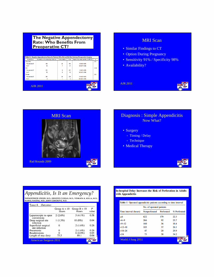
4
AJR 2011
MRI Scan
• Similar Findings to CT
• Option During Pregnancy
• Sensitivity 91% / Specificity 98%
• Availability?
AJR 2011
MRI Scan
Rad Rounds 2009
Diagnosis : Simple AppendicitisNow What?
• Surgery• Timing / Delay
• Technique
• Medical Therapy
American Surgeon 2011 World J Surg 2011

5
Ann Surg 2011 JACS 2011
Ann Surg 2006
Laparoscopic vs Open Surgery
• 67 Studies and 6000 Patients
• Better with Laparoscopic Approach• Lower Wound Infection Rate
• Less POD # 1 Pain
• Shorter Hospital Stay
• Quicker Return Bowel Function
Cochrane Review 2010
Laparoscopic vs Open Surgery
• 67 Studies and 6000 Patients
• Worse with Laparoscopic Approach• Higher Rate of Intra-abdominal Abscess
• Longer Operative Time
• Higher Operative and Hospital Costs
Cochrane Review 2010
• Retrospective, Observational Study
• 40,000 Appendectomies• 2006 - 2008
• Complicated and Uncomplicated
• Overall Favored Lap Appendectomy
Annals Surg 2011
Operative Findings
• Normal Appendix• Look for other pathology
• IBD / PUD
• Diverticulitis / TOA
• Remove Appendix• Microscopic Findings
• Future RLQ pain
• Cecal Inflammation?

6
Appendiceal Neoplasms
• Very Rare: < 1% of Appendectomies• Often Found on microscopic review
• Carcinoid• Right Hemicolectomy for >2 cm
• Primary Adenocarcinoma• Right Hemicolectomy
• Cystic / Pseudomyxoma• Avoid spillage as able
Medical Therapy for Appendicitis
• First Described in 1959
• Recent Studies – New Life
• Prospective Randomized Studies• Few
• Poorly Described
• Cochrane Review 2011• Inconclusive
Lancet 2011
• Randomized Trial• Antibx Group
• 12% appendectomy within one month• 29% appendectomy within one year• Fecolith as risk factor
• Conclusion - Noninferior?
Lancet 2011
JACS 2011
• Retrospective Study• Postop Antibx
• No change in SSIs• Increased UTI / c. diff / diarrhea• Increased hospital LOS
Complicated Appendicitis
• Phlegmon
• Abscess
• Surgery depends on presentation• Sepsis / Peritonitis – Surgery
• Indolent – Antibx +/- IR drain• Interval appendectomy?

7
Complicated AppendicitisMeta-Analysis
• Conservative Therapy• Fewer Complications
• No Difference LOS or Antibiotic Course
• Results valid• Recent Studies
• Pediatric Patients
Surgery 2010
Interval Appendectomy?
• Adults (mean age 54)• 25.5% Recurrence Rate
• 83% within six months
• 3% Colon Cancer (not at cecum)
• 8% New Bowel Diagnoses
• 84% Specimens with Inflammation
World J Surg 2006J Surg Research 2010
Interval Appendectomy?
• Children• Different Pathophysiology?
• Risk of colon cancer minimal
• Recurrence Rate ~ 8%• 7.5 year follow-up
• Procedure itself is safe
J Ped Surg 2007
Interval Appendectomy?
• What to do?• Children – Weigh risk of lifetime
• Women of Child Bearing Age• Risk of Appendicitis during preganancy
• Older patients• Colonscopy – then personalized care
J Ped Surg 2007
Appendicitis in Pregnancy
• Most common GS problem in pregnancy• Incidence about 0.1%
• More common in 2nd trimester
• Presentation may be atypical
• Imaging often called for
JACS 2006
Imaging in Pregnancy
• Ultrasound - No Radiation• Decreased ability to visualize appendix
• CT Scan - Radiation• Reasonable Results
• MRI Scan – No Radiation• 91% Sensitivity / 98% Specificity
AJR 2011

8
Pregnancy Considerations
• Technique• Laparoscopy largely acceptable
• Studies equivalent results
• Fetal loss rate may be slightly higher• ~ 5% – 6 %
• Complicated Appendicitis• VERY limited data
WJS 2009 / J Soc Lap Surgeons 2009
Outcomes in Pregnancy
• Delay in Dx > 24 hours • Increases perforation rate
• Increases fetal loss (36% v 1.5%)
• Increased peritonitis
• Increased early delivery
• Spontaneous abortion / Premature labor• Even in uneventful procedure
JACS 2006
JACS 2011
• 3 year follow-up • 52 patients• 2% fetal loss rate• No developmental delays
• Trimester of surgery – no effect
Appendicitis in Elderly
• More likely to have gangrene / perforation• Biology of peritonitis
• Delays to presentation
• Diagnostic difficulties
• Laparoscopy safe
• Concern for cancer
Can J Surg 1996 / WJS 2009
Am J Surg 2011 Annals Surg 2011

9
Annals Surg 2011 Annals Surg 2012
Annals Surg 2012 JAMA 2011
Summary
• Ongoing Research
• New Approaches
• Great Opportunities for
Acute Care Surgery

1/18/2012
1
Rao R. Ivatury M.D., F.A.C.S.VCU Medical Center
Virginia Commonwealth UniversityRichmond, Virginia
THEOPEN ABDOMEN
“THE OPEN ABDOMEN”1.Review history
2.Outline indications, benefits and risks3. Discuss the patho-physiology of the
open abdomen ( Open abdomen as a motor for SIRS )
4. Discuss delayed primary & definitive repair
5. Outline long term outcomes
“THE OPEN ABDOMEN”
1.Review history 2.Outline indications, benefits and risks3. Discuss the patho-physiology of the
open abdomen ( Open abdomen as a motor for SIRS )
4. Discuss delayed primary & definitive repair
5. Outline long term outcomes
“THE OPEN ABDOMEN” : History and Evolution
Zipper closure of the abdominal wall in the treatment of recurrent IAA
Doody DP et al ,1986
On leaving the peritoneal cavity open ingeneralized suppurative peritonitis
Steinberg D, 1979
Open peritoneal drainage as effective treatment of advanced peritonitis
Maetani S and Tobe T, 1981
The septic abdomen : open managementwith Marlex mesh with a zipper
Hedderich GS et al, 1986

1/18/2012
2
Open management of the septic abdomen
Ivatury RR et al, Crit Care Med 1989 17:511-7
30 patients with abdominal sepsis
Groups I (Trauma) : 112 (Pancreatic abscess) : 5
3: (acute GI path) : 14
27 patients had MODS16 (53%) of the 30 survived
73% in Group 1, 60% in Group 2, 36% in Group 3
Survival correlated well with :age < 50
Absence of MODS
Open management of the post-traumatic septic abdomen
Ivatury R et al, Am Surg. 1990
13 patients with abdominal trauma and sepsisresistant to conventional methods
11 had MODS
10/13 patients (76.9%) survived,significantly better than predicted by APACHE (50%)
Necrotizing infections
Uncontrolled sepsis
Recalcitrant abscess
“for he who fights and runs away will live to fight another day:But he who is in battle slain
will never rise and fight again”
Oliver Goldsmith, 1761
“for he who fights and runs away will live to fight another day:But he who is in battle slain
will never rise and fight again”
Oliver Goldsmith, 1761
Damage-control surgeryAbbreviated laparotomy
Staged laparotomy“bail-out surgery”
Termination of the initial operation Return for completion
Temporary Abdominal Closure
(TAC)

1/18/2012
3
Open abdomen :Staged Management
Primary Closure Delayed primary Closure
Stage IVDefinitive reconstruction
Stage IIIPlanned ventral hernia
Stage II
Stage ITAC
ICU
Skin-grafting and Planned ventral hernia
Barker’s Vac-Pac“Poor man’ s VAC”
Separation of bowel
from fasciaJ-Ps to suction
Ioban
Negative Pressure therapy V.A.C
Polyurethane foam dressing -175 mm Hg suction
Vacuum Assisted Fascial Closure
Garner et al , 2001 : 13 of 14, 92%
Miller et al, 2002 : 59 of 83, 71%
Suliburk et al, 2003 : 25 0f 29, 86%
Clothren et al, 2006 : 14 of 14, 100%
“THE OPEN ABDOMEN”1.Review history
2.Outline indications, benefits and risks
3. Discuss the patho-physiology of the open abdomen ( Open abdomen as a
motor for SIRS )4. Discuss delayed primary & definitive
repair5. Outline long term outcomes

1/18/2012
4
“THE OPEN ABDOMEN”
Indications
“Damage-control” proceduresIntra-abdominal hypertension &
ACSSevere suppurative peritonitis
Proposed “second-look”Necrotic abdominal wall
“THE OPEN ABDOMEN”BenefitsReduce:
Intra-abdominal hypertension &Abdominal Compartment Syndrome
MODSFacilitate :Irrigation
confirm bowel viability
Incidence of IAH (23 of 70, 32.8%)
Group I (mesh closure):10 of 45, 22.2%
Group II (fascial suture)13 of 25, 52%
p=0.012 (Fisher’s)
INTRAABDOMINAL HYPERTENSION
Ivatury et al, 1998
Mortality 4 (8.5%) 10 (43.5%) 0.006
MODS 0.8 ± 1.9 4.3 ± 3.7 0.0001
No IAH IAHp
(n=47) (n=23)
INTRAABDOMINAL HYPERTENSION
Ivatury et al, 1998
Raeburn et al, 2001
Mortality 12% 43% 0.01
MOF 8% 32% 0.01
NO ACS ACSp(n=49) (n=28)
ACS, MOF and Mortality
Balogh et al, 2003
ACS & MOF
ACS an independent risk factor for
MOF ( OR 9.2, CI 3.8-22.8, p<0001)
Mortality (OR 8.4, CI 3.5-20.6, p<0.001)

1/18/2012
5
“THE OPEN ABDOMEN”Risks
Fluid lossesNursing problems
“Tertiary peritonitis”“EA” fistulasHernial defect
“THE OPEN ABDOMEN”
Serositis Fistulas
“Entero-atmospheric fistula”Principles of Management
1. Prevention
2. Attempt to seal the fistula
3. Control fistula effluent
4. Cover fistula with vascularized soft tissue
5. Nutritional support
6. Resect chronic fistula when timely
Prevention
Protect bowel with omentumAvoid serosal injury
Avoid edematous bowelVacuum-suction devicesRe-close abdomen ASAP
Nutritional support
TPNRarely enteral feedingMonitor :wound statusPrealbumin
Control sepsis
Attempt to seal the fistula

1/18/2012
6
Control fistula effluent
Duodenal fistula
“retroperitoneal laparostomy”
Attempt to seal the fistula
Fibrin glueHuman dermisSkin graft
20%-25% chance of success
Resect chronic fistula
All wounds healedOptimal nutritional state
6 months to a year
Entero-Atmospheric fistulas
Prevent !! Protect bowel with omentum
Avoid serosal injuryAvoid edematous bowelVacuum-suction devicesRe-close abdomen ASAP
Classification of open abdomen
GRADE Description
I A Clean, no adhesion between bowel and abdominal wall
IB Contaminated without adhesion / fixity
2A Clean , developing adhesion / fixity
2B Contaminated , developing adhesion / fixity
3 Complicated by fistula
4 Frozen, adhesed bowel unable to close
Bjorck M et al, 2009
“THE OPEN ABDOMEN”1.Review history
2.Outline indications, benefits and risks
3. Discuss the patho-physiology of the open abdomen ( Open
abdomen as a motor for SIRS )4. Discuss delayed primary & definitive
repair5. Outline long term outcomes

1/18/2012
7
Toxic lymph (Deitch)
Ligate mesenteric lymph ductLung Injury
Neutrophil ActivationMortality
Mesenteric lymph EN Apoptosis & Permeability
Two-Hit Injury Swine Model
• First Hit - Superior mesenteric artery (SMA) was isolated and clamped for 30 minutes to induce intestinal ischemia/reperfusion
• Second Hit- Enterotomy made in cecum and fecal clot created and placed in the abdomen to induce severe sepsis
• Two Groups: 1) NPT and 2) Passive Drain
Kubiak et al Critical Care, 2011
Plasma TNF
Plasma IL-6
Plasma IL-12
Plasma IL-1
Histopathology
Kubiak et al Crit. Care.med, 2011
NPT vs. PD Data
50%
“THE OPEN ABDOMEN”1.Review history
2.Outline indications, benefits and risks3. Discuss the patho-physiology of the
open abdomen ( Open abdomen as a motor for SIRS )
4. Outline delayed primary & definitive repair
5. Outline long term outcomes

1/18/2012
8
A. There are no level I recommendations
B. Any TAC technique must provide for easy re-exploration, a high rate of definitive closure, and be cost
effective (level II)
C. Multiple techniques of TAC are safe including Bogota´bag, Wittman Patch, and Vacuum pack (VP) (level II)
D. Permanent mesh (i.e., polypropylene [PPE]) should not be used for TAC, as it is associated with high fistula rates
(level III)
E. The 3-layer VP is considered the current standard by which to measure other devices (level III)
Temporary Abdominal ClosureEAST PMG : Diaz J et al, 2010 Barker’s Vac-Pac
“Poor man’ s VAC”Separation of bowel
from fasciaJ-Ps to suction
Ioban
Negative Pressure therapy V.A.C
Polyurethane foam dressing -175 mm Hg suction
AbThera™
AbThera™ AbThera™Mechanism of Action of Protective Layer
Provides medial tension, helps minimize fascial retraction and loss of domain
Actively removes fluid and reduces edema
Enhances fluid removal from paracolic gutters

1/18/2012
9
Ab Thera vs VAC-PAC
280patients enrolled from 20 study sites 138 : at least 48 hours of consistent TAC therapy
(94 ABThera, 44 VP)
PFC rate : 76% vs. 57% (p=0.03)30-day all-cause mortality : 15% vs 34% (p=0.01)
Logistic regression : patients with anABThera were 2.8 times more likely to achieve PFC
(p=0.01) 4.0 times more likely to survive compared to VP
(p<0.01)
Cheatham M et al , 2012
“Component Separation”
DiCocco JM et al, 2010
Results of MCS +/- Prosthesis
•Female gender, BMI
&
VAC 8 251 60%(54 – 66)Vacuum pack 15 1186 52%(49 – 54)Artificial burr 4 180 90%(86 – 95)Silo 3 109 29% (20 – 37)Mesh/sheet 16 1176 23% (20 – 25)Loose packing 1 18 11%Skin only 2 101 43% (34 – 53)Zipper 7 135 39% (31 – 47)
Boele van HensbroeK P et al, 2008
Weighted closure rates
“THE OPEN ABDOMEN”1.Review history
2.Outline indications, benefits and risks3. Discuss the patho-physiology of the
open abdomen ( Open abdomen as a motor for SIRS )
4. Discuss delayed primary & definitive repair
5. Outline long term outcomes
Long term outcomes
? Physical health
Mental well beingQuality of lifeemployment

1/18/2012
10
Long term outcomes of OA(Cheatham et al, 2008)
324 consecutive patients in 6 years
EAF more with STSG patientsPrimary fascial closure best withProphylactic decompression or
Damage control groups
ACS, fascial dehiscence , sepsis:More skin only closure
Resource use of OA(Cheatham et al, 2008)
Primary closure :
Least resource utilizationLowest mechanical ventilation days
Lowest ICU daysLowest hospital stay
Lowest hospital charges
Long term outcomes SF-36(Cheatham M et al, 2008)
Prospective study of 44 patients , 2-years after open abdomen
6 months: physical and social functions significantly down in those with hernia
but not with fascial closure
18 months: patients with hernias comparable to general populationQALY comparable to those with
fascial closure, similar employability
Secrets to success with open adomen
Choose open abdomen wisely
Resuscitate complete & fastDO NOT OVER-RESUSCITATE
Avoid “ fluid-creep”!Avoid edematous bowel
Secrets to success with open adomen
Pay attention to IAPOptimize IAP
Aggressively fight IAH
Never see ACS!
Re-close abdomen ASAP
Long term outcome:
What have I done to this patient?I resuscitated him I controlled his IAP
I supported his organ functionI managed his open abdomen well
I closed his fasciaI sent him back to his family!
He is back at work , mentally ok```

1/18/2012
11
Temporary and permanent closureof the open abdomen
&
Conclusion
Recent innovations have provided a variety of techniques to achieve temporary and permanent closure,
accrue all the benefits and minimize the potential complications
of the open abdomen approach