Embed Size (px)
Citation preview

Pan-American Journal of Aquatic Sciences (2009), 4(2): 158-178
Cortisol and Glucose: Reliable indicators of fish stress?
MARCEL MARTÍNEZ-PORCHAS1, LUIS RAFAEL MARTÍNEZ-CÓRDOVA2 & ROGELIO RAMOS-ENRIQUEZ3
1Centro de Investigaciones Científicas y de Educación Superior de Ensenada. CICESE. Departamento de Acuicultura. Km. 107 Carretera Tijuana–Ensenada. 22860. Ensenada Baja California, México. Email: [email protected] 2Universidad de Sonora. Departamento de Investigaciones Científicas y Tecnológicas de la Universidad de Sonora. DICTUS. Bldv. Luis Donaldo Colosio. 83000. Hermosillo, Sonora 3Universidad de Sonora. Laboratorio de Análisis Clínicos e Investigación de la Universidad de Sonora. LACIUS. Bldv. Luis Donaldo Colosio. 83000. Hermosillo, Sonora.
Abstract. Stress in fish has been widely studied. Cortisol and glucose are two of the most common stress indicators. In spite of the extended use of these indicators and their acceptance, some inconsistencies have been reported in the results of several experimental studies, much of them associated to undefined and uncontrolled variables which may alter the response in secretion of cortisol and glucose into the bloodstream. Most of those factors are not considered as direct stressors but have an effect on the intensity of the response which makes them a source of error. Some of those factors are related to metabolic changes in the organisms as an adaptation or acclimation mechanism; other are extrinsic to the fishes; other sources of error are caused unconsciously by the researcher during manipulation or due to inadequate control of variables, and may lead to intrinsic changes. The present paper is a contribution on the review of the most evident factors that may affect results when using cortisol and/or glucose as fish stress indicators. Some suggestions to avoid or minimize erroneous results in such investigations are also presented. Keywords: Blood chemistry, blood parameters, blood sugar, biochemical responses, corticoids, stress indicators. Resumen. ¿Cortisol y glucosa: fiables indicadores de estrés de los peces? El estrés en los peces ha sido ampliamente estudiado. El cortisol y la glucosa son dos de los indicadores de estrés más comunes. A pesar del extenso uso de estos indicadores y su aceptación, se han reportado algunas inconsistencias en los resultados de muchos experimentos, algunos de ellos asociados a variables que no son controladas, las cuales pueden alterar la respuesta de secreción de cortisol y glucosa. Muchos de esos factores no son considerados estresores directos, pero tienen un efecto en la intensidad de respuesta, lo cual los vuelve una fuente de error. Algunos de esos factores están relacionados con cambios metabólicos en los organismos, como un mecanismo de aclimatación o adaptación; otros son extrínsecos a los peces y otros más son causados inconscientemente por el investigador mediante la manipulación o inadecuado control de variables, lo cual puede provocar cambios intrínsecos. El presente manuscrito es una contribución en la revisión de los factores más evidentes que pueden afectar los resultados cuando se usa cortisol y/o glucosa como indicadores de estrés en peces y a su vez se mencionan algunas sugerencias para evitar o minimizar resultados erróneos. Palabras clave: Azúcar sanguínea, corticoides, indicadores de estrés, parámetros sanguíneos, química sanguínea, respuestas bioquímicas.
Introduction
In recent years the concept of stress as applied to fish has awaked the interest among
scientists dedicated to the research of environmental influences on health (Barreto et al. 2006).
There are discrepancies from the different authors about the stress definition. One of the most

Cortisol and Glucose: Reliable indicators of fish stress?
Pan-American Journal of Aquatic Sciences (2009), 4(2): 158-178
159
accepted is described as chemical and physical factors causing body reactions that may contribute to disease and-or death (Rottmann et al. 1992). Stress is also known as “the nonspecific response of the body to any demand made upon it” (Selye 1973). Although there are several definitions, most of them refer to an “altered state” which increases the energy demand. According to Selye (1985) stress should be divided into two phases: “eustress” or the healthy stress and “distress” or bad stress. Eustress occur as a response of the organism undergoing situations that provoke physiological changes that optimize its biological performance, for example exercise. Distress occurs when certain factor promotes physiological changes into an organism that may compromise organism’s integrity. Major part of stress research is focused on distress phase.
The response to stress in fish is characterized by the stimulation of the hypothalamus, which results in the activation of the neuroendocrine system and a subsequent cascade of metabolic and physiological changes (Wedemeyer 1990, Lowe & Davison 2005). These changes enhance the tolerance of an organism to face an environmental variation or an adverse situation while maintaining a homeostasis status (Mazeaud et al. 1977, Pickering, 1981).
Under conditions of stress, the body of the fish emits immediate responses recognized as primary and secondary responses. The primary response is the perception of an altered state by the central nervous system (CNS) and the release of the stress hormones, cortisol and catecholamines (adrenaline and epinephrine) into the bloodstream by the endocrine system (Randall & Perry 1992). Secondary responses occur as a consequence of the released stress hormones (Barton & Iwama 1991), causing changes in the blood and tissue chemistry, e.g. an increase of plasma glucose (Barton 1997, Begg & Pankhurst 2004). This entire metabolic pathway produces a burst of energy to prepare the fish for an emergency situation (Rottmann et al. 1992).
Some plasma chemicals may be useful tools to evaluate the health and/or stress condition of the fishes (Sadler et al. 2000a, Campbell 2004, Wagner & Congleton 2004). Because stress has been reported to elevate plasma cortisol (Pottiner & Mosuwe 1994, Wendelaar-Bonga 1997, Pottinger et al. 2003, Haukenes et al. 2008) and glucose levels (Silbergeld 1974, Wedemeyer & Yasutake 1977, David et al. 2005), many researchers consider as a “rule of thumb” that fishes undergoing stressful situations exhibit plasmatic increases of cortisol and glucose (Hattingh 1977, Balm et al. 1989, Barcellos
et al. 1999). In spite of the extensive use of cortisol and glucose levels as stress indicators, there are some inconsistencies in the results of various experiments that in some cases would be attributed to unknown situations.
This is a review on the effectiveness of glucose and cortisol as stress indicators in fish and we attempt to identify possible errors within different scenarios and make some recommendations.
Cortisol. Cortisol is the principal glucocorticoid
secreted by the interrenal tissue (steroidogenic cells) located in the head-kidney of teleost fish (Iwama et al. 1999). This hormone is released by the activation of the hypothalamus-pituitary-interrenal axis (HPI axis) (Mommsen et al. 1999). When an organism undergoes stress conditions, the hypothalamus releases corticotropin-releasing factor (CRF) toward blood circulation. This polypeptide further stimulates secretion of adrenocorticotrophic hormone (ACTH) from the anterior pituitary gland (Fryer & Lederis 1986) which finally activates the release of cortisol by the interrenal tissue (Mommsen et al. 1999).
Cholesterol is the precursor of cortisol; this sterol is transformed to pregnenolone by the action of the enzyme P450 side chain cleavage (P450SCC) in the inner mitochondrial membrane. Then pregnenolone is further converted into 11-deoxycortisol by steroidogenic enzymes and this product is finally converted to cortisol by enzyme 11b-hydroxylase (Miller 1998, Castillo et al. 2008).
The secretion of cortisol is slower than catecholamines, but its effects are more prolonged (Gamperl et al. 1994a, b; Waring et al. 1996), combining mineral and glucocorticoid actions to restore homeostasis (Wendelaar-Bonga 1997, Maule et al. 1993, Colombe et al. 2000). Cortisol activates glycogenolysis and gluconeogenesis processes in fish; but also causes that chromaffin cells increase the release of catecholamines which further increase glycogenolysis and modulate cardiovascular and respiratory function (Reid et al. 1992, Reid et al. 1998). This whole process increases the substrate levels (glucose) to produce enough energy according with the demand.
Factors that can affect the intensity of response. The intensity of response is not always caused by a specific stressor in any experiment; instead it may be modulated or affected by alien factors that are not considered as direct stressors (Frisch & Anderson 2005) but that may further impact cortisol secretion (Fig. 1). Those factors that affect/modulate the response may be from intrinsic
MARTINEZ-PORCHAS ET AL.
Pan-American Journal of Aquatic Sciences (2009), 4(2): 158-178
160
nature when some factors depend basically on the genotype or phenotype of the organism and from extrinsic nature when response is affected by external factors.
Figure 1. Briefly view of the dynamics of cortisol and catecholamine in the production of glucose. (+) means positive modulation and (–) means negative modulation.
Intrinsic. Heritability is considered as a modulator with progeny groups of high response and low response showing a similar intensity of cortisol secretion as their ancestors (Pottinger & Carrick 1999). Also age has been identified as one of those factors (Pottinger & Mosuwe 1994), for example, Sakakura et al. (2002) reported an increase of immunoreactive cortisol concentrations during the transition from larval to juvenile stage of yellowtail (Seriola quinqueradiata). Pottinger et al. (1995) identified sexual maturity as a factor related with the intensity of response in fishes. Gilmour et al. (2005) mentioned that cortisol response is variable even in salmonid fishes of the same stock and that subordinate organisms showed a higher response than dominant ones; this is in agreement with Doyon et al. (2003) report, where they documented that socially subordinate salmonids exhibit enhanced CRF mRNA in the preoptic area.
Another factor that may affect results is the fact that in some cases cortisol is rapidly converted into cortisone (Kime 1978) which is significantly less immunoreactive than cortisol. Some authors have reported increases in the concentrations of plasma cortisone of stressed fishes (Weisbart & McGowan 1984, Patiño et al. 1987, Pottinger et al. 1992).
Extrinsic. Extrinsic factors may affect a variety of biochemical functions within the fish organism such as cortisol biosynthesis and release rates. Environmental color is reported to have an effect on cortisol secretion (Van der Salm et al. 2004). A higher intensity of cortisol response is documented in Pargus pargus acclimated in black
tanks as compared with those in gray and white tanks when fish were exposed to crowding stress (Rotllant et al. 2003).
In some species the magnitude of the stress response varies with respect of a previous thermal acclimation or acclimatization (Strange et al. 1977, Stouthart et al. 1998, Lankford et al. 2003). As an example Koldkjær et al. (2004) reported differences in plasma cortisol of rainbow trout (Oncorhynchus mykiss) when comparing results in warm months versus cold months. Also Stouthart et al. (1998) hypothesized that the rearing temperature for eggs and larvae of fish can influence the induction of cortisol response.
Differences in the intensity of response might occur in domesticated organisms as compared with non-domesticated ones (Jentoft et al. 2005). Nutritional status (Pottinger 1998; Pottinger et al. 2003) is another factor that may affect the response; for instance, serotonin which is a HPI axis regulator increases when administered dietary tryptophan (serotonin precursor) (Lepage et al. 2002, 2003).
Reid et al. (1998) made a review about the adrenergic response in fish and mentioned that the regulation in the production of stress hormones is influenced by adverse internal or external conditions in the history of the fish (anoxia, pollution, nutritive stress, physical stress). This last argument can be explained because those organisms require energy and necessitate an “alteration in the capacity to express the adrenergic stress response”.
The rate of cortisol clearance is another step in the cortisol cycle that may be influenced by environmental factors. Liver is the key organ for cortisol disposal with the hepato-biliary system as the main biochemical pathway for cortisol clearance (Wilson et al. 1998, Vijayan & Leatherland 1990). However the efficiency of that process is reported to be altered by stress, salinity, maturity, nutritional state, etc (Mommsen et al. 1999).
If a modulator of response is not identified, experiments may provide erroneous results, thereby it is indispensable to know cortisol basal levels of any experimental species. There are species-specific and stressor-specific cortisol values that may serve as general guidelines (Barton & Imawa 1991, Gamperl et al. 1994b, Iwama et al. 2006) to avoid over or sub estimating the cortisol response (Table I). We suggest standardizing physiological and biochemical status off all experimental organisms previously to the beginning of any experiment. For instance: a prior acclimation of experimental fishes to laboratory conditions (temperature, dissolved oxygen, water quality, nutritional status,

Cortisol and Glucose: Reliable indicators of fish stress?
Pan-American Journal of Aquatic Sciences (2009), 4(2): 158-178
161
photoperiod, size, weight, color and shape of experimental containers); organisms should be from the same progenies or at least from the same place of collection. It also has been suggested that high variability in response from one organism to another (even from the same species) may be avoided by using clonal groups of fishes (Plaut & Gordon 1994). However this is a difficult task and would not be possible in many cases and also clonal groups may be only useful in specific studies, for example to test genetically modified organisms.
Although these are common sense suggestions, sometimes these are not followed, leading to abnormal results.
To illustrate this, fishes from polluted sites may have a different response than those acclimated to laboratory conditions. Organisms accustomed to the harassing of predators will show a weak stress response during the experiments as compared with others that thrive in environments without predators. There are other several situations that have an effect on the stress response.
Acute and chronic stress. In experiments of acute stress, the cortisol response is rapid but regularly becomes weak or disappears some hours after the exposure to stress (Davis Jr. & McEntire 2006).
In most fishes, cortisol reaches highest concentration 1 hour after being stressed, and returns to basal levels after 6 hours (Iwama et al. 2006). Cortisol levels of red drum during some handling procedures grew rapidly, but decreased to the basal state within 48 hours (Robertson et al. 1987). Common dentex (Dentex dentex) increased its glucose and cortisol levels immediately after handling and then returned to the basal level after 8 hours (Morales et al. 2005). Carp (Cyprinus carpio) increased plasma cortisol when retained in anglers´ keep nets but returned to basal levels within 4 hours (Pottinger 1998).
It has been suggested that after stress, the cortisol levels of fishes return to basal levels to avoid tissue damage (Wendelaar-Bonga 1997). This damage has been observed in salmons, where high levels of cortisol cause death in Pacific salmon (Oncorhynchus spp) by tissue degeneration and damage of homeostatic mechanisms (Dickhoff 1989, Stein-Behrens & Sapolsky 1992). Thus, cortisol test is a good option in acute stress experiments, but it is indispensable to measure cortisol immediately after stress and over time, because a single and/or a late test will have a high probability to be far from the real response.
In chronic-stress experiments some fish showed a weak increase of cortisol (Barton et al.
2005, Fast et al. 2008) probably caused by exhaustion of the endocrine system as a result of prolonged hyperactivity (Hontela et al. 1992) or an habituation of the organism to that condition. For example, when an organism undergoes suboptimal conditions for a considerable period of time, the release of cortisol decreases because the interrenal tissue of stressed fishes becomes less sensitive to the action of ACTH or other pituitary hormones (Vijayan and Leatherland 1990, Mommsen et al. 1999). This culminates in less cortisol secretion than expected. In consonance, Barton et al. (1987) observed that cortisol levels of juvenile rainbow trout increased in acute exposure to stress, but returned to basal after a chronic exposure.
Although there are exceptions (Gil-Barcellos et al. 2006, Ramsay 2006), because in the absence of the ACTH some other pituitary hormones can increase the secretion of cortisol (Wilson et al. 1998).
For instance, different hormones such as alpha-melanocyte-stimulating hormone (MSH), endorphin from the pars intermedia (PI) (Lamers et al. 1992, 1994; Metz et al. 2005) and some sympathetic nerve fibers (Arends et al. 1999) have been implicated in cortisol release during the chronic phase in fishes, functioning as an emergency system. However if the sub-optimal condition persists this system may be also depleted.
Therefore, cortisol response would not be a sufficient but rather a less reliable tool to examine stress status after chronic stress experiments.
Previous experience to stress conditions should also be considered as a chronic exposure, which is another source of error that appears when the fish has been acclimated to conditions of stress or was acclimatizated to a certain stress factor in its environment that the collector did not notice. Barton et al. (2005) observed a less intense response of fish acclimated to chronic confinement (70 ng mL-1) than fish acclimated to low density (139 ng mL-1) when submitted both to acute handling stress. Rainbow trout (O. mykiss) submitted to a 6-week exercise program showed less cortisol concentration than unexercised trout when both were in rest condition (Woodward & Smith 1985). Pickering & Pottinger (1987b) hypothesized an acclimation of the HPI axis assessed by changes in plasma cortisol levels. Perhaps this means that the fish used to stress require a lesser amount of cortisol to reach the same quantity of energy (glucose).
On the other hand, Selye (1936) reported that during the first 6-48 hours after an organism undergoes adverse conditions it suffers changes in blood chemicals (cortisol increase), which is called

MA
RTIN
EZ-PO
RC
HA
S ET AL.
Pan-American Journal of Aquatic Sciences (2009), 4(2): 158-178
162Table I. Plasma cortisol values of different species of fishes before and after being stressed.
References
Lyytikäinen et al. (2002)
Bowers et al. (2000)
Sadler et al. (2000b)
Sadler et al. (2000b)
Benfey & Biron (2000)
Benfey & Biron (2000)
Ruane et al. (2002)
Barton et al. (2000)
Barton et al. (2000)
Benguira et al. (2002)
Benfey & Biron (2000)
Clements et al. (2002)
Clements et al. (2002)
Ortuño et al. (2001)
Barton et al. (2003)
Exposure
Acute
Chronic
Acute
Acute
Acute
Acute
Acute
Acute
Acute
Chronic
Acute
Acute
Acute
Chronic
Acute
Cortisol (nmol/l)
Poststress
449
339
151
124
242
146
206
16
8
110
698
380
764
358
380-480
Prestress
5
99
27
27
19
2
19
5
5
49
77
16
57
13
33-315
Stressor
Handling
Sea lice challenge
Confinement
Confinement
Handling and confinement
Handling and confinement
Density
Confinement
Handling
Chemical exposure
Handling and confinement
Trapping
Trapping
Crowding
Capture and transport
Species
Atlantic char Salvelinus alpinus
Atlantic salmon Salmo salar
Atlantic salmon (diploid) Salmo salar
Atlantic salmon (triploid) Salmo salar
Brook trout (diploid) Salvelinus fontinalis
Brook trout (triploid) Salvelinus fontinalis
Common carp Cyprinus carpio
Pallid sturgeon
Scaphirhynchus albus
Pallid sturgeon Scaphirhynchus albus
Rainbow trout Oncorhynchus mykiss
Rainbow trout (diploid) Oncorhynchus mykiss
Rainbow trout male Oncorhynchus mykiss
Rainbow trout female Oncorhynchus mykiss
Sea bream Sparus aurata
Walleyes Stizostedion vitreum
Cortisol and Glucose: Reliable indicators of fish stress?
Pan-American Journal of Aquatic Sciences (2009), 4(2): 158-178
163
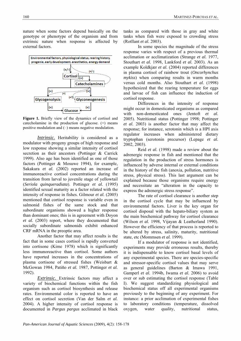
“general alarm reaction” (GAR). If those conditions continue, blood chemicals return to normal (general adaptation syndrome) due to some modifications in
the metabolism. But, if the stressful environment prevails, the GAR symptoms appear again caused by energy depletion (Fig. 2).
BEGINNING OF STRE
SS EXP
OSU
RE GENERAL ALARM REACTION
GENERAL ALARM REACTION
GENERAL ADAPTATION SYNDROME
EXHAUSTION
TIME
BIOCH
EMICAL PA
RAMETER
S
Figure 2. Biochemical responses of fishes undergoing chronic stress (Selye 1936). The responses change with respect to time; the fish modifies and regulate the biochemical processes to restore the homeostasis (GAR), but the duration of this mechanism depends on the availability of energy reserves.
When Pickering & Pottinger (1987b) experimented with salmonids under crowding conditions, they concluded that changes in cellular composition were better stress indicators than plasma cortisol levels. Likewise we agree with those authors and recommend that cortisol may be a primary stress indicator in acute rather than chronic experiments. However cortisol may be reported as complementary data of chronic experiments.
Chemicals. Several pollutants can stress the fish, activating alarm reactions producing a primary and a secondary response (Brown 1993). In Atlantic salmon (Salmo salar), cortisol and glucose levels increased after being exposed to high aluminum concentrations (Ytrestøyl et al. 2001). Roche & Bogué (1996) argued that one of the most frequent responses in fish blood to specific chemical intoxication is cortisolemia.
Nevertheless Wendelaar-Bonga (1997) explained that “the exposure to chemicals may directly compromise the stress response by interfering with specific neuroendocrine control
mechanisms”. Some chemicals affect metabolic pathways which eventually will influence neural and interrenal tissue functions. In agreement with this finding, it has been observed that cortisol secretion
can be affected by environmental contaminants because xenobiotic chemicals such as DDT are toxicants targeting multiple sites along the HPI axis, resulting in secretion of less bioactive ACTH, which in turn will promote a minor cortisol release from the interrenal tissue (Aluru et al. 2004; Hontela 1997).
Several studies have corroborated the impairment in the cortisol synthesis and secretion due the action of chemicals. Gravel & Vijayan (2006) studied the impacts of three pharmaceuticals (acetaminophen, ibuprofen, and salicylic acid) in rainbow trout and supported the hypothesis that these pharmaceuticals disrupt steroidogenesis in fish interrenal tissue. These findings were also tested in vitro and observed that salicylic acid produced a depression of ACTH stimulation in cortisol secretion and a lower gene expression of steroidogenic acute regulatory (StAR) protein, which is involved in steroidogenesis of cortisol (Hontela 2006); the same author also stated that StAR protein may be a sensitive target of many environmental pollutants, ranging from pesticides to pharmaceuticals. Also, the expression of StAR and P450SCC decreased in fish exposed to xenobiotics because they bind aryl hydrocarbon–receptor (AhR), a cytosolic ligand-

MARTINEZ-PORCHAS ET AL.
Pan-American Journal of Aquatic Sciences (2009), 4(2): 158-178
164
induced transcription factor, with a consequent depress of steroidogenic enzyme activity and finally altering the cortisol production and secretion (Aluru et al. 2005). Therefore many pollutants halt cortisol secretion and even if the fish is under stress this will probably not be reflected in cortisol response.
Pickering & Pottinger (1987a) observed that the exposure of brown trout (Salmo trutta) to poor quality water resulted in a 50% suppression of the cortisol release. Brodeur et al. (1997) reported an impairment of the cortisol stress-response in the yellow perch (Perca flavescens) from polluted sites. Langiano & Martínez (2008) did not observed any change in plasma cortisol of neotropical fish Prochilodus lineatus when exposed to different concentrations of a glyphosphate-based herbicide at different periods, while difference in glucose levels were assessed.
These results suggest that the interrenal response may be contaminant specific. Thus the estimation of cortisol as a toxic stress indicator may be in doubt, and if no response is observed in fishes under obvious stressful conditions, then cortisol should be replaced by more useful tests to evaluate the effect of any chemical compound on any species. To mention some examples: medium lethal dose (LD50) (Sprague 1969), behavior (Sprague 1971), histopathological indicators (Schwaiger et al. 1997), blood parameters (Iwama et al. 1995) or enzymatic activity (oxidative stress enzymes) (Pedrajas et al. 1995, Gorbi & Regoli 2004).
Anesthesia and Sampling. When sampling cortisol in fishes, it is necessary to handle the organisms. This handling eventually aware the organism and provokes an alarm reaction altering the level of pituitary hormones and thus increases the possibilities to obtain less precise results. Anesthetics have been used to reduce pain and awareness, and thus avoid metabolism enhancement (increase of cortisol or other parameters) in fish. To this respect, Small (2003) documented that anesthetics reduce or block the activation of HPI axis, so blood chemicals would not be altered at sampling process.
However, Flodmark et al. (2002) mentioned that some anesthetics per se (i.e. tricaine and 2-phenoxyethanol) are stressful and may raise plasma cortisol. Similar conclusion was assessed by Barton & Peter (1982) when observed that 2-phenoxiethanol and tricaine increased blood cortisol in the trout Salmo gairdneri. A possible explanation for these abnormal results may be that oxygen concentration in water significantly decreases when an anesthetic is added and HPI axis is activated rather than blocked (Bolasina 2006). Palić et al.
(2006) evaluated the effectiveness of three anesthetics (MS 222, metomidate and enguenol) in fathead minnows, concluding that MS 222 did not block the activation of HPI axis, instead they had better results using metomidate. Although 2-phenoxiethanol and tricaine are the two most used anesthetics, we do not recommend their use for cortisol and glucose evaluation because of the possibility of erroneous increased results.
Some anesthetics though have been shown to halt secretion of cortisol. Clove oil showed to be a strong blocker of cortisol increase in channel catfish (Ictalurus punctatus) (Small 2003). In contrast, it was documented that clove oil did not block cortisol secretion in stressed sea bream (Pagrus major).
Isoeugenol was shown to diminish 60% blood cortisol in channel catfish exposed to confinement, whereas metomidate showed greater effectiveness in blocking cortisol release under high ammonium concentrations (Small 2004). Olsen et al. (1995) intraperinoteally injected ACTH into Atlantic salmon to promote cortisol secretion; they reported that metomidate blocked cortisol release, whilst high cortisol levels were found when using tricaine (MS 222). Metyrapone has also shown successful results as a cortisol synthesis blocker (Hopkins et al. 1995). However, metyrapone and etomidate halt cortisol secretion by inhibiting 11β-hydroxylase, a key enzyme in the conversion of 11-deoxycortisol to cortisol (Dang & Trainer 2007). This hindrance in the biochemical pathway of cortisol generation may compromise physiological status of organisms limiting adaptation capacity, and perhaps leading them to death. In this case, these anesthetics may be used if a single sample of every fish is required, for example when fishes are sacrificed while sampling (heart puncture), such as the case of small size fishes. But, if more than one sample (from caudal vein) of every fish is needed or the investigator does not intend to sacrifice the animal (animal management ethics), then it is not recommended to use metyrapone nor isoenguenol, and maybe other anesthetics constitute better options, perhaps sacrificing precision for the survival of the experimental organisms.
Results are very contrasting, the efficacy of anesthetics seems to be species-specific and prior tests would be required and report how much of the cortisol response is due to anesthetic and how much is due to the stressor.
It is also worth to mention that some of the above studies only tested a single dose of anesthetic. Thus it is unknown if the kind of anesthetic per se is inefficient or the dose was not adequate. In a recent experiment Welker et al. (2007) tested four
Cortisol and Glucose: Reliable indicators of fish stress?
Pan-American Journal of Aquatic Sciences (2009), 4(2): 158-178
165
concentrations of MS 222 (0, 90, 120 and 180mg·l-1) in the channel catfish and reported that the highest cortisol level was found in the treatment without anesthetic (0mg·l-1), but the treatment with the highest level of anesthetic (180mg·l-1) also increased the cortisol concentrations. The best results were obtained with the dose of 90mg·l-1. Therefore, prior tests should consider not only anesthetics themselves, but also explore the adequate dose. Also it is important to establish the time at which samples should be taken, because apparently the time after applying the anesthetic has an effect on the secretion of cortisol. Welker et al. (2007) found that the MS 222 effectively blocked cortisol secretion of channel catfish during the first 20 minutes after anesthetizing, but the levels tended to increase after 25 minutes.
Water temperature is another point of concern at the time of administering any anesthetic. Park et al. (2008) proved the efficacy of clove oil in anesthetizing fishes and their physiological responses when administered it at different concentrations and temperatures, finding that the optimal dose (lower cortisol and glucose secretion) decreased at higher temperatures.
Non invasive methods. Non invasive methods have been used as indirect indicators of stress. In 1994, Sorensen & Scott found that goldfish released steroid to the water, and that one of those steroids was cortisol. After that, the measurement of cortisol in water to evaluate stress status in fish was proposed (Scott et al. 2001). Ellis et al. (2004) measured free cortisol released into water by rainbow trout. Lower et al. (2005) tagged two species of fishes, the common carp (Cyprinus carpio) and the rutilus (Rutilus rutilus) reporting that cortisol in water increased from 70 to 400 and from 170 to 2000 pg/g/h respectively.
This method has the advantage that fishes are not stressed up when sampling due to null or minimal intervention. Moreover there is no necessity to bleed and hurt the animal to measure cortisol. However Scott & Ellis (2007) pointed out that in some cases cortisol in water is too low to be measured by conventional methods, being necessary to extract and concentrate cortisol from water, because the highest proportion of cortisol is eliminated through hepatic processes, while renal and branchial routes play a secondary role in steroid elimination (Idler & Truscott 1972, Butler 1973). Scott & Ellis also suggested that only free cortisol and not conjugated steroid fractions (sulphated and glucuronidated steroids) have to be measured to evaluate stress response, because “the concentration of free steroid in the water equates to the
concentration of ‘physiologically active’ steroid in the plasma, which is very close to the moment in time that the sample is taken.
This method also faces the problem of fish mass and water flow rate, because cortisol secretion is in direct proportion to fish biomass and flow rate modifies cortisol concentration. Thus, very similar biomasses are required in every experimental unit, together with calculations considering flow rate (see Scott & Ellis 2007). Furthermore, the method can not be used to measure individual cortisol levels, unless tests of single organisms are carried out. Despite those related problems, this method emerges as an interesting alternative to substitute cortisol measurements in plasma.
Another non invasive method to measure cortisol is to measure it in feces. This procedure has been reported by some authors, but with limited success (Oliveira et al. 1999, Turner et al. 2003). Also the major part of free cortisol releasing occurs through the gills (Ellis et al. 2005). Despite the advantage of being non intrusive, this method does not have yet the precision of direct evaluations (plasma cortisol levels; Huntingford et al. 2006) or water cortisol measurement for what its use is of limited practical value.
Glucose. Glucose is a carbohydrate that has a major
role in the bioenergetics of animals, being transformed to chemical energy (ATP), which in turn can be expressed as mechanical energy (Lucas 1996).
In suboptimum or stressful conditions (internal or external) the chromaffin cells release catecholamine hormones, adrenaline and noradrenaline toward blood circulation (Reid et al. 1998). Those stress hormones in conjunction with cortisol mobilize and elevate glucose production in fish through glucogenesis and glycogenolysis pathways (Iwama et al. 1999) to cope with the energy demand produced by the stressor for the “fight of flight” reaction. This glucose production is mostly mediated by the action of cortisol which stimulates liver gluconeogenesis and also halts peripheral sugar uptake (Wedemeyer et al. 1990). Glucose is then released (from liver and muscle) toward blood circulation and enters into cells through the insulin action (Nelson & Cox 2005).
Regardless of the wide use of glucose as an indicator of stress, some authors (Mommsen et al. 1999, Flodmark et al. 2001) emphasized that care has to be taken when using plasma glucose as the only indicator. It has been reported that glucose content is a less precise indicator of stress than cortisol (Wedemeyer et al. 1990, Pottinger 1998).
MARTINEZ-PORCHAS ET AL.
Pan-American Journal of Aquatic Sciences (2009), 4(2): 158-178
166
Mommsen et al. (1999) were skeptical about the use of glucose as a stress indicator, whereas Simontacchi et al. (2008) stated that glucose and cortisol “cannot be considered itself as reliable stress indicators”.
Factors that affect the intensity of response. Similar to cortisol, some factors can indirectly alter the response of glucose levels in blood. Vijayan & Moon (1994) suggests that “the rearing history including nutritional status may affect the stress response and glucose clearance rates”. That affirmation is supported by other authors who concluded that blood glucose results have to be interpreted with care, taking into account extrinsic factors such as diet, life stage, time since last feeding and season of the year, etc., because they may affect liver glycogen stores (Nakano & Tomlinson 1967, Barton et al. 1988, McLeay 1977, Wedemeyer et al. 1990).
Nutritional status is a factor that can have an effect in the glucose response. The intake of diets with different lipid and protein content resulted in different responses of blood glucose of the orange-spotted Grouper (Epinephelus coioides) when it was exposed to cold stress (Cheng et al. 2006). The channel catfish under fasting conditions evidenced hyperglycemia after 30 days of experiment (22.8 versus 4.7 ng·ml-1 in the control group) (Peterson & Small 2004).
Glucose also varies between species and stage of development (Iwama et al. 2004, Hemre et al. 2002). Woodward & Strange (1987) observed that wild rainbow trout experienced a cortisol increase 3 times greater than hatchery fish when exposed to net confinement and electroshock. To avoid erroneous results we also suggest prior standardization of organisms before any stress experiment.
On the other hand as previously stated, stress hormones such as catecholamines, cortisol and others may be influenced by internal or external conditions in the history of the fish (anoxia, pollution, nutritive stress, physical stress) (Reid et al. 1998). Cortisol is known to increase blood glucose, herein a disruption in cortisol secretion may conclude in an altered glucose response.
However there have been observed increases in blood glucose whilst cortisol secretion is impaired (Costas et al. 2008). Some authors suggest that this increase in glucose may be attributed to a different mechanism of the action of cortisol, (catecholamine action for instance) (Vijayan et al. 1991, 1994; Trenzado et al. 2006). To this respect, it has been demonstrated that catecholamines itself can increase sugar levels (Wagner et al. 2003). Catecholamines
promote the phosphorylation of the enzyme glycogen phosphorylase which results in a glycogenolysis increase (Vijayan & Moon 1992); then, if catecholamine production or secretion is modified also glucose response may be affected. When Nilsson (1989) exposed crucian carp (Carassius carassius) to anoxic conditions (76-169 h), a significantly decrease in stored noradrenaline in kidney head was observed. However Reid et al. (1998) also concluded that those effects are reversible under normoxic conditions.
As in the cortisol case it is indispensable to know basal or pre-stress levels of the species to be studied (Table II).
Energy demand. As mentioned, sugar levels increase during stress, however some authors reported a weak rise of glucose (Davis Jr. & McEntire 2006), others found no change (Rotllant & Tort 1997, Jentoft et al. 2005), and even a decrease (Wood et al. 1990).
Sometimes no significant changes in plasma glucose may be observed, because under stress the fish is rapidly consuming the energetic substrates generated (glucose) since the main function of the central nervous system (CNS) is to maintain homeostasis. West et al. (1993) argued that during peak activity glucose use can increase by almost 30-fold. However it is possible that fish exposed to chronic stress suffer substrate depletion that leads to a decrease on plasma glucose (Fig. 2).
The freshwater fish rohu (Labeo rohita) exposed to high fenvalerate (an insecticide) concentrations, presented the maximum glucose level in the fourth day of exposure, but the level began to decrease over time until depleted (David et al. 2005). From those results, a weak or no change in plasma glucose may be attributed to a high energy demand so that glucose cannot be accumulated (acute experiments) or the organism be habituated (chronic experiments). A decrease of glucose is linked to depletion of reserve energy. If it is not part of the experiment, fishes should not be exercised during experimental or acclimation period to avoid an increase in energy demand; this caution will reduce the risk to obtain abnormal results, nonetheless glucose response is still more variable than cortisol response.
Rapid measurements. Normally the increase of glucose in plasma is not as rapid as for cortisol. Many researchers documented an increase of glucose minutes or days after the stress (Pratap & Wendelaar-Bonga 1990, Hemre & Krogdahl 1996, Barcellos et al. 1999, Falahatkar & Barton 2007) because cortisol triggers glucose production. Measuring glucose just after an acute experiment is

Cortisol and G
lucose: Reliable indicators of fish stress? Pan-Am
erican Journal of Aquatic Sciences (2009), 4(2): 158-178
167
Table II. Plasma glucose values of different species of fishes before and after being stressed. In every line is indicated if the kind of exposure to the stressor was acute or chronic. When Chronic/Acute appears, it indicates that the fish was first acclimated to any condition and thereafter exposed to an acute stressor.
References
Siikavuopio & Sæther (2006) Lowe & Davison (2005)
Welker et al. (2007)
Frisch & Anderson (2005)
Lowe & Davison (2005)
Urbinati & Carneiro (2006)
Barreto & Volpato (2006)
Barreto & Volpato (2006)
Miller et al. (2007)
Gagnon et al. (2006)
Davis & Peterson (2006)
Zuccarelli et al. (2008)
Exposure
Chronic
Chronic
Acute
Chronic
Chronic
Acute
Acute
Acute
Acute
Chronic/Acute
Chronic/Acute
Acute
Glucose (mmol/l)
Poststress
0.23
10
2.8
7.9
7.4
7.5
10
6.4
6.7
9
7.2
10.5
1.7
Prestress
0.17
4.5
1.7
1.6
1.9
1.5
2.8
2.2
1.9
4.2
5.1
6.1
1.6
Stressor
Nitrite exposure
Temperature
Handling
Capture and handling
Temperature
Handling and transportation
Electroshock
Social stressor
Pollutant
Copper and air exposure
Temperature and confinement
Air exposure
Species
Atlantic cod Gadus morhua
Bald notothen Pagothenia borchgrevinki
Channel catfish Ictalurus punctatus
Coral trout
Plectropomus maculatus
Plectropomus leopardus
Emerald rockcod Trematomus bernacchii
Matrinxã Brycon amazonicus
Nile tilapia Oreochromis niloticus
Nile tilapia
Rainbow trout Oncorhynchus mykiss
Rainbow trout
Sunshine bass Morone chrysops x saxatilis
White sturgeon Acipenser transmontanus
MARTINEZ-PORCHAS ET AL.
Pan-American Journal of Aquatic Sciences (2009), 4(2): 158-178
168
considered a source of error, because there is the probability of not measuring any change. For instance, Perez-Casanova et al. (2008) increased water temperature of Atlantic cod (Gadus morhua) at a rate of 2°C·h-1 and measured glucose every 2°C, not finding statistical differences from 10 to 24°C (a critical temperature). These results appeared probably because the change in blood glucose levels might occurrs minutes, hours or even days later (Langiano & Martínez 2008). Thus single measures of glucose are not a real indicator, rather it is recommended to measure glucose over time as in the cortisol case.
For chronic experiments, the acclimation period should be long enough to ensure that in case of observing a null or weak glucose response, that response is not caused by the general adaptation syndrome (Fig. 2). Some authors suggest 3 weeks of acclimation for laboratory experiments (Houston 1982; Johnsson et al. 2003), however this is not a rule and may vary among species and stressors. Fast et al. (2008) did not observe a rise of glucose in the Atlantic salmon during acute experiments, but reported an increase when the fish were exposed to prolonged stress. A 3-week period of crowding stress elevated cortisol and glucose in gilthead seabream (Sparus auratus) (Tort et al. 1996). Therefore, unlike in acute experiments, glucose can be measured immediately after a chronic exposure to stressful conditions.
Blood samples can be extracted during the chronic experiments, but if the experimental units are limited and all the samples have to be taken from the same tanks, it is not recommendable to measure cortisol over time, because the consequent handling and manipulation of organisms may lead to erroneous results in the future samples. Nevertheless, if it is necessary to measure glucose over time, it is recommended that sampling is not very frequent, while a limited number of samplings should be established.
Anesthesia and sampling. Some irregularities have been reported in glucose response when anesthetics are used before sampling. The ideal role of anesthetics is to minimize fish stress response, prevent any negative impact on performance and thus measure real values of glucose or other blood components (Pickering 1998). Nevertheless, some anesthetics do not fit with that role. Ortuño et al. (2002) tested four anesthetics (MS 222, benzocaine, 2-phenoxytehanol and quinaldine) in gilthead seabream and reported that basal glucose level was 3.6 mmol·l-1 and increased to 5.6, 11.1, 11.9 and 16.4 mmol·l-1 when exposed to MS 222, benzocaine, 2-phenoxytehanol and quinaldine
respectively; also immune response was depressed by benzocaine and 2-phenoxyethanol, but not by MS 222 or quinaldine. In another experiment, Iversen et al. (2003) exposed Atlantic salmon to other four anesthetics (metomidate, clove oil, Aqui-S™ and Benzoak®) not finding any increase in plasma glucose. On the other hand, Velíšek et al. (2005) reported a significant increase in rainbow trout glucose when anesthetized with clove oil. Also clove oil and MS 222 blocked cortisol secretion but increased glucose in another experiment with rainbow trout (Wagner et al. 2003). Those controversial results suggest that perhaps anesthetic efficacy in halting glucose increase is species-dependant and also previous tests may be required (see Anesthesia and sampling in cortisol section).
Other stress indicators. Previously we documented that many
studies utilized cortisol and glucose as sole stress indicators of stress in fish; however regarding the several factors that can affect these responses give us to consider that cortisol and glucose are not enough as stress indicators. Iwama et al. (2004) argued that “none of the current indicators of stress, including the stress hormones, are 100% suitable in reflecting stressed states in fish”; in consonance with that, it is recommendable to complement cortisol and glucose with other stress indicators to establish a more complete profile of the experimental organism.
In the case of cortisol it is known that in some fishes a small increase in plasma cortisol leads to an alteration in amino acid metabolism (Hopkins et al. 1995), for this reason it is plausible to consider that the activity of those enzymes involved in amino acid metabolism would be a complement or also a more accurate indicators of stress even if cortisol response is weak. Glutamine synthase for example, has been observed to increase with small response of cortisol (Reid et al. 1998).
Otherwise, it is possible to measure intermediate enzymes of glycolysis (phosphoenol pyruvate carboxykinase, fructose 1,6-biphosphate, glucose 6-phosphatase), since cortisol and catecholamines positively influence that process. For instance, Vijayan et al. (2003) exposed rainbow trout to cortisol treatments and reported an increase in the abundance of phosphoenolpyruvate carboxykinase (PEPCK) mRNA. Dziewulska-Szwajkowska et al. (2003) documented an increase of glucose 6-phosphatase when injected a high dose of cortisol in the common carp.
However, there are other important parameters that should be taken into account to study stress. For instance catecholamines are recognized as a stress indicator; adverse conditions

Cortisol and Glucose: Reliable indicators of fish stress?
Pan-American Journal of Aquatic Sciences (2009), 4(2): 158-178
169
activates the HPI axis and catecholamines are released into blood stream (Iwama 2007). Melanocyte stimulating hormone (α-MSH) is a peptide produced in the pituitary cells of several fishes (Kawauchi et al. 1984; Lamers et al. 1991) and proved to increase in stressful conditions (Arends et al. 1999, 2000). Lactate is a chemical compound that plays a role in anaerobic metabolism of animals, produced from pyruvate via the enzyme lactate dehydrogenase during exercise and is considered as a stress indicator in fishes because its levels are enhanced in under adverse situations (Thomas et al. 1999; Grutter & Pankhurst 2000).
Eventually, fish also respond at the cellular level to stressors. This response comprises some protein changes, for example an enhancement in heat shock protein (hsps) synthesis (Iwama et al. 1998, 2004). In cells of stressed organisms there is an increase in the production of hsps which are required to assist the folding of nascent polypeptide chains, act as a molecular chaperone and mediate the repair and degradation of altered or denatured proteins to maintain homeostasis.
On the other hand, different non-invasive methods can be used to complement the biochemical and molecular parameters. The ventilation rate and oxygen consumption represent an adequate alternative because they are indicators of the metabolic rate due to high activity, stress, etc. However, Alvaenga & Volpato (1995) stated that metabolic differences derived from social stress usually show data with high variance, masking important differences among treatments and that the oxygen consumption and ventilation rate can complement the stress studies. This method is very simple and if the water is clear enough the measurements can simply be determined visually. It has been observed that the ventilation rate and oxygen consumption increase with different stressors such as, presence of predators, air exposure, light intensity, etc. (Brown et al. 2005, Sager et al. 2000, Thompson et al. 2008). However despite ventilation rate is a very sensitive test, it has the limitation that the severity of any stimulus or stressor is not reflected in this parameter (Barreto & Volpato 2004) and thus, it is only useful to indicate if the fish is being stressed or not, but not how much.
Other non-invasive methods are the measurement of the excretion of nitrogenous compounds such as ammonia and urea, gas exchange (carbon dioxide) and others (Walsh et al. 1994, Evans et al. 2003). These non-invasive methods are good candidates to complement the cortisol and glucose as stress indicators, although they have the same limitation as the non-invasive
methods to measure cortisol (see above). These suggestions are not a rule of thumb
and may be replaced or complemented with others, depending on the nature of the stressor. In that manner, false results obtained by using any particular stress test may be validated or contrasted with others.
Conclusions
Cortisol and glucose cannot be eliminated from the stress indicators list, but due to their high variability they must be complemented with other measurements such as other stress hormones, hsps, blood-cell counts (preferably in chronic experiments), non-invasive methods and/or others, in order to have a more complete profile about the stress status of any fish.
Cortisol may be useful only in acute stress experiments and monitored throughout time. To be used as stress indicator, the physiological status of organisms should be standardized.
Anesthetics efficiency shows many inconsistencies and there is controversy about the convenience to use an effective metabolism blocker anesthetic or a non harmful anesthetic due to animal management ethics. It is important, as long as possible, to select an adequate anesthetic according to the species, which effectively blocks metabolism while causing a minimum damage to the animal integrity. The dose of the anesthetic and the water of temperature are subjects of concern.
Non invasive methods such as measuring cortisol in water are a suitable alternative to avoid anesthetic problems.
Glucose measurements show many inconsistencies and should be a complement of stress tests rather than a main indicator. It also can be used as a tool to provide a point of reference for a particular species.
Results may be more realistic if standardization of organisms and experimental conditions are done, and organisms are not exercised prior sampling blood.
Repeated glucose measures have to be done during or after acute exposures, but during chronic experiments the sampling should not be very frequent, because the handling and manipulation of organisms may affect the future measurements.
In using both cortisol and glucose as stress indicators the researcher needs to be careful to identify possible situations or factors that may influence the stress response of the fish as long as possible and to be certain they are not part of the experiment. On the other hand, the use of these two indicators as pollution or toxic stress indicators is
MARTINEZ-PORCHAS ET AL.
Pan-American Journal of Aquatic Sciences (2009), 4(2): 158-178
170
not adequate, rather behavior and oxidative stress tests are recommended.
Finally in the scientific task of monitoring stress, the reliability of the results may be significantly increased if adequate and enough number of tests is carried out. References Aluru. N., Jorgensen. E. H., Maule, A. G. &
Vijayan, M. M. 2004. PCB disruption of the hypothalamus-pituitary-interrenal axis involves brain glucocorticoid receptor downregulation in anadromous arctic charr. American Journal of Physiology - Regulatory, Integrative and Comparative Physiology, 287: 787–793.
Aluru, N., Renaud, R., Leatherland, J.F. & Vijayan, M. M. 2005. Ah receptor–mediated impairment of interrenal steroidogenesis involves StAR protein and P450scc gene attenuation in rainbow trout. Toxicological Sciences, 84: 260–269.
Alvarenga, C.M.D., Volpato, G.L., 1995. Agonistic profile and metabolism in alevins of the Nile tilapia. Physiological Behavior, 57: 75–80.
Arends, R. J., Mancera, J. M., Munoz, J. L., Wendelaar Bonga, S. E. & Flik, G. 1999. The stress response of the gilthead sea bream (Sparus aurata L.) to air exposure and confinement. Journal of Endocrinology, 163: 149–157.
Arends, R. J., Rotllant, J., Metz, J. R., Mancera, J. M., Wendelaar Bonga, S. E. & Filk, G. 2000. α-MSH acetylation in the pituitary gland of the sea bream (Sparus aurata L.) in response to different backgrounds, confinement and air exposure. Journal of Endocrinology, 166: 427–435.
Balm, P. H. M., Lambert, J. D. G. & Wendelaar-Bonga, S. E. 1989. Corticosteroid biosynthesis in the interrenal cells of the teleost fish, Oreochromis mossambicus. General and Comparative Endocrinology, 76: 53-62.
Barcellos, L. J. G., Nicolaiewsky, S., de Souza, S. M. G. & Lulhier, F. 1999. Plasmatic levels of cortisol in the response to acute stress in Nile tilapia, Oreochromis niloticus (L.), previously exposed to chronic stress. Aquaculture Research, 30: 437-444.
Barreto, R. E. & Volpato, G. L. 2006. Stress responses of the fish Nile tilapia subjected to electroshock and social stressors. Brazilian Journal of Medical and Biological Research, 39: 1605-1612
Barton, B. A. & Peter, R. E. 1982. Plasma cortisol stress response in fingerling rainbow trout, Salmo gairdneri Richardson, to various transport conditions, anesthesia, and cold shock. Journal of Fish Biology, 20: 39–51.
Barton, B. A., Schreck, C. B. & Barton, L. D. 1987. Effects of chronic cortisol administration and daily acute stress on growth, physiological conditions, and stress responses in juvenile rainbow trout. Diseases of Aquatic Organisms, 2:173-185.
Barton, B. A., Schreck, C. B. & Fowler, L. G. 1988. Fasting and diet content affect stress-induced changes in plasma glucose and cortisol in juvenile chinook salmon. The Progressive Fish Culturist. 50: 16-22.
Barton, B. A. & Iwama, G. K. 1991. Physiological changes in fish from stress in aquaculture with emphasis on the response and effects of corticosteroids. Annual Reviews of Fish Diseases, 1: 3-26.
Barton, B. A. 1997. Stress in finfish: past, present and future – a historical perspective. In: Iwama, G. K., Pickering, A. D., Sumpter, J. P. & Schreck, C. B. (eds). Fish Stress and Health in Aquaculture. Cambridge University Press, Cambridge.
Barton, B. A., Bollig, H., Hauskins, B. L. & Jansen, C. R. 2000. Juvenile pallid (Scaphirhynchus albus) and hybrid pallid×shovelnose (S. albus×platorynchus) sturgeons exhibit low physiological responses to acute handling and severe confinement. Comparative Biochemistry and Physiology, 126: 125-134.
Barton, B. A., Haukenes, A. H., Parsons, B. G. & Reed, J. R. 2003. Plasma cortisol and chloride stress responses in juvenile walleyes during capture, transport, and stocking procedures. North American Journal of Aquaculture, 65: 210–219.
Barton, B. A., Ribas, L., Acerete, L. & Tort, L. 2005. Effects of chronic confinement on physiological responses of the juvenile gilthead sea bream, Sparus aurata L. to acute handling. Aquaculture Research, 36: 172-179.
Barreto, R. E. & Volpato-Braz, G. L. 2006. Stress responses of the fish Nile tilapia subjected to electroshock and social stressors. Journal Biological and Medical Research, 39: 1605-1612.
Begg, K. & Pankhurst, N. W. 2004. Endocrine and metabolic responses to stress in a laboratory population of the tropical damselfish Acanthochromis polyacanthus. Journal of

Cortisol and Glucose: Reliable indicators of fish stress?
Pan-American Journal of Aquatic Sciences (2009), 4(2): 158-178
171
Fish Biology, 64: 133–145. Benfey, T. J. & Biron, M. 2000. Acute stress
response in triploid rainbow trout (Oncorhynchus mykiss) and brook trout (Salvenis fontinalis). Aquaculture, 184: 167-176.
Benguira, S., Leblond, V. S., Weber, J-P. & Hontela, A. 2002. Loss of capacity to elevate plasma cortisol in rainbow trout (Oncothynchus mykiss) treated with a single injection of o,p′-Dichlorodiphenyldichloroethane. Environmental Toxicology and Chemistry, 21: 1753–1756.
Bolasina, S. N. 2006. Cortisol and hematological response in Brazilian codling, Urophycis brasiliensis (Pisces, Phycidae) subjected to anesthetic treatment. Aquaculture International, 14: 569-575.
Bowers, J. M., Mustafa, A., Speare, D. J., Conboy, G. A., Brimacombe, M., Sims, D. E. & Burka, J. F. 2000. The physiological response of Atlantic salmon, Salmo salar L., to a single experimental challenge with sea lice, Lepeophtheirus salmonis. Journal of Fish Diseases, 23: 165–172.
Brodeur, J. C., Sherwood, G., Rasmussen, J. B. & Hontela, A. 1997. Impaired cortisol secretion in yellow perch (Perca flavescens) from lakes contaminated by heavy metals: in vivo and in vitro assessment. Canadian Journal of Fisheries and Aquatic Sciences, 54: 2752–2758.
Brown, C., Gardner, C. & Braithwaite, V.A., 2005. Differential stress responses in fish from areas of high and low-predation pressure. Journal of Comparative Physiology, 175, 305–312.
Butler, D. G. 1973. Structure and function of the adrenal gland of fishes. American Zoologist, 13: 839-379.
Brown, J. A. 1993. Endocrine responses to environmental pollutants. In: Rankin, J. C. & Jensen, F. B. (eds). Fish Ecophysiology. Chapman & Hall, London. 276–296p.
Campbell, T. W. 2004. Blood biochemistry in lower vertebrates. In: 55th Annual Meeting of the American College of Veterinary Pathologists (ACVP) & 39th Annual Meeting of the American Society of Clinical Pathology (ASVCP), ACVP and ASVCP (ed)
Castillo, J., Castellana, B., Acerete, L., Planas, J. V., Goetz, F. W., Mackenzie, S. & Tort, L. 2008. Stress-induced regulation of steroidogenic acute regulatory protein expression in head kidney of Gilthead seabream (Sparus aurata). Journal of Endocrinology, 196: 313–322.
Cheng, A. C., Chen, C. Y., Liou, C. H. & Chang, C. F. 2006. Effects of dietary protein and lipids on blood parameters and superoxide anion production in the grouper, Epinephelus coioides (Serranidae: Epinephelinae). Zoological Studies, 45: 492-502.
Clements, S. P., Hicks, B. J., Carragher, J. F. & Dedual, M. 2002. The effect of a trapping procedure on the stress response of wild rainbow trout. North American Journal Fisheries Management, 22: 907–916.
Colombe, L., Fostier, A., Bury, N., Pakdel, F. & Guiguen, Y. 2000. A mineralocorticoid-like receptor in the rainbow trout, Oncorhynchus mykiss: cloning and characterization of its steroid binding domain. Steroids, 65: 319-328.
Costas, B., Aragão, C., Mancera, J. M., Dinis, M. T. & Conceição, L. E. C. 2008. High stocking density induces crowding stress and affects amino acid metabolism in Senegalese sole Solea senegalensis (Kaup 1858) juveniles. Aquaculture Research, 39: 1-9.
Dang, C. N. & Trainer, P. 2007. Pharmacological management of cushing’s syndrome: An update. Arquivos Brasileiros de Endocrinologia & Metabologia, 51: 1339-1348.
David, M., Shivakumar, R., Mushigeri, S. B. & Kuri, R. C. 2005. Blood glucose and glycogen levels as indicators of stress in the freshwater fish, Labeo rohita under fenvalerate intoxication. Journal of Ecotoxicology & Environmental Monitoring, 15: 1-5.
Davis, K. B. & Peterson, B. C. 2006. The effect of temperature, stress, and cortisol on plasma IGF-I and IGFBPs in sunshine bass. General and Comparative Endocrinology, 149: 219–225.
Davis Jr, K. B. & McEntire, M. E. 2006. Comparison of the cortisol and glucose stress response to acute confinement and resting insulin-like growth factor-I concentrations among white bass, striped bass and sunshine bass. Aquaculture America Book of Abstracts p 79.
Dickhoff, W. W. 1989. Salmonids and annual fishes: Death after sex. In: Schreibman, M. P. & Scanes, C. G. (eds) Development, maturation, and senescence of neuroendocrine systems: A comparative approach. Academic press, New York.
Doyon, C., Gilmour, K. M., Trudeau, V. L. & Moon, T. W. 2003. Corticotropin-releasing factor and neuropeptide Y mRNA levels are elevated in

Pan-America
172
the prainbEndo
DziewulskaM., AA. 20glucobisphfructocarp ComPart
Ellis, T., Ja2004measwaterBiolo
Evans, D. HThe mgas regulwaste
Falahatkar, obseracutebelug38: 1
Fast, M. D.,O. Brelatesalarterm 24: 1
Frisch, A. Jstress(PlecmacuPhys
Flodmark, LArnekB. S.juvenflow Fish
Fryer, J. corticAmer
Gagnon, A.of Cuby a(Onco78: 5
Gamperl, A1994cortis
an Journal of A
preoptic areow trout. G
ocrinology, 1a-SzwajkowsAdamowicz, 003. The effeose-6-phosphhosphatase ose-2,6-bisph
tissues parative BiB, 135: 485-
ames, J. D.,. A non-invaurement of fr by rainboogy, 65: 1233H., Piermarinmultifunction
exchange, lation, and e. Physiologi
B. & Bartorvations of e handling aga Huso huso786-1789. , Hosoya, S.,
B. 2008. Cored effects or Linnaeus) sstress. Fish 94-204.
J. & Andersos responses octropomus leulatus). Comiology Part L. E. W., Ukleiv, J. V., 2001. Corti
nile brown trregime in anBiology, 60:L. & Ledecotropin serican Zoolo, Jumarie, C
u on plasma cadrenocorticaorhynchus m9-65.
A. K., Vijayaa. Epinephsol concen
Aquatic Scien
ea of sociaGeneral an133: 260–27ska, D., ozA., Wojtasz
ect of high dohatase andactivity, anhosphate c
(Cyprinus iochemistry -491. Stewart, Casive stress afree cortisol ow trout. J3–1252. ni, P. M. & Cnal fish gill:
osmoregulaexcretion
ical Reviewson, B. A. 20
physiologicand confinemo L. Aquacu
, Johnson, S.rtisol responof Atlantic subjected to and Shellfis
on, T. A. 200of two specieopardus an
mparative BiA, 140: 317-
Urke, H. A., Vøllestad, L
isol and glucrout subjecten artificial str: 238–248. eris, K. 19ecretion in gist, 26: 101. & Hontela,cortisol and al cells of
mykiss). Aqu
an, M. M. &hrine, norepntrations
nces (2009), 4
ally subordnd Compara
1. zi ska-Gab
zek, J. & Dzose of cortisod fructosend glucose concentration
carpioand Physio
. & Scott, Aassay based ureleased into
Journal of
Choe, K. P. 2Dominant si
ation, acid-of nitroge
s, 85: 97-177007. Prelimi
cal responsement in juvulture Resea
. C. & Afonsnse and imm
salmon (Sshort- and l
sh Immunol
05. Physioloies of coral nd Plectropoiochemistry-327. Halleraer, J
L. A. & Poléocose responsd to a fluctuream. Journ
986. Controteleost fi
17–1026. A. 2006. Efcortisol secr
f rainbow uatic Toxicol
& Boutilier, Rpinephrine, in cannu
(2): 158-178
dinate ative
bska, ugaj, ol on
e-1,6-and
n in L.).
ology
A. P. upon o the Fish
2003. ite of -base enous 7. inary es to venile arch,
so, L. mune-Salmo long-logy,
ogical trout omus and
J. H., o, A.
ses in ating
nal of
ol of shes.
ffects etion trout logy,
R. G. and
ulated
Gam
Gill-
Gilm
Gorb
Grav
Grut
Hatt
Hau
Hem
Hem
seawater-a(OncorhynconfinemeJournal o
mperl, A. K.,1994b. hormone applicatioFisheries,
-Barcellos, LQuevedo, does not additionalquelen, QAquacult
mour, K. M.2005. Phyof social and Com
bi, S. & cytochrommetaboliteanguilla: variabilityMarine E515.
vel, A. & disrupts iglycocortitrout. Tox
tter, A. S. &of captuectoparasiglucose aHemigymnBiology, 5
tingh, J. 197stress in t(Smith). J195.
ukenes, A. HCortisol yellow plipopolysa72: 780-78
mre, G-I. &handling in carbosalmon, Nutrition
mre, G-I., MoCarbohydgrowth, enzymes. 194.
M
acclimated nchus mykisent and of Fish Biolo, Vijayan, MExperimentalevels in
ns. Reviews, 4: 215-255.L. G., Kreu
R. 2006. Palter the c
l acute stresQuoy and ture, 253: 31., DiBatista, ysiological cstatus in salparative BioRegoli, F.
me P4501Aes in EuSeasonal, d
y in field anEnvironmen
Vijayan, Minterrenal steicoid receptoxicological S
& Pankhurst, ure, handlinite load on pand lactate nus melapte57: 391–40177. Blood sthe freshwatJournal of F
., Barton, B.responses operch foloaccharide. Jo84.
& Krogdahland fish size
ohydrate mSalmo sa
n, 2: 249–252ommsen, T. P
drates in fisglucose meAquacultu
MARTINEZ-PO
rainbowss) followingepinephrine
ogy, 45: 313-M. M. & Boual control fishes: techs on Fish B.
utz, L. C. &Previous chcortisol respssor in jundi
Gaimard) 7-321. J. D. & Th
causes and colmonid fish. ology, 45: 26. 2005. In
A and biuropean eedose- and timnd laboratoryntal Researc
M. M. 2006eroidogenesior expressionSciences, 93:
N. W. 2000.ng, confin
plasma levelsin the cora
erus. Journ. ugar as an ter fish, LabFish Biolog
. A. & Bolliof pallid stwing challournal of Fi
l, Å. 1996.e on secondetabolism
alar L. A2. P. & Krogda
sh nutrition:etabolism a
ure Nutritio
ORCHAS ET AL.
w troutg black-box
injection.-324. utilier, R. G.
of stresshniques andBiology and
& Mezzalira-hronic stressponse to aniá (Rhamdia
fingerlings.
homas, J. B.onsequencesIntegrative
63-273. nduction ofliary PAH
el Anguillame-response
y conditions.ch, 58: 511–
6. Salicylateis and brainn in rainbow41–49.
. The effectsement ands of cortisol,al reef fish
nal of Fish
indicator ofbeo capensisgy, 10: 191–
gs, H. 2008.turgeon andlenge withish Biology,
. Effect ofdary changesin Atlantic
Aquaculture
ahl, Å. 2002. effects on
and hepaticon, 8: 175-
t x .
. s d d
-s n a .
. s e
f H a e .
–
e n w
s d ,
h h
f s –
. d h ,
f s c e
. n c -
Cortisol and Glucose: Reliable indicators of fish stress?
Pan-American Journal of Aquatic Sciences (2009), 4(2): 158-178
173
Hontela, A., Rasmussen, J. B., Audet, C. & Chevalier, G. 1992. Impaired cortisol stress response in fish from environments polluted by PAHs, PCBs, and mercury. Archives of Environmentan Contamination and Toxicology, 22: 278-283.
Hontela, A. 1997. Endocrine and physiological responses of fish to xenobiotics: Role of glucocorticosteroid hormones. Reviews in Toxicology, 1: 1-46.
Hontela, A. 2006. Corticosteroidogenesis and StAR protein of rainbow trout disrupted by human-use pharmaceuticals: Data for Use in Risk Assessment. Toxicological Sciences, 93: 1–2.
Hopkins, T. E., Wood, C. M. & Walsh, P. J. 1995. Interactions of cortisol and nitrogen metabolism in the ureogenic Gulf toadfish, Opsanus beta. Journal of Experimental Biology, 198: 2229–2235.
Houston, A. H. 1982. Thermal effects upon fishes. Publication of the Environmental Secretariat 18566, 1–200.
Huntingford, F. A., Adams, C., Braithwaite, V. A., Kadri, S., Pottinger, T. S., Sandøe, P. & Turnbull, J. F. 2006. Current issues in fish welfare. Journal of Fish Biology, 68: 332-372.
Idler, D. R. & Truscott, B. 1972. Corticosteroids in fish. In: Idler, D. R. (ed) Steroids in Nonmammalian Vertebrates. Academic Press, New York, pp. 127–211.
Iversen, M., Finstad, B., McKinley, R. S. & Eliassen, R. A. 2003. The efficacy of metomidate, clove oil, Aqui-S™ and Benzoak® as anaesthetics in Atlantic salmon (Salmo salar L.) smolts, and their potential stress-reducing capacity. Aquaculture, 221: 549-566.
Iwama, G. K., Morgan, J. D. & Barton, B. A. 1995. Simple field methods for monitoring stress and general condition of fish. Aquaculture Research, 26: 273-282.
Iwama, G. K., Thomas, P. T., Forsyth, R. B. & Vijayan, M. M. 1998. Heat shock protein expression in fish. Reviews on Fish Biology and Fisheries, 8: 35-56.
Iwama, G. K., Vijayan, M. M., Forsyth, R. B. & Ackerman, P. A. 1999. Heat shock proteins and physiological stress in fish. American Zoologist, 39: 901-909.
Iwama, G. K., Afonso, L. O. B. & Vijayan, M. M. 2006. Stress in fishes. In: Evans, D. H. & Claiborne, J. B. The Physiology of fishes. 319-342. Taylor and Francis, 3rd edition. 601 p. USA.
Iwama, G. K., Afonso, L. O. B., Todgham, A., Ackerman, P. & Nakano, K. 2004. Are hsps suitable for indicating stressed states in fish?. Journal of Experimental Biology, 207:15-19.
Iwama, G. K. 2007. The welfare of fish. Diseases of Aquatic Organisms, 75: 155–158.
Jentoft, S., Aastveit, A. H., Torjesen, P. A. & Andersen, Ø. 2005. Effects of stress on growth, cortisol and glucose levels in non-domesticated Eurasian perch (Perca fluviatilis) and domesticated rainbow trout (Oncorhynchus mykiss). Comparative Biochemistry and Physiology Part A, 141: 353-358.
Johnsson, J. I., Parkkonen, J. & Förlin, L. 2003. Reduced territorial defense in rainbow trout fry exposed to a paper mill effluent: using the mirror image stimulation test as a behavioral bioassay. Journal of Fish Biology, 62: 959-964.
Kawauchi, H., Kawazoe, I., Adachi, Y., Buckley, D. I. & Ramachandran, J. 1984. Chemical and biological characterization of salmon melanocyte-stimulating hormone. General and Comparative Endocrinology, 53: 37-48.
Kime, D. E. 1978. The hepatic catabolism of cortisol in teleost fish – adrenal origin of 11-oxotestosterone precursors. General and Comparative Endocrinology, 35: 322-328.
Koldkjær, P., Pottinger, T. G., Perry, S. F. & Cossins, A. R. 2004. Seasonality of the red blood cell stress response in rainbow trout (Oncorhynchus mykiss). Journal of Experimental Biology, 207: 357-367.
Lamers, A. E., Balm, P. H. M., Haenen, H. E., Jenks, B. G. & Wendelaar-Bonga, S. E. 1991. Regulation of differential release of α-melanocytestimulating hormone forms from the pituitary of a teleost fish, Oreochromis mossambicus. Journal of Endocrinology, 129: 179-187.
Lamers, A. E., Flik, G., Atsma, W. & Wendelaar-Bonga, S. E. 1992. A role for di-acetyl alpha-melanocyte-stimulating hormone in the control of cortisol release in the teleost Oreochromis mossambicus. Journal of Endocrinology, 135: 285–292.
Lamers, A. E., Flik, G. & Wendelaar-Bonga, S. E. 1994. A specific role for TRH in release of diacetyl alpha-MSH in tilapia stressed by acid water. American Journal of Physiology- Regulatory, Integrative and Comparative Physiology, 267: 1302–1308.
Langiano, V. C. & Martínez, C. B. R. 2008. Toxicity

MARTINEZ-PORCHAS ET AL.
Pan-American Journal of Aquatic Sciences (2009), 4(2): 158-178
174
and effects of a glyphosphate-based herbicide on the neotropical fish Prochilodus lineatus. Comparative Biochemistry and Physiology Part C, 147:2 22-231.
Lankford, S. E., Adams, T. E. & Cech, J. J. Jr. 2003. Time of day and water temperature modify the physiological stress response in green sturgeon, Acipenser medirostris. Comparative Biochemistry and Physiology, 135: 291-302.
Lowe, C. J. & Davison, W. 2005. Plasma osmolarity, glucose concentration and erythrocyte responses of two Antarctic nototheniid fishes to acute and chronic thermal change. Journal of Fish Biology, 67: 752–766.
Lower, N., Moore, A., Scott, A. P., Ellis, T., James, J. & Low, P. J. 2005. A non-invasive method to assess the impact of electronic tag insertion on stress levels in fishes. Journal of Fish Biology, 67: 1202–1212.
Lucas, A. 1996. Physical concepts of bioenergetics. In: Lucas, A. (ed). Bioenergetics of aquatic animals. English edition, Taylor & Francis, France.
Lyytikäinen, T., Pylkkö, P., Ritola, O. & Lindström-Seppä, P. 2002. The effect of acute stress and temperature on plasma cortisol and ion concentrations and growth of Lake Inari Arctic charr, Salvelinus Alpinus. Environmental Biology of Fishes, 64: 195-202.
Maule, A. G., Shreck, C. B. & Sharpe, C. 1993. Seasonal changes in cortisol sensitivity and glucocorticoid receptor affinity and number in leukocytes of coho salmon. Fish Physiology and Biochemistry, 10: 497-506.
Mazeaud, M. M., Mazeaud, F. & Donaldson, E. M. 1977. Primary and secondary effects of stress in fish: some new data with a general review. Transactions of the American Fisheries Society, 106: 201-212.
McLeay, D. J. 1977. Development of a blood sugar bioassay for rapidly measuring stressful levels of pulpmill effluent to salmonid fish. Journal of the Fisheries Research Board of Canada, 34: 477-485.
Metz, J. R., Geven, E. J.W., van den Burg, E.H. & Flik, G. 2005. ACTH, MSH, and control of cortisol release: cloning, sequencing, and functional expression of the melanocortin-2 and melanocortin-5 receptor in Cyprinus carpio. American Journal of Physiology - Regulatory, Integrative and Comparative Physiology, 289: 814-826.
Miller, W. L. 1988. Molecular biology of steroid hormone synthesis. Endocrine Reviews, 9: 295–318.
Miller, L. L., Wang, F., Palace, V. P. & Hontela, A. 2007. Effects of acute and sub chronic exposures to waterborne selenite on the physiological stress response and oxidative stress indicators in juvenile rainbow trout. Aquatic Toxicology, 83: 263-271.
Mommsen, T. P., Vijayan, M. M. & Moon, T. W. 1999. Cortisol in teleosts: dynamics, mechanisms of action, and metabolic regulation. Reviews on Fish Biology and Fisheries, 9: 211–268.
Morales, A. E., Cardenete, G., Abellán, E. & García-Rejón, L. 2005. Stress-related physiological responses to handling in common dentex (Dentex dentex Linnaeus, 1758). Aquaculture Research, 36: 33-40.
Nakano, T. & Tomlinson, N. 1967. Catecholamine and carbohydrate concentrations in rainbow trout (Salmo gairdneri) in relation to physical disturbance. Journal of the Fisheries Research Board of Canada, 24: 1701-1715.
Nelson, D. L. & Cox, M. M (eds). 2005. Lehninger Principles of Biochemistry. 4th ed.; WH Freeman and Co. New York. 1013p.
Nilsson, G. E. 1989. Effects of anoxia on catecholamine levels in brain and kidney of the crucian carp. American Journal of Physiology, 257: 10–14.
Olsen, Y. A., Einarsdottir, I. E. & Nilssen, K. J. 1995. Metomidate anaesthesia in Atlantic salmon, Salmo salar, prevents plasma cortisol increase during stress. Aquaculture, 134: 155-168.
Oliveira, R. F., Canario, A. V. M. & Bsharry, R. 1999. Hormones, behavior and conservation of littoral fishes: current status and prospects for future research. In: Almada, V. C., Oliveira, R. F., Gonçalves, E. J. (eds) Behaviour and Conservation of Littoral Fishes, Blackwell Publishing, Lisboa. 149-178p.
Ortuño, J., Esteban, M. A. & Meeguer, J. 2001. Effects of short-term crowding stress on the gilthead steabream (Sparus aurata L.) innate immune response. Fish & Shellfish Imumunology, 11: 187-197.
Ortuño, J., Esteban, M. A. & Meeguer, J. 2002. Effects of four anaesthetics on the innate immune response of gilthead seabream (Sparus aurata L.). Fish & Shellfish Imumunology, 12: 49–59.
Palić, D., Herolt, D. M., Andreasen, C. B., Menzel,

Cortisol and Glucose: Reliable indicators of fish stress?
Pan-American Journal of Aquatic Sciences (2009), 4(2): 158-178
175
B. W. & Roth, J. A. 2006. Anesthetic efficacy of tricaine methanesulfonate, metomidate and eugenol: Effects on plasma cortisol concentration and neutrophil function in fathead minnows (Pimephales promelas Rafinesque, 1820). Aquaculture, 254: 675–685.
Park, M. O., Hur, W. J., Im, S-Y., Seol, D-W., Lee, J. & Park, I-S. 2008. Anaesthetic efficacy and physiological responses to clove oil-anaesthetized kelp grouper Epinephelus bruneus. Aquaculture Research, 39: 877-884.
Patiño, R., Redding, J. M. & Schreck, C. B. 1987. Interrenal secretion of corticosteroids and plasma cortisol and cortisone concentrations after acute stress and during seawater acclimation in juvenile coho salmon (Oncorhynchus kisutch). General and Comparative Endocrinology, 68: 431–439.
Pedrajas, J. R., Peinado, J. & López-Barea, J. 1995. Oxidative stress in fish exposed to model xenobiotics. Oxidatively modified forms of Cu, Zn-superoxide dismutase as potential biomarkers. Chemico-Biological Interactions, 98: 267-282.
Pérez-Casanova, J. C., Afonso, L. O. B., Johnson, S. C., Currie, S. & Gamperl, A. K. 2008. The stress and metabolic responses of juvenile Atlantic con Gadus morhua L. to an acute thermal challenge. Journal of Fish Biology, 72: 899-916.
Peterson, B. C. & Small, B. C. 2004. Effects of fasting on circulating IGF-binding proteins, glucose, and cortisol in channel catfish (Ictalurus punctatus). Domestic Animal Endocrinology, 26: 231-240.
Pickering, A. D. 1981. Introduction: the concept of biological stress. In: Pickering, A. D. (ed) Stress and Fish. Academic Press, London.
Pickering, A. D. & Pottinger T. G. 1987a. Poor water quality suppresses the cortisol response of salmonid fish to handling and confinement. Journal of Fish Biology, 30: 363-374.
Pickering, A. D. & Pottinger, T. G. 1987b. Crowding causes prolonged leucopenia in salmonid fish, despite interrenal acclimation. Journal of Fish Biology, 30: 701-712.
Pickering, A. D. 1998. Stress responses of farmed fish. In: Black, K. D. & Pickering, A. D. (eds). Biology of Farmed Fish. Sheffield Academic Press, Sheffield. 222–255p.
Plaut, I. & Gordon, M. S. 1994. Swimming metabolism of wild type and cloned zebrafish Brachydanio rerio. Journal of Experimental Biology, 194: 209-223.
Pottinger, T. G., Moran, T. A. & Cranwell, P.A. 1992. The biliary accumulation of corticosteriods in rainbow trout, Oncorhynchus mykiss, during acute and chronic stress. Fish Physiology and Biochemistry, 10: 55–66.
Pottinger, T.G. & Mosuwe, E. 1994. The corticosteroidogenic response of brown and rainbow trout alevins and fry to environmental stress during a critical period. General and Comparative Endocrinology, 95: 350-362.
Pottinger, T. G., Balm, P. H. & Pickering, A. D. 1995. Sexual maturity modifies the responsiveness of the pituitary-interrenal axis to stress in male rainbow trout. General and Comparative Endocrinology, 98: 311-320.
Pottinger, T. G. 1998. Changes in blood cortisol, glucose and lactate in carp retained in anglers’ keepnets. Journal of Fish Biology, 53: 728-742.
Pottinger, T. G. & Carrick, T. R. 1999. Modification of the plasma cortisol response to stress in rainbow trout by selective breeding. General and Comparative Endocrinology, 116: 122-132.
Pottinger, T. G., Carrick, T. R., Appleby, A. & Yeomans, W. E. 2000. High blood cortisol levels and low cortisol receptor affinity: is the chub, Leuciscus cephalus, a cortisol-resistant species?. General and Comparative Endocrinology, 120: 108–117.
Pottinger, T. G., Rand-Weaver, M. & Sumpter, J. P. 2003. Overwinter fasting and re-feeding in rainbow trout: plasma growth hormone and cortisol levels in relation to energy mobilization. Comparative Biochemistry and Physiology Part B, 136: 403-417.
Pratap, H. B. & Wendelaar-Bonga, S. E. 1990. Effects of water-borne cadmium on plasma cortisol and glucose in the cichlid fish Oreochromis mossambicus. Comparative Biochemistry and Physiology, 95:313-317.
Ramsay, J. M., Feist, G. W., Varga, Z. M., Westerfield, M., Kent, M. L. & Schreck, C. B. 2006. Whole-body cortisol is an indicator of crowding stress in adult zebrafish, Danio rerio. Aquaculture, 258: 565-574.
Randall, D. J. & Perry, S. F. 1992. Catecholamine. In: Hoar, W. S., Randall, D. J., Farrell, T. P. (eds) Fish Physiology, Vol. XII, Academic Press, New York.
Reid, S. D., Moon, T. W. & Perry, S. F. 1992. Rainbow trout hepatocyte beta-adrenoceptors, catecholamine responsiveness, and effects of cortisol. American Journal of Physiology,
MARTINEZ-PORCHAS ET AL.
Pan-American Journal of Aquatic Sciences (2009), 4(2): 158-178
176
262: 794-799. Reid, S. G., Bernier, N. J. & Perry, S. F. 1998. The
adrenergic stress response in fish: control of catecholamine storage and release. Comparative Biochemistry and Physiology Part C, 120: 1-27.
Robertson, L., Thomas, P., Arnold, C. R. & Trant, J. M. 1987. Plasma cortisol and secondary stress responses of red drum to handling, transport, rearing density, and a disease outbreak. The Progressive Fish Culturist, 49: 1-12.
Roche, H. & Bogué, G. 1996. Fish blood parameters as a potential tool for identification of stress caused by environmental factors and chemical intoxication. Marine Environmental Research, 41: 27-43.
Rotllant, J. & Tort, L. 1997. Cortisol and glucose responses after acute stress by net handling in the sparid red porgy previously subjected to crowding stress. Journal of Fish Biology, 51: 21–28.
Rotllant, J., Tort, L., Montero, D., Pavlidis, M., Martinez, M. & Wendelaar-Bonga, S. E. & Balm, P. H. 2003. Background colour influence on the stress response in cultured red porgy Pagrus pagrus. Aquaculture, 223: 129-139.
Rottmann, R. W., Francis-Floyd, R. & Durborow, R. 1992. The role of stress in fish disease. SRAC Publication No 474. 4p.
Ruane, N. M., Carballo, E. C. & Komen, J. 2002. Increased stocking density influences the acute physiological stress response of common carp Cyprinus carpio (L.). Aquaculture Research, 33: 777-784.
Sadler, J., Wells, R. M. G., Pankhurst, P. M. & Pankhurst, N. W. 2000a. Blood oxygen transport, rheology and hematological responses to confinement stress in diploid and triploid Atlantic salmon, Salmo salar. Aquaculture, 184: 349-361.
Sadler, J., Pankhurst, N. W., Pankhurst, P. M. & King, H. 2000b. Physiological stress responses to confinement in diploid and triploid Atlantic salmon. Journal of Fish Biology, 56: 506-518.
Sager, D. R., Hocutt, C. H. & Stauffer, J.R. 2000. Base and stressed ventilation rates for Leiostomus xanthurus Lacepede and Morone americana (Gmelin) exposed to strobe lights. Journal of Applied Ichthyology, 16: 89–97.
Schwaiger, J., Wanke, R., Adam, S., Pawert, S., Honnen, W. & Trienskorn, R. 1997. The use of histopathological indicators to evaluate contaminant-related stress in fish. Journal of
Aquatic Ecosystem Stress and Recovery, 6: 75-86.
Scott, A. P., Pinillos, M. & Ellis, T. 2001. Why measure steroids in fish plasma when you can measure them in water?. In: Goos, H. J. T., Rastogi, R. K., Vaudry, H. & Pierantoni, R. (eds). Perspectives in Comparative Endocrinology: Unity and Diversity. The Proceedings of the 14th International Congress of Comparative Endocrinology, Sorrento, Italy, 26-30 May, 2001. Monduzzi Editore S.p.A.-Medimond Inc., Bologna, Italy. pp. 1291–1295.
Scott, A. P. & Ellis, T. 2007. Measurement of fish steroid in water – a review. General and Comparative Endocrinology, 153: 392-400.
Selye, H. 1936. A syndrome produced by diverse nocuous agents. Nature, 138:32-32.
Selye, H. 1973. The evolution of the stress concept. American Scientist, 61: 692–699.
Selye, H. 1985. Stress: Eustress, distress and human perspectives. In: Day, S. (ed). Life stress. Vol III. Van Nostrand Reinhold, Cincinnati.
Siikavuopio, S. I. & Sæther, B-J. 2006. Effects of chronic nitrite exposure on growth in juvenile Atlantic cod, Gadus morhua. Aquaculture, 255: 351–356.
Simontacchi, C., Poltronieri, C., Carraro, C., Bertotto, D., Xiccato, G., Trocino, A. & Radaelli 2008. Alternative stress indicators in sea bass Dicentrarchus labrax, L. Journal of Fish Biology, 72: 747-752.
Small, B. C. 2003. Anesthetic efficacy of metomidate and comparison of plasma cortisol responses to tricaine methanesulfonate, quinaldine and clove oil anesthetized channel catfish Ictalurus punctatus. Aquaculture, 218: 177-185.
Small, B. C. 2004. Effect of isoeugenol sedation on plasma cortisol, glucose, and lactate dynamics in channel catfish Ictalurus punctatus exposed to three stressors. Aquaculture, 238:469-481.
Sorensen, P. W. & Scott, A. P. 1994. The evolution of hormonal sex pheromones in teleost fish: poor correlation between the pattern of steroid release by goldfish and olfactory sensitivity suggests that these cues evolved as a result of chemical spying rather than signal specialization. Acta Physiologica Scandinavica, 152: 191-205
Sprague, J. B. 1969. Measurement of pollutant toxicity to fish I. Bioassay methods for acute toxicity. Water Research, 3: 793-821.
Sprague, J. B. 1971. Measurement of pollutant toxicity to fish III. Bioassay methods for acute

Cortisol and Glucose: Reliable indicators of fish stress?
Pan-American Journal of Aquatic Sciences (2009), 4(2): 158-178
177
toxicity. Water Research, 5: 245-266. Stein-Behrens, B. A. & Sapolsky, R. M. 1992.
Stress, glucocorticoids, and aging. Aging, 4:197-210.
Stouthart, A. J. H. X., Lucassen, E. C. H. E. T., Van Strien, F. J. C., Balm, P. H. M., Lock, R. A. C., Wendelaar-Bonga, S. E. 1998. Stress responsiveness of the pituitary-interrenal axis during early life stages of common carp (Cyprinus carpio). Journal of Endocrinology, 157: 127–137.
Silbergeld, E. K. 1974. Blood glucose: A sensitive indicator of environmental stress in fish. Bulletin of Environmental Contamination and Toxicology, 11: 20-25.
Strange, R. J., Schreck, C. B. & Golden, J. T. 1977. Corticoid stress response to handling and temperature in salmonids. Transactions of the American Fisheries Society, 106: 213-218.
Thomas, P. M., Pankhurst, N. W. & Bremner, H. A. 1999. The effect of stress and exercise on post-mortem biochemistry of Atlantic salmon and rainbow trout. Journal of Fish Biology, 54: 1177–1196.
Thompson, L. A., Cooke, S. J., Donaldson, M. R., et al. 2008. Physiology, behavior, and survival of angled and air-exposed largemouth bass. North American Journal of Fisheries Management, 28: 1059-1068.
Tort, L., Sunyer, J. O., Gómez, E. & Molinero, A. 1996. Crowding stress induces changes in serum hemolytic and agglutinating activity in the gilthead sea bream Sparus aurata. Veterinary Immunology and Immunopathology, 51: 179-188.
Tort, L., Puigcerver, M., Crespo, S. & Padrós, F. 2002. Cortisol and haematological response in sea bream and trout subjected to the anaesthetics clove oil and 2-phenoxyethanol. Aquaculture Research, 33: 907-910.
Turner, J. W., Nemeth, R. & Rogers, C. 2003. Measurement of fecal glucocorticoids in parrotfishes to assess stress. General and Comparative Endocrinology, 133: 341–352.
Trenzado, C. E., Carrick, T. R. & Pottinger, T. G. 2003. Divergence of endocrine and metabolic responses to stress in two rainbow trout lines selected for differing cortisol responsiveness to stress. General and Comparative Endocrinology, 133: 332-340.
Urbinati, E. C. & Carneiro, P. C. F. 2006. Sodium chloride added to transport water and physiological responses of Matrinxã Brycon amazonicus (Teleost: Characidae). Acta
Amazonica, 24: 569-572. Van der Salm, A. L., Martínez, M., Flik, G. &
Wendelaar-Bonga, S. E. 2004. Effects of husbandry conditions on the skin colour and stress response of red porgy, Pagrus pagrus. Aquaculture, 241: 371-386.
Velíšek, J., Svobodová, Z. & Piačková, V. 2005. Effects of clove oil anaesthesia on rainbow trout (Oncorhynchus mykiss). Acta Veterinaria Brno, 74:139-146.
Vijayan, M. M. & Leatherland, J. F. 1990. High stocking density affects cortisol secretion and tissue distribution in brook char, Salvelinus fontinalis. Journal of Endocrinology, 124: 311–318.
Vijayan, M. M., Ballantyne, J. S. & Leatherland, J. F. 1991. Cortisol-induced changes in some aspects of the intermediary metabolism of Salvelinus fontinalis. General and Comparative Endocrinology, 82: 476-486.
Vijayan, M. M. & Moon, T. W. 1992. Acute handling stress alters hepatic glycogen metabolism in food-deprived rainbow trout (Oncorhynchus mykiss). Canadian Journal of Fisheries and Aquatic Sciences, 49: 2260-2266.
Vijayan, M. M., Pereira, C. & Moon, T. W. 1994. Hormonal-stimulation of hepatocyte metabolism in rainbow-trout following an acute handling stress. Comparative Biochemistry and Physiology Part C, 108: 321-329.
Vijayan, M. M. & Moon, T. W. 1994. The stress response and the plasma disappearance of corticosteroid and glucose in a marine teleost, the sea raven. Canadian Journal of Zoology, 72: 379–386.
Vijayan, M. M., Raptis, S. & Sathiyaa, R. 2003. Cortisol treatment affects glucocorticoid receptor and glucocorticoid-responsive genes in the liver of rainbow trout. General and Comparative Endocrinology, 132: 256-263.
Wagner, G. N., Singer, T. D. & McKinley, R. S. 2003. The ability of clove oil and MS-222 to minimize handling stress in rainbow trout (Oncorhynchus mykiss Walbaum). Aquaculture Research, 34: 1139-1146.
Wagner, T. & Congleton, J. L. 2004. Blood chemistry correlates of nutritional condition, tissue damage, and stress in migrating juvenile chinook salmon (Oncorhynchus tshawytscha). Canadian Journal of Fisheries and Aquatic Sciences, 61: 1066–1074.
Walsh, P. J., Tucker, B. C. & Hopkins, T. E. 1994. Effects of confinement/crowding on
MARTINEZ-PORCHAS ET AL.
Pan-American Journal of Aquatic Sciences (2009), 4(2): 158-178
178
ureogenesis in the Gulf toadfish Opsanus beta. Journal of Experimental Biology, 191: 195-206.
Waring, C. P., Stagg, R. M. & Poxton, M. G. 1996. Physiological responses to handling in the turbot. Journal of Fish Biology, 48: 161–173.
Wedemeyer, G. A. & Yasutake, W. T. 1977. Clinical methods for the assessment of the effects of environmental stress on fish health. Tech Pap USFWS no 89.
Wedemeyer, G. A., Barton, B. A. & McLeay, D. J. 1990. Stress and acclimation. In: Schreck, C. B. & Moyle, P. B. (eds). Methods for Fish Biology. MD: American Fisheries Society, Bethesda. 491-527p.
Welker, T.L., Lim, C., Yildrim-Askoy, M. & Kelsius, P.H. 2007. Effect of buffered and unbuffered tricaine methanesulfonate (MS-222) at different concentrations on the stress responses of channel catfish, Ictalurus punctatus Rafinesque. Journal of Applied Aquaculture, 19: 1-18.
Weisbart, M. & McGowan, L. K. 1984. Radioimmunoassay of cortisone in the adult Atlantic salmon Salmo salar L. General and Comparative Endocrinology, 55: 429–436.
Wendelaar-Bonga, S. E. 1997. The stress response in fish. Physiological Reviews, 77: 591-625.
West, T. G., Arthur, P. G., Suarez, R. K., Doll, C. J. & Hochachka, P. W. 1993. In vivo utilization of glucose by heart and locomotory muscles of exercising rainbow trout (Oncorhynchus mykiss). Journal of Experimental Biology,
177: 63–79. Wilson, J. M., Vijayan, M. M., Kennedy, C. J.,
Iwama, G. K. & Moon, T. W. 1998. Naphthoflavone abolishes interrenal sensitivity to ACTH stimulation in rainbow trout. Journal of Endocrinology, 157: 63–70.
Wood, C. M., Walsh, P. J., Thomas, S. & Perry, S. F. 1990. Control of red blood cell metabolism in rainbow trout after exhaustive exercise. Journal of Experimental Biology, 154: 491-507.
Woodward, J. J. & Smith, L. S. 1985. Exercise training and the stress response in rainbow trout, Salmo gairdneri Richardson. Journal of Fish Biology, 26: 435-447.
Woodward, C. C. & Strange, R. J. 1987. Physiological stress responses in wild and hatchery-reared rainbow trout. Transactions of the American Fisheries Society, 116: 574–579.
Ytrestøyl, T., Finstad, B. & McKinley, R. S. 2001. Swimming performance and blood chemistry in Atlantic salmon spawners exposed to acid river water with elevated aluminum concentration. Journal of Fish Biology, 58: 1025–1038.
Zuccarelli, M. D., Kusakabe, M. K., Nakamura, I., Prentice, E. F., Young, G. & Ingermann, R. L. 2008. Acute stress response of Kootenai River white sturgeon Acipenser transmontanus Richardson reflected in peritoneal fluid and blood plasma. Journal of Fish Biology, 72: 1831–1840.
Received January 2009 Accepted March 2009
Published online May 2009


![[inserm-00630697, v1] The chemokine CCL2 protects against ... · The chemokine CCL2 protects against methylmercury neurotoxicity. David Godefroy, Romain-Daniel Gosselin, Akira Yasutake,](https://img.dokumen.tips/doc/110x75/5f071b327e708231d41b5617/inserm-00630697-v1-the-chemokine-ccl2-protects-against-the-chemokine-ccl2.jpg)