Embed Size (px)
Citation preview

S1
Supporting Information
Structure of (E)-4-hydroxy-3-but-2-enyl 4-diphosphate reductase, the terminal
enzyme of the non-mevalonate pathway
Ingo Rekittke*#, Jochen Wiesner*#, Rene Röhrich*, Ulrike Demmer†, Eberhard
Warkentin†, Weiya Xu§, Kathrin Troschke*, Martin Hintz*, Joo Hwan No∞, Evert C.
Duin§, Eric Oldfield∞¶‡, Hassan Jomaa*‡ and Ulrich Ermler†‡
*Institut für Klinische Immunologie und Transfusionsmedizin, Justus-Liebig-Universität
Giessen, Langhansstrasse 7, 35385 Giessen, Germany; †Max Planck Institute für
Biophysik, Max-von-Laue-Straße 3, D-60438 Frankfurt am Main, Germany; §Department
of Chemistry and Biochemistry, 179 Chemistry Building, Auburn University, Alabama
36849; ∞Center for Biophysics and Computational Biology, 607 South Mathews Avenue,
Urbana, Illinois 61801; and ¶Department of Chemistry, University of Illinois at Urbana-
Champaign, 600 South Mathews Avenue, Urbana, Illinois 61801
Date received: August XX, 2008
Running Title: x-ray of LytB
‡To whom correspondence may be addressed. E-mail: [email protected];

S2
Supporting methods and materials
Work under oxygen exclusion. All work requiring anaerobic conditions was carried out
in a tent (Coy Laboratories, Inc., Grass Lake, USA) under an atmosphere of N2 / H2
(95 % / 5 %). Residual O2 inside the tent was removed with palladium catalysts. Buffers
were degassed in an ultrasonic bath by passing a stream of helium through the liquid.
Plasmid construction. A synthetic gene encoding the LytB protein of A. aeolicus in the
preferred codon usage of E. coli was produced by custom synthesis (Eurofins
Medigenomix):
ccATGGTGGATATCATTATTGCCGAACATGCCGGTTTCTGTTTCGGTGTCAAACGTGCAGTTAAGCTGGCGGAAGAGTCCCTGAAAGAGAGCCAAGGCAAAGTCTATACCCTGGGCCCGATCATTCATAATCCGCAAGAAGTGAACCGCCTGAAAAACCTGGGCGTGTTTCCATCTCAGGGTGAAGAATTTAAAGAAGGCGATACCGTCATTATCCGCTCACATGGCATTCCGCCGGAAAAAGAAGAAGCGCTGCGCAAAAAAGGCCTGAAGGTGATTGACGCGACGTGCCCTTACGTGAAAGCGGTTCACGAAGCGGTGTGTCAACTGACGCGTGAAGGTTACTTTGTGGTCCTGGTCGGCGAGAAAAATCACCCGGAAGTGATCGGTACGCTGGGCTATCTGCGCGCGTGCAACGGCAAGGGTATTGTCGTTGAAACTCTGGAGGATATCGGTGAGGCACTGAAACATGAACGTGTGGGTATCGTTGCGCAAACGACCCAGAATGAAGAATTTTTTAAGGAAGTGGTGGGTGAAATTGCCCTGTGGGTGAAAGAGGTTAAAGTTATCAATACCATCTGTAACGCAACCTCGCTGCGCCAGGAATCTGTTAAAAAACTGGCCCCAGAGGTTGACGTCATGATTATTATTGGTGGCAAGAACAGTGGCAACACCCGCCGTCTGTATTACATCTCAAAAGAACTGAATCCGAATACTTATCACATTGAAACTGCCGAGGAGCTtCAGCCGGAATGGTTCCGCGGCGTGAAGCGTGTTGGCATTTCGGCAGGTGCGTCCACCCCTGATTGGATCATCGAACAGGTCAAAAGCCGTATCCAGGAAATCTGCGAGGGTCAGCTGGTCAGCAGTagatct
The synthetic gene was cloned into the pQE60 vector (Qiagen) issuing a T5 promoter/lac
operator element, using Nco I and Bgl II restriction sites. After this construct proved to
be unstable in E. coli cells (presumably due to leaky expression), the gene was re-cloned
into the tetracycline inducible pASK-IBA33-plus vector (IBA, Göttingen, Germany). The

S3
coding sequence was amplified by polymerase chain reaction (PCR) using the
oligonucleotides Aaeo_LytBsynth_pASK_FP (5’-ATG GTA GGT CTC AAA TGG TGG
ATA TCA TTA TTG CCG AAC ATG-3’) and Aaeo_LytBsynth_pASK_RP (5’-ATG
GTA GGT CTC AGC GCT GCT GAC CAG CTG ACC CTC GC-3’). The PCR product
was first ligated in the pCR2.1-TOPO-vector (Invitrogen) and then sub-cloned into
pASK-IBA33-plus (IBA, Göttingen, Germany) using BsaI restriction sites, in-frame with
the vector-derived C-terminal His6 stretch. The resulting plasmid (pASK-AaeoLytB)
provided stringent repression and was stably propagated in E. coli cells.
Protein purification and Fe/S cluster reconstitution. E. coli TOP 10 cells (Invitrogen)
were transformed with pASK-AaeoLytB and grown in LB broth supplemented with 150
µg ml-1 ampicillin and 300 µM FeCl3 at 37 °C until an OD600 of 0.6. Induction was
achieved with 200 µg l-1 anhydrotetracycline for 15 h at 30 °C. The cells were harvested
by centrifugation (17700 g, 15 min, 4 °C) and stored at -30 °C until use. All subsequent
purification steps were carried out under oxygen exclusion. The cell pellet (15 g) was
resuspended in 200 ml 100 mM NaCl, 30 mM Tris-HCl (pH 7.5), 10 mM imidazole, and
disintegrated by ultrasonic treatment at 0 °C. After centrifugation (75600 g, 25 min, 4 °C)
the supernatant was filtered through a 0.22 µm filter (Millipore). Using a Pharmacia
FPLC device equipped with a Waters 996 Photodiode Array Detector and a Waters 474
Scanning Fluorescence Detector, the His6-LytB fusion protein was loaded on a TALON
superflow immobilized cobalt column (50 ml bed volume; BD Clonetech, Heidelberg,
Germany) at a flow rate of 10 ml min-1. After two washing steps with 10 mM and 20 mM
imidazole, respectively, each 15 min, the protein was eluted with 150 mM imidazole. The

S4
LytB peak fraction was subjected to in vitro reconstitution of the Fe/S cluster, basically
as described1. The protein solution was first adjusted to 1.5 mM Na2S, 1.4 mM cysteine
and 3.5 mM dithiothreitol (DTT). Then, FeCl3 was added from a 150 mM stock solution
to a concentration of 1.5 mM. The mixture was gently agitated, and the addition of FeCl3
repeated after 20 min and 40 min. In order to remove the precipitate that formed, the
mixture was centrifuged (2440 g, 10 min) and the supernatant filtered through a 0.22 µm
filter. The filtrate was diluted 1:10 with 30 mM Tris-HCl (pH 7.5) and loaded on a
Source 15 Q strong anion exchange chromatography column (20 ml bed volume;
Amersham) using a Waters 600S HPLC device. The column was developed with a linear
gradient of 0 - 500 mM NaCl in 1h at a flow rate of 5 ml min-1. The homogeneity of the
eluted LytB fractions was analyzed by size exclusion chromatography on a Superdex 200
analytical grade 10/300 column (Amersham) with 150 mM NaCl, 30 mM Tris-HCl (pH
7.5) as eluent, at a flow rate of 0.4 ml min-1. Fractions containing the LytB dimer with no
apparent amounts of higher aggregates were combined and concentrated using Centricon
ultrafiltration units with 30 kDa cut-off (Millipore). The concentration of the final LytB
preparation was adjusted to 20 mg ml-1 with 40 mM NaCl, 30 mM Tris-HCl (pH 7.5).
Protein concentrations were determined with the BCA Protein Assay Kit (Pierce).
Crystallization and data collection. Crystallization was performed at a temperature of
18°C under exclusion of oxygen using the hanging drop vapor diffusion method. Initial
screening experiments with the Hampton and Jena Bioscience crystallization kits resulted
in a hit in drop C6 of the JBScreen Classic Nr. 4 kit. Best crystals grew by mixing 1.5 µl
enzyme solution and 1.5 µl reservoir solution composed of 10 % (w/w) PEG 8000, 0.1 M

S5
Tris-HCl (pH 8). Their space group is P21 with unit cell parameters of 60.3 Å, 87.6 Å,
72.1 Å and 95.1°, best compatible with two monomers in the asymmetric unit (VM=2.3
Å3/Da). X-ray measurements were performed at the PXII beamline of the Swiss Light
Source (Villigen, Switzerland). Native data were collected at 1.65 Å resolution. A peak,
inflection and remote data set at 2.5 Å resolution was recorded at the iron edge to solve
the phase problem by means of the multiple anomalous dispersion (MAD) method. Data
processing was performed by using HKL2 and XDS3.
Phase determination and refinement. The coordinates of the two [3Fe-4S] clusters in
the asymmetric unit were calculated from the anomalous information of the peak data set
using SHELXD4, and further refined using SHARP5. Phases were subsequently
calculated with SHARP at 2.6 Å, resolution including the peak, inflection and remote
data, and improved by solvent flattening assuming a solvent content of 52 %6. The
quality of the electron density was sufficient to identify the individual iron atoms in the
cluster, and to improve thereby the phases. Partial model building of the two molecules in
the asymmetric unit allowed the definition of non-crystallographic symmetry operators.
Twofold molecular averaging within DM resulted in a clearly interpretable electron
density map of lobes A and B7 and the model could be built in automatically8. The more
flexible and variable lobe C was modelled manually using O9 and COOT10. Refinement
was started with CNS11 and brought to convergence with REFMAC512. Except for
residues 290 and 291 at the C-terminal end, the complete polypeptide chain was visible in
the electron density. Moreover, a glycerol molecule was identified at the surface of lobe
B at the same site in both monomers. The quality of the data was assessed by using the

S6
programs CNS and PROCHECK13. Figures were produced with PYMOL (DeLano
Scientific).
Ligand docking. HMBPP was docked to LytB using the Glide program14. The
diphosphate was constrained to hydrogen bond to His42 in the imidazolium form, while
the HMBPP 4-OH was modeled as the alkoxide, since this RO-group is formally
analogous to the HO-group thought to bind to Fe(4) in aconitase. The fourth ion was
positioned by using homology with the Fe4S4 cluster in a HiPIP protein, PDB file 1HPI.

S7
Supporting x-ray crystallographic data
Table S1. Data acquisition and structure refinement statistics for LytBa
Native MAD Data set high resolution peak Inflection Remote Data collection Wavelength 0.9828 1.7374 1.7402 1.716
Space group P21 P21 P21 P21
Resolution (Å) 30.0 - 1.65 (1.65-1.7)
30.0 - 2.1 (2.1-2.14)
30.0 - 2.5 (2.5-2.6)
30.0 - 2.8 (2.8-2.87)
Cell axis (Å) (°)
60.4,87.8,72.4 95.1
60.3,87.6,72.1 95.1
60.4,87.5,72.1 95.0
60.0,87.0,71.7 94.9
Completeness (%) 90.9 (58.3) 99.6 (97.9) 99.7 (100.0) 99.5 (99.8)
Rsym (%) 4.6 (34.5) 6.6 (37.5) 5.5 (20.7) 6.2 (18.9) I/sigI 15.3 (3.0) 13.2 (3.3) 19.1 (7.8) 17.0 (7.8)
Redundancy 3.5(2.3) 3.6 (2.8 ) 3.6 (3.6) 3.5 (3.5) Refinement
No. mon. a.u. 2
Resolution (Ǻ) 20.0 – 1.65
No. reflections 77992
Rworking, Rfree (%) 19.4, 22.8
No. atoms protein [3Fe-4S] clusters water + glycerol
4589 14 561
B-factors protein [3Fe-4S] clusters Water+glycerol
35.1 28.2 44.2
R.m.s deviation bond lengths (Å) bond angles (°)
0.017 1.45
a All data were measured under cryogenic conditions. Rsym and completeness for the last resolution shell are given in parentheses. 5% of the data were set aside for Rfree calculation.
S8
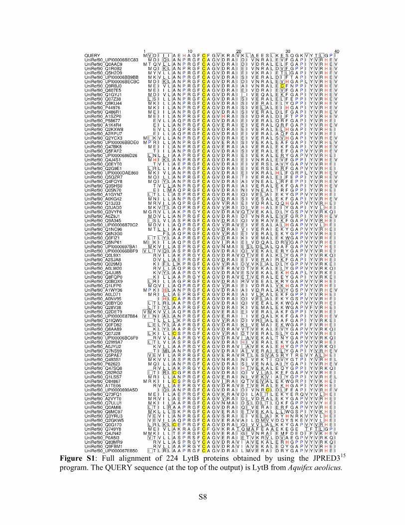
Figure S1: Full alignment of 224 LytB proteins obtained by using the JPRED315 program. The QUERY sequence (at the top of the output) is LytB from Aquifex aeolicus.

S9
Figure S1 (continued).

S10
Figure S1 (continued).

S11
Figure S1 (continued).

S12
Figure S1 (continued).

S13
Figure S1 (continued).

S14
Figure S1 (continued).

S15
Figure S1 (continued).

S16
Figure S1 (continued).

S17
Figure S1 (continued).

S18
Figure S1 (continued).

S19
Figure S1 (continued).

S20
Figure S1 (continued).

S21
Figure S1 (continued).

S22
Figure S1 (continued).

S23
Figure S1 (continued).

S24
Figure S1 (continued).

S25
Figure S1 (continued).

S26
Figure S1 (continued).

S27
Figure S1 (continued).

S28
Figure S1 (continued).

S29
Figure S1 (continued).

S30
Figure S1 (continued).

S31
Figure S1 (continued).

S32
References
(1) Tong, W.-H.; Jameson, G. N. L.; Huynh, B. H.; Rouault, T. A. Proc. Natl. Acad. Sci.
U.S.A. 2003, 100, 9762-9767.
(2) Otwinowski, Z.; Minor, W. Methods Enzymol. 1996, 276, 307-326.
(3) Kabsch, H. J. Appl. Cryst. 1993, 26, 795-800.
(4) Schneider, T. R.; Sheldrick, G. M. Acta Crystallogr. 2002, D58, 1772-1779.
(5) De la Fortelle, E.; Bricogne, G. Methods Enzymol. 1997, 276, 472-494.
(6) Abrahams, J. P.; Leslie, A. G. W. Acta Crystallogr. 1996, D52, 30-42.
(7) Cowtan, K. D. Joint CCP4 and ESF-EACBM Newsletter on Protein Crystallography
1994, 31, 83-91.
(8) Terwilliger, T. C. Acta Crystallogr. 2003, D59, 38-44.
(9) Jones, T. A.; Zou, J. Y.; Cowan, S. W.; Kjeldgaard, M. Acta Crystallogr. 1991, A47,
110-119.
(10) Emsley, P.; Cowtan, K. Acta Crystallogr. 2004, D60, 2126-2132.
(11) Brünger, A.; Adams, P. D.; Clore, G. M.; Delano, W. L.; Gros, P.; Grosse-
Kunstleve, R.; Jiang, J.-S.; Kuszewski, J.; Nilges, M.; Pannu, N. S.; Read, R. J.;
Rice, L. M.; Simonson, T.; Warren, G. L. Acta Crystallogr. 1998, D54, 905-921.
(12) Murshudov, G. N.; Vagin, A. A.; Dodson, E. J. Acta Crystallogr. 1997, D53, 240-
255.
(13) Laskowski, R. A.; MacArthur, M. W.; Moss, D. S.; Thornton, J. M. J. Appl. Cryst.
1993, 26, 283-291.
(14) Glide 4.5 ; Schrodinger, LLC: New York, NY, 2007

S33
(15) Cuff, J. A.; Clamp, M.E.; Siddiqui, A. S.; Finlay, M.; Barton, G. J. Bioinformatics
1998, 14, 892-893.


![Asymmetric crystallization during cooling and heating in ... · metastable melt is cooled to the same temperature T f [13,14]. Asymmetries in the crystallization time scales upon](https://img.dokumen.tips/doc/110x75/5f843efad874866685756322/asymmetric-crystallization-during-cooling-and-heating-in-metastable-melt-is.jpg)