Embed Size (px)
Citation preview

1
lytA-based identification methods can misidentify Streptococcus pneumoniae
Alexandra S. Simõesa,1,#, Débora A. Tavaresa,#, Dora Rolob,c,2, Carmen Ardanuyb,c,
Herman Goossensd,e, Birgitta Henriques-Normarkf,g, Josefina Linaresb,c,, Hermínia de
Lencastreh,i, and Raquel Sá-Leãoa,*
aLaboratory of Molecular Microbiology of Human Pathogens, Instituto de Tecnologia
Química e Biológica António Xavier (ITQB), Universidade Nova de Lisboa (UNL), Oeiras,
Portugal
bMicrobiology Department, Hospital Universitari de Bellvitge, Universitat de Barcelona-10
IDIBELL, L’Hospitalet de Llobregat, Barcelona, Spain
cCIBERES (Ciber de Enfermedades Respiratorias), ISCIII, Madrid, Spain
dDepartment of Medical Microbiology, University of Antwerp, Antwerp, Belgium
eVaccine and Infectious Disease Institute, University of Antwerp, Antwerp, Belgium
fDepartment of Microbiology, Tumor and Cell Biology, Karolinska Institutet, Stockholm,
Sweden
gDepartment of Laboratory Medicine, Division of Clinical Microbiology, Karolinska University
Hospital, Stockholm, Sweden
hLaboratory of Molecular Genetics, ITQB, UNL, Oeiras, Portugal
iLaboratory of Microbiology and Infectious Diseases, The Rockefeller University, New York, 20
NY, USA
Running Tittle: lytA-based assays can misidentify pneumococci
*Corresponding Author.
1 Present Address: Global Health and Tropical Medicine, Instituto de Higiene e Medicina Tropical, UNL, Lisboa, Portugal 2 Present Address: ISGlobal, Barcelona Ctr. Int. Health Res. (CRESIB), Hospital Clínic - Universitat de Barcelona, Barcelona, Spain

2
Laboratory of Molecular Microbiology of Human Pathogens
Instituto de Tecnologia Química e Biológica António Xavier
Av. da República, 2780‐157 Oeiras, Portugal
Tel.: + 351 214469872
E‐mail address: [email protected] (R Sá-Leão) 30
#These authors contributed equally to this work
Word Counts for Abstract: 145
Word Counts for Body of the text: 3,556
3
Abstract
During surveillance studies we detected, among over 1,500 presumptive pneumococci, 11
isolates displaying conflicting or novel results when characterized by widely accepted
phenotypic (optochin susceptibility and bile solubility) and genotypic (lytA-BsaAI-RFLP and 40
MLST) identification methods. We aimed to determine the genetic basis for the unexpected
results given by lytA-BsaAI-RFLP and investigate the accuracy of the WHO recommended
lytA real-time PCR assay to classify these 11 isolates. Three novel lytA-BsaAI-RFLP
signatures were found (one in pneumococcus and two in S. mitis). In addition, one
pneumococcus displayed the atypical lytA-BsaAI-RFLP signature characteristic of non-
pneumococci and two S. pseudopneumoniae displayed the typical lytA-BsaAI-RFLP pattern
characteristic of pneumococci. lytA real-time PCR misidentified these three isolates. In
conclusion, identification of pneumococci by lytA real-time PCR, and other lytA-based
methodologies, may lead to false results. This is of particular relevance in the increasingly
frequent colonization studies relying solely on culture-independent methods. 50
Keywords
S. pneumoniae; S. pseudopneumoniae; lytA; real-time PCR; identification; molecular
methods.
4
1. Introduction
Streptococcus pneumoniae (pneumococcus) is a major human pathogen, causing a wide
range of infections from otitis media to bacteremia and meningitis. Routine identification of
pneumococcus (colony morphology on blood agar plates, susceptibility to optochin, cell wall
lysis by 1% of sodium deoxycholate (bile solubility), and assignment of a capsular type by 60
serotyping) is not always straightforward since some isolates may give atypical results in
one or more of these assays (Arbique, et al., 2004; Balsalobre, et al., 2006; Bosshard, et al.,
2004; Nunes, et al., 2008; Sá-Leão, et al., 2006; Simões, et al., 2010; Whatmore, et al.,
2000).
As a human colonizer, pneumococci co-habit the nasopharynx with several other bacterial
species, including its closest relatives: S. pseudopneumoniae, S. mitis, and S. oralis. The
exchange of genetic elements between pneumococci and its closest relatives has been
described and increases the difficulties in species identification (Denapaite, et al., 2010;
Donati, et al., 2010; Johnston, et al., 2010). Although isolates of closely related species have 70
been implied in disease episodes, pneumococcus is the most important disease-causing
species of the mitis group (formed by pneumococcus, S. pseudopneumoniae, S. mitis, and
S. oralis, among others) (Bochud, et al., 1994; Douglas, et al., 1993; Keith, et al., 2006;
Rolo, et al., 2013). For this reason, a correct identification of pneumococcus is crucial for an
accurate diagnosis and treatment. In fact, misidentification of pneumococcus could falsely
increase the rates of pneumococci non-susceptible to antimicrobials since high rates of
penicillin-resistant and multidrug-resistant S. mitis isolates have been described (Ioannidou,
et al., 2001; Simões, et al., 2010; Wester, et al., 2002).
In recent years, several molecular methods have been proposed to differentiate 80
pneumococcus from closely related species. The presence of the lytA gene – the major
autolysin and a ubiquitous virulence factor – has been proposed to identify pneumococci
(Messmer, et al., 1997). However, homologues of the lytA gene have been detected in

5
strains of closely related streptococcal species (Denapaite, et al., 2010; Romero, et al.,
2004; Whatmore, et al., 2000). A lytA-BsaAI-RFLP strategy to differentiate pneumococcus
from closely related species based on signatures characteristic of pneumococcal (typical)
lytA or non-pneumococcal (atypical) lytA has been proposed and successfully used (Llull, et
al., 2006). Also, based on DNA sequence differences between the pneumococcal lytA and
its homologues, real-time PCR assays for the specific identification of pneumococcus have
been developed (Carvalho, et al., 2007). Nowadays, the lytA real-time PCR strategy 90
developed by the CDC is currently the WHO recommended culture-independent method to
detect pneumococci (Carvalho, et al., 2007; Satzke, et al., 2013).
In addition, multilocus sequence typing (MLST) and multilocus sequence analysis (MLSA)
strategies have been validated as tools for reliable species identification among streptococci
of the viridans group (Hanage, et al., 2005a; Bishop, et al., 2009).
During surveillance studies, we detected 11 presumptive pneumococcal isolates displaying
conflicting or novel results when characterized by the combination of optochin susceptibility,
bile solubility, lytA-BsaAI-RFLP and, MLST. In this study, we aimed to determine the genetic 100
basis for the unexpected results given by lytA-BsaAI-RFLP. Also, considering the increasing
and wide use of lytA real-time PCR for the identification of pneumococci, we also aimed to
investigate the accuracy of this method in the classification of these 11 isolates.
2. Materials and methods
2.1. Ethics statement
In the present study a sub-set of bacterial isolates selected from different studies was
characterized. All samples have been coded numerically upon collection and processed
anonymously. No human subjects, human material or human data were used, thus excusing
the requirement for an ethical approval. Approval for the original studies was obtained from: 110
i) the Portuguese Ministry of Education; the study was registered and approved at the Health

6
Care Centre of Oeiras that reports to Administração Regional de Saúde (ARS; “Regional
Health Administration”) of Lisboa and Vale do Tejo from the Ministry of Health (PT coded
isolates); signed informed consent was obtained from parents/guardians of participating
children; ii) the “Comité Ètic d'Investigació Clínica del Hospital Universitari de Bellvitge”
(Spain coded isolates); and iii) research sites involved in the European project GRACE
(Genomics to Combat Resistance against Antibiotics in Community-acquired LRTI in
Europe) obtained ethical and competent authority approval from their local organizations.
Patients who fulfilled the inclusion criteria were given written and verbal information on the
study and asked for informed consent (GRA coded isolates). 120
2.2. Study isolates
Isolates were selected from biological samples under the framework of other studies aimed
to identify pneumococci. These included surveillance carriage studies performed in Portugal
(obtained between 2011-2014, n=1,226 isolates), a study of lower respiratory tract infections
in several European countries (obtained between 2007-2010, n=204 isolates; GRACE -
Genomics to Combat Resistance against Antibiotics in Community-acquired LRTI in Europe;
www.gracelrti.org), and a collection of disease isolates with atypical properties from Spain
(obtained between 1991-2009, n=132; (Rolo, et al., 2013)). The assays performed for
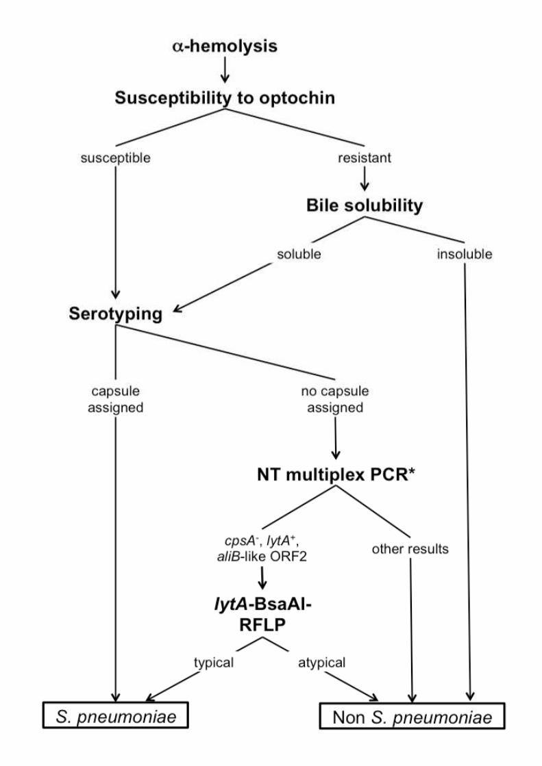
species identification are described below and are summarized in Fig. S1. Briefly, isolates 130
were presumptively identified as pneumococci based on presence of α-hemolysis when
grown in gentamycin blood agar plates and optochin susceptibility. If optochin resistance
was observed, bile solubility was performed. Presumptive pneumococci were then serotyped
by the Quellung reaction and/or by multiplex PCR. When the assignment of a serotype was
not possible, a multiplex PCR designed to detect non-encapsulated pneumococci and lytA-
BsaAI-RFLP were performed as described below (Simões, et al., 2011).
Among the 1,562 isolates mentioned above and presumptively identified as pneumococci,
lytA-BsaAI-RFLP was performed for 247 isolates (99 from Portugal, 25 from GRACE, and
7
123 from Spain). Of these, 61 were identified as non-encapsulated pneumococci with a 140
typical lytA-BsaAI-RFLP pattern (29 from Portugal and 32 from Spain) and 175 were
identified as non S. pneumoniae with an atypical lytA-BsaAI-RFLP pattern (66 from Portugal,
22 from GRACE, and 87 from Spain). The 11 isolates reported here (four from Portugal,
three from GRACE, and four from Spain) exhibited conflicting (or novel) results when
presumptive identification based on optochin susceptibility and bile solubility was compared
to the one suggested by the lytA-BsaAI-RFLP typing system..
2.3. Optochin susceptibility in CO2 and in O2 atmosphere
Optochin susceptibility was tested by disk diffusion using commercially available optochin
discs (5 µg; 6 mm; Oxoid, Hampshire, England). Discs were applied to overnight cultures 150
plated in blood agar (trypticase soy agar supplemented with 5% defibrinated sheep blood).
Plates were incubated overnight at 37°C in 5% CO2 atmosphere or in ambient atmosphere
as described by Arbique et al. (Arbique, et al., 2004). Isolates were considered resistant to
optochin when they displayed inhibition zones smaller than 14 mm.
2.4. Bile solubility test
The bile solubility assay was performed according to standard procedures: colonies from an
overnight culture were suspended in 1 mL of a 0.85% NaCl (w/v) solution to a turbidity equal
to 0.5-1 McFarland standard (Rouff, et al., 2003). This suspension was distributed into two
tubes (500 µL each tube) and 200 µL of a 10% deoxycholate solution were added to one 160
tube while the other received 200 µL of a 0.85% NaCl (w/v) solution (control). Both tubes
were incubated at 37ºC for up to 2h. A sample was considered soluble in bile when clearing
of the turbidity occurred in the tube with deoxycholate but not in the control.
2.5. Capsular typing
Capsular type assignment was performed by the Quellung reaction and by multiplex PCR as
previously described (Brito, et al., 2003; Pai, et al., 2006; Simões, et al., 2011).

8
2.6. lytA-BsaAI-RFLP signatures
The entire lytA gene was amplified by PCR using primers previously described (LA5_Ext: 5’-170
AAGCTTTTTAGTCTGGGGTG-3’ and LA3_Ext: 5’-AAGCTTTTTCAAGACCTAATAATATG-
3’), yielding a PCR product of approximately 1,200 bp (Obregon, et al., 2002). Typical
(characteristic of pneumococcal lytA) or atypical (characteristic of non-pneumococcal lytA)
RFLP signatures were determined as described before by digesting the PCR product with
BsaAI and separating the fragments by agarose gel electrophoresis (Llull, et al., 2006).
2.7. Real-time PCR targeting lytA and piaA
The presence of the lytA gene was tested by real-time PCR using previously described
primers (lytA_F: 5’-ACGCAATCTAGCAGATGAAGCA-3’ and lytA_R: 5’-
TCGTGCGTTTTAATTCCAGCT-3’) and probe (5’-FAM-GCCGAAAACGCTTGATACAG 180
GGAG-3’-BHQ1) (Carvalho, et al., 2007). The presence of the piaA gene was tested by real-
time PCR using previously described primers (piaF: 5’-CATTGGTGGCTTAGTAAGTGCAA-
3’ and piaR: 5’-TACTAACACAAGTTCCTGATAAGGCAAGT-3’) and probe (5’-FAM-
TGTAAGCGGAAAAGCAGGCCTTACCC-3’-BHQ1) (Trzcinski, et al., 2013). The assays
were carried out in a final volume of 25 µL using the FastStart TaqMan Probe Master
(Roche) containing 2.5 µL of 0.2 ng/µL DNA, 0.15 µM of each primer, and 0.075 µM of
probe. The assay was performed three times on different days and DNA from S.
pneumoniae TIGR4 (positive control) and S. pseudopneumoniae ATCC BAA-960 (negative
control) was used in every run. DNA was amplified with CFX96 real time system (Bio-Rad)
with the cycling parameters previously described (Carvalho, et al., 2007). Samples were 190
considered positive when cycle threshold (Ct) values were below 35.
2.8 DNA sequencing
9
Sequencing reactions needed for the methods described below were conducted at
Macrogen, Inc. (Amsterdam, The Netherlands). Sequencing analysis was done with
DNAStar (Lasergene).
2.9. lytA sequencing analysis
lytA PCR products of 1,200 bp were obtained as described above (Obregon, et al., 2002).
Sequencing was conducted at Macrogen, Inc. (Amsterdam, The Netherlands) and 200
subsequent analysis of the sequences was done with DNAstar (Lasergene). lytA sequences
were also obtained for strains TIGR4 (pneumococcus, NCBI accession number AE
005672.3) and ATCC BAA-960 (S. pseudopneumoniae, NCBI accession number
AM113495.1), to be used for comparison. Nucleotide sequences of lytA gene described in
this study were deposited at the GenBank database with the accession numbers KT253593-
KT253603.
2.10. Multilocus sequence typing (MLST)
Amplification of internal fragments of the seven housekeeping genes – aroE, gdh, gki, recP,
spi, xpt, and ddl - was done according to the MLST scheme developed by Enright and Spratt 210
for S. pneumoniae (Enright and Spratt, 1998). Sequencing analysis was done with DNAStar
(Lasergene). Allele number assignment was done at the international MLST database for S.
pneumoniae (www.mlst.net).
2.11. Multilocus sequence analysis (MLSA) for viridans group streptococci
Amplification of internal fragments of the seven housekeeping genes – map, pfl, ppaC, pyk,
rpoB, sodA, and tuf – was done according to the scheme developed and validated for
viridans group streptococci by Bishop et al., except for the primer sodA-dn, that had an R to
Y substitution (5’-AYRTARTAMGCRTGYTCCCARACRTC-3’) based on published
sequences of strains TIGR4 (S. pneumoniae, accession number AE005672.3), B6 (S. mitis, 220
accession number NC_013853.1), and IS7493 (S. pseudopneumoniae, accession number
10
CP002925.1) (Bishop, et al., 2009). Phylogenetic analysis of the concatenated sequences of
strains analyzed in this study and the ones deposited at the eMLSA database (427 strains of
the viridans group of Streptococcus; http://www.emlsa.net/) was performed using MEGA6.06
(http://www.megasoftware.net/): sequences were aligned by ClustalW using default
parameters (gap opening penalty of 15 and gap extension penalty of 6.66 for both pairwise
and multiple alignment; IUB as DNA weight matrix, with a transition weight of 0.5). A
minimum-evolution phylogenetic tree was constructed using default parameters (maximum
composite likelihood was used as the substitution model of nucleotides, with transitions and
transversions as the substitutions to include; uniform rates among sites; homogeneous 230
pattern among lineages; complete deletion of gaps and missing data and close-neighbor-
interchange was used as the ME heuristic method, based on an initial tree by neighbor-
joining). Different concatenated sequences of the strains analyzed were arbitrarily named
viridans MLSA profiles 1 to 10 and species assignment was inferred based on clustering
analysis of the study isolates with the strains from the MLSA database.
3. Results
3.1. Phenotypic and genotypic characterization of the strains
Characteristics of the strains studied are described in Table 1, which also shows a summary
of all results. All strains grew in gentamycin blood agar and displayed pneumococcus-like 240
colony morphology albeit some were optochin resistant. The presence of cpsA or of other
capsular genes (screened by PCR serotyping as described in the Material and Methods
section) could not be detected in any of the strains, suggesting the absence of a
pneumococcal capsule.
3.2. Atypical and novel lytA-BsaAI-RFLP patterns found in pneumococci
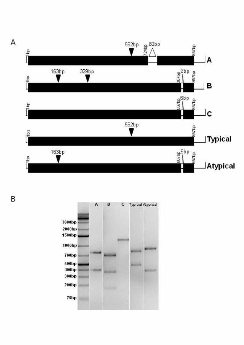
One pneumococcal strain (GRA218B) displayed an atypical lytA-BsaAI-RFLP pattern,
usually associated with non-pneumococci (Fig. 1) (Llull, et al., 2006). This strain was both
optochin susceptible and bile soluble, belonged to ST8073, and clustered with pneumococci
11
by MLSA (Table 1, Fig. 2). Sequence analysis of the lytA gene confirmed a 98% similarity of 250
the amino acid sequence with the LytA homologue of ATCC BAA-960, the S.
pseudopneumoniae strain used as reference (Fig. S2).
Three other pneumococcal strains (GRA036B, Spain6220, and Spain7582) displayed a lytA-
BsaAI-RFLP pattern not previously described (pattern A, Fig. 1). These strains were both
optochin susceptible and bile soluble. Also, these strains belonged to ST508 and clustered
with pneumococci by MLSA (Table 1, Fig. 2). Such similarities between these strains
suggest that they belong to a common lineage that may be disseminated. Sequence
analysis of the lytA gene revealed a 60bp deletion at 724bp, resulting in the loss of 20 amino
acids (KKIAEKWYYFDGEGAMKTGW). Apart from this deletion, the LytA amino acid 260
sequence shared a 99% similarity with that of TIGR4, the pneumococcal strain used as
reference (Fig. S2).
3.3. Typical and novel lytA-BsaAI-RFLP patterns found in non-pneumococci
Two non-pneumococcal strains (Spain2270 and Spain9880) displayed a typical lytA-BsaAI-
RFLP pattern, usually associated with pneumococci (Fig. 1) (Llull, et al., 2006). These
strains were optochin resistant in CO2 atmosphere, optochin susceptible in aerobic
conditions, and bile soluble. These strains could not be assigned to an ST according to the
S. pneumoniae MLST database and clustered with S. pseudopneumoniae by MLSA (Table
1, Fig. 2). Such similarities between these strains suggest that they belong to a common 270
lineage. Sequence analysis of the lytA gene confirmed a 99% similarity of the amino acid
sequence with the LytA of TIGR4, the pneumococcal strain used as reference (Fig. S2).
Five other non-pneumococcal strains displayed two lytA-BsaAI-RFLP patterns not previously
described (patterns B and C, Fig. 1). All these strains were bile soluble and while one was
susceptible to optochin, the others were resistant. None of the strains could be assigned to
an ST according to the S. pneumoniae MLST database and they all clustered with S. mitis

12
by MLSA (Table 1, Fig. 2). Sequence analysis of the lytA gene of strains displaying pattern B
(GRA254A, PT8543, PT8638 and PT9018) revealed a silent C329T mutation resulting in an
extra BsaAI cutting site. On the other hand, sequence analysis of the lytA gene of the strain 280
displaying pattern C (PT8238) revealed a silent C163T mutation resulting in the loss of the
BsaAI cutting site. Apart from these mutations, the LytA amino acid sequences of these
strains were 97-98% similar to that of the homologue of ATCC BAA-960, the S.
pseudopneumoniae strain used as reference.
3.4. Misidentifications by real-time PCR targeting lytA and piaA
Pneumococcal identification by lytA real-time PCR failed for three of the 11 strains in this
study: the pneumococcal strain harboring the S. pseudopneumoniae homologue of the lytA
gene (no amplification) and the two S. pseudopneumoniae strains harboring (the
pneumococcal) lytA (Ct 24 for both, Table 1). For all other cases described, the mutations 290
found were located outside the annealing sites of the real-time PCR primers and probe and
no misidentifications by real-time PCR were found (Fig. S2).
However, when pneumococcal identification by real-time PCR targeting lytA was
complemented by real-time PCR targeting piaA, only the pneumococcal strain harboring the
S. pseudopneumoniae homologue of the lytA gene remained misidentified, as this strain was
also negative for piaA (Table 1).
4. Discussion
In this study we aimed to determine the genetic basis for the unexpected results given by 300
lytA-BsaAI-RFLP and to investigate the accuracy of the lytA real-time PCR to classify 11 α-
hemolytic streptococcal isolates displaying conflicting or novel results when characterized by
the combination of optochin susceptibility, bile solubility, lytA-BsaAI-RFLP, and MLST. The
main findings of our work were: (i) the identification of one pneumococcal carriage strain
harboring a non-pneumococcal homologue of lytA; (ii) the identification of two invasive

13
(cerebrospinal fluid and sputum) S. pseudopneumoniae strains harboring the characteristic
pneumococcal lytA; and (iii) the misidentification of these three strains just referred to above
by the commonly used lytA real-time PCR. In addition, novel lytA-BsaAI-RFLP patterns were
identified and these were due to deletions or point mutations in lytA.
310
This is, to the best of our knowledge, the first report of a pneumococcal isolate harboring a
non-pneumococcal homologue of lytA. Isolates with similar properties may have passed
undetected in culture-independent studies relying solely on detection of lytA to identify
pneumococci. On the other hand, non-pneumococcal isolates originating false positive
results by the lytA real time PCR strategy have been described (Carvalho, et al., 2013). In
particular, two copies of lytA (one copy of pneumococcal lytA and one copy of the non-
pneumococcal homologue) were described in the genome of a clinical isolate (IS7493) of S.
pseudopneumoniae (Shahinas, et al., 2011). However, lytA real-time PCR amplification of
strain IS7493 did not retrieve a positive result (Tavares et al., unpublished).
320
The use of piaA real-time PCR to complement lytA real-time PCR has been proposed as
piaA has been described as a pneumococcal specific non-ubiquitous gene that appears to
be present in the majority of pneumococcal isolates (a notable exception are some non-
encapsulated pneumococci) (Tavares, et al., 2015; Trzcinski, et al., 2013; Whalan, et al.,
2006). Whalan et al. found this gene in all encapsulated pneumococci tested (39 isolates
covering 27 serotypes) and in six out of eight (75%) non-typeable pneumococci (Whalan, et
al., 2006). However, in a recent study of non-typeable pneumococci circulating in Portugal,
we have only detected piaA in 12 out of 35 (34%) non-typeable pneumococci suggesting
absence of this gene is common among these strains (Tavares, et al., 2015). Nevertheless,
the strategy of targeting piaA in addition to lytA clearly enhances the specificity of 330
pneumococcal identification (Trzcinski, et al., 2013). Even so, in the present study, one
pneumococcal strain could not be identified based on these two real-time PCR assays.
14
The occurrence of genetic exchange between oral Streptococcus species has been well
documented and horizontal gene transfer has been suggested as an important attenuator of
putative species barriers (Chi, et al., 2007; Denapaite, et al., 2010; Donati, et al., 2010;
Hanage, et al., 2005b; Johnston, et al., 2010). In fact, a smooth transition between
pneumococcus species and its close relatives has been proposed (Hakenbeck, et al., 2001).
On the other hand, Kilian et al. have proposed that both pneumococcus and S. mitis have
evolved divergently from a pathogenic common ancestral: while pneumococcus maintained 340
most of the ancestral virulence genes, S. mitis evolved to become a commensal (Kilian, et
al., 2008, Kilian, et al., 2014).
It is important to highlight that, although exceptions have been reported here and elsewhere
(Arbique, et al., 2004; Balsalobre, et al., 2006; Bosshard, et al., 2004; Nunes, et al., 2008;
Sá-Leão, et al., 2006; Simões, et al., 2010; Whatmore, et al., 2000), susceptibility to
optochin, bile solubility, and assignment of a capsular type are, each one of them, excellent
presumptive methods to identify the majority of pneumococcal isolates. In this report, all but
one S. mitis strain were correctly identified based on optochin susceptibility. For dubious
cases, the assignment of specific lytA-BsaAI-RFLP signatures and a multiplex PCR strategy 350
have been proposed (Llull, et al., 2006; Simões, et al., 2011). However, this study suggests
that with these methods, although rarely, misidentification can still occur. The assignment of
a sequence type by the S. pneumoniae MLST scheme or the viridans MLSA scheme,
although very useful as tools for species identification, are time-consuming and expensive
for routine laboratories, thus often being used only in selected cases (Bishop, et al., 2009;
Hanage, et al., 2005). Alternatively, the determination of a pneumococcal-specific sequence
signature of 16S rRNA has also been proposed as an inexpensive identification tool
(Scholz, et al., 2012).
One possible limitation of our study is that we did not systematically study the frequency at 360
which isolates with the new or conflicting characteristics described in this study occur in

15
collections of pneumococcal clinical and carriage isolates. This aim was beyond the scope of
this study. Although all isolates were characterized by optochin susceptibility and bile
solubility, lytA-BsaAI-RFLP was applied only with isolates presumptively identified as
pneumococci and for which a serotype could not be assigned. Still, based on our
experience, the frequency of such isolates appears to be low. In particular, among the
colonization isolates from Portugal (n=1,226) we have systematically performed the lytA-
BsaAI-RFLP assay for presumptive pneumococcal isolates for which a capsular type could
not be assigned (n=99). Of these, four S. mitis isolates (described in this study),
corresponding to c.a. 4.0% of the tested samples, had unusual lytA patterns. In addition, in 370
our collections the unusual pattern A was found in three of 61 (4.9%) S. pneumoniae
isolates tested by the lytA-BsaAI-RFLP.
The future is heading towards automated screenings of unprocessed samples. Currently,
lytA real-time PCR is the culture-independent methodology of choice and the association
with a second gene such as piaA as proposed by Trzcinski et al. is an interesting strategy
(Carvalho Mda, et al., 2007; Satzke, et al., 2013; Trzcinski, et al., 2013). Whole genome
sequencing (WGS) is being increasingly used and an approach combining automated
extraction of WGS information with MLST-extended schemes will undoubtedly reveal itself
very useful for the unambiguous classification of strains as the ones described in this study 380
(Sabat, et al., 2013).
In conclusion, identification of pneumococci based on lytA detection, including real-time
PCR, may lead to false results. This is of particular relevance in the increasingly frequent
colonization studies relying solely on culture-independent methods targeting lytA.
Funding
This work was funded by Fundação para a Ciência e a Tecnologia, Portugal, through grants
PTDC/BIA-MIC/64010/2006 and PTDC/BIA-BEC/098289/2008 to RSL,

16
SFRH/BD/70147/2010 to DAT, SFRH/BD/27325/2006 to ASS, and Pest-390
OE/EQB/LAO004/2011 to Laboratório Associado de Oeiras. The funding agency had no
involvement in the study design, collection, analysis, and interpretation of data, writing of the
article, nor in the decision to submit the study for publication.
Conflicts of interest
None to declare.
Acknowledgments
We thank Sónia T. Almeida for assistance with MLST.
400
References
Arbique JC, Poyart C, Trieu-Cuot P, Quesne G, Carvalho Mda G, Steigerwalt AG, Morey
RE, Jackson D, Davidson RJ, Facklam RR (2004) Accuracy of phenotypic and genotypic
testing for identification of Streptococcus pneumoniae and description of Streptococcus
pseudopneumoniae sp. nov. J Clin Microbiol 42:4686-4696 doi: 10.1128/JCM.42.10.4686-
4696.2004.
Balsalobre L, Hernandez-Madrid A, Llull D, Martin-Galiano AJ, Garcia E, Fenoll A, de la
Campa AG (2006) Molecular characterization of disease-associated streptococci of the mitis
group that are optochin susceptible. J Clin Microbiol 44:4163-4171 doi: 10.1128/JCM.01137-
06. 410
Bosshard PP, Abels S, Altwegg M, Bottger EC, Zbinden R (2004) Comparison of
conventional and molecular methods for ientification of aerobic catalase-negative gram-
positive cocci in the clinical laboratory. J Clin Microbiol 42:2065-2073 doi:
10.1128/JCM42.5.2065-2073.2004.
Bishop CJ, Aanensen DM, Jordan GE, Kilian M, Hanage WP, Spratt BG (2009) Assigning
strains to bacterial species via the internet. BMC Biol 7:3 doi: 10.1186/1741-7007-7-3.

17
Bochud PY, Calandra T, Francioli P (1994) Bacteremia due to viridans streptococci in
neutropenic patients: a review. Am J Med 97:256-264 doi: 0002-9343(94)90009-4
Brito DA, Ramirez M, de Lencastre H (2003) Serotyping Streptococcus pneumoniae by
multiplex PCR. J Clin Microbiol 41:2378-2384 doi: 10.1128/JCM.41.6.2378-2384.2003. 420
Carvalho Mda G, Tondella ML, McCaustland K, Weidlich L, McGee L, Mayer LW,
Steigerwalt A, Whaley M, Facklam RR, Fields B, Carlone G, Ades EW, Dagan R, Sampson
JS (2007) Evaluation and improvement of real-time PCR assays targeting lytA, ply, and
psaA genes for detection of pneumococcal DNA. J Clin Microbiol 45:2460-2466 doi:
10.1128/JCM.02498-06.
Carvalho Mda G, Pimenta FC, Moura I, Roundtree A, Gertz Jr RE, Li Z, Jagero G, Bigogo G,
Junghae M, Conklin L, Feikin DR, Breiman RF, Whitney CG, Beall BW (2013) Non-
pneumococcal mitis-group streptococci confound detection of pneumococal capsular
serotype-specific loci in the upper respiratory tract. PeerJ 1:e97 doi: 10.7717/peerj.97.
Chi F, Nolte O, Bergmann C, Ip M, Hakenbeck R (2007) Crossing the barrier: evolution and 430
spread of a major class of mosaic pbp2x in Streptococcus pneumoniae, S. mitis and S.
oralis. Int J Med Microbiol 297:503-512 doi: 10.1016/j.ijmm.2007.02.009.
Denapaite D, Bruckner R, Nuhn M, Reichmann P, Henrich B, Maurer P, Schahle Y,
Selbmann P, Zimmermann W, Wambutt R, Hakenbeck R (2010) The genome of
Streptococcus mitis B6--what is a commensal? PLoS One 5:e9426 doi:
10.1371/journal.pone.0009426.
Donati C, Hiller NL, Tettelin H, Muzzi A, Croucher NJ, Angiuoli SV, Oggioni M, Hotopp JCD,
Hu FZ, Riley DR, Covacci A, Mitchell TJ, Bentley SD, Kilian M, Ehrlich GD, Rappuoli R,
Moxon ER, Masignani V (2010) Structure and dynamics of the pan-genome of
Streptococcus pneumoniae and closely related species. Gen Biol 11:R107 doi: 10.186/gb-440
2010-11-10-r107.
18
Douglas CW, Heath J, Hampton KK, Preston FE (1993) Identity of viridans streptococci
isolated from cases of infective endocarditis. J Med Microbiol 39:179-182 doi:
10.1099/00222615-39-3-179.
Enright MC, Spratt BG (1998) A multilocus sequence typing scheme for Streptococcus
pneumoniae: identification of clones associated with serious invasive disease. Microbiology
144:3049-3060 doi: 10.1099/00221287-144-11-3049.
Hanage WP, Kaijalainen T, Herva E, Saukkoriipi A, Syrjanen R, Spratt BG (2005a) Using
multilocus sequence data to define the pneumococcus. J Bacteriol 187:6223-6230 doi:
10.1128/JB.187.17.6223-6230.2005. 450
Hanage WP, Fraser C, Spratt BG (2005b) Fuzzy species among recombinogenic bacteria.
BMC Biol 7:6 doi: 10.1186/1741-7007-3-6.
Hakenbeck R, Balmelle N, Weber B, Gardes C, Keck W, de Saizieu A (2001) Mosaic genes
and mosaic chromosomes: intra- and interspecies genomic variation of Streptococcus
pneumoniae. Infect Immun 69:2477-2486 doi: 10.1128/IAI.69.4.2477-2486.2001.
Ioannidou S, Tassios PT, Kotsovili-Tseleni A, Foustoukou M, Legakis NJ, Vatopoulos A
(2001) Antibiotic resistance rates and macrolide resistance phenotypes of viridans group
streptococci from the oropharynx of healthy Greek children. Int J Antimicrob Agents 17:195-
201 doi: 10.1016/S0924-8579(00)00338-1.
Johnston C, Hinds J, Smith A, van der Linden M, Eldere JA, Mitchell T (2010) Detection of 460
large numbers of pneumococcal virulence genes in streptococci of the mitis group. J Clin
Microbiol 48:2762-2769 doi: 10.1128/JCM.01746-09.
Keith ER, Podmore RG, Anderson TP, Murdoch DR (2006) Characteristics of Streptococcus
pseudopneumoniae isolated from purulent sputum samples. J Clin Microbiol 44:923-927 doi:
10.1128/JCM.44.3.923-927.2006.

19
Kilian M, Poulsen K, Blomqvist T, Havarstein L, Bek-Thomsen M, Tettelin H, Sorensen UBS
(2008) Evolution of Streptococcus pneumoniae and its close commensal relatives. PLoS
One 3:e2683 doi: 10.1371/journal.pone.0002683.
Kilian M, Riley DR, Jensen A, Bruggemann H, Tettelin H (2014) Parallel evolution of
Streptococcus pneumoniae and Streptococcus mitis to pathogenic and mutualistic lifestyles. 470
MBio 5:e01490-01414 doi: 10.1128/mBio.01490-14.
Llull D, Lopez R, Garcia E (2006) Characteristic signatures of the lytA gene provide a basis
for rapid and reliable diagnosis of Streptococcus pneumoniae infections. J Clin Microbiol
44:1250-1256 doi: 10.1128/JCM.44.4.1250-1256.2006.
Messmer TO, Whitney CG, Fields BS (1997) Use of polymerase chain reaction to identify
pneumococcal infection associated with hemorrhage and shock in two previously healthy
young children. Clin Chem 43:930-935.
Nunes S, Sá-Leão R, de Lencastre H (2008) Optochin resistance among Streptococcus
pneumoniae strains colonizing healthy children in Portugal. J Clin Microbiol 46:321-324 doi:
10.1128/JCM.02097-07. 480
Obregon V, Garcia P, Garcia E, Fenoll A, Lopez R, Garcia JL (2002) Molecular peculiarities
of the lytA gene isolated from clinical pneumococcal strains that are bile insoluble. J Clin
Microbiol 40:2545-2554 doi: 10.1128/JCM.40.7.2545-2554.2002.
Pai R, Gertz RE, Beall B (2006) Sequential multiplex PCR approach for determining
capsular serotypes of Streptococcus pneumoniae isolates. J Clin Microbiol 44:124-131 doi:
10.1128/JCM.44.1.124-131.2006.
Rolo D, Simões AS, Domenech A, Fenoll A, Linares J, de Lencastre H, Ardanuy C, Sa-Leão
R (2013) Disease isolates of Streptococcus pseudopneumoniae and non-typeable S.
pneumoniae presumptively identified as atypical S. pneumoniae in Spain. PLoS One
8:e57047 doi: 10.1371/journal.pone.0057047. 490
20
Romero P, Lopez R, Garcia E (2004) Characterization of LytA-like N-acetylmuramoyl-L-
alanine amidases from two new Streptococcus mitis bacteriophages provides insights into
the properties of the major pneumococcal autolysin. J Bacteriol 186:8229-8239 doi:
10.1128/JB.186.24.8229-8239.2004.
Rouff K, Whiley RA, Beighton D (2003) Streptococcus. In P. R. Murray, E. J. Barron, J. H.
Jorgensen, M. A. Pfaller & R. H. Yolken (Eds, Manual of Clinical Microbiology (8th ed., pp.
405-421). Washington, D.C.: American Society for Microbiology.
Sá-Leão R, Simões AS, Nunes S, Sousa NG, Frazão N, de Lencastre H (2006)
Identification, prevalence and population structure of non-typable Streptococcus
pneumoniae in carriage samples isolated from preschoolers attending day-care centres. 500
Microbiology 152:367-376 doi: 10.1099/mic.0.28596-0.
Sabat AJ, Budimir A, Nashev D, Sá-Leão R, van Dijl J, Laurent F, Grundmann H, Friedrich
AW (2013) Overview of molecular typing methods for outbreak detection and
epidemiological surveillance. Euro Surveill 18:20380.
Satzke C, Turner P, Virolainen-Julkunen A, Adrian PV, Antonio M, Hare KM, Henao-
Restrepo AM, Leach AJ, Klugman KP, Porter BD, Sá-Leão R, Scott JA, Nohynek H, O'Brien
KL (2013) Standard method for detecting upper respiratory carriage of Streptococcus
pneumoniae: updated recommendations from the World Health Organization Pneumococcal
Carriage Working Group. Vaccine 32:165-179 doi: 10.1016/j.vaccine.2013.08.062.
Scholz CFP, Poulsen K, Kilian M (2012) Novel molecular method for the identification of 510
Streptococcus pneumoniae applicable to clinical microbiology and 16S rRNA sequence-
based microbiome studies. J Clin Microbiol 50:1968-1973 doi: 10.1128/JCM.00365-12.
Shahinas D, Tamber GS, Arya G, Wong A, Lau R, Jamieson F, Ma JH, Alexander DC, Low
DE, Pillai DR (2011) Whole-genome sequence of Streptococcus pseudopneumoniae isolate
IS7493. J Bacteriol 193:6102-6103 doi: 10.1128/JB.06075-11.
21
Simões AS, Sá-Leão R, Eleveld MJ, Tavares DA, Carriço JA, Bootsma HJ, Hermans PW
(2010) Highly penicillin-resistant multidrug-resistant pneumococcus-like strains colonizing
children in Oeiras, Portugal: genomic characteristics and implications for surveillance. J Clin
Microbiol 48:238-246 doi: 10.1128/JCM.01313-09.
Simões AS, Valente C, de Lencastre H, Sá-Leão R (2011) Rapid identification of 520
noncapsulated Streptococcus pneumoniae in nasopharyngeal samples allowing detection of
co-colonization and reevaluation of prevalence. Diagn Microbiol Infect Dis 71:208-216 doi:
10.1016/j.diagmicrobio.2011.07.009.
Tavares DA, Simões AS, Bootsma HJ, Hermans PW, de Lencastre H, Sá-Leão R (2015)
Non-typeable pneumococci circulating in Portugal are of cps type NCC2 and have genomic
features typical of encapsulated isolates. BMC Genomics 15:863 doi: 10.1186/1471-2164-
15-863.
Trzcinski K, Bogaert D, Wyllie A, Chu ML, van der Ende A, Bruin JP, van den Dobbelsteen
G, Veenhoven RH, Sanders EA (2013) Superiority of trans-oral over trans-nasal sampling in
detecting Streptococcus pneumoniae colonization in adults. PLoS One 8:e60520 doi: 530
10.1371/journal.pone.0060520.
Wester CW, Ariga D, Nathan C, Rice TW, Pulvirenti J, Patel R, Kocka F, Ortiz J, Weinstein
RA (2002) Possible overestimation of penicillin resistant Streptococcus pneumoniae
colonization rates due to misidentification of oropharyngeal streptococci. Diagn Microbiol
Infect Dis 42:263-268 doi: 10.1016/S0732-8893(01)00358-3.
Whalan RH, Funnell SG, Bowler LD, Hudson MJ, Robinson A, Dowson CG (2006)
Distribution and genetic diversity of the ABC transporter lipoproteins PiuA and PiaA within
Streptococcus pneumoniae and related streptococci. J Bacteriol 188:1031-1038 doi:
10.1128/JB.188.3.1031-1038.2006.
Whatmore AM, Efstratiou A, Pickerill AP, Broughton K, Woodard G, Sturgeon D, George R, 540
Dowson CG (2000) Genetic relationships between clinical isolates of Streptococcus

22
pneumoniae, Streptococcus oralis, and Streptococcus mitis: characterization of "atypical"
pneumococci and organisms allied to S. mitis harboring S. pneumoniae virulence factor-
encoding genes. Infect Immun 68:1374-1382 doi: 10.1128/IAI.68.3.1374-1382.2000.
23
Figures
Figure 1. lytA-BsaAI-RFLP patterns. (A) Schematic representation of the DNA fragments
produced by digestion of lytA gene with BsaAI. Solid triangles show the restriction sites of
BsaAI. Open triangles represent deletions. (B) Separation of BsaAI-digested fragments by
agarose gel electrophoresis. 550
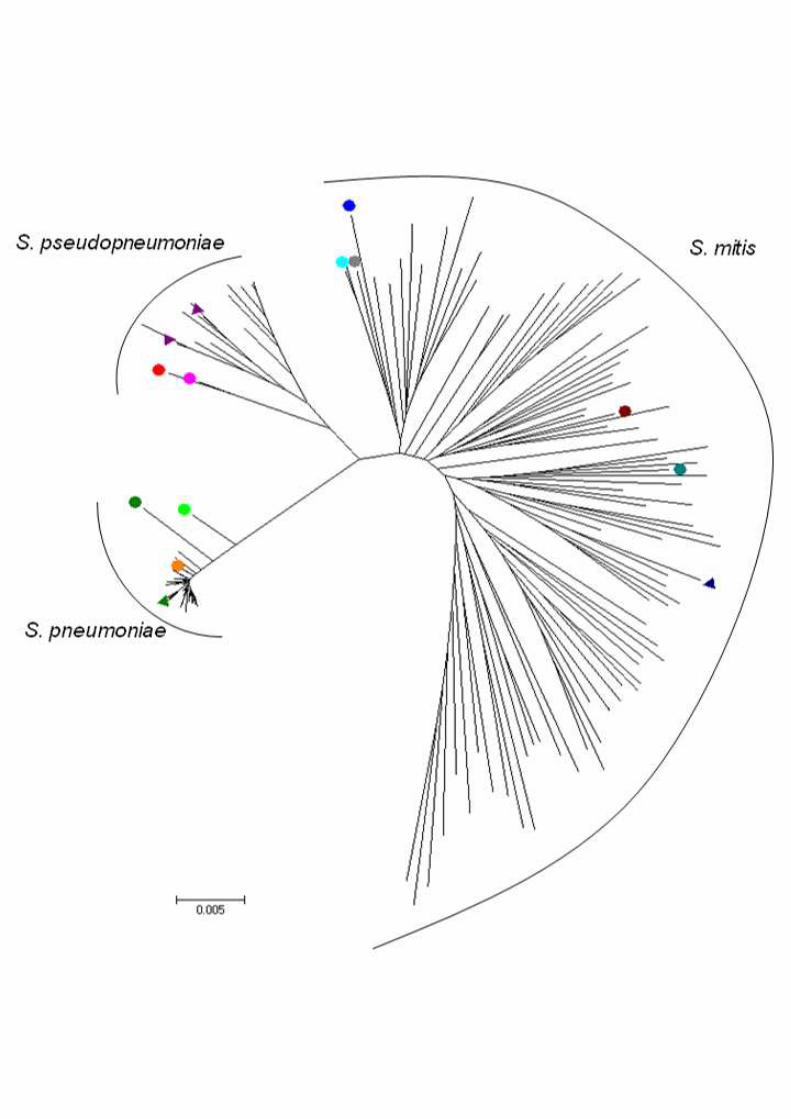
Figure 2. Genetic relationships of the strains analyzed in this study and S.
pneumoniae, S. pseudopneumoniae, and S. mitis strains deposited at the eMLSA
database. Orange circle – GRA036B and Spain7582, light green circle – Spain6220, dark
green circle – GRA218B, dark blue circle – GRA254A, blue-green circle – PT8543, maroon
circle – PT8638, cyan circle – PT9018, gray circle – PT8238, rose circle – Spain 2270, red
circle – Spain9880, green triangle – S. pneumoniae reference strain TIGR4, purple triangle –
S. pseudopneumoniae reference strains ATCC-BAA 960 and IS7493, blue triangle – S. mitis
reference strain DSM12643, no markers – S. pneumoniae, S. pseudopneumoniae, and S.
mitis strains deposited at eMLSA database. 560
Supporting Information
Figure S1. Routine laboratory workflow for the identification of S. pneumoniae. *As
described in (Simões, et al., 2010).
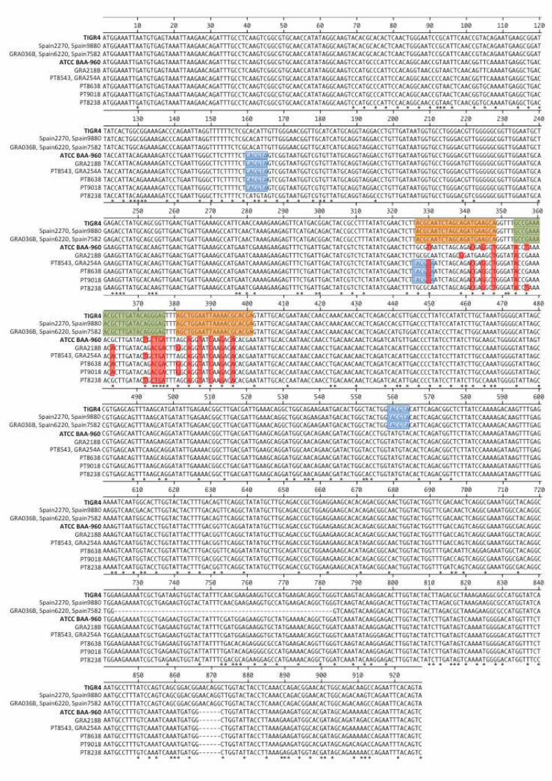
Figure S2. lytA sequences of the strains analyzed in this study. In bold are the lytA
sequences used as control (S. pseudopneumoniae ATCC BAA-960 and S. pneumoniae
TIGR4); blue shadow indicates the restriction sites for BsaAI; green and orange shadows
indicate probe and primers annealing sites (respectively) of the lytA real-time PCR assay; *
indicates base substitutions; red shadow indicates substitutions in the annealing regions 570
(primers and probe) of the lytA real-time PCR assay.
24
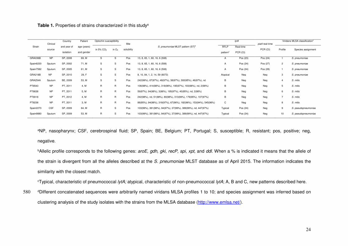
Table 1. Properties of strains characterized in this studya
aNP, nasopharynx; CSF, cerebrospinal fluid; SP, Spain; BE, Belgium; PT, Portugal; S, susceptible; R, resistant; pos, positive; neg,
negative.
bAllelic profile corresponds to the following genes: aroE, gdh, gki, recP, spi, xpt, and ddl. When a % is indicated it means that the allele of
the strain is divergent from all the alleles described at the S. pneumoniae MLST database as of April 2015. The information indicates the
similarity with the closest match.
cTypical, characteristic of pneumococcal lytA; atypical, characteristic of non-pneumococcal lytA; A, B and C, new patterns described here.
dDifferent concatenated sequences were arbitrarily named viridans MLSA profiles 1 to 10; and species assignment was inferred based on 580
clustering analysis of the study isolates with the strains from the MLSA database (http://www.emlsa.net/).
Strain Clinical
source
Country
and year of
isolation
Patient
age (years)
and gender
Optochin susceptibility Bile
solubility S. pneumoniae MLST pattern (ST)b
lytA piaA real-time
PCR (Ct)
Viridans MLSA classificationd
in 5% CO2 in O2 RFLP
patternc
Real-time
PCR (Ct) Profile Species assignment
GRA036B NP SP, 2008 69, M S S Pos 13, 8, 65, 1, 60, 16, 6 (508) A Pos (23) Pos (24) 1 S. pneumoniae
Spain6220 Sputum SP, 2002 71, M S S Pos 13, 8, 65, 1, 60, 16, 6 (508) A Pos (24) Pos (27) 2 S. pneumoniae
Spain7582 Sputum SP, 2005 61, M S S Pos 13, 8, 65, 1, 60, 16, 6 (508) A Pos (24) Pos (28) 1 S. pneumoniae
GRA218B NP SP, 2010 29, F S S Pos 8, 10, 84, 1, 2, 14, 59 (8073) Atypical Neg Neg 3 S. pneumoniae
GRA254A Sputum BE, 2009 53, M S S Pos 242(99%), 67(97%), 48(97%), 38(97%), 393(95%), 46(97%), nd B Neg Neg 4 S. mitis
PT8543 NP PT, 2011 4, M R R Pos 106(98%), 414(98%), 318(96%), 195(97%), 193(96%), nd, 2(98%) B Neg Neg 5 S. mitis
PT8638 NP PT, 2011 5, M R R Pos 59(97%), 94(98%), 3(96%), 195(97%), 40(95%), nd, 2(98%) B Neg Neg 6 S. mitis
PT9018 NP PT, 2012 4, M R R Pos 242(98%), nd, 51(98%), 80(98%), 313(99%), 179(95%), 107(97%) B Neg Neg 7 S. mitis
PT8238 NP PT, 2011 3, M R R Pos 88(95%), 94(98%), 318(97%), 67(96%), 182(96%), 153(94%), 545(96%) C Neg Neg 8 S. mitis
Spain2270 CSF SP, 2009 64, M R S Pos 103(99%), 381(98%), 94(97%), 37(99%), 389(99%), nd, 447(97%) Typical Pos (24) Neg 9 S. pseudopneumoniae
Spain9880 Sputum SP, 2009 53, M R S Pos 103(99%), 381(98%), 94(97%), 37(99%), 389(99%), nd, 447(97%) Typical Pos (24) Neg 10 S. pseudopneumoniae