Embed Size (px)
Citation preview
General Pathology VPM 152
Enrique Aburto http://people.upei.ca/eaburto Winter 2015
Lecture 6 Carcinogenic agents (cont’d); Local &
Systemic effects of Neoplasms
Disorders of Cell Growth & Neoplasia
Lymphoma
Spinal cord compression
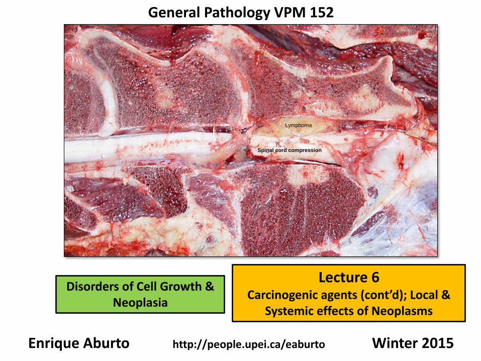
Radiation Carcinogenesis
• ionizing radiation, either weak (UV rays) or strong (medical) can induce neoplasia.
• many skin tumors are induced by UV light exposure.
• degree of risk associated with: type of UV rays (esp UV-B),
intensity of exposure (eg equator, high altitude)
amount of protective pigmentation (esp white regions)
• most damaged cells are either repaired (NER pathway) or undergo apoptosis.
• excessive sun exposure overwhelms the NER pathway (inability to repair DNA)
• carcinogenicity is due to mutations arising from pyrimidine dimer formation.
• p53 and RAS are particularly prone to mutation by UV light.
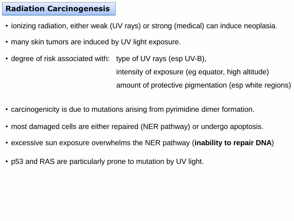
The tumor most frequently associated with prolonged exposure to UV light in domestic animals is
squamous cell carcinoma. They typical occur in non-pigmented locations with sparse hair coat.
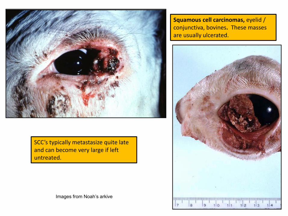
Squamous cell carcinomas, eyelid / conjunctiva, bovines. These masses are usually ulcerated.
SCC’s typically metastasize quite late and can become very large if left untreated.
Images from Noah’s arkive
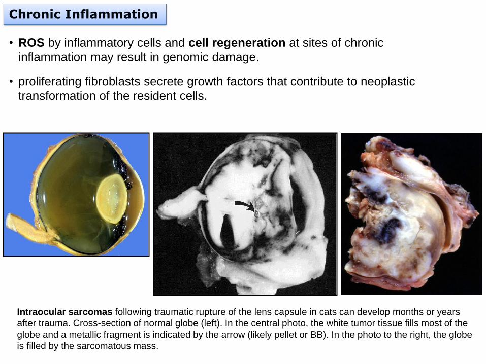
Chronic Inflammation
• ROS by inflammatory cells and cell regeneration at sites of chronic
inflammation may result in genomic damage.
• proliferating fibroblasts secrete growth factors that contribute to neoplastic
transformation of the resident cells.
Intraocular sarcomas following traumatic rupture of the lens capsule in cats can develop months or years
after trauma. Cross-section of normal globe (left). In the central photo, the white tumor tissue fills most of the
globe and a metallic fragment is indicated by the arrow (likely pellet or BB). In the photo to the right, the globe
is filled by the sarcomatous mass.
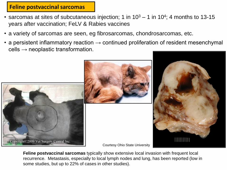
• sarcomas at sites of subcutaneous injection; 1 in 103 – 1 in 104; 4 months to 13-15
years after vaccination; FeLV & Rabies vaccines
Feline postvaccinal sarcomas
• a variety of sarcomas are seen, eg fibrosarcomas, chondrosarcomas, etc.
• a persistent inflammatory reaction → continued proliferation of resident mesenchymal
cells → neoplastic transformation.
Feline postvaccinal sarcomas typically show extensive local invasion with frequent local
recurrence. Metastasis, especially to local lymph nodes and lung, has been reported (low in
some studies, but up to 22% of cases in other studies).
Courtesy Ohio State University

Spirocercosis, dog. Submucosal nodules can transform to become malignant tumors e.g fibrosarcoma (arrow heads). A section of the parasite is shown by the arrows
Pathologic Basis of Veterinary Disease (2006), 4thed.
Spirocerca lupi typically causes nodular granulomatous inflammation in the submucosa at the distal third of the esophagus (e) which eventually may evolve into fibrosarcomas (F) in domestic and wild canids.
F
F
e
stomach
e

Effects of Neoplasms in the host
Local Effects: Compression of Adjacent Structures
• expansile growth of benign pituitary / brain tumors can compress adjacent structures.
Adenoma, pituitary gland (sagittal section), dog. A large
pituitary adenoma (A) has extended dorsally and compresses
the overlying brain. The optic chiasm (arrow) is also severely
compressed. The adenohypophysis, neurohypophysis, and
hypothalamus have been destroyed by the neoplasm.
Pathologic Basis of Veterinary Disease(2006), 4th ed.
Compression of the spinal cord (SC) by a
subdural lymphoma (L), bovine. Noah’s
arkive.
L
SC

Intestinal lymphoma (top) and intestinal adenocarcinoma
(bottom). There is obstruction due to luminal obliteration or
stenosis (arrows). The intestinal segment proximal to the site
of obstruction is dilated (d).
Squamous cell carcinoma, skin, ventral thorax /
abdomen dog. The tumor is completely ulcerated
d
• benign or malignant tumors can cause local
obstruction of tubular organs (intestinal or
urinary tract).
• tumors on organ surfaces can have
ulceration, bleeding, 2o infections.
Local Effects: Obstruction, ulceration and
infection

Local effects: Rupture or Infarction of Tumor
Hemangiosarcomas are frequently often found on the right atrium / auricle (right). They are prone to rupture with subsequent hemopericardium and cardiac tamponade (left).
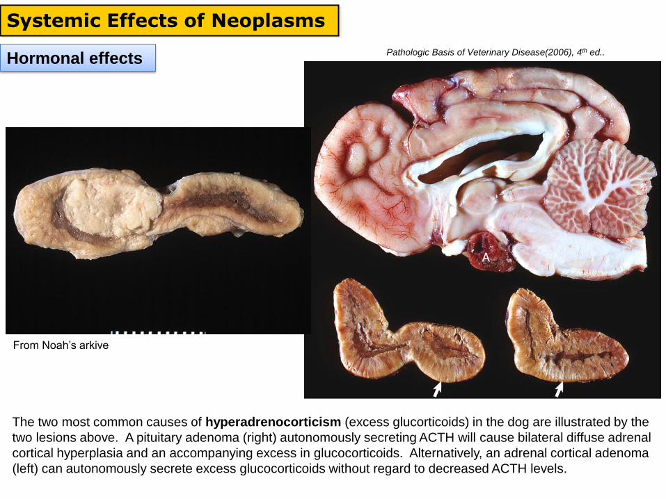
Hormonal effects
Systemic Effects of Neoplasms
The two most common causes of hyperadrenocorticism (excess glucorticoids) in the dog are illustrated by the
two lesions above. A pituitary adenoma (right) autonomously secreting ACTH will cause bilateral diffuse adrenal
cortical hyperplasia and an accompanying excess in glucocorticoids. Alternatively, an adrenal cortical adenoma
(left) can autonomously secrete excess glucocorticoids without regard to decreased ACTH levels.
Pathologic Basis of Veterinary Disease(2006), 4th ed..
From Noah’s arkive
Pathologic Basis of Veterinary Disease(2006), 4th ed.
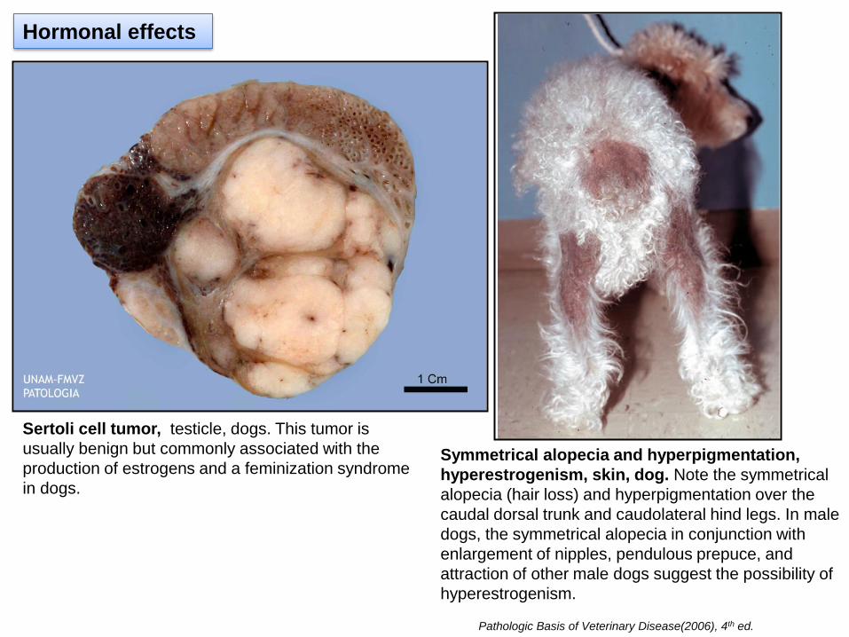
Hormonal effects
Sertoli cell tumor, testicle, dogs. This tumor is
usually benign but commonly associated with the
production of estrogens and a feminization syndrome
in dogs.
Symmetrical alopecia and hyperpigmentation,
hyperestrogenism, skin, dog. Note the symmetrical
alopecia (hair loss) and hyperpigmentation over the
caudal dorsal trunk and caudolateral hind legs. In male
dogs, the symmetrical alopecia in conjunction with
enlargement of nipples, pendulous prepuce, and
attraction of other male dogs suggest the possibility of
hyperestrogenism.
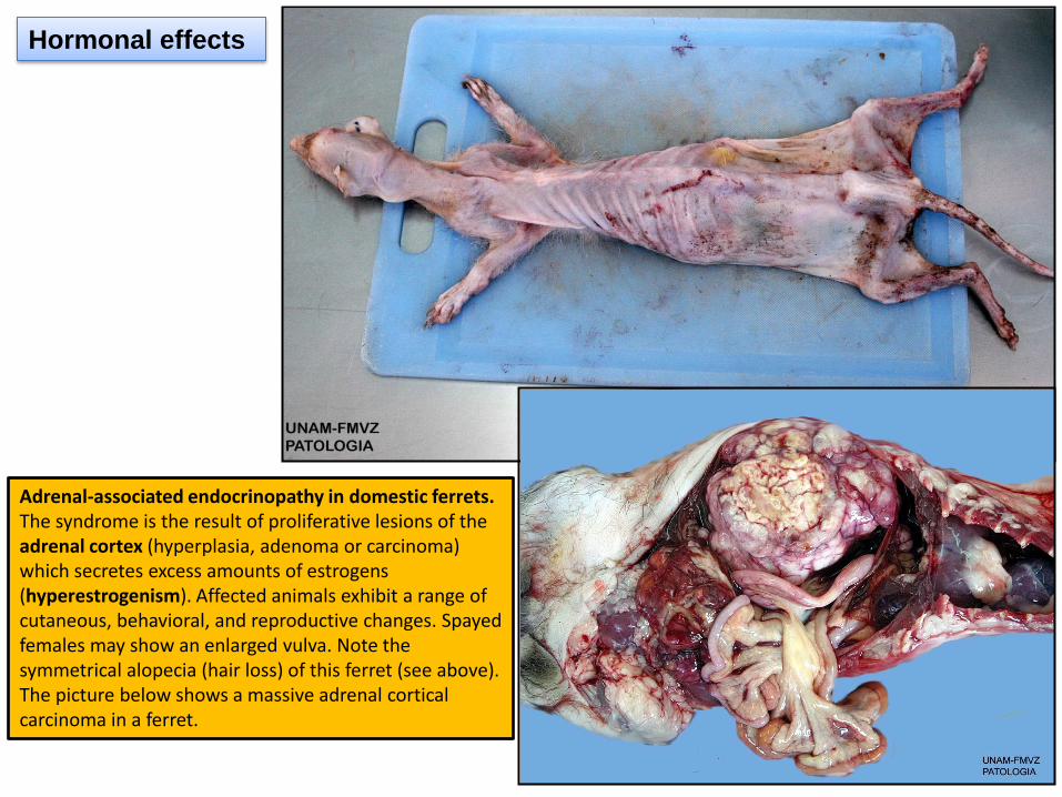
Hormonal effects
Adrenal-associated endocrinopathy in domestic ferrets. The syndrome is the result of proliferative lesions of the adrenal cortex (hyperplasia, adenoma or carcinoma) which secretes excess amounts of estrogens (hyperestrogenism). Affected animals exhibit a range of cutaneous, behavioral, and reproductive changes. Spayed females may show an enlarged vulva. Note the symmetrical alopecia (hair loss) of this ferret (see above). The picture below shows a massive adrenal cortical carcinoma in a ferret.
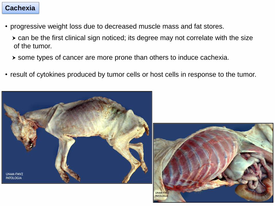
Cachexia
• progressive weight loss due to decreased muscle mass and fat stores.
• result of cytokines produced by tumor cells or host cells in response to the tumor.
can be the first clinical sign noticed; its degree may not correlate with the size
of the tumor.
some types of cancer are more prone than others to induce cachexia.

Other Cytokines
• other cytokines, IL-1 & IF-γ act synergistically with TNF.
• proteolysis inducing factor (PIF) can directly catabolize fat & muscle.
Cachexia
Cancer Cells as Metabolic Parasites
• revert to anaerobic metabolism (converting glucose to lactate), a wasteful form of
energy production (2 vs 34 ATP)
• utilize 5-10 times as much glucose as normal tissues (negative energy balance)
Tumor Necrosis Factor (TNF)
• TNF-α produced by macrophages or tumor cells, a mediator of wasting in
malignancies and chronic infectious diseases.
• TNF-α induces a net catabolic state (↑ catabolism of muscle & fat).
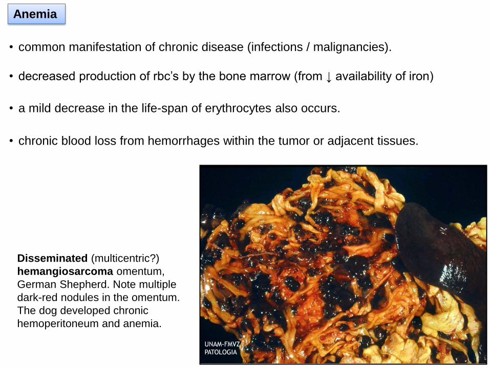
Anemia
• common manifestation of chronic disease (infections / malignancies).
• decreased production of rbc’s by the bone marrow (from ↓ availability of iron)
• a mild decrease in the life-span of erythrocytes also occurs.
• chronic blood loss from hemorrhages within the tumor or adjacent tissues.
Disseminated (multicentric?)
hemangiosarcoma omentum,
German Shepherd. Note multiple
dark-red nodules in the omentum.
The dog developed chronic
hemoperitoneum and anemia.
Paraneoplastic Syndromes
• “manifestations in cancer-bearing patients that cannot readily be explained, either by
the local or distant spread of the tumor or by the elaboration of hormones indigenous
to the tissue from which the tumor arose"
• in ~15% of human patients with advanced malignant disease
• can be the main presenting sign in animals.
Paraneoplastic Endocrine Syndromes
• systemic effects that mimic an endocrinopathy.
• occur when tumors secrete hormones or hormone-like substances that are not
normally produced by the organ / tissue of origin.
Paraneoplastic Syndromes
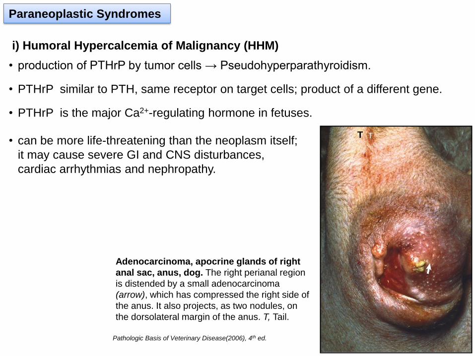
• can be more life-threatening than the neoplasm itself;
it may cause severe GI and CNS disturbances,
cardiac arrhythmias and nephropathy.
i) Humoral Hypercalcemia of Malignancy (HHM)
Adenocarcinoma, apocrine glands of right
anal sac, anus, dog. The right perianal region
is distended by a small adenocarcinoma
(arrow), which has compressed the right side of
the anus. It also projects, as two nodules, on
the dorsolateral margin of the anus. T, Tail.
Pathologic Basis of Veterinary Disease(2006), 4th ed.
T
• production of PTHrP by tumor cells → Pseudohyperparathyroidism.
• PTHrP similar to PTH, same receptor on target cells; product of a different gene.
• PTHrP is the major Ca2+-regulating hormone in fetuses.

i) Humoral Hypercalcemia of Malignancy (HHM)
Paraneoplastic Syndromes
• most commonly seen in:
adenocarcinoma of apocrine glands of the anal sac (dogs)
lymphoma (dogs & cats)
squamous cell carcinoma (cats)
gastric carcinoma (horses)
Adenocarcinoma, apocrine glands, anal sac,
dorsal plane, dog. A 1-cm-diameter nodule
(arrows) derived from apocrine glands of the wall
of the right anal sac protrudes into the lumen of
the anal sac. Anal sacs (A) are present on both
sides of the rectum (R).
Pathologic Basis of Veterinary Disease(2006), 4th ed.

Paraneoplastic Syndromes
Some Other Paraneoplastic Syndromes
i) Cutaneous paraneoplastic syndromes
• a variety of paraneoplastic dermatosis with obscure pathogenesis (dogs & cats)
• other hormone-like factors produced by tumors: ACTH-like substance, TSH-like substance,
insulin-like substance, erythropoietin.
Feline Paraneoplastic Alopecia – this syndrome is seen on rare occasions in cats with internal malignancies;
particularly pancreatic, biliary or intestinal adenocarcinomas. The bilaterally symmetric hair loss (alopecia) seen
above is from a cat with pancreatic adenocarcinoma.
Vet Dermatol 1997
ii) Other paraneoplastic endocrine syndromes

Paraneoplastic Syndromes
Nodular dermatofibrosis and cystic renal
adenocarcinomas, dog. Note the presence of multiple
cutaneous nodules composed of collagen (N). These growths
have been described in German shepherds and other breeds
associated with renal tumors which are often multiple, bilateral
and cystic (cystadenocarcinomas, C). Uterine leiomyomas
(often pedunculated) are also seen in females.
N
N
C
C
Paraneoplastic Syndromes
ii) Paraneoplastic neurologic syndromes
iii) Coagulation abnormalities associated with thrombocytopenia
iv) Myasthenia gravis in cats with thymoma
v) Hypoglycemia associated with intraabdominal leiomyomas / leiomyosarcomas
vi) Persistent leukocytosis (often neutrophilia) associated with carcinomas
Some other Paraneoplastic Syndromes

Best wishes in your final exam