Embed Size (px)
Citation preview

Lupeol induces S-phase arrest and mitochondria-mediatedapoptosis in cervical cancer cells
NUPOOR PRASAD1, AKASH SABARWAL2, UMESH C S YADAV
1 and RANA P SINGH2*1School of Life Sciences, Central University of Gujarat, Gandhinagar, Gujarat, India
2Cancer Biology Laboratory, School of Life Sciences, Jawaharlal Nehru University, New Delhi, India
*Corresponding author (Email, [email protected], [email protected])
MS received 1 August 2017; accepted 27 February 2018; published online 22 March 2018
Cervical cancer is fourth most common fatal cancer in women worldwide. Lupeol is a dietary triterpenoid and has shown itsanticancer efficacy against various cancer types with selectivity in targeting cancer cells. In the present study, anticancerefficacy and mechanism of action of a phytochemical, lupeol, in human cervical carcinoma (HeLa) cells has been examined.The anticancer efficacy of lupeol was assessed by trypan blue cell counting, annexin Vassay, cell cycle analysis, expressionof apoptotic proteins by RT-PCR and Western blotting and assessment of mitochondrial ROS generation by mitosox andmitotracker assays. Our results demonstrated that lupeol decreased cell proliferation and viability of HeLa cells significantly(p\0.001). Lupeol induced S-phase cell cycle arrest and also decreased the expression of S-phase Cyclins and CDKs andincreased the expression of cyclin-dependent kinase inhibitors, p21 at transcriptional and translational level. Further, lupeolinduced apoptosis and increased the expression of apoptosis markers such as cleaved PARP and Bax:Bcl-2 ratio. Fur-thermore, mitosox and mitotracker dye incubation followed by FACS analysis showed an increase in mitochondrialsuperoxide generation and reduction in healthy mitochondrial mass. These results suggest that lupeol could be an effectivechemotherapeutic agent against cervical carcinoma due to its growth inhibitory activity through induction of S-phase cellcycle arrest and apoptosis.
Keywords. Apoptosis; cell cycle; cervical cancer; lupeol; mitochondria; ROS
Abbreviations: CDKI, cyclin-dependent kinase inhibitor; NAC, N-acetyl cysteine; PARP, poly(ADP-ribose) polymerase;ROS, reactive oxygen species
1. Introduction
Cervical cancer is the fourth most common fatal cancer inwomen globally. With approximately 26% incidence, cer-vical cancer is the most common cancer and top most reasonof mortality among Indian women due to cancer (Siegelet al. 2013). HPV infection is the major risk factor forintraepithelial neoplasia and invasive cervical cancer. About99% of cervical cancer occurs due to infection with HPV-16and HPV-18 strains (Walboomers et al. 1999). HPV infec-tion releases oncogenic proteins such as E6 and E7 in thehost and are responsible for increased cell proliferation,survival and invasive cervical cancer development (Yim andPark 2005). Hence, regulation and control of cell cycle andcell proliferation is important for checking the tumourprogression.
Among various treatment modalities such as surgery,radiotherapy and chemotherapy, the latter is most practisedapproach for the treatment of advanced invasive cervicalcancer. However, major challenges with this treatmentstrategy is development of chemo-resistance and related
toxic side effects (Janicek and Averette 2001). Thus, astrategy having minimal side effects for the treatment ofthese cancers is warranted. Phytochemicals have emerged asimportant chemopreventive agents due to their ability totarget multiple pathways specifically in the cancer cells andcauses little harm to the normal cells. Dietary agents presentin various fruits and vegetables has been shown to beeffective against various cancer types (Surh 2003). Theplant-derived molecules are known to cause cell cycle arrestand apoptosis induction in cancer cells (Singh et al. 2002)and thus could be promising treatment strategy for control-ling cancer prevention and progression.
One such phytochemical, Lupeol is a dietary triterpenoidbelonging to group lupane which is present in various typesof fruits and vegetables including mango, strawberry, olive,fig, red grapes, cabbage, pepper, cucumber and tomato. Itpossesses strong antioxidant, anti-inflammatory, anti-arthritic and anti-malarial activity in vitro and in vivo system(Saleem 2009). Lupeol is a potent inhibitor of proteinkinases and serine proteases and inhibits the activity of DNAtopoisomerase-II which is a known target for anti-cancer
http://www.ias.ac.in/jbiosci 249
J Biosci Vol. 43, No. 2, June 2018, pp. 249–261 � Indian Academy of SciencesDOI: 10.1007/s12038-018-9743-8

chemotherapy (He et al. 2011). It has been shown to haveanticancer activity against melanoma (Saleem et al. 2008),prostate (Saleem et al. 2005), epidermoid (Prasad et al.2009), pancreatic (Murtaza et al. 2009), hepatocellular (Leeet al. 2011) and head and neck carcinoma (Lee et al. 2007).The major mode of action by which lupeol represents anti-cancer activity includes induction of cell cycle arrest andapoptosis. Toxicity study performed in mice showed thatoral administration of lupeol at a dose of 2g/kg had noadverse effects in mice and no mortality was observed up to96 h (Saleem 2009). Although, effect of lupeol on varioustypes of cancer has been investigated, yet very limitedstudies have been performed on its effect on cervical cancerand its mechanism of action on cervical cancer cells.
Therefore, in the present study we have examined theanticancer potential of lupeol in regulating the cell cycleprogression and cell viability and induction of apoptosis inHeLa cervical cancer cells. The results indicate that lupeoltreatment of cervical cancer cells induced S-phase cell cyclearrest and increased the expression of apoptosis markers andalso increased mitochondrial superoxide generation, whichsuggest its effectiveness in cervical cancer chemopreventionand chemotherapy.
2. Materials and methods
2.1 Chemicals
Lupeol, dimethyl sulfoxide (DMSO), propidium iodide (PI),saponin, bromophenol blue, trypan blue, EDTA, b-actin andCyclin A antibodies were obtained from Sigma Aldrich (St.Louis, MO, USA). HeLa (cervical cancer) cell lines werepurchased from National Centre for Cell Sciences (NCCS,Pune, India). Antibodies against p21/Cip1, Bcl2, Bax, andcleaved-PARP were bought from Cell Signalling Technol-ogy (Danvers, Massachusetts, USA), and antibodies againstCyclin E and CDK2 were bought from Santa CruzBiotechnology (Dallas, TX, USA). The cell culture growthmedia Minimum Essential media (MEM) and antibioticspenicillin-streptomycin-amphotericin B cocktail wasobtained from HiMedia laboratories (Mumbai, Maharashtra,India). Other chemicals were of analytical grades andobtained from Sisco Research Laboratories (Mumbai,Maharashtra, India).
2.2 Cell culture and treatments
The HeLa cells were grown in MEM containing glutamineand supplemented with 10% FBS and 1% antibiotics andfungicide cocktail (penicillin-streptomycin-amphotericin B)by incubating in a humidified atmosphere supplied with 5%CO2 and at 37�C temperature.
Fresh stock solution of lupeol (30 mM) was prepared bydissolving it in warm ethanol and DMSO in a 1:1 ratio everytime for the treatment of the cells. Final concentration ofDMSO used for treatment of the cells was 0.16 % (v/v) inthe culture medium, which was found non-toxic to the cells.Aliquots from stock solution were diluted with the media tomake final working concentrations for treatment.
2.3 Cell viability assay
The cells were seeded in 60-mm cell culture dishes atapproximately 105 cells/dish density and allowed to adherefor 24 h. Subsequently, the cells were treated with freshcomplete medium containing appropriate amount of eithervehicle (DMSO & ethanol, 1:1) alone or lupeol (25, 50, or100 lM) and incubated for different time points. After 24,48 and 72 h of incubation with lupeol, cells were trypsinized,and cell viability was examined using trypan blue exclusionassay (Nambiar et al. 2013). Total cell number were calcu-lated by adding live and dead cells and percent cell deathwas calculated by dividing dead cell with total cell numbermultiplied by 100. Experiment was performed three timesand samples were taken in triplicate for each group.
2.4 FACS analysis for cell cycle distributions
The cells were seeded as described above and after 24h cellswere first kept in 0.5% serum media for 18 h in order tosynchronize the cells. After 18 h, cells were treated witheither vehicle (DMSO:ethanol, 1:1) or lupeol (25, 50 and100 lM) and incubated for 24h. At the end of incubationcells were harvested and cell cycle analysis was performedas reported recently (Sabarwal et al. 2017). Briefly, aftertrypsinization, the cell pellets were resuspended in 0.5 mL ofa buffer cocktail (0.3% saponin (w/v), 25 lg/mL PI (w/v),0.1 mM EDTA and 10 lg/mL RNase A (w/v) in PBS), andincubated for 4 h at 4�C in dark. The cells were subjected toFACS analysis using FACS Aria III (BD Biosciences, SanJose, CA, USA) and subsequently data analysis was donewith ModFit software. Experiment was performed twice andsamples for each group were taken in duplicate.
2.5 Annexin-V assay for apoptosis
Approximately 19105 cells were seeded in 60 mm culturedishes and after they adhered, the cells were treated withlupeol or the carrier. At the end of 24 and 48 h of incuba-tions, both floating and adherent cells were harvested andapoptosis assay was performed using Annexin V-FITCapoptosis detection kit following manufacturer’s protocol(BD Pharmingen, San Jose, CA, USA) using FACS Aria III
250 Nupoor Prasad et al.

(BD Biosciences, San Jose, CA, USA). Experiment wasperformed twice and samples for each group were taken intriplicate.
2.6 RT-PCR
Total RNA from the cells was extracted using TRIZOLreagent and manufacturer’s instructions (Life Technology,Carlsbad, CA, USA). Equal amount of total RNA (5 lg)was used to prepare cDNA using Takara cDNA synthesiskit according to the manufacturer’s protocol (Takara-Bio,Japan). Further, reaction mixture was prepared usingtemplate (3 lL), forward primer (10 pmol/lL), reverseprimer (10 pmol/lL), dNTP mix (10 mM), 10X PCRbuffer containing Mg?? (25 mM) and Taq DNA poly-merase (3U/lL) in final volume of 25 lL. The reactionwas set for 25 cycles of regular PCR reaction in Eppen-dorf’s Thermocycler (Hamburg, Germany). PCR productswere analyzed on 1% agarose gel electrophoresis andvisualized under GelDoc system (Applied Biosystems,Foster City, CA, USA). The following primer sequenceswere used for RT-PCR:
1) p21Forward primer- 50-CCC AGT GGA CAG CGA GCAGC-30
Reverse primer- 50-TCT GCC GCC GTT TTC GACCC-30
2) CDK2Forward primer- 50-AGG CCC GTG ATC CCC ACAGT- 30
Reverse primer- 50-TGG TGG GGG TGC CTT GTCCA-30
3) BCL-2Forward primer- 50-AAG CGG TCC CGT GGA TAGA-30
Reverse primer- 50-TCC GGT ATT CGC AGA AGTCC-30
4) BAXForward primer- 50-AGG ATG CGT CCA CCA AGAAG-30
Reverse primer- 50-CCA GTT GAA GTT GCC GTCAGA-30
5) Cyclin EForward primer- 50-ACT GCT GCT GCC TTG TGCCA-30
Reverse primer- 50-GCC TCC ATT GCA CAC TGGTGA CA-30
6) Cyclin AForward primer- 50-GCA GAC GGC GCT CCA AGAGG-30
Reverse primer- 50-AGG GGT GCA ACC CGT CTCGT-30
7) GAPDHForward primer- 50-GCC TTC CGT GTC CCC ACTGC-30
Reverse primer- 50-CAA TGC CAG CCC CAG CGTCA-30
2.7 Western blotting
Approximately 59105 cells were seeded in 100 mm petri-plates and after 24 h, cells were treated with different con-centrations of either vehicle or lupeol (0–100 lM). Aftercompletion of different incubation time points, cells werewashed with cold PBS and lysed with RIPA lysis buffer (50mM Tris, 1% Triton X-100, 0.1% sodium dodecyl sulfate,150 mM NaCl) containing protease and phosphatase inhi-bitors (Roche Applied Sciences, Indianapolis, IN, USA) onice. The protein was quantified and equal amount of proteinswere electrophoresed onto 10–12% SDS-PAGE gels. Theseparated proteins were transferred on to nitrocellulosemembrane followed by incubation in blocking solutioncontaining 5% non-fat milk for 1 h at room temperature orovernight at 4�C. Membranes were probed using specificprimary antibodies against Cyclin A, Cyclin E, CDK2,p21/cip1, Bax, Bcl-2 and cleaved-PARP, followed by incu-bation with horse radish peroxidase-conjugated appropriatesecondary antibodies, and visualized by the ECL detectionsystem. Image J software was used for densitometry of theblots.
2.8 MitoTracker Red for mitochondrial mass reduction
Approximately 19105 cells were seeded in 60 mm culturedishes and after they adhered, the cells were treated withlupeol (100 lM) or the carrier as described above. Aftercompletion of incubations as indicated, cells were treatedwith 200 nM MitoTracker Red and incubated for 60 min.Thereafter, cells were collected by trypsinization and washedwith cold PBS. The cell Pellets were resuspended in 0.5 mLPBS and mitochondrial degradation was determined usingFACS Aria III (Tal et al. 2009).
2.9 MitoSOX for superoxide generation
For quantification of mitochondrial superoxides approxi-mately 19105 cells were seeded in 60 mm culture dishes andafter they adhered, the cells were treated with lupeol (100lM) or the carrier as described above. Subsequently, thecells were incubated with 5 lM MitoSOX Red dye andincubated for 30 min, harvested by trypsinization. The cellpellets were washed with cold PBS and fluorescence was
Lupeol induces mitochondria-mediated apoptosis 251

detected using FACS Aria III (Mukhopadhyay et al. 2007;Hahm et al. 2011).
2.10 Statistical and densitometry analyses
Statistical analysis was done using GrapPad Prism 5 soft-ware version 5. Quantitative data are presented asMean±SEM. Statistical significance between control andtreated group was determined by one-way ANOVA followedby Student’s t-test and p\0.05 was considered significant.
3. Results
3.1 Lupeol decreased cell growth and viability of HeLacells and SiHa cells
We first evaluated the effect of lupeol on cell growth andviability of cervical cancer (HeLa) cells. At 24 h, lupeoltreatment resulted in 71% decrease in total cell number and36% increase in cell death at the 100 lM concentration.Further, lupeol treatment for 48 h resulted in 28% and 85%decrease in total cell number, and 16% and 41% increase inpercent cell death at 50 lM and 100 lM concentrations,respectively (figure 1A and B). There was 53% and 95%decrease in total cell number, and 28% and 40% increase inpercent cell death after 72 h of lupeol treatment at 50 lMand 100 lM concentrations, respectively (figure 1A and B).
Further, we also performed cell viability assay usinglupeol on another cervical adenocarcinoma cell line, SiHacells. As shown in the figure 1C and D, lupeol (0–100 lM)treatment of SiHa cells for 0–48 h resulted in significantdecrease in the cell number by up to 80% at 100 lM dose at48h (p\0.001) and increased the cell death by up to 50% (±)in 100 lM dose at 48h (p\0.001) (figure 1C & 1D).
These results show that lupeol strongly inhibits cellgrowth and induces cell death in two different cervicalcancer cells i.e. HeLa and SiHa cells. These findings suggestthat lupeol concentration- and time-dependently inhibitedthe growth and survival, and induced cell death in in vitromodel of human cervical cancer cells.
3.2 Lupeol induced S-phase arrest in HeLa cells
Since lupeol showed decreased cell proliferation andincreased cell death, we next examined whether growthinhibitory effect of lupeol is due to induction of cell cyclearrest. The cells were serum starved (0.5% FBS) to syn-chronize and then treated with the carrier or the lupeol for 24or 48 h and cell cycle analysis was performed. As shown infigure 2A and 2B, no significant change at 25 lM concen-tration of lupeol was observed at 24 h however, at 48 h, the
cells showed decrease in G0/G1 cell population to 55% ascompared to 67% in control (p\0.001) and increasedS-phase cell population to 78% as compared to 27% incontrol. In the group with 50 lM concentration of lupeol at24 h, G0/G1 cell population decreased to 59% (p\0.05) ascompared to 64% in control and S-phase cell populationincreased to 36% (p\0.01) as compared to 29% in control.In the same group at 48 h duration cells population in G0/G1phase decreased to 53% as compared to 67% in control(p\0.001), whereas cells in S-phase increased to 41% ascompared to 27% in control (p\0.001) while in G2-M phasecell decreased to 5% as compared to 9% in control (p\0.05)(figure 2B). Similarly, at 100 lM concentration of lupeol,cells population in G0/G1 phase decreased to 54% as com-pared to 64% in control (p\0.001), S-phase cells increasedto 40% as compared to 29% in control (p\0.001) at 24 h,while at 48 h, cell population in G0/G1 phase decreased to52% as compared to 67% in control (p\0.001) and S-phasepopulation increased to 44% as compared to 27% in control(p\0.001). These results clearly suggest that lupeol treat-ment of HeLa cells induced S-phase cell cycle arrest at 24and 48 h at different doses which may be responsible fordecreased cell growth.
3.3 Lupeol modulated the expression of cell cycleregulatory proteins in HeLa cells
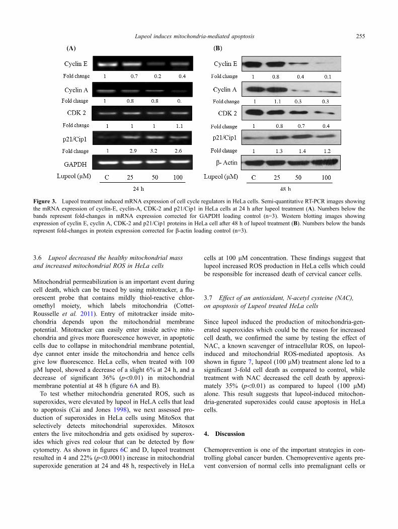
Since lupeol induced a significant increase in the HeLa cellsin S-phase and a moderate increase in G2-M phase of cellcycle, we next examined the regulation of cell cycle genesinvolved in the induction of S-phase arrest by lupeol attranscription and translation levels. First, the effect of lupeolon the expression of cell cycle regulatory proteins at thetranscriptional level was examined. Semi-quantitative RT-PCR analysis showed a significant decrease in the expres-sion of Cyclin E (80% and 60% at 50 and 100 lM con-centrations, respectively) and Cyclin A (20% and 80% at 50and 100 lM concentrations, respectively) by lupeol at 24 h,while no significant effect on CDK2 level was observed atboth the concentrations. Further, significant increase in theexpression of p21/Cip1 by 2.9-, 3.2- and 2.6-fold at 25, 50and 100 lM concentrations were observed (figure 3A).These observations in the m-RNA expression of cell cycleregulatory genes suggest that lupeol could modulate theirexpression in HeLa cells at the transcriptional level.
Next, we assessed the effect of lupeol on the regulation ofexpression at the protein level by immunoblotting. As shownin figure 3B, treatment with different concentrations oflupeol resulted in decrease in the protein expression levels ofCyclin E by 20% in 25 lM, 60% in 50 lM and 90% in 100lM at 48 h. Further, approximately 70% decrease in theexpression of Cyclin Awas observed in both 50 and 100 lMdoses. Lupeol treatment of HeLa cells also resulted in
252 Nupoor Prasad et al.

decreased expression of CDK-2 protein by 20, 30 and 60%at 25, 50 and 100 lM doses, respectively. We also examinedthe expression of CDK inhibitor and observed that lupeolinduced significant increase in the expression of CDKinhibitor p21/Cip1 at 48 h by 1.3, 1.4 and 1.2 fold at 25, 50and 100 lM doses, respectively. These findings suggest thatlupeol regulated the expression of cell cycle regulatoryproteins which lead to S-phase cell cycle arrest and thuscould be responsible for lupeol-induced decrease in HeLacell growth.
3.4 Lupeol induced apoptotic cell death in HeLa cells
Since cell cycle arrest usually results in the blockage of cellgrowth, which may lead to cell death via apoptosis and couldplay important role in decreased tumour growth (Pucci et al.2000). Accordingly, to assess whether lupeol induced celldeath as observed in trypan blue dye exclusion assay couldbe due to apoptosis, we next performed Annexin V assay. Asshown in the figure 4A and 4B, lupeol (100 lM) treatmentresulted in more than 2-fold increase in apoptotic cells at 24
h, while approximately more than 5-fold increase in apop-totic cells at 48 h as compared to respective controls. Therewas no significant effect in the apoptotic cell population at25 and 50 lM concentrations of lupeol as compared torespective controls (figure 4B).
3.5 Lupeol modulated the expression of apoptosisregulating molecules in HeLa cells
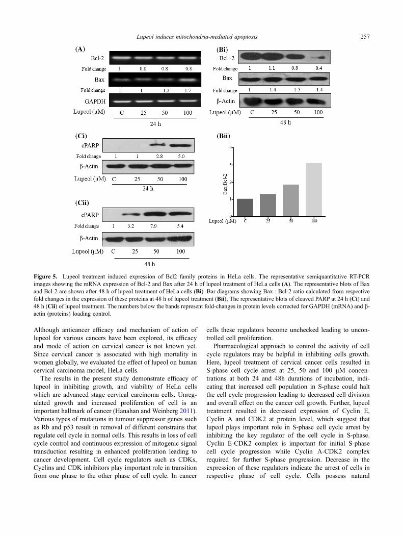
Next, we assessed the effect of lupeol on the expression ofapoptotic markers in HeLa cells. As shown in figure 5A,although only slight decrease in mRNA expression of anti-apoptotic protein Bcl-2 was observed, there was a significantincrease (1.7-fold in 100 lM) in mRNA level of pro-apop-totic protein, Bax. Similarly, immunoblotting of Bcl-2 pro-tein showed a concentration-dependent decrease in itsexpression, showing maximum decrease (by *60%) at 100lM, while a slight change in the expression of Bax wasobserved at different lupeol doses (figure 5Bi). However, theBax:Bcl-2 ratio (fold-change of Bax protein divided withfold-change of Bcl-2 protein) showed a significant increase
Figure 1. Growth inhibitory effects of lapel on human cervical carcinoma (HeLa and SiHa) cells. Viable cells and dead cells were countedusing Trypan blue dye exclusion method in haemocytometer. Representative value of total cell number (A) and percent cell death (B) ofHeLa cells are shown as bar diagrams at 24, 48 and 72 h. The total cell number of SiHa cells (C) and percent cell death (D) of SiHa cells at24 and 48 h are shown as bar diagrams. The cell growth data shows mean ± SEM (n=3); *p\0.05, **p\0.01, ***p\0.001 vs. respectivecontrols analysed.
Lupeol induces mitochondria-mediated apoptosis 253

by 2- and 3-fold at 50 and 100 lM concentrations, respec-tively (figure 5Bii). This ratio when near one suggest healthycells while higher than 1 value indicates decreased surviv-ability of the cells suggesting lupeol-induced cell death/apoptosis in HeLa cells.
Since cleavage of Poly ADP-ribose polymerase (PARP),a 116 kilodalton (kDa) protein, is one of the importantbiomarkers of apoptosis (Mullen 2004), we next examinedthe effect of lupeol on PARP cleavage. Initially, weexamined level of cleaved PARP at 24 h by Westernblotting and found increased level of cleaved protein by 2.8
and 5 fold at 50 and 100 lM concentrations, respectively(figure 5Ci). Further, we the HeLa cells with lupeol for 48h and assessed the level of cleaved PARP. We observed asignificant increase in PARP cleavage as evident byincreased level of 89 kDa cleaved PARP at 25, 50 and 100lM concentrations by 3.2-, 7.9- and 5.4-fold, respectively.However, 100 lM concentration at 48 h showed a slightdecrease in the level of cleaved PARP as compared to 50lM concentration which may be likely due to the fact thatduring longer incubations of 48 h the cleaved PARP couldhave degraded.
Lupeol (0 μM) Lupeol (25 μM) Lupeol (50 μM) Lupeol (100 μM)
Lupeol (0 μM) Lupeol (25 μM) Lupeol (50 μM) Lupeol (100 μM)
24 h
48 h
(A)
(B)
Figure 2. Lupeol treatment induced cell cycle arrest in HeLa cells. Cell population in different cell cycle phases are shown as histogramanalysed by ModFit software (A). Quantification of the cell population (in percent) in different cell cycle phases shown as bar diagramsrepresenting mean ± SEM (n=6) (B). *p\0.05, **p\0.01, ***p\0.001 vs. respective controls.
254 Nupoor Prasad et al.
3.6 Lupeol decreased the healthy mitochondrial massand increased mitochondrial ROS in HeLa cells
Mitochondrial permeabilization is an important event duringcell death, which can be traced by using mitotracker, a flu-orescent probe that contains mildly thiol-reactive chlor-omethyl moiety, which labels mitochondria (Cottet-Rousselle et al. 2011). Entry of mitotracker inside mito-chondria depends upon the mitochondrial membranepotential. Mitotracker can easily enter inside active mito-chondria and gives more fluorescence however, in apoptoticcells due to collapse in mitochondrial membrane potential,dye cannot enter inside the mitochondria and hence cellsgive low fluorescence. HeLa cells, when treated with 100lM lupeol, showed a decrease of a slight 6% at 24 h, and adecrease of significant 36% (p\0.01) in mitochondrialmembrane potential at 48 h (figure 6A and B).
To test whether mitochondria generated ROS, such assuperoxides, were elevated by lupeol in HeLA cells that leadto apoptosis (Cai and Jones 1998), we next assessed pro-duction of superoxides in HeLa cells using MitoSox thatselectively detects mitochondrial superoxides. Mitosoxenters the live mitochondria and gets oxidised by superox-ides which gives red colour that can be detected by flowcytometry. As shown in figures 6C and D, lupeol treatmentresulted in 4 and 22% (p\0.0001) increase in mitochondrialsuperoxide generation at 24 and 48 h, respectively in HeLa
cells at 100 lM concentration. These findings suggest thatlupeol increased ROS production in HeLa cells which couldbe responsible for increased death of cervical cancer cells.
3.7 Effect of an antioxidant, N-acetyl cysteine (NAC),on apoptosis of Lupeol treated HeLa cells
Since lupeol induced the production of mitochondria-gen-erated superoxides which could be the reason for increasedcell death, we confirmed the same by testing the effect ofNAC, a known scavenger of intracellular ROS, on lupeol-induced and mitochondrial ROS-mediated apoptosis. Asshown in figure 7, lupeol (100 lM) treatment alone led to asignificant 3-fold cell death as compared to control, whiletreatment with NAC decreased the cell death by approxi-mately 35% (p\0.01) as compared to lupeol (100 lM)alone. This result suggests that lupeol-induced mitochon-dria-generated superoxides could cause apoptosis in HeLacells.
4. Discussion
Chemoprevention is one of the important strategies in con-trolling global cancer burden. Chemopreventive agents pre-vent conversion of normal cells into premalignant cells or
Figure 3. Lupeol treatment induced mRNA expression of cell cycle regulators in HeLa cells. Semi-quantitative RT-PCR images showingthe mRNA expression of cyclin-E, cyclin-A, CDK-2 and p21/Cip1 in HeLa cells at 24 h after lupeol treatment (A). Numbers below thebands represent fold-changes in mRNA expression corrected for GAPDH loading control (n=3). Western blotting images showingexpression of cyclin E, cyclin A, CDK-2 and p21/Cip1 proteins in HeLa cell after 48 h of lupeol treatment (B). Numbers below the bandsrepresent fold-changes in protein expression corrected for b-actin loading control (n=3).
Lupeol induces mitochondria-mediated apoptosis 255

suppress the further progression of premalignant cells intoinvasive cancer (Scott et al. 2009). Several phytochemicalsincluding resveratrol, genistein, silibinin and curcumin haveproved their pre-clinical and mechanistic efficacy aschemopreventive agent. From a safety point of view,chemopreventive phytochemicals have shown minimal
toxicity at a given concentration which prevents damage ofnormal cells and hence are attractive agents. Lupeol, apentacyclic triterpenoid, is one such phytochemical that hasshown its potent chemopreventive efficacy against manytypes of cancers including prostate, pancreatic, head andneck, hepatocellular and skin cancers (Saleem 2009).
Figure 4. Lupeol treatment induced apoptosis of HeLa cells. The representative dot blot images after 24 and 48 h of lupeol (100 lM)treatment of HeLa cells obtained from flow cytometry of Annexin V stained cells are shown (A); Quantification of the apoptotic cellpopulation (in percent) shown as bar diagrams representing mean ± SEM (n=6) (B). *p\0.05 ***p\0.001, vs. respective controls.
256 Nupoor Prasad et al.
Although anticancer efficacy and mechanism of action oflupeol for various cancers have been explored, its efficacyand mode of action on cervical cancer is not known yet.Since cervical cancer is associated with high mortality inwomen globally, we evaluated the effect of lupeol on humancervical carcinoma model, HeLa cells.
The results in the present study demonstrate efficacy oflupeol in inhibiting growth, and viability of HeLa cellswhich are advanced stage cervical carcinoma cells. Unreg-ulated growth and increased proliferation of cell is animportant hallmark of cancer (Hanahan and Weinberg 2011).Various types of mutations in tumour suppressor genes suchas Rb and p53 result in removal of different constrains thatregulate cell cycle in normal cells. This results in loss of cellcycle control and continuous expression of mitogenic signaltransduction resulting in enhanced proliferation leading tocancer development. Cell cycle regulators such as CDKs,Cyclins and CDK inhibitors play important role in transitionfrom one phase to the other phase of cell cycle. In cancer
cells these regulators become unchecked leading to uncon-trolled cell proliferation.
Pharmacological approach to control the activity of cellcycle regulators may be helpful in inhibiting cells growth.Here, lupeol treatment of cervical cancer cells resulted inS-phase cell cycle arrest at 25, 50 and 100 lM concen-trations at both 24 and 48h durations of incubation, indi-cating that increased cell population in S-phase could haltthe cell cycle progression leading to decreased cell divisionand overall effect on the cancer cell growth. Further, lupeoltreatment resulted in decreased expression of Cyclin E,Cyclin A and CDK2 at protein level, which suggest thatlupeol plays important role in S-phase cell cycle arrest byinhibiting the key regulator of the cell cycle in S-phase.Cyclin E-CDK2 complex is important for initial S-phasecell cycle progression while Cyclin A-CDK2 complexrequired for further S-phase progression. Decrease in theexpression of these regulators indicate the arrest of cells inrespective phase of cell cycle. Cells possess natural
Figure 5. Lupeol treatment induced expression of Bcl2 family proteins in HeLa cells. The representative semiquantitative RT-PCRimages showing the mRNA expression of Bcl-2 and Bax after 24 h of lupeol treatment of HeLa cells (A). The representative blots of Baxand Bcl-2 are shown after 48 h of lupeol treatment of HeLa cells (Bi). Bar diagrams showing Bax : Bcl-2 ratio calculated from respectivefold changes in the expression of these proteins at 48 h of lupeol treatment (Bii); The representative blots of cleaved PARP at 24 h (Ci) and48 h (Cii) of lupeol treatment. The numbers below the bands represent fold-changes in protein levels corrected for GAPDH (mRNA) and b-actin (proteins) loading control.
Lupeol induces mitochondria-mediated apoptosis 257

inhibitors which regulate the activity of CDKs for example,p21/Cip1 is one of the many important CDK inhibitors thatbinds to CyclinA-CDK2 and CyclinE-CDK2 complexesand inactivate them (Hinds 2003). Our results demonstratethat lupeol increased the expression of p21/Cip1 at bothmRNA and protein levels, which could lead to cell cyclearrest and prevent further cell cycle progression of HeLacells.
Cancer cells can resist cell death and have increasedsurvival capacity. Induction of apoptosis by chemopreven-tive agent is an effective way to control cancer growth.Chemopreventive agent are known to induce apoptosis byhalting the repair mechanism in cancer cells, an effectivemechanism to decrease viability of cancer cells (Toshiyaet al. 2012). Here, we observed that lupeol treatment resultedin significant induction (2- and 5-fold) of apoptosis in cer-vical cancer cells at 100 lM concentration at 24 and 48 h,respectively which could suggest its anti-cancer property incervical carcinoma.
The excess of anti-apoptotic proteins make cancer cellsresistant to death. The ratio of Bax, a pro-apoptotic proteinand Bcl-2, an anti-apoptotic protein decides the release ofthe cytochrome c from the mitochondria which is animportant event during mitochondria-mediated apoptosis.High than one Bax:Bcl-2 ratio favours mitochondria-me-diated apoptosis whereas lower than one indicates survival.Our results show that lupeol treatment (100 lM) caused amild increase in Bax level but a strong decrease in Bcl-2protein levels. This resulted in a huge increase in Bax:Bcl-2 ratio, which could be responsible for significant inductionof apoptosis in HeLa cells. Further, increased Bax:Bcl-2ratio implicates mitochondria-mediated apoptosis in HeLacells.
Cleavage of Poly(ADP-ribose) polymerase (PARP), apolymerase that recognizes nicks in DNA and repairs thedamaged DNA, is one of the important biomarkers ofapoptosis (Mullen 2004). During apoptosis 116 kDa PARPprotein is cleaved by active caspase-3 into a 89 kDa
Figure 6. Lupeol treatment induced mitochondria-mediated apoptosis. Cell population showing mitotracker (A) and Mitosox(C) fluorescence in lupeol (100 lM) treated HeLa cells for 24 and 48 h respectively, are shown as representative histogram with theirrespective quantification (percent mitochondrial mass) (B) and (percent superoxide generation) (D) shown as bar diagrams representingmean ± SEM (n=6). *p\0.05, **p\0.01 vs. respective controls.
258 Nupoor Prasad et al.

C-terminal fragment with reduced catalytic activity and a 24kDa N-terminal peptide containing DNA binding domain.The cleavage of PARP prevents repair of damaged DNA andas a result cells undergo apoptosis. Lupeol treatment (100
lM) caused an increase in cleavage of PARP up to *5-foldat 24 h. Nonetheless, lupeol-induced increased cleavage ofPARP further confirmed the effectiveness of lupeol ininducing cell death in HeLa cells.
Figure 7. Lupeol treatment induced mitochondria-mediated apoptosis prevented by NAC. The HeLa cells were pre-treated with vehicleor NAC (5 mM) for 5 h followed by lupeol (100 lM) or vehicle treatment for 48 h. After the completion of incubation, the lightmicroscopic images were obtained followed by Annexin-V FITC staining. The representative microscopic images of are shown,magnification 1009 (A). Quantification of the apoptotic cell population (in percent) obtained from flow cytometry of Annexin V stainedcells from above experiment are shown as bar diagrams (B). Bars show mean ± SEM (n=6). *p\0.05 vs. Control; **p\0.01 vs. lupeoltreated group.
Lupeol induces mitochondria-mediated apoptosis 259

During apoptosis, the increase in proapoptotic proteinse.g. Bax results in the formation of pores in outer mito-chondrial membrane which results in collapse in mitochon-drial membrane potential followed by release ofcytochrome-c from mitochondria (Green and Reed 1998).The cytochrome-c further binds with apoptotic proteaseactivating factor 1 (APAF1) resulting in formation ofapoptosome complex which leads to execution of variouscaspases and apoptosis induction. In the present study lupeol(100 lM) significantly decreased mitochondrial membranepotential which indicates mitochondria’s role in lupeol-in-duced apoptosis in HeLa cells.
Excessive production of superoxides from mitochondriaalters the cellular redox potential by acting on several redoxsensitive sites in mitochondrial membranes. This also facil-itates release of cytochrome c from mitochondria (Susinet al. 1998) indicating that production of ROS by mito-chondria is an important event during apoptosis. Lupeoltreatment of HeLa cells caused a significant increase inmitochondrial superoxide generation, which further supportsthat increase in apoptotic cell death could be via mito-chondria-mediated apoptosis. N-Acetyl cysteine, a ROSscavenger, is an aminothiol and synthetic precursor of
intracellular cysteine and glutathione (GSH). GSH produc-tion by NAC is responsible for scavenging ROS which mayblock mitochondrial ROS-mediated apoptosis (Sun 2010).We also observed that the pre-treatment of HeLa cells withNAC caused a significant decrease in apoptotic cell death by35%, which confirmed the role of ROS in lupeol -mediatedapoptosis induction in Hela cells.
In summary, results in the present study show that lupeolcould inhibit the growth of cervical carcinoma cells throughinduction of cell cycle arrest and mitochondria-mediatedapoptosis inHeLa cells (figure 8), suggesting that lupeol couldbe a potential molecule for intervention of cervical cancer.
Acknowledgements
NP is recipient of UGC Non-NET Fellowship from CentralUniversity of Gujarat, Gandhinagar, UCSY is recipient ofRamanujan Fellowship from Department of Science andTechnology (DST)/Science Engineering and Research Board(SERB), India.
References
Cai J and Jones DP 1998 Superoxide in apoptosis mitochondrialgeneration triggered by cytochromec loss. J. Biol. Chem. 27311401–11404
Cottet-Rousselle C, Ronot X, Leverve X. and Mayol JF 2011Cytometric assessment of mitochondria using fluorescentprobes. Cytometry Part A. 79 405–425
Green D R and Reed JC 1998 Mitochondria and apoptosis. Science281 1309–1312
Hahm ER, Moura MB, Kelley EE, Van Houten B, Shiva S andSingh SV 2011 Withaferin A-induced apoptosis in human breastcancer cells is mediated by reactive oxygen species. PLoS ONE6 e23354
Hanahan D and Weinberg RA 2011 Hallmarks of cancer: the nextgeneration. Cell 144 646–674
He Y, Liu F, Zhang L, Wu Y, Hu B, Zhang Y, Li Y and Liu H 2011Growth inhibition and apoptosis induced by lupeol, a dietarytriterpene, in human hepatocellular carcinoma cells. Biol.Pharm. Bull. 34 517–522
Hinds PW 2003 Cdk2 dethroned as master of S phase entry. CancerCell 3 305–307
Janicek MF and Averette HE 2001 Cervical cancer: prevention,diagnosis, and therapeutics. CA: Cancer J. Clin. 51 92–114
Lee TK, Poon RT, Wo JY, Ma S, Guan XY, Myers JN, Altevogt Pand Yuen AP 2007 Lupeol suppresses cisplatin-induced nuclearfactor-jB activation in head and neck squamous cell carcinomaand inhibits local invasion and nodal metastasis in an orthotopicnude mouse model. Cancer Res. 67 8800–8809
Lee TKW, Castilho A, Cheung VCH, Tang KH, Ma S and Ng IOL2011 Lupeol targets liver tumor-initiating cells through phos-phatase and tensin homolog modulation. Hepatology 53160–170
Figure 8. A schematic diagram depicting the potential mode ofaction of lupeol on cervical adenocarcinoma (HeLa) cells.
260 Nupoor Prasad et al.

Mukhopadhyay P, Rajesh M, Hasko G, Hawkins BJ, Madesh M andPacher P 2007 Simultaneous detection of apoptosis and mito-chondrial superoxide production in live cells by flow cytometryand confocal microscopy. Nature. Protocol. 2 2295–2301
Mullen P 2004 PARP cleavage as a means of assessing apoptosis.Methods Mol. Med. 88 171–181
Murtaza I, Saleem M, Adhami VM, Hafeez BB &Mukhtar H 2009.Suppression of cFLIP by lupeol, a dietary triterpene, is sufficientto overcome resistance to TRAIL-mediated apoptosis inchemoresistant human pancreatic cancer cells. Cancer Res. 691156–1165
Nambiar D, Prajapati V, Agarwal R and Singh RP 2013 In vitro andin vivo anticancer efficacy of silibinin against human pancreaticcancer BxPC-3 and PANC-1 cells. Cancer Lett. 334 109–117
Prasad S, Madan E, Nigam N, Roy P, George J and Shukla Y 2009Induction of apoptosis by lupeol in human epidermoid carci-noma A431 cells through regulation of mitochondrial, Akt/PKBand NF-kappaB signaling pathways. Cancer Biol. Ther. 81632–1639
Pucci B, Kasten M and Giordano A 2000 Cell cycle and apoptosis.Neoplasia 2 291–299
Sabarwal A, Agarwal R and Singh RP 2017. Fisetin inhibitscellular proliferation and induces mitochondria-dependent apop-tosis in human gastric cancer cells. Mol. Carcinog. 56 499–514
Saleem M 2009 Lupeol, a novel anti-inflammatory and anti-cancerdietary triterpene. Cancer Lett. 285 109–115
Saleem M, Kweon MH, Yun JM, Adhami VM, Khan N, Syed DNand Mukhtar RH 2005 A novel dietary triterpene Lupeol inducesfas-mediated apoptotic death of androgen-sensitive prostatecancer cells and inhibits tumor growth in a xenograft model.Cancer Res. 65 11203–11213
Saleem M, Maddodi N, Zaid MA, Khan N, Bin Hafeez B, Asim M,Suh Y, Yun JM, Setaluri V and Mukhtar H 2008 Lupeol inhibits
growth of highly aggressive human metastatic melanoma cellsin vitro and in vivo by inducing apoptosis. Clin. Cancer Res. 142119–2127
Scott EN, Gescher AJ, Steward WP and Brown K 2009 Develop-ment of dietary phytochemical chemopreventive agents:biomarkers and choice of dose for early clinical trials. CancerPrev. Res. 2 525–530
Siegel R, Naishadham D and Jemal A 2013 Cancer statistics 2013CA: CancerJ. Clinic. 63 11–30
Singh RP, Dhanalakshmi S and Agarwal R 2002 Phytochemicals ascell cycle modulators a less toxic approach in halting humancancers. Cell Cycle 1 155–160
Sun SY 2010 N-acetylcysteine, reactive oxygen species andbeyond. Cancer Biol. Ther. 9 109–110
Surh YJ 2003 Cancer chemoprevention with dietary phytochem-icals. Nat. Rev. Cancer 3 768–780
Susin SA, Zamzami N and Kroemer G 1998 Mitochondria asregulators of apoptosis: doubt no more. Biochim. Biophys. Acta1366 151–165
Tal MC, Sasai M, Lee HK, Yordy B, Shadel GS and Iwasaki A2009 Absence of autophagy results in reactive oxygen species-dependent amplification of RLR signaling. Proc. Natl. Acad. Sci.USA 106 2770–2775
Toshiya K, Testuya T, Akira H and Takuji T 2012 Cancerchemoprevention through the induction of apoptosis by naturalcompounds J. Biophys. Chem. 3 157–173
Walboomers JM, Jacobs MV, Manos MM, Bosch FX, Kummer JA,Shah KV, Snijders PJ, Peto J, Meijer RCJ and Munoz N 1999Human papillomavirus is a necessary cause of invasive cervicalcancer worldwide. J. Pathol. 189 12–19
Yim EK and Park JS 2005 The role of HPV E6 and E7oncoproteins in HPV-associated cervical carcinogenesis. CancerRes. Treat. 37 319–324
Corresponding editor: SORAB DALAL
Lupeol induces mitochondria-mediated apoptosis 261