Embed Size (px)
Citation preview

Lung ultrasoundin the critically ill patient
Pneumothorax
Rohit Patel, MDUniversity of Florida Health
Director, Critical Care Ultrasound Surgical ICUCenter for Intensive Care
Gainesville, Florida
Sunday, August 14, 16

Critical Care UltrasoundA lines Bat sign B lines
B3 lines B7 lines BLUE protocol
BLUE points Comet tails Sinusoid sign
Jellyfish sign Lung point Merlin's space
PLAPS Point Quad sign Seashore sign
Shred sign Stratosphere sign
Tissue like sign Z lines
Sunday, August 14, 16

What can I find?Pneumothorax
Hemothorax
Other pleural effusions
Alveolar consolidation
Pulmonary edema and/or extravascular lung water
Pulmonary embolism?
Sunday, August 14, 16

What are the questions?
Focused assessment with sonography in trauma
Focused abdominal sonography in trauma
Shock, hypoxia, oligoanuria, fever, etc.?
Sunday, August 14, 16

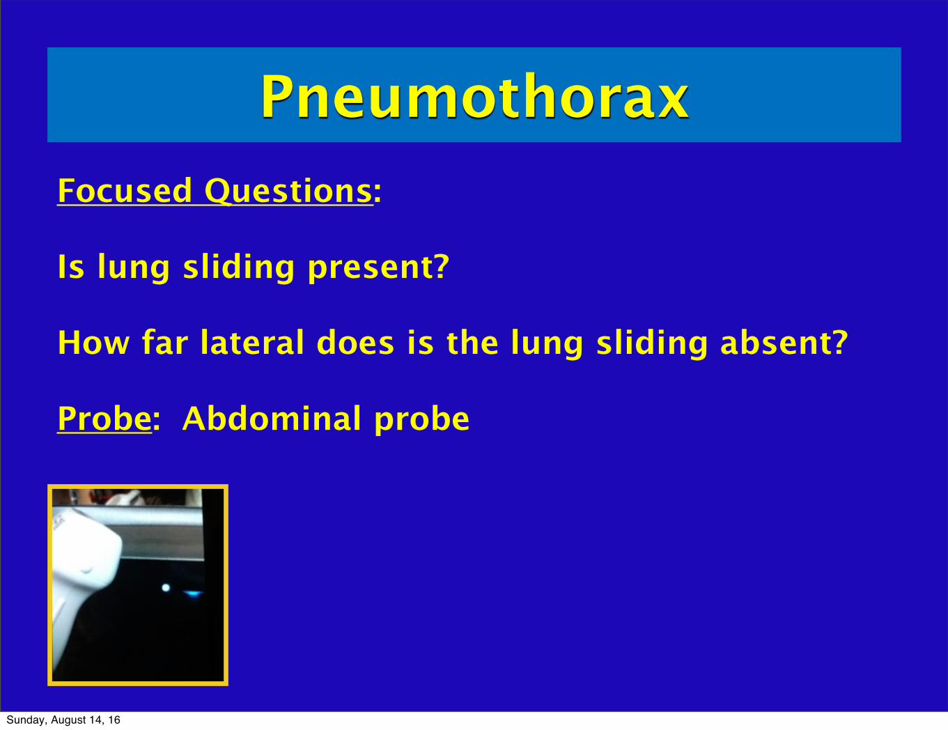
PneumothoraxFocused Questions:
Is lung sliding present?
How far lateral does is the lung sliding absent?
Probe: Abdominal probe
Sunday, August 14, 16

Ultrasound for pneumothorax
Kirkpatrick, J Trauma 2004
First described in a horse in 1986
In a normal lung, the visceral and parietal pleura are closely associated, and ultrasound shows shimmering or sliding at the pleural interface during respiration; absence of this indicates a pneumothorax
In trauma, US shown to be more than twice as sensitive for detecting occult pneumothorax with similarly high specificity (98%)
Comet tails are ultrasound artifacts that arise when ultrasound encounters a small air fluid interface
Sunday, August 14, 16

Ultrasound for pneumothorax
Zhang M. Crit Care. 2006Lichtenstein. Chest. 1995
Chest radiography?
US relies on fact that free air is lighter than normal aerated lung tissue, accumulates in nondependent areas of thoracic cavity
Multiple studies show ultrasound to be more sensitive than supine chest radiography (CT gold standard)
Sensitivity 86 to 100%Specificity 92 to 100%Negative predictive value of 100% (Lichtenstein study)
Zhang study: sensitivity 86% vs 27% AND time to obtain study 2.3 minutes vs 19.9 minutes
Sunday, August 14, 16

Ultrasound for pneumothorax
Lichtenstein D. Intensive Care Med. 2000
Supine
High frequency linear array best
Midclavicular line at third through fifth intercostal space to ID pleural line, but should look through several intercostal spaces
Lung point: area where pneumothorax interfaces with chest wall
Sunday, August 14, 16

Lung SlidingParietal and visceral pleura can be seen sliding to each other
Graphically depicted using M-mode
Absence can also be seen in COPD bleb, consolidated pneumonia, atelectasis, main stem intubation
Sunday, August 14, 16

Sunday, August 14, 16

Sunday, August 14, 16

Video
Perera P. "http://www.sound-bytes.tv"Sunday, August 14, 16

Video
Perera P. "http://www.sound-bytes.tv"Sunday, August 14, 16

No sliding
Sunday, August 14, 16

Barcode or Stratosphere Sign
Sunday, August 14, 16

Sliding NO sliding
Sunday, August 14, 16

Sunday, August 14, 16

Lung Pulse
Sunday, August 14, 16

Apical
Moderate
Large
Once past mid clavicle or nipple; in trauma patient can assume it is at least a moderate size pneumothorax and chest
tube should be strongly considered
EFAST - Looking for pneumothorax in the trauma patient
Sunday, August 14, 16
PneumothoraxFocused Questions:
Is lung sliding present?
How far lateral does is the lung sliding absent?
Probe: Abdominal probe
Sunday, August 14, 16