Embed Size (px)
Citation preview

www.jcn.co.uk
For what mattersin practice
Volume 2Number 1
2016
SKIN CARE TODAYSKIN CARE TODAY
© 2016
Wou
nd C
are P
eople
Ltd

The TalkingEczema tool helps by quickly and easily providing a snapshot of the physical and psychological impact of their condition.
The Tool encourages holistic, patient-centred care, ensuring tailored treatment plans, more empowered patients and ultimately better outcomes. In a pilot, 100% of patients said they would recommend the Tool to others with eczema.
Eczema can have a significant effect on a patient’s quality of life. But with limited time, we know it can be hard to get a clear picture of how they’re managing.
The TalkingEczema tool has been developed and supported by Thornton & Ross Dermatology
It lays the foundation for a much more effective consultation, using the short time available for each patient to focus on the issues highlighted as most pertinent. It ensures a much more targeted use of time.
Dr Brian Malcolm GPwSI in Dermatology
I feel it can have a tremendous benefit. It helps you quickly find out how your patient is feeling, how they are coping and how they are managing treatment, which means together you can identify those knowledge gaps which will lead to better eczema management.
Julie Van Onselen Independent Dermatology Nurse
Ordering materials couldn’t be easier, simply visit www.talkingeczematool.co.uk
Scratch beneath the surface of eczemaA new tool, developed and tested by a panel of dermatology specialists, to help you make the most of your consultation time with eczema patients.
Better consultations, better skinTalking toolEczema
© 2016
Wou
nd C
are P
eople
Ltd

EDITORIAL
One of the features of Skin Care Today is that it highlights why good skin care is relevant to many areas of nursing practice, not just dermatology nurses. When skin fails,
the physical symptoms include pain, soreness and itch, but it is not only the physical pain which is distressing — the psychological factors of embarrassment, stigmatisation and social isolation are also debilitating. In this issue, we consider both the physical and psychological aspects of skin care, alongside the usual practical advice that makeSkin Care Today such a useful journal.
Tissue viability and dermatology should always work closely together. Looking at it crudely, tissue viability focuses on compromised or absent skin, whereas dermatology deals with skin that is starting to exhibit problems. In both fields, key health promotion messages involve protecting the skin, preventing its breakdown and enhancing its natural functions. This relationship between dermatology and tissue viability is featured here in articles on caring for the periwound skin and skin tears. In both cases, avoiding skin breakdown is preferable, but where it does occur, rapid intervention can minimise the effects.
Itching is a common symptom of many skin conditions and three of the articles here offer approaches to dealing with pruritus. Successful management of atopic eczema, for example, usually involves emollients and topical corticosteroids along with support for parents and children. Topical corticosteroids often cause grave concern and the practical tips offered here should help readers to allay any fears. As we approach summer we should also be aware of promoting sun awareness messages (while heeding them ourselves, of course). Finding shade at the hottest times of the day and wearing a hat, close weave protective clothing and sunglasses form the bedrock of this advice. Sunscreens are also critical, however, and patients should be reminded that applying sunscreen is not an excuse to spend longer in the sun, nor to be out in it at the hottest times of the day.
As revalidation looms large for all nurses, we hope that this issue provides you with plenty of material to build your portfolio. Next time you care for someone with a skin problem you may consider reflecting on how the articles here have helped how you look after them.
Rebecca Penzer, dermatology specialist nurse, Bedford Hospital;visiting lecturer, University of Hertfordshire, April, 2016
Managing directorNicola [email protected] [email protected] [email protected] managerAlec O’[email protected] 282827Sales managerSam [email protected]
Opinions expressed in the articles are those of the authors and do not necessarily reflect those of Wound Care People Limited. Any products referred to by the authors should only be used as recommended by manufacturers’ data sheets.
Good skin care is not just about dermatology
© Wound Care People Limited 2016First Floor, Unit G, Wixford Park, George’s Elm Lane, Bidford on Avon, Alcester B40 4JS
ISSN 2058-6760
t: +44(0) 1789 582000e: [email protected]://www.jcn.co.uk
All rights reserved. No part of the Skin Care Today journal may be reproduced, stored in a retrieval system or transmitted by any means electronic or mechanical, photocopied or otherwise without the prior written permission of Wound Care People Limited.
Printed in England by Blackmore Ltd, Shaftesbury
Contents4 Skin care matters: how much
sun is too much?8 Skin tears at a glance10 Managing the periwound skin
Annemarie Brown
18 Managing atopic eczema in the community settingAnn Joy
26 Managing itch — a bio-psychosocial approach to carePolly Buchanan, Zoe Chouliara
33 Top tips: topical corticosteroid useTanya Flavell
SKIN CARE TODAY 2016, Vol 2, No 1 3
© 2016
Wou
nd C
are P
eople
Ltd

heard the sayings — a little of what you fancy will do you good; what doesn’t kill you makes you stronger etc, except now, when it comes to sunbathing, it seems that even moderation may be too much.
A new report from the National Institute for Health and Care Excellence (NICE), has stated that in fact, there is no safe way to suntan (even if you already have a ‘base’ tan), and that the benefits of absorbing vitamin D from the sun must be balanced with the danger of contracting skin cancer (‘No safe way to suntan, new NICE guidance warns’ — www.bbc.co.uk).
DANGER SIGNS
Just how dangerous is sunbathing? It depends on which research you read, but the general consensus seems to be, ‘pretty
dangerous’. The Daily Mail highlights a report stating that sunbathing is more dangerous than driving a car, with figures showing that more people die from skin cancer every year in the UK than are killed in traffic accidents (‘How sunbathing is more dangerous than driving: skin
In each issue of Skin Care Today we investigate a hot topic currently affecting our readers. In this issue we ask...
How much sun is too much?
The list of things we can’t do seems to be getting longer by the day. It used to be just
smoking and drinking that were the main vices, but now there seems to be no limit to the dangers involved in simply living your life. Chocolate? Too fattening. Exercise?
4 SKIN CARE TODAY 2016, Vol 2, No 1
SKIN CARE MATTERS
Risk of arthritis. Coffee? Bad for your heart. Sunbathing? Danger of skin cancer.
How do you extract any pleasure from life when everything that tastes or feels good is forbidden? Moderation, that’s how. We’ve all
© 2016
Wou
nd C
are P
eople
Ltd

cancer kills thousands more people than car accidents each year, study finds’ — www.dailymail.co.uk).
Part of the problem is the rise of tanning salons, which according to the Daily Mail, use powerful ultraviolet (UV) rays (a primary cause of melanoma, the most serious type of skin cancer), and can be set hotter than the Mediterranean sun.
Another report in the Huffington Post, also looks at the new NICE guidelines and highlights the ongoing confusion around sun cream strength, which many people still find difficult to interpret (‘New sunbathing advice: there’s “no safe way to tan’’’ — www.huffingtonpost.co.uk). The report quotes the new guidance, advising people to always
wear at least sun protection factor (SPF)15 when in the sun and clarifies that higher strengths of sunscreen (such as SPF30) do not necessarily mean you won’t burn.
MIXED SIGNALS
So, the best way to avoid the dangers of sun exposure is to stay out of the sun altogether, right? Well, not exactly. As the NICE guidance itself highlights, it is not as simple as staying in the shadows, with many adults in the UK experiencing low levels of vitamin D, which is particularly important for healthy bones and teeth. The NICE guidance, while stating that there is no safe level of tanning, also recommends limited exposure to sunlight to help build vitamin D levels.
SKIN CARE TODAY 2016, Vol 2, No 1 5
A suntan is a sign that your skin has been damaged by the ultraviolet (UV) radiation present in sunlight (and which is reproduced by sunbeds), and is trying to protect itself by making itself darker. This natural form of protection is not adequate to prevent further UV damage, however, and the darker the skin becomes, the more harm it has suffered.
Skin cancer is the most common form of cancer in the UK, and incidence rates are rising. While the latest survey by the British Association of Dermatologists (BAD) shows that the majority of the British public
are aware of the dangers posed by the sun, most do not take appropriate precautions to protect themselves. Protective clothing such as long-sleeved t-shirts, wide-brimmed hats and sunglasses, is the first line of defence against harmful UV rays. As is spending time in the shade during the hottest part of the day, which is often between 11.00am and 3.00pm in the UK.
Sunscreen is an additional line of defence. A sunscreen with a sun protection factor (SPF) of at least 30 and which also offers UVA protection should be applied liberally half an hour before going into the sun, again just before leaving the house, and then again every two hours or straight after any activity that may accidentally remove the protection, such as swimming or towel drying. As most people do not apply enough sunscreen to achieve the advertised SPF, using a product with an SPF of lower than 30 is not advisable.
Short exposures of the arms and face will help to maintain adequate vitamin D levels. If an individual is concerned that they might be suffering from low levels of vitamin D they should visit their GP. Amending the diet to include foods rich in vitamin D and taking vitamin D supplements are safer alternatives to increasing vitamin D levels than unprotected exposure to the sun.
Nina Goad, head of communications at the British Association of Dermatologists (BAD)
SKIN CARE MATTERS
As nurses, where does that leave you? What advice should you give patients on how to experience the benefits of moderate sun exposure while avoiding the dangers of skin cancer? Luckily, the NICE guidance makes a number of practical recommendations that you can pass on to patients:
To build up vitamin D, people should expose their arms and legs to the sun for short periodsUnfortunately there is no healthy way to tan and any tanning increases risk of skin cancer, even so-called ‘base tans’People who should take particular care in the sun, include children, those with fair-skin, people with extensive moles or freckles and the immunosuppressed (who may
© 2016
Wou
nd C
are P
eople
Ltd

This is certainly a topic worth raising and making a few clarifications as guidance can be a little confusing. It represents one area where skin
care really does matter. The bottom line is that skin cancer is a significant health problem in the UK, with sun exposure being the single most important contributing factor in the aetiology. Skin cancer prevention is within the remit of every nurse and healthcare professional.
Sound and realistic advice is the order of the day. Patients should be told to enjoy the benefits of fine weather but to use use hats, clothing and sunscreens so as not to put themselves at risk of skin cancer. They should also avoid sunburn at all costs. Basking in the sun, accelerating tanning and extended time in the sun is now regarded as an unhealthy behaviour — this applies to artificial sunlight also. Being outdoors in fine weather will aid Vitamin D synthesis and some sun exposure provides a balance for physical and psychological wellbeing. However, primary prevention strategies are important and must be supported with early recognition of suspicious lesions. This begins with self-skin examination (SSE) and seeking medical advice if there are concerns about a changing or new lesion on the skin.
Vanity and fashion and the desire for a dark tan has played a part in the rise of skin cancer incidence. However, models (males and females) often achieve their ‘bronzed skin’ through tanning sprays, creams and cosmetics. Many are well aware that tanning by exposure to UV light (both natural and artificial) will prematurely age their skin, thus reducing their ‘looks’ and potentially shortening their working life. It is a fact of life that they will not get work if their skin appears dry, blotchy, and wrinkled by long-term UV exposure. The skin has a memory and remembers every ounce of sunshine — and will display it eventually. Remember, tanning was originally a word that meant ‘turning hide into leather’. That should provide ample food for thought.
Finally, then, can vanity play a part in skin cancer prevention? Of course. But if patients protect their skin from excessive UV light, and eat and drink well they can stay looking younger and healthier for longer.
Polly Buchanan, lead nurse, research and development, NHS Fife; chair, Scottish Dermatological Nursing Society
have less resistance to skin problems because of disease or drugs). Those with a family history of skin cancer should also be cautiousSun creams with a higher SPF may offer better protection but do not mean you can spend limitless amounts of time in the sun without burningApplying sunscreen too thinly means the amount of protection is reducedSunscreen needs to be reapplied
6 SKIN CARE TODAY 2016, Vol 2, No 1
SKIN CARE MATTERSSKIN CARE MATTERS
liberally and frequently, including straight after swimming (even if using water-resistant cream) and after towel drying, sweating or where the cream may have rubbed offBabies should be kept out of direct strong sunlight.
While this list is not exhaustive (for the full guidance, visit ‘Sun exposure: risks and benefits’ — www.nice.org.uk), it seems that when it comes to the sun, a balance of light
exposure and good skin protection is the order of the day, although this nuanced advice may not be too convenient for nurses faced with patients who want definitive guidance about what they can and cannot do.
As with many other lifestyle choices, moderation is the key to sun exposure. Except, that is, when it comes to tanning, where just like smoking, a little is definitely too much. SCT
© 2016
Wou
nd C
are P
eople
Ltd

Register now to use the new, free JCN revalidation zone
JCN’s online revalidation zone helps you follow NMC CPD rules for nurses and midwives:
Managing your portfolio is easy and FREE
Store all your work in one place
Compile your evidence with a simpleCPD hours calculator
See your progress at a glance
www.jcn.co.uk/revalidation
and register today
Visit
© 2016
Wou
nd C
are P
eople
Ltd

Skin tears are commonly seen in the elderly and very young, due to the fragile nature of the skin in these patient populations. With an increasing number of people living to older ages, the incidence of skin tears is also likely to increase. Healthcare professionals need to understand the importance of identifying those at risk, as well as effectively assessing and managing skin tears.
Assessing and managing skin tears can seem challenging...
8 SKIN CARE TODAY 2016, Vol 2, No 1
WHAT IS A SKIN TEAR?
Skin tears are traumatic wounds that usually occur in the elderly or those with fragile skin (Stephen-Haynes and Greenwood, 2014). They can be:› Partial-thickness (where the
epidermis separates from the dermis)
› Full-thickness (where both the epidermis and dermis separate from underlying structures) (LeBlanc and Baronoski, 2011).
WHERE AND WHY DO THEY OCCUR?
Although they can occur anywhere on the body, they are most commonly located on the extremities, such as the arms and lower leg, as well as the dorsal surface of the hand.
They are usually caused by an accidental bump/knock, or from friction and shear forces, and often occur during routine care procedures. Thus, many are considered to be preventable.
In the very young, they can be caused by adhesives or as a result of
trauma from devices (LeBlanc and Baranoski, 2011).
Risk factorsIdentifying risk factors for skin tears in each individual patient plays an important part in prevention. These can include:› Previous history of skin tears› Elderly or very young skin› Poor mobility› Inadequate nutrition/hydration› Cognitive/sensory impairment› Other underlying comorbidities,
i.e. chronic heart failure, renal failure
› Long-term steroid use› Highly dependent patients,
i.e. those who need help when showering, etc
› Dry, fragile skin, or skin that is in a poor condition.
ASSESSMENT AND CATEGORISATION
If a person has a skin tear, the first step is to take a full patient history to establish if this has occurred before. The person’s underlying medical condition and health status should also be considered (Stephen-Haynes, 2012). Although the cause of a skin tear cannot always be determined (Benbow, 2009), if this is possible, e.g. due to trauma or friction/shear forces, this can help to ensure that measures are put in place to prevent recurrence.
The skin around the tear should also be examined, as this will help to decide which dressings can be used. For example, if the surrounding skin is very fragile, a soft silicone-coated dressing will help to stabilise the skin flap without having to use adhesives (Beldon, 2006).
It is also important to consider
SKIN TEARS AT A GLANCE›
the patient’s nutritional status, any medication that they might be on, wound location and the size and category of skin tear.
There is no one standard classification system for skin tears. However, using one helps both with assessment and to guide the treatment plan (Battersby, 2009). Two commonly used systems are:› Payne-Martin, 1993› Skin Tear Audit Research (STAR)
(Carville et al, 2007).
Both systems identify three different categories, but the STAR system is more detailed with regard to the amount of epidermal loss and condition of the epidermal
Figure 1. A typical skin tear.
❛❛ Prevention: top tips...
Avoid wearing jewellery that could ‘snag’ the skin
Keep nails short
Follow good manual-handling techniques
Use appropriate aids to transfer patients
Never apply adhesives directly to the skin
Avoid soaps that dry the skin
Keep skin well hydrated
Ensure good lighting
Remove any small tables/chairs that might be in the way
Cover any sharp corners of furniture with soft materials
Advise patients to wear long sleeves, trousers, or knee-high socks, etc to protect fragile skin
© 2016
Wou
nd C
are P
eople
Ltd
Box 1:
STAR classification system
Category 1: without tissue loss Descriptiona. Linear Edges can be realigned without stretching
Skin/flap is not pale, dusky or darkened
b. Linear Edges can be realigned without stretching
Skin/flap is pale, dusky or darkened
Category 2: with partial tissue loss Descriptiona. Less than 25% Edges cannot be realigned
Skin/flap is not pale, dusky or darkened
b. More than 25% Edges cannot be realigned
Skin/flap is pale, dusky or darkened
Category 3: with entire tissue loss DescriptionA skin tear where the skin flap is totally absent
Refer to tissue viability team
SKIN TEARS AT A GLANCE
tissue (Box 1; Stephen-Haynes and Greenwood, 2014).
In 2013, the International Skin Tear Advisory Panel also introduced a toolkit to prevent, assess and treat skin tears (LeBlanc et al, 2013).
MANAGEMENT
This falls into four main stages (Stephen-Haynes and Carville, 2011):
Cleansing the woundReapproximating the skin flapDressing the woundReviewing/reassessing.
CleansingSaline or running tap water should be used to gently irrigate the wound and remove any dirt/debris. The wound should be carefully patted dry, not rubbed, in order not to damage the periwound skin.
Reapproximating the skin flapIf the skin flap is viable, gently ease it back into position using tweezers or gloved fingers and consider using this skin as an improvised dressing.
Top tip:
Marking dressings with an arrow to show the direction that they should be removed will help to
Red Flag
Always avoid using staples, sutures and traditional adhesive strips when reapproximating the skin flap, as these may cause traction and further trauma.
If it is difficult to align, use a moistened swab for 5–10 minutes to help rehydrate the area.
If the flap is large, wound closure strips can be used on robust skin and micro-adherent closure products on fragile skin.
The surrounding skin should always be protected with the use of a barrier product.
Whichever method is used for reapproximation, it should always be documented in the patient’s notes.
Dressing the woundTo secure the flap, apply a non-adherent or atraumatic dressing, leaving a 2cm overlap. The condition of the wound, i.e. the volume of exudate being produced will determine wear time, but it is important to leave it in place for as long as possible so as not to disturb the flap.
Reviewing/reassessingAfter 3–7 days, depending on the condition of the wound, gently lift the dressing, working away from the skin flap. Silicone-based adhesive removers can help to lessen any trauma to the surrounding skin. It is important to note the colour of the skin flap, because if it is pale, dusky/darkened it should be reassessed within 24–48 hours to check for
any further skin breakdown. It is also important to check for signs of infection and, if present, treat appropriately (European Wound Management Association [EWMA], 2013). Again, always document any changes.
If, however, the wound has healed, stop dressings and instigate a good skin cleansing regimen to prevent recurrence.
CONCLUSION
Skin tears are common in the elderly. However, prevention largely lies in taking a commonsense approach to identifying risks and following a routine skin care regimen that involves cleansing and moisturising to maintain skin integrity.
REFERENCES
Battersby L (2009) Nurs Times 105(16): 22–6
Beldon P (2006) Wound Essentials 1: 108–9
Benbow M (2009) J Community Nurs 23(1): 14–18
Carville K, Lewin G, Newall N, et al (2007)Primary Intention 15(1): 18–28
EWMA (2013) Antimicrobials and Non-healing Wounds. Available online: http://ewma.org/fileadmin/user_upload/EWMA.org/Project_Portfolio/EWMA_Documents/Antimicrobial.pdf
LeBlanc K, Baranoski S (2011) Adv Skin Wound Care 24(9): 2–15
LeBlanc K, Baranoski S, Christensen D, et al (2013) Adv Skin Wound Care 26(10):459–76
Payne RL, Martin ML (1993) Ostomy Wound Manage 39(5): 16–26
Stephen-Haynes J, Greenwood M (2014) Wound Care Today 1(1): 58–64
Stephen-Haynes J (2012) Br J Community Nurs Suppl March: S6, S8, S10 passim
Stephen-Haynes J, Carville K (2011) Skin Tears Made Easy. Wounds International
SCT
SKIN CARE TODAY 2016, Vol 2, No 1 9
© 2016
Wou
nd C
are P
eople
Ltd

10 SKIN CARE TODAY 2016, Vol 2, No 1
When undertaking wound assessment, nurses are inclined to focus on the
wound itself without taking the condition of the periwound skin into consideration. The integrity of this fragile skin surrounding the wound, however, is easily breached if conditions within the wound are not managed effectively. By far the biggest challenge is the effective management of wound exudate (Figure 1), where the application of inappropriate dressing products can result in a deterioration and increase wound size (Mudge et al, 2008).
This article will discuss factors that can impact on the condition of the periwound skin and strategies which nurses can employ to minimise damage to this vulnerable and frequently overlooked area.
Managing the periwound skin
(Thompson and Stephen-Haynes, 2007; Hollinworth, 2009). In acute wounds, these enzymes work to break down proteins and clear away any debris in the wound bed; once this has been achieved and healing is underway, the amount of exudate gradually diminishes. In chronic wounds, however, this process is more prolonged, meaning that enzymes contained within the exudate are present for longer (White and Cutting, 2003). As a result, the enzymes can begin to break down the wound bed itself, which results in an extended inflammatory phase and excessive exudate production (Schuren et al, 2005).
Chronic wounds also frequently contain a high level of bacteria, which are also associated with increased exudate production (Cameron, 2004).
Annemarie Brown, lecturer, BSc Adult Nursing, University of Essex, Southend
IN BRIEF
Maceration and other conditions affecting the periwound area can be avoided through careful assessment and product selection.Overlooking the vulnerable area of skin around the wound can result in trauma and pain for the patient and a deteriorating and extending wound.Continuous assessment is the guiding principle in managing wounds and nurses need to be aware that exudate volume may change over time.It is important to select the most appropriate dressing product, which provides the optimum environment for wound healing.
KEY WORDS:
Maceration Skin stripping Exudate management Periwound skin Dressings
Annemarie Brown
Figure 1. Maceration as a result of poor exudate management.
Did you know?
Maceration usually develops when the dressing is unable to handle the volume of exudate produced by the wound, which
onto the surrounding skin.
CAUSES OF PERIWOUND SKIN DAMAGE
ExudateThe aim of effective wound management is to ensure that the wound environment is conducive to healing, however, managing wound exudate can pose a significant challenge. The importance of keeping a wound moist to promote healing was demonstrated by Winter (1962), however, the volume of exudate within the wound must be carefully managed to maintain an optimum moisture balance — not too wet and not too dry.
Exudate is produced as part of the inflammatory phase and is beneficial as it is rich in the enzymes and growth factors necessary to facilitate the wound-healing process
FOCUS ON PERIWOUND SKIN
© 2016
Wou
nd C
are P
eople
Ltd

© 2016
Wou
nd C
are P
eople
Ltd

FOCUS ON PERIWOUND SKIN
12 SKIN CARE TODAY 2016, Vol 2, No 1
As a result, chronic wound exudate has been referred to as a ‘corrosive cocktail’ or ‘toxic soup’ and is very damaging to the periwound area if not contained within the dressing (Coutts et al, 2010).
MacerationMaceration develops when the wound dressing is unable to handle the volume of exudate, which, as a result, overflows onto the surrounding skin (Figure 2). It can be seen as a white ‘soggy’ discolouration within four centimetres of the wound edge and develops as a result of overhydration of the keratocytes in the skin and a loss of epithelium (Cutting and White, 2002; Cameron, 2004; Thompson and Stephen-Haynes, 2007).
A common ‘everyday’ example of maceration is that seen on the skin after prolonged bathing, for example. Macerated skin is weaker than non-macerated skin and is easily damaged by trauma and corrosive wound fluid (Hollinworth, 2009). Macerated skin also has a higher pH than normal skin and is therefore at increased risk of bacterial and fungal infections due to the humid conditions created by dressings (Langoen and Bianchi, 2013). It is important that nurses understand how to protect the periwound skin by ensuring that the moisture balance within the wound is well managed. Otherwise, the wound can deteriorate and increase in size (Mudge et al, 2008).
Erythematous macerationAs a result of prolonged contact with wound exudate, the periwound skin may become red, inflamed and also shows signs similar to irritant contact dermatitis (Cameron, 2004; Schofield, 2013). The patient may also report burning, stinging and itching around the affected area and the application of a topical corticosteroid may be needed for a few days to dampen the inflammatory response before using a skin protectant (Cameron, 2004). The potency of the steroid is dependent on the severity of the condition and as the area improves, the potency and frequency of application should be reduced accordingly. Nurses should apply any topical steroid sparingly to the periwound area, taking care that it is not in direct contact with
the wound, as this has been found to delay healing (Marks et al, 1983).
Skin strippingThe repeated action of removing and applying adhesive tapes and dressings to the wound site will eventually result in stripping of the stratum corneum, the outermost layer of the epidermis responsible for maintaining the skin’s integrity and barrier function (Langoen and Bianchi, 2013).
Certain dressing types, such as hydrocolloids, films and tapes made of traditional adhesives are best avoided in patients with very fragile, vulnerable skin (Cutting, 2008). Extra care should be taken when treating patients who are undergoing radiotherapy, which can render the skin particularly vulnerable to trauma (Goldberg and Mcgynn-Byer, 2000; Hollinworth, 2009).
There are adhesive removal products available on the market that are designed to reduce the trauma of dressing removal. Some of these products contain silicone, which helps to minimise the pain and trauma of skin stripping. These products are particularly useful for patients with very fragile skin, patients with epidermolysis bullosa, or patients who experience painful dressing changes (Stephen-Haynes, 2008). Some nurses have been known to cut the adhesive border from dressings before applying them as a pragmatic solution to the problem of protecting the fragile periwound skin (Stephen-Haynes, 2008).
However, this is not recommended since the dressing will still need to be retained in
position and as Hollinworth (2009) notes, using adhesive tape or other film dressings to secure the primary dressing will merely result in damage to another area of skin.
Incorrect removal of wound dressings can also result in skin stripping. As a general rule, when removing a dressing, the surrounding skin should be supported by one hand and the dressing gently lifted off with the other hand; loosening the edges first may also help and some nurses find applying water to the dressing edges to break the adhesive bond helps this process. Another strategy may be to encourage the patient to remove the dressing themselves; this is particularly helpful where dressing changes are painful. However, if pain and trauma on dressing removal persist, the nurse may need to consider using alternatives such as silicone-containing dressings, which are designed to come away more easily from the skin (Cutting, 2008).
TREATMENT OPTIONS
Barrier productsTraditionally, protecting the periwound
Practice points...Periwound skin should form part of a wound assessment.
Assess exudate levels before selecting a dressing product.
Be aware that exudate levels may vary; HCPs need to change the type of dressing accordingly.
Consider the use of barrier products when exudate levels are high.
Change the dressings according to the recommended wear time.
Figure 2. Maceration from a highly exuding wound.
© 2016
Wou
nd C
are P
eople
Ltd

Advancis Medical
@AdvancisMedical
+44 (0)1623 751500
Highly absorbent, breathable non-strikethrough backing, so no need to layer dressings
Thin and conforming for comfort enhancing the patients quality of life
Reduced dressing changes, saving nurse time
Perfect for use in conjunction with our range of Medical Grade 100% Activon Manuka Honey products
© 2016
Wou
nd C
are P
eople
Ltd
14 SKIN CARE TODAY 2016, Vol 2, No 1
FOCUS ON PERIWOUND SKIN
area from maceration involved the application of barrier products such as zinc oxide paste BP and emollients containing petrolatum products (Coutts et al, 2010; White and Cutting, 2003; Hollinworth, 2009). Zinc oxide paste can be effective, particularly in protecting the periwound skin in venous leg ulcers; however, Schuren et al (2005) warn that this will obscure the wound, making assessment difficult; furthermore, it tends to be ‘messy’ to apply and remove.
Alternatively, emollients containing liquid paraffin BP form an external barrier that repels moisture and are cost-effective; however, they have a tendency to ‘melt’ due to body temperature and may leak into the wound itself (Schuren et al, 2005). Nurses need to be mindful that these products are incompatible with silver-containing wound dressings as the paraffin contained in them can de-activate the silver (Schuren et al, 2005) — it is always important to check the manufacturer’s instructions before application.
The main disadvantage of using
Top tip:
As a general rule, when removing a dressing the surrounding skin should be supported with one hand and the dressing gently lifted away with the other hand; loosening
as will applying water to the dressing edges to break the adhesive bond.
products such as zinc oxide paste and petrolatum-containing products is that some patients, in particular those with longstanding venous leg ulcers, may become sensitised to their ingredients, resulting in an allergic rash (Cameron, 2004). For this reason, patch-testing is recommended when introducing a new emollient (Newton and Cameron, 2003; Tavadia et al, 2003; Moffatt, 2007).
Moffatt (2007) recommended applying a small amount of the product to normal skin away from the affected area, this should be removed on day three and a further assessment for any potential reaction performed on day five. This, however, results in a delay in effective treatment of periwound maceration, by which time the actual wound may have deteriorated. Other disadvantages of using emollients containing paraffin and zinc include:
The need for frequent reapplicationTheir flammability (Hollinworth, 2009) which the patient must be warned about (National Patient Safety Agency, 2007)The difficulty of accurately applying with ‘greasy’ gloves.
Acrylate skin barriersMore recently, liquid-forming acrylate skin barrier products have been introduced. These products are delivered via a liquid spray and convert into a solid barrier film on the skin’s surface, which is impermeable to wound fluid (Schuren et al, 2005). They are also available as creams, sponge-tipped applicators and wipes (Hollinworth, 2009; Coutts et al, 2010). The barrier film versions are
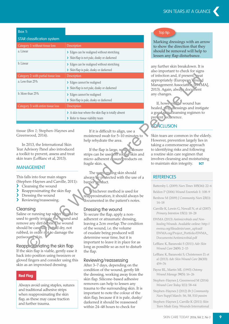
suitable for use on both intact (Figure 3) and broken skin, are alcohol-free and non-stinging and need to be reapplied every 72 hours (Houser et al, 2010). The cream version provides protection for intact skin against bodily fluids; has moisturising properties and needs to be reapplied every three days.
Whichever product is chosen, nurses need to ensure that they select the correct type of barrier products for the wound and patient they are dealing with. The cream version is recommended for skin protection in incontinence-related skin conditions, for example, whereas the film version is designed to protect the periwound area.
However, inappropriate use of barrier creams for periwound skin protection has the potential to damage skin further since they may considerably enhance the dressing’s ability to stick to the skin, and, if frequent dressing changes are required, may then damage the stratum corneum. Consequently, barrier creams should be used with caution in older adults, babies and children, and those with fragile skin due to dermatological conditions such as eczema (Cutting, 2008; Hollinworth, 2009).
Studies comparing acrylate skin barrier products with the more traditional skin protectants have produced positive results, although both were used on superficial pressure damage as opposed to general periwound skin (Bale et al, 2004; Bliss et al, 2007; Ermer-Seltun, 2011). However, Coutts et al (2010) compared acrylate skin barrier products to zinc oxide and petrolatum ointments and found that all of them were effective in the management of periwound maceration. Nurses also need to be aware that some barrier products are contraindicated on areas where fungal infection is suspected and that no more than one product should be used at any one time (Wounds UK, 2012).
Wound dressingsThe easiest way to protect the periwound skin is to prevent wound Figure 3. Skin flap injury with intact periwound skin.
© 2016
Wou
nd C
are P
eople
Ltd

FOCUS ON PERIWOUND SKIN
Additional dressingsWhere effective exudate management is a problem, it may be necessary to apply an alginate or Hydrofiber™
under the main dressing in order to prolong its wear time. These are available in sheet or rope form (designed to fill cavities) and absorb the exudate to form a gel, which maintains a moist rather than a wet wound environment.
Nurses need to be familiar with the mode of absorption of these dressings since this varies between the two product types. Alginate dressings absorb exudate laterally into their fibres and therefore should be cut to the shape of the wound. If an alginate dressing becomes over-saturated with exudate and is left in place for too long, maceration to the
fluid from coming into contact with the skin in the first place (Lawton, 2009). There are various wound management products that are designed to manage differing volumes of exudate and it is important to select one which can manage exudate while maintaining an optimum level of moisture within the wound (Hollinworth, 2009).
Foam dressings Foam dressings are designed to manage a variety of exudate volumes and are available in different absorbency levels. These products have been called ‘intelligent’ dressings, since they are capable of adapting the volume of exudate they absorb by allowing exudate to evaporate from the dressing into the atmosphere, so that a moist rather than a wet wound environment can be maintained.
This process is called the moisture vapour transmission rate (MVTR) (White and Cutting, 2003) and varies from product to product. In order to facilitate this process, the surface area of a foam dressing should not be covered with other occlusive dressings, such as films.
Superabsorbent dressings Superabsorbent dressings comprise multiple layers including a wound contact layer that interfaces with the wound bed, an inner layer or core with fluid-handling properties comprising absorbent fibres, powders, crystals or gelling agents, and a fluid-repellent backing layer that prevents exudate leaking from the dressing. Some superabsorbent dressings absorb exudate via osmosis; others use a capillary action where the exudate is absorbed and retained within the hydrophilic layer of the dressing, thus ensuring that the wound bed is kept moist but not too wet (Wound Care Today, 2015).
Wear timeA further consideration is the recommended wear time of the dressing and nurses are advised to consult the recommended wear times on the packaging or their local trust formulary. Unfortunately, there are no standard tools currently available to measure exudate volume and nurses tend to use terms such as ‘light’, ‘moderate’ or ‘heavy’, which can be very subjective (Cameron, 2004). Table 1 outlines how to assess the efficacy of dressings absorbency.
Table 1: Clinical indicators of dressing absorbency
Wound status IndicatorDry Wound bed may be dry
There is no evidence of exudate on the dressing which is unmarked?The dressing may have stuck to the wound bed
NB: This may be appropriate for an ischaemic wound (that develops as a result of arterial insufficiency)
Moist Some evidence of moisture on wound bedSmall marks on dressing but dressing has not been breachedFrequency of dressing change appropriate for level of exudate
Wet Small amount of exudate visible when dressing is removedThe primary dressing is considerably marked but there is no strikethrough evidentExudate contained according to frequency of dressing changes
Saturated Primary dressing is wet and there is evidence of strikethroughWounds need more frequent dressing changes and there is evidence of maceration to periwound area Rule out wound infection
Leaking Dressing is saturated and there is leakage of exudate from the dressing onto clothes and beyondVery frequent dressing changes are required to contain exudate Rule out wound infection
* Adapted from World Union of Wound Healing Societies (WUWHS, 2009)
Figure 4. Maceration in a venous leg ulcer. Note that the maceration is at the lower end of the wound due to gravity.
Figure 5. Venous leg ulcer with well-managed exudate.
SKIN CARE TODAY 2016, Vol 2, No 1 15
© 2016
Wou
nd C
are P
eople
Ltd

16 SKIN CARE TODAY 2016, Vol 2, No 1
FOCUS ON PERIWOUND SKIN›periwound skin will result (Flanagan, 2013). Hydrofibers on the other hand, absorb exudate horizontally and can be left to overlap the wound margins as the dressing surrounding the wound will remain dry.
It must be noted, however, that dressings alone may not be sufficient to manage large quantities of exudate. For example, in the case of ‘leaking legs’ or venous leg ulcers, dressings should be used as an adjunct to treatment with compression therapy as treating the cause of the oedema will result in a reduction in exudate volume (Figures 4 and 5).
CONCLUSION
Maceration and other conditions affecting the periwound area can be avoided through careful assessment and product choice. Overlooking the vulnerable area of skin around a wound can result in trauma and pain for the patient and a deteriorating and extending wound.
Continuous assessment is the guiding principle when managing wounds and nurses need to be aware that exudate volume may change over time and ensure that they select the most appropriate product to ensure the optimum environment to enable the wound to heal. SCT
REFERENCES
Bale S, Tebble N, Jones V, Price P (2004)The benefits of implementing a new skin care protocol in nursing homes. J Tissue Viability 14(2): 44–50
Bliss DZ, Zehrer C, Savik K, et al (2007) An economic evaluation of four skin damage prevention regimens in nursing home residents with incontinence. Product and labour costs. J Wound Ostomy Continence Nurs 34(2): 143–52
Cameron J (2004) Exudate and care of the periwound skin. Nurs Stand 19(7): 62–8
Coutts P, Sibbald GR, Queen D (2010) Periwound ski protection: a comparison of a new skin barrier vs traditional therapy in wound management. Poster Presentation at the CAWC Conference,
London Ontario, Canada
Cutting K, White R (2002) Maceration of the skin and wound bed 1: its nature and causes. J Wound Care 11(7): 275-8
Cutting K (2008) Impact of adhesive surgical tape and wound dressings on the skin, with reference to skin stripping. J Wound Care 17(4): 157-62
Ermer-Seltun J (2011) Practical prevention and treatment of incontinence–associated dermatitis — a risk factor for pressure ulcers. Ostomy Wound Management Available online: www.o–wm.com/content/practicalprevention–and–treatment–incontinence–associated–dermatitis–%E2%80%94–risk–factor–pressure (accessed 5 February, 2016)
Flanagan F (2013) Wound Healing and Skin Integrity. Principles and Practice. Wiley-Blackwell, Sussex
Goldberg M, Mcgynn-Byer P (2000) Oncology-related skin damage. In: Bryant R, ed. Acute and Chronic Wounds. 2nd edn. Mosby, St. Louis
Hollinworth H (2009) Challenges in protecting periwound skin. Nurs Stand 24(7): 53–62
Houser T, Grove GL, Zerweck C (2010) A comparison of the durability of four barrier film products over a 72-hour period on human volunteers. Poster presentation at Advances in Skin and Wound Care Conference (ASWC), Canada.
Langoen A, Bianchi J (2013) Maintaining skin integrity. In Flanagan F, ed. Wound Healing and Skin Integrity. Principles and Practice. Wiley-Blackwell, Sussex
Lawton S (2009) Assessing and managing vulnerable periwound skin. Worldwide Wounds Available online: www.worldwidewounds.com/2009/October/Lawton-Langoen/vulnerable-skin-2.html (accessed 4 February, 2016)
Marks JG Jnr, Cano C, Leitzel K, Lipton A (1983) Inhibition of wound healing by topical steroids. J Dermatol Surg Oncol 9(10): 819–21
Moffatt C (2007) Compression Therapy in Practice. Wounds UK, Aberdeen
Mudge E J, Meaume S, Woo K, Sibbald R G, Price EP (2008) Patients’ experience of wound-related pain: an international
perspective. European Wound Management Association J 8(2): 19–28
National Patient Safety Agency (2007) Fire Hazard with Paraffin-based Skin Products. NPSA, London
Newton H, Cameron J (2003) Skin Care in Wound Management. Medical Communications UK, Holsworthy
Schofield J (2013) Skin integrity and dermatology. In Flanagan F, ed. Wound Healing and Skin Integrity. Principles and Practice. Wiley-Blackwell, Sussex
Schuren J, Becker A, Sibbald RG (2005) A liquid film-forming acrylate for periwound protection: a systematic review and meta-analysis (3M™ Cavilon™ no-sting barrier film) Int Wound J 2(3): 230–8
Stephen-Haynes J (2008) Skin integrity and silicone: Appeel ‘no-sting’ medical adhesive remover. Br J Nurs 17(12): 792–5
Tavadia S, Bianchi J, Dawe M, et al (2003) Allergic contact dermatitis in venous leg ulcer patients. Contact Dermatitis 48: 26–65
Thompson G, Stephen-Haynes J (2007) An overview of wound healing and exudate management. Br J Community Nurs 12(12 Suppl): S22–30
Winter GC (1962) Formation of the scab and rate of epithelialization of superficial wounds in the skin of the young domestic pig. Nature 193: 293–4
White RJ, Cutting KF (2003) Interventions to avoid maceration of the skin and wound bed. Br J Nurs 12(20): 1186–1201
World Union of Wound Healing Societies (2009) Principles of best practice. Wound exudate and the role of dressings. A consensus document. MEP Ltd, London
Wound Care Today (2015) Product Pyramid — superabsorbent dressings. Available online: woundcare-today.co.uk/categories-pyramid/superabsorbent-dressings (accessed 3 February, 2016)
Wounds UK (2012) Cavilon skin care products made easy. Available online: www.wounds-uk.com/made-easy/cavilon-skin-care-products-made-easy/page-2 (accessed 5 February, 2016)
© 2016
Wou
nd C
are P
eople
Ltd
PRESCRIBINGPOLICY ACUTE CAREHEALTH PROMOTIONLONG-TERM CONDITIONS
To receive your free copy and access to online resources (including the new revalidation zone), register at: www.journalofpracticenursing.co.uk
Journal of General Practice Nursing
Journal of General Practice Nursing
Promoting practice to improve patient health and quality of life
Published in association with Education for Health, this free journal (both online and in print) helps challenge and develop practice within primary care.
Regular features include; debate and discussion on the latest hot topics, an educational learning zone (linked to the online GPN learning zone units), as well as clinical articles on long-term conditions, health promotion and prescribing, to name but a few.
© 2016
Wou
nd C
are P
eople
Ltd

FOCUS ON ATOPIC ECZEMA
18 SKIN CARE TODAY 2016, Vol 2, No 1
Atopic eczema commonly begins in infancy with approximately 60%
of children growing out of the condition by adolescence (Baron et al, 2012). However, in about one-third of people a chronic persistent course of atopic eczema continues into adulthood (Garmhausen et al, 2013).
Skin disease accounts for 15–20% of a GP’s workload (All Party Parliamentary Group on Skin [APPGS], 2013), which means that community nurses are ideally placed to support patients in controlling their condition, particularly through good self-care.
This article aims to provide nurses with practical advice on the management of atopic eczema in adults and adolescents.
ATOPIC ECZEMA
Atopic eczema usually presents in childhood but affects 2–10% of
Managing atopic eczema in the community setting
homeostasis) has been identified as a factor in the development of atopic eczema (Palmer et al, 2006). Filaggrin has a pivotal role in maintaining the skin barrier, and loss of this function results in dry skin and a strong predisposition to eczema (Williams and Grindlay, 2009). In addition, the skin’s contact with detergents and soaps can have a further detrimental effect on barrier function, causing dryness (Cork et al, 2006).
Other factors include exposure to allergens such as house dust mites, Staphylococcus aureus
infections, excessive heat and contact with irritants that disrupt the barrier function of the skin (British Association of Dermatologists [BAD], 2013).
Ann Joy, senior staff nurse, Dermatology Outpatient Department, Queen Margaret Hospital, Fife
IN BRIEF
Atopic eczema is a frustrating and complex skin condition that has no cure.With good support, education and the correct application of topical treatments, atopic eczema can be well controlled.Nurses in primary care can provide patients with information about their condition, how to apply topical treatment effectively
the eczema and patient quality of life.
KEY WORDS:
Dermatology Atopic eczema Skin care guidance Topical treatment Patient education Quality of life
Ann Joy
adults in the UK and its prevalence continues to rise (Baron et al, 2012). The condition involves environmental and genetic factors (Bieber, 2008) and the term atopic, in particular, is used when there is a strong family history of eczema, asthma and hay fever.
Atopic eczema is a complex condition caused by several factors involving genetic mutations and skin hyperactivity to environmental stimuli. Mutations in the gene that encodes filaggrin (a protein that plays a role in epidermal
Practice point
Any advice provided to patients should be reinforced with written information and a treatment plan, with follow-up support offered as needed.
Copy
right
: Fed
orov
Ivan
Ser
geev
ich
© 2016
Wou
nd C
are P
eople
Ltd

For patients who want the convenience of self-selection,
handy sized packs are available for purchase
in pharmacies
Please refer to the full SPC text before prescribing this product. Adverse events should be reported. Reporting forms and information can be found at www.mhra.gov.uk/yellowcard. Adverse events should also be reported to Bayer plc, Consumer Care Division.
Date of preparation: September 2015 Code: AWB-1924932177Copyright © 2015 Bayer. All rights reserved unless otherwise indicated. All trademarks are owned by Bayer, and its affiliates, or licensed for its use.
Diprobase Product Information
Uses: Diprobase Cream and Ointment are emollients, with moisturising and protective properties, indicated for follow-up treatment with topical steroids or in spacing such treatments. They may also be used as diluents for topical steroids. Diprobase products are recommended for the symptomatic relief of red, inflamed, damaged, dry or chapped skin, the protection of raw skin areas and as a pre-bathing emollient for dry/eczematous skin to alleviate drying effects. Dosage: The cream or ointment should be thinly applied to cover the affected area completely, massaging gently and thoroughly into the skin. Frequency of application should be established by the physician. Generally, Diprobase Cream and Ointment can be used as often as required. Contra-indications: Hypersensitivity to any of the ingredients. Side-effects: Skin reactions including pruritus, rash, erythema, skin exfoliation, burning sensation, hypersensitivity, pain, dry skin and bullous dermatitis have been reported with product use. Package Quantities: Cream: 50g tubes, 500g pump dispensers; Ointment: 50g tubes, 500g tubs. Basic NHS Costs: Cream: 50g tube = £1.28, 500g pump = £6.32; Ointment: 50g tube = £1.28, 500g tub = £5.99. Legal Category: GSL. Marketing Authorisation Numbers: Cream: PL 00010/0658; Ointment: PL 00010/0659. Marketing Authorisation Holder: Bayer plc, Consumer Care Division, Bayer House, Strawberry Hill, Newbury, Berkshire, RG14 1JA, U.K. Date of Revision of Text: March 2015
Active Ingredients: None. Legal Category: Medical device. Uses: Diprobase Lotion is an emollient with moisturising and protective properties, recommended for the management of eczema and other dry skin conditions. Relieves and soothes dry or eczematous skin. Side-effects: No skin reactions have been reported with product use. Contra-indications: Hypersensitivity to any of the ingredients. Dosage: Apply to affected area as often as required. Package Quantities: 300ml pump pack, 50ml tubes. NHS Price: 300ml £3.49, 50ml £1.28. Recommended Retail Price: 300ml £7.99, 50ml £3.99. Date of preparation: December 2014. For further information contact Bayer plc, Consumer Care Division, Bayer House, Strawberry Hill, Newbury, Berkshire, RG14 1JA, U.K.
Essential Information: Diprobase Lotion
N.V. Organon, Molenstraat 110, 5342 CC, Oss, The Netherlands.
MSD Consumer Care, Inc.3030 Jackson Avenue, Memphis, TN 38112, USA.
“Sleepless nights...constant itching... I wish bedtime was more peaceful ”
Spread CalmSoothing, calming and protecting,
Diprobase has been helping
people with eczema to hydrate
their skin, relieve symptoms
and live more peaceful lives
for over 30 years.
© 2016
Wou
nd C
are P
eople
Ltd

20 SKIN CARE TODAY 2016, Vol 2, No 1
FOCUS ON ATOPIC ECZEMA›The key to supporting patients in
managing atopic eczema is:› Providing time to explain
the condition› Discussing the application of
topical treatment, the avoidance of exacerbating factors
› Reviewing and assessing their progress to treatment.
COMPLETE EMOLLIENT THERAPY
In atopic eczema the defect in barrier function results in a lack of natural oils. This leads to water loss and increased susceptibility to irritants and penetration of allergy-inducing substances causing itch and inflammation. Further drying and irritation of the skin can be lessened by the avoidance of soaps and detergents, which contain perfumes and preservatives.
Complete emollient therapy is the mainstay of treatment (BAD, 2013). Regular application of a moisturiser, washing with a soap substitute and using a moisturising bath or shower oil will provide moisture to the skin, helping to prevent further water loss (DermNet NZ, 2013a) and the desire to scratch.
BATHING
Daily bathing is advised to cleanse and hydrate the skin and reduce the risk of infection. Certain bath additives contain antimicrobial, antiseptic and/or antipruritic properties designed to reduce bacterial load and calm the intense itch provoked by atopic eczema. Antiseptic/antimicrobial preparations
should not be used regularly unless infection is recurrent or widespread (British Medical Association and Royal Pharmaceutical Society of Great Britain [BMA/RPS], 2013).
Bath emollients are added to running bath water and some can be applied to wet skin and showered off. These products leave a layer of oil on the skin that seals-in moisture. Bathing should ideally be restricted to 15 minutes to prevent disruption of the skin’s barrier function (British Dermatological Nursing Group [BDNG], 2012).
Soap substitutes are used to cleanse the skin without removing natural oils and they can be applied directly to the skin and rinsed off. A majority of cream-based moisturisers can be used as soap substitutes and a wide range of products are available including gels, lotions, ointments and creams. After bathing or showering, the skin should be gently patted dry as vigorous rubbing can lead to increased irritation (BDNG, 2012).
MOISTURISING
Leave-on moisturisers are available in lotion, cream, ointment, gel and spray formulations. Due to the defective skin barrier function in atopic eczema, moisturisers should be applied to the skin in its entirety and not restricted to affected areas. Moisturisers should be applied even when the skin is clear (National Institute for Health and Clinical Excellence [NICE], 2007). They should also be prescribed in large quantities — at least 600g is recommended for an adult for a weekly, twice-daily whole-body application (BMA/RPS, 2013).
Ointment preparations can be more effective than creams as they are greasier and do not contain preservatives. This prevents stinging when applied to inflamed skin. However, patient preference is vital in the choice of moisturiser and often a greasy product is not practical for use during the day or while at work. Thus, a cream-based product is advised for daytime
use and a greasier ointment for overnight application. It is important to find a moisturiser that suits each individual and this can be achieved by providing a variety of sample moisturisers for the patient to try out.
Aqueous cream was designed as a soap substitute, which is applied to the skin and then washed off (Cork and Danby, 2009). However, it has an irritant effect due to the sodium lauryl sulphate content, which is a harsh surfactant (substances such as detergents that, when added to liquids, increase their ‘spreading’ and ‘wetting’ ability) (Cork and Danby, 2009). It is now recommended that aqueous creams are not used even as washing products (Moncrieff et al, 2013) as they have been shown to weaken the skin barrier and increase transepidermal water loss (Danby et al, 2011).
Spray-on emollients can improve treatment concordance (MacKenzie and Schofield, 2013), especially in adolescents, as they have a non-touch application technique and are quick and easy to apply.
Any moisturiser should be allowed to absorb into the skin before the application of an additional therapeutic product, such as topical steroids (BDNG, 2012). Practical tips for application of emollients are provided in Table 1.
› Practice point
The ‘itch-scratch cycle’ refers to the situation where itching results in scratching, but scratching itself can subsequently aggravate the itch. Also, when someone has lived with eczema for a long time, the scratching can become a habit. Distraction techniques can sometimes help to interrupt these cycles.
› The facts...
Atopic eczema is a chronic condition, causing pruritic inflammation of the epidermis and dermis resulting in thickened, dry cracking skin, which can also be painful. Acute flares can arise resulting in erythema, vesicles and oedema, which can produce scaly, crusty skin prone to infection. Pruritis can be severe causing scratching, further exacerbation, infection and pain. Diagnosis is usually obtained from patient history, family history of atopy and clinical appearance.
© 2016
Wou
nd C
are P
eople
Ltd

SKIN CARE TODAY 2016, Vol 2, No 1 21
FOCUS ON ATOPIC ECZEMA ›TOPICAL STEROIDS
Topical steroids are effective in reducing the symptoms of inflammation through their anti-inflammatory, immunosuppressive, antiproliferative and vasoconstrictive actions (Ersser and Van Onselen, 2010). Topical steroid preparations are available in lotion, cream, ointment, gel, impregnated tape and mousse form, and in four different strengths:› Mild› Moderate› Potent› Very potent.
Application should be performed in conjunction with a good moisturising regimen to enhance absorption, efficacy and ease of application. Topical steroids should be applied 30 minutes after applying a moisturiser to avoid diluting the steroid (BDNG, 2012).
Topical application is once-daily (Scottish Intercollegiate Guidelines Network [SIGN], 2011), with the strength of the steroid tailored to the severity, age and site of the eczema (Primary Care Dermatology Society [PCDS], 2015).
A step-up and step-down approach is recommended (NICE, 2007), which matches the strength of topical steroid to the severity of the eczema — once the eczema settles, the strength of steroid is decreased, rather than being withdrawn altogether. If the eczema then flares-up again, the strength can be stepped-up (NICE, 2007).
The use of a ‘steroid ladder’ aids the identification of steroid strength and a stepped-approach to treatment (Page and Robertson, 2004). The steroid ladder groups the topical steroids according to their strength — ‘very potent’, ‘potent’, ‘moderate’ and ‘mild’ — for ease of identification. The theory is illustrated as a step ladder to advise the user to ‘step-up or down’ the ladder but not to ‘jump off’. In other words, it reminds patients to reduce the strength of steroid rather than ceasing treatment.
As a general rule, a weak preparation should be used on the face and genital areas, with a moderate or potent steroid applied elsewhere on the body (Baron et al, 2012).
The amount of corticosteroid is measured in finger-tip units (FTUs), which comprise the distance from the tip of the adult index finger to the first joint. One finger-tip unit will adequately treat the surface area of two adult palms (Long and Finlay, 1991).
For frequent eczema flares, it is suggested that a potent topical steroid be applied to areas of inflammation once-daily for two weeks, then on alternate days for two weeks (PCDS, 2015). Once benefit is seen, the potency of steroid is reduced until they are discontinued.
As with moisturisers, steroid ointment preparations are preferable to creams due to the lack of stinging pain on application, which can enhance concordance (Baron et al, 2012). However, creams are advised in ‘weepy’ eczema (exhibiting exudate), due to their more effective drying action (see ‘Top tips on topical corticosteroid use’, pp 33–35).
A clear care plan will enhance understanding and concordance with treatment, both in patients, but also in staff where different nurses might be involved in treatment, for
instance. This is particularly relevant as fears about using topical steroid therapy (due to its strength) can often lead to under-treatment, subsequent treatment failure and disillusionment (Smith et al, 2010).
Nurses should take time to thoroughly explain the benefits of topical treatments and formulate a treatment plan that fits into the patient’s daily life. A demonstration of the amount of topical treatment to apply to certain areas, along with application techniques, can improve patient confidence. Written information can also help patients’ understanding and concordance with treatment regimens.
TOPICAL CALCINEURIN INHIBITORS
Topical calcineurin inhibitors are immuno-modulating agents licensed for the treatment of atopic eczema (BMA/RPS, 2013). Their main benefit is that they are not steroid-based and do not cause skin atrophy. They are considered if topical steroid treatment has failed or where there is a risk of adverse effects from further topical steroid use (NICE, 2004). Treatment is usually initiated by a dermatologist (BAD, 2013).
Topical calcineurin inhibitors include creams and ointments incorporating tacrolimus and
Table 1:
Practical tips for application of emollients (BDNG, 2012)
Use complete emollient therapy, comprising bath additives, soap substitutes and leave-on emollients
Bath oils can be added to the bath water or applied directly to damp skin as a soap substitute in the shower or bath and then rinsed off
Apply liberal amounts of topical leave-on moisturisers — approximately 500–600g per week
Apply leave-on moisturisers throughout the day
Always apply emollients in a downward motion following the direction of the hair to avoid folliculitis and excess rubbing
Use emollients directly after a bath/shower
Creams should be applied to ‘weepy’ skin
Greasy ointments are best applied to dry, scaly, lichenified and/or fissured skin
Do not stop emollients when eczema resolves. A daily emollient routine can help prevent flares
Always remember that the best emollients are the ones patients like, as they are more likely to apply them
To prevent moisturisers becoming contaminated, use clean implements to scoop products out of tubs or use sealed pump dispensers
Warn patients that paraffin-based products are flammable
© 2016
Wou
nd C
are P
eople
Ltd

22 SKIN CARE TODAY 2016, Vol 2, No 1
pimecrolimus. The commonest side-effect of topical calcineurin inhibitors is stinging on application, which usually settles after a few applications, but they should be discontinued if the skin becomes infected (BAD, 2013). Twice-weekly applications of tacrolimus ointment in patients with stable eczema can prevent further exacerbations (BMA/RPS, 2013).
MANAGING INFECTED ECZEMA
Atopic eczema often flares due to infection. This is commonly associated with Staphylococcus aureus, however, the herpes simplex virus can also be involved (BAD, 2013). Signs of bacterial infection include:
WeepingYellow crustsPustulesErythemaExcoriationRapidly worsening eczemaFailure of eczema to respond to treatment.
In the case of infected eczema, combined antimicrobial and corticosteroid ointments can be applied for short periods. However, if the infection is widespread, a seven-day course of an oral antibiotic such as flucloxacillin may be required (PCDS, 2015), in conjunction with a topical steroid and emollient therapy. Bath additives with antimicrobial agents can also be useful in reducing the bacterial load on the skin.
Once the infection has settled, antibacterial products should be avoided unless infection is evident or a frequent complication (BMA/RPS, 2013), otherwise plain bath products are advised.
FOCUS ON ATOPIC ECZEMA
Patients with atopic eczema are more prone to viral infections, particularly involving the herpes simplex virus. It is important for patients to recognise the signs of herpes simplex infection, as this can spread rapidly, causing severe eczema herpeticum (DermNet NZ, 2013b). Eczema herpeticum is indicated by the presence of grouped vesicles, punched-out erosions and generalised illness and can cause a painful widespread flare of eczema. Prompt treatment is, therefore, essential. Topical steroids should be stopped and SIGN (2011) recommends an emergency dermatology referral.
AVOIDING ENVIRONMENTAL IRRITANTS
Nurses should allow time for discussions with the patient on any potential exacerbating factors, and extremes in temperature and clothing containing synthetic fibres or wool are best avoided. Practical tips include:
Keeping the fingernails short to prevent damage from scratchingUsing comfortable cotton clothing and beddingAvoiding soap and detergents, which might cause further drying of the skin (National Eczema Society [NES], 2013).
An understanding of the causes of atopic eczema and the common factors that can cause flares in the condition — such as general ill-health — can forewarn the patient of the need to step-up treatment application at certain times (Baron et al, 2012). Also, an acceptance of the need to continually apply emollient therapy — even when the skin is clear of eczema — to maintain good skin barrier function will help to prevent flares (Baron et al, 2012).
OTHER TREATMENT STRATEGIES
Medicated bandages impregnated with zinc paste or ichthammol are effective for soothing and cooling inflamed, excoriated eczema (Ersser, 2010), and softening chronic thickened or lichenified skin (NICE, 2007). These products can provide some relief from itching and provide protection from scratching.
Paste bandages can also be useful in occluding chronically excoriated areas and can be applied over moisturisers and topical steroid therapy. They should be applied in an incomplete spiral fashion, folding back or cutting the bandage into individual strips to prevent tightening — this is then secured with a further tubular bandage. They can be left in place for 12–24 hours. The occlusion provided by bandaging will increase skin hydration and improve the penetration of topical steroids, however, the potency of topical steroid used should be taken into consideration due to enhanced absorption (McAleer et al, 2012). However, occlusive dressings should not be used on weepy, infected eczema (PCDS, 2015) and application can be time-consuming and messy.
In atopic eczema, the short-term use of sedating antihistamines at night may be useful if flares are causing sleep disturbance (SIGN, 2011). There seems to be no need for non-sedating antihistamines unless for co-existent hay fever (PCDS, 2015). Behaviour modification techniques, such as habit reversal may be useful and complement conventional therapies — this approach aims to heighten the patient’s awareness of scratching and helps to break the itch-scratch cycle (see ‘Practice points box’, p. 20).
PSYCHOLOGICAL AND SOCIAL IMPACT
Eczema can cause a great deal of distress for patients and their families, with the physical discomfort, need to apply time-consuming ‘messy’ treatments and sleep loss all combining to have a deleterious impact (APPGS, 2013).
Society’s increasing emphasis on appearance and pressure to attain the ‘perfect’ body place greater strain on people with skin conditions. In adolescents, when considerable focus is placed on appearance, skin disease can be devastating to self-esteem and psychological wellbeing (APPGS, 2013). Patients may isolate themselves to avoid comments from others, preventing participation in
Top tip:
Information should be appropriate, written and compatible with the patient’s lifestyle, beliefs and cultural practices. As eczema is episodic, it is vital that patients/carers are provided with the details of when and how to step treatment up or down (Roberts, 2015).
© 2016
Wou
nd C
are P
eople
Ltd

SKIN CARE TODAY 2016, Vol 2, No 1 23
FOCUS ON ATOPIC ECZEMA ›
SCT
British Association of Dermatologists (2013) Atopic Eczema. BAD, London
Baron SE, Cohen SN, Archer CB (2012) Guidance on the diagnosis and clinical management of atopic eczema. Clin Exp Dermatol 37(1): 7–12
British Dermatological Nursing Group (2012) Best Practice in Emollient Therapy. A Statement for Healthcare Professionals. BDNG, London
Bieber T (2008) Atopic dermatitis. New Eng J Med 358: 1483–94
British Medical Association/Royal Pharmaceutical Society (2013) British National Formulary: 64. BMA/RPS, London
Cork MJ, Danby S (2009) Skin barrier breakdown: a renaissance in emollient therapy. Br J Nurs 18(14): 872–7
Cork MJ, Robinson DA, Vasilopoulos Y, et al (2006) New perspectives on epidermal barrier dysfunction in atopic dermatitis: gene-environment interactions. J Allergy Clin Immunol 118(1): 3–21
Danby SG, Al-Enezi T, Sultan A, Chittock J, Kennedy K, Cork MJ (2011) The effect of aqueous cream BP on the skin barrier in volunteers with a previous history of atopic dermatitis Br J Dermatol 165(2): 329–34
DermNet NZ (2013a) The causes of atopic dermatitis. Available online: www.dermnetnz.org/dermatitis/atopic-causes.html (accessed 21 March, 2016)
DermNet NZ (2013b) Complications of atopic dermatitis. Available online: www.dermnetnz.org/dermatitis/atopic-complications.html
Ersser SJ (2010) Protecting the skin and preventing breakdown. In: Penzer R, Ersser S, eds. Principles of Skin Care: A Guide for Nurses and Other Health Care Professionals. Wiley-Blackwell, Oxford
Ersser S, Van Onselen J (2010) Eczema. In: Penzer R, Ersser S, eds. Principles of Skin Care: A Guide for Nurses and Other Health Care Professionals. Wiley-Blackwell, Oxford
Garmhausen D, Hagemann T, Bieber T, et al (2013) Characterisation of different courses of atopic dermatitis in adolescent and adult patients. Eur J Allergy Clin Immunol 68: 498–506
Long C, Finlay A (1991) The fingertip unit: a new practical measure. Clin Exp Dermatol 16: 444–7
MacKenzie A, Schofield O (2013)
Recommended diagnosis and management of atopic eczema. Prescriber Nov: 18–26
McAleer MA, Flohr C, Irvine AD (2012) Management of difficult and severe eczema in childhood. Br Med J 345: 35–41
Moncrieff G, Cork M, Lawton S, et al (2013) Use of emollients in dry-skin conditions: consensus statement. Clin Exp Dermatol 38: 231–8
National Eczema Society (2013) Itching and Scratching. How to control eczema-related itching and sleep disturbance. NES, London
National Institute for Health and Care Excellence (2004) Tacrolimus and Pimecrolimus for Atopic Eczema. Technology Appraisal Guidance 82. NICE, London
National Institute for Health and Care Excellence (2007) Atopic Eczema in Children. Management of atopic eczema in children from birth up to the age of 12 years. Clinical guideline 57. NICE, London
Page B, Robertson S (2004) Hands on...topical corticosteroids identified. Derma Nurs 3(2): 16–17
Palmer CAN, Irvine AD, Terron-Kwiatkowski A, et al (2006) Common loss-of-function variants of the epidermal barrier protein filaggrin are a major predisposing factor of atopic dermatitis. Nature Genetics 38(4): 441–6.
Primary Care Dermatology Society (2015) Eczema: atopic eczema. Available online: www.pcds.org.uk/clinical-guidance/atopic-eczema
Roberts A (2015) Childhood eczema: a parent’s perspective. J GPN 1(1): 48–53
Scottish Intercollegiate Guidelines Network (2011) Management of Atopic Eczema in Primary Care. A National Guideline. SIGN, Edinburgh
Smith SD, Hong E, Fearns S, Blaszczynski A, Fischer G (2010) Corticosteroid phobia and other confounders in management of childhood atopic dermatitis explored using parent focus groups. Australas J Dermatol 51: 168–74
Williams HC, Grindlay DJ (2009) What’s new in atopic eczema? An analysis of systemic reviews published in 2007 and 2008. Part 1. Definitions, causes and consequences of eczema. Clin Exp Dermatol 35: 12–15
social activities and sports. Eczema can also place a strain on intimate relationships and limit professional opportunities (APPGS, 2013).
Financial hardship may also be a factor because of absence from work due to flares, attending medical appointments or the cost of obtaining treatments. These obstacles may cause anxiety and depression, resulting in some patients not treating their skin, leading to further exacerbation. The adverse effects on quality of life for patients and their families should always be considered when assessing atopic eczema (SIGN, 2011).
WHEN TO REFER TO SECONDARY CARE
Patients should be referred to a dermatologist when there is uncertainty about the diagnosis, failure to respond to appropriate topical therapy or poor control of the condition, recurrent secondary infection and sleep problems, or psychological upset (SIGN, 2011). Treatment options in this setting include phototherapy and oral immunosuppressant agents.
CONCLUSION
Atopic eczema is a frustrating and complex condition, which has no cure. However, with good support, education and correct application of topical treatments the condition can be well-controlled.
Community nurses can provide patients with information about their condition, how to apply their topical treatment effectively and how to manage flares and maintain a routine — all strategies that will improve the eczema and the patient’s quality of life. Any information should also be reinforced with written information and treatment plans, with follow-up support offered as needed.
REFERENCES
All Party Parliamentary Group on Skin (2013) The Psychological and Social Impact of Skin Disease on People’s Lives. APGS, London
© 2016
Wou
nd C
are P
eople
Ltd

STUDY DAYS & FREE ATTENDANCE FOR ALL PRIMARY CARE NURSES IN YOUR AREA
SIX EDUCATIONAL SESSIONS DELIVERED IN ONE DAY
PRESENTED BY LEADING CLINICIANS
COVERS THEORY AND PRACTICAL SKILLS FOR COMMUNITY PRACTICE
EXHIBITION WITH LATEST PRODUCTS, DEMONSTRATIONS AND SAMPLES
ATTENDANCE CONTRIBUTES TO REVALIDATION
IN ASSOCIATION WITH:
HELPING NURSES WORKING IN PRIMARY CARE TO UNDERSTAND
NEW FOR 2016: THE NEW ONLINE JCN REVALIDATION TOOL INCLUDING A SESSION ON WHAT REVALIDATION MEANS, WHAT YOU NEED TO DO, AND
© 2016
Wou
nd C
are P
eople
Ltd

EXHIBITION 2016AND MEET THE NEW REVALIDATION REQUIREMENTS
HOW TO USE THE NEW FREE JCN REVALIDATION TOOL TO SUPPORT YOUR LEARNING
event venue dateCardiff Village Hotel Wednesday 24 February
Blackpool Village Hotel Wednesday 16 March
Newcastle Village Hotel Wednesday 13 April
Walsall Walsall Football Club Wednesday 27 April
Leeds/Bradford Cedar Court Hotel Wednesday 18 May
Bristol University of the West of England Wednesday 8 June
Exeter Exeter Race Course Wednesday 22 June
Peterborough Holiday Inn Wednesday 6 July
Elstree Holiday Inn Wednesday 21 September
Ashford Ashford International Hotel Wednesday 12 October
Sheffi eld Hilton Doubletree Wednesday 16 November
Norwich Norwich City Football Club Wednesday 7 December
EVENTS CALENDAR
© 2016
Wou
nd C
are P
eople
Ltd

26 SKIN CARE TODAY 2016, Vol 2, No 1
Itch is most often associated with inflammatory and non-inflammatory skin conditions,
but can also manifest in other non-dermatological illnesses such as haematological conditions, malignant disease, endocrine disorders, hepatic and renal disease, human immunodeficiency virus (HIV), pregnancy, and psychiatric conditions (Karim, 2011). Managing chronic itch remains a major nursing challenge, despite advances in medical science and nursing practice. This article provides an overview of itch from a biopsychosocial perspective (where biological, psychological and social factors all play a significant role in disease), as well as encouraging an holistic approach to care.
PATHOPHYSIOLOGY OF ITCH
For many years, scientists and healthcare professionals regarded
Managing itch — a biopsychosocial approach to care
identified and include histamine, substance P, serotonin, cytokines, proteinases, neuropeptides and opioids (Karim, 2011; Garibyan, 2013). Raap et al (2011) also suggested that cells within the skin
(keratinocytes, mast cells and eosinophils) contribute to the detection, mediation and transmission of itch sensation and form part of an innate defence mechanism (see ‘Science’ box for causes of itch).
PruritusPruritus, or severe itching, can manifest as a superficial (i.e. close to the skin’s surface), localised and spontaneous sensation, which is relieved
easily by gentle scratching or rubbing; or a deep defuse
unrelenting itch, which elicits such a
Polly Buchanan, lead nurse, research and development, NHS Fife Queen Margaret Hospital, Dunfermline; chair, Scottish Dermatological Nursing Society ([email protected]); Zoe Chouliara, associate professor in counselling, transformation and professional practice, Edinburgh Napier University ([email protected])
IN BRIEF
Itch (pruritus) is an uncomfortable and subjective sensation experienced by individuals that causes a desire to scratch.
psychological burden which can affect quality of life (Kini et al, 2011; Chen, 2012).It is important that clinicians understand and assess itch and implement nursing strategies to relieve patients’ discomfort and prevent complications.
KEY WORDS:
Pruritus Itch Quality of life Psychosocial Scratching Pain
Polly Buchanan, Zoe Chouliara
Figure 1. Skin affected by conditions such as microbial eczema can be vulnerable to pruritus.
Did you know?
Pruritus can be a local sensation, which is relieved easily by scratching; or a deep unrelenting itch with a strong urge to scratch causing skin breakdown and pain.
itch as a sub-category of pain, sharing similar nerve pathways within the peripheral and central nervous system (‘selectivity’ theory suggests neurons are selective for both pain and itch). It is only relatively recently
that research has identified the sensation of itch as being a separate entity to pain, although some similarities exist (Raap et al, 2011). This is known as ‘labelled line’ theory, where a peripheral itch relates to specific neuronal fibres. Garibyan et al (2013) suggest both theories may be relevant in the transmission of the itch sensation.
As with pain, the mediators of itch (known as pruritogens) have been
FOCUS ON ITCH
© 2016
Wou
nd C
are P
eople
Ltd

Alliance Pharmaceuticals Ltd, Avonbridge House, Bath Road, Chippenham, Wiltshire SN15 2BBTel: 01249 466 966 Fax: 01249 466 977 www.alliancepharma.co.uk
AL/2073/12.15/0.001 Date of preparation: December 2015
Switch off from dry skin and eczema...
...with Hydromol Ointment
z z z
z
PRESCRIBING INFORMATION
Hydromol® Ointment
Contra-
Legal Packs and basic NHS
Go to www.hydromol.co.uk for more information and to view our Hydromol Ointment Product Demonstration Video
3 in 1 all purpose emollient, bath additive and soap substitute
Soothes, calms and protects sensitive skin - suitable for all ages
25% less expensive than Epaderm ointment
© 2016
Wou
nd C
are P
eople
Ltd

28 SKIN CARE TODAY 2016, Vol 2, No 1
FOCUS ON ITCH
THE SCIENCE — CAUSESLike pain, the sensation
of itch is experienced following noxious stimuli from within the body itself (e.g. release of histamine) or from an external source (such as insect sting or allergen). Free nerve endings within the skin transmit itch sensation messages via the afferent C-fibres to the spinal cord via the dorsal root ganglia. Nerve transmissions then ascend the spinal cord to the thalamus and cerebral cortex where the sensation is interpreted as itch. Scratch is the resultant behaviour used to relieve the unpleasant sensation of itch.
strong urge to scratch that it results in skin breakdown and pain before any relief is experienced.
It is this deep unrelenting itch or chronic pruritus that is associated with inflammatory skin conditions such as:
EczemaPsoriasisUrticaria: so called ‘hives’Bullous pemphigoid: a blistering skin diseaseMastocytosis: excess number of mast cellsCutaneous T-cell lymphoma: rare type of non-Hodgkin lymphoma affecting the skinCutaneous drug reactionsInfestations such as scabies and lice.
Pruritus is also a commonly reported symptom of certain non skin-related systemic diseases, such as diabetes mellitus, malignancies, thyroid disease, and renal and biliary disease.
CLASSIFICATION
Itch can be acute (lasting less than six weeks), or chronic (lasting over six weeks), localised or generalised, and can be classified into four main categories that relate to the nature and origin of the itch (Garibyan et al, 2013):
Pruritoceptive: originates in the skin usually due to an inflammatory or other pathological cutaneous processNeuropathic: originates from a malfunction of the impulse-conducting cells anywhere along the nerve/spinal cord/brain afferent pathwayPsychogenic: originates from a psychological disorderNeurogenic: originates in the central nervous system but not from damage to the nerve tissue itself.
This article will focus on pruritoceptive itch, which is commonly associated with dermatological conditions.
BIOPSYCHOSOCIAL IMPACT OF PRURITOCEPTIVE ITCH
The consequences of chronic pruritus can be physical, psychological and social (van Os-Mendendorp et al, 2006), the impact of which can have a profound effect on the individual’s and family’s quality of life (Chrostowska-Plak et al, 2013).
Chronic itch causes local and widespread skin damage. The patient’s appearance can be altered through skin excoriation, inflammation, ‘scabbing’ and ‘crusting’, scaring, and lichenification (thickening of the skin), as well as being associated with stigma, social isolation and loss of employment and social life (Bundy, 2012; Stumpf et al, 2013).
Profound and prolonged itch can lead to psychological
morbidities such as anxiety and depression as a consequence of sleep deprivation, sleep disturbance, loss of concentration, feelings of helplessness, frustration, loss of function or daily activities, and suicidal thoughts (Metz et al, 2013).
Secondary infection involving bacterial, viral and/or fungal microorganisms is a common complication. With a high risk of recurrent bacterial infections, antibiotic resistance is a major concern. Therefore, preventative measures to reduce or avoid infection are paramount (Department of Health [DH], 2013), including the use of topical antiseptic/antimicrobial agents (benzoylkonium chloride and chlorhexidine hydrochloride) rather than antibiotic agents and non-medical strategies such as habit reversal to prevent scratching. This requires knowledge and skill in assessing and managing itch as part of an overall skin examination and early intervention programme.
ASSESSMENT
The International Forum for the Study of Itch (IFSI) has developed a special interest group for scoring itch in clinical trials and provides a comprehensive review with recommendations for the measurement of itch using scales and/or questionnaires. These can be used in research and also translated to clinical settings (Stander et al, 2013).
A number of itch scales are available and have been validated for use in clinical trials and daily practice. Multidimensional questionnaires
Itch — facts...
The term pruritus is of Latin origin — prurire meaning ‘to itch’.
Itch is most often associated with inflammatory and non-inflammatory skin conditions.
Itch can also manifest in other non-dermatological conditions such as haematological conditions, malignant disease, endocrine disorders, hepatic and renal disease, HIV, pregnancy and psychiatric conditions.
Managing chronic itch remains a major nursing challenge, despite advances in medical science and nursing practice.
© 2016
Wou
nd C
are P
eople
Ltd

attempt to measure a range of itch factors, e.g. intensity, duration, location, consequences, treatment and distress. The multidimensional 5-D itch severity scale (Elman et al, 2010) is a patient self-report questionnaire that uses five dimensions — degree, duration, direction, disability and distribution. Similarly, the Leuven itch scale (Haest et al, 2011) measures all aspects of the itch experience including:
FrequencyDurationSeverityCircumstancesTreatmentConsequencesSensory characteristicsDistressLocation.
The Leuven itch scale is designed to measure pain over a four-week period and is a patient self-report questionnaire.
These multidimensional tools are also very useful in clinical practice as they represent patient-reported outcomes (PROs) and are brief and simple to implement. It is recommended that all clinical audits include PRO measures (PROMs) or patient-reported experiences measures (PREMs) (Ricketts, 2012; Westerby, 2012).
Unidimensional scales on the other hand measure only one aspect of itch, most commonly severity or intensity. The most commonly used unidimensional tools are the itch severity scale (Majeski et al, 2006); visual analogue scales (where patients mark a point on a linear scale with 0 being the lowest and 10 being the highest severity), or numerical rating scales (usually a scale of 0–10, with 0 representing
Top tip:
Essentially any treatment plan should ascertain the type and cause of the itch and take steps to reduce exposure to triggers to lessen the risk of infection while providing symptom relief.
‘no itch’ and 10 being ‘worst itch experienced’ (Stander et al, 2013). The visual analogue and numerical rating scales have been adapted from similar pain scales and are useful for assessing one aspect of the itch experience, such as pain intensity. To be of value, sequential measurements are required over a period of time.
The value of assessing the severity of the patient’s itch cannot be underestimated and should be integral to the overall skin examination and history taking. Diagnostic assessment of pruritus should always include a medical/skin history, and full clinical examination and may also require further laboratory investigation, diagnostic procedures and imaging (Raap et al, 2011), which may help to diagnose the following, for example:
Scabies: dermoscopy (examination of the skin using skin surface microscopy)Bacterial and viral infection: skin swabs for microbiology or virology respectivelyFungal conditions: skin scrapings for fungal microscopy and culture Underlying systemic disease:
imaging, i.e. magnetic resonance imaging (MRI).
Using a multidimensional assessment tool, such as the 5-D itch questionnaire or the Leuven itch scale can aid clinical decision making as well as providing a method of evaluating any interventions. Perhaps most importantly, it also demonstrates clearly to the patient that effort is being made to address their symptoms.
Multidimensional tools provide an indication of the psychosocial impact of itch and aid decision making in addressing psychosocial needs and interventions. Helping the patient understand and cope with the burden of itch are also important aspects of care and will be discussed below.
MEDICAL TREATMENT
Traditionally, treatment for pruritus includes topical preparations and systemic medications. The principles of care are to:
Manage the itch, e.g. pruritoceptive itchManage the underlying condition, e.g. eczema
FOCUS ON ITCH
Table 1: Three-step approach to the management of itch*
Step Treatment optionsStep 1 Emollient therapies and/or antipruritic agents
Avoid triggers, e.g. medications, foods, spices, alcohol, hot drinksProvide symptomatic relief, e.g. topical corticosteroids, and/or antihistaminesPossibly provide psychosocial support/interventions
Step 2 Symptomatic relief related to underlying cause and/or psychosocial interventions
Step 3 — when response is poor or cause is unknown
Further topical symptomatic relief, e.g. capsaicin, ultraviolet (UV) light therapySystemic treatment, e.g. calcineurin inhibitors, cannabinoid agonists, opioid antagonists, anticonvulsants, immunosuppressives, antidepressantsPsychosocial interventions
Ongoing multidisciplinary approach
Treat cause of itch, e.g. dermatological, hepatic or renal diseaseTreat complications:
infections: antibiotics, antivirals, antifungals, antisepticsscratching behaviour: habit reversal, relaxation, distraction, CBT; knowledge deficit and ineffective coping strategies – psychoeducational programmespsychological morbidity: biopsychosocial interventionspsychogenic itch: psychiatric consultation.
* adapted from Raap et al, 2011
SKIN CARE TODAY 2016, Vol 2, No 1 29
© 2016
Wou
nd C
are P
eople
Ltd

30 SKIN CARE TODAY 2016, Vol 2, No 1
FOCUS ON ITCH
focusing on the skin condition as a result of the itch-scratch cycle and the resulting distress may account for the cosmetic concerns experienced by people with pruritus (Bundy, 2012; Stumpf et al, 2013).
However, the very nature of the intense scratching response and subsequent inflammation worsens the appearance of the skin, which unavoidably adds to people’s cosmetic concerns and distress (Stumpf et al, 2013). Similarly, anxiety and depression can lower an individual’s tolerance of pruritus, which further contributes to the itch-scratch cycle (Stangier and Ehlers, 2000; Chrostowska Plak et al, 2013).
Feelings of helplessness, low perceived control and catastrophising (excessive negative thinking), along with higher levels of fatigue and limited social support tend to be the most important predictors of psychosocial morbidity in patients with pruritic chronic disease, while ineffective coping strategies and perceived loss of control have been shown to intensify the sensation of itch (van Os-Medendorp et al, 2006).
Previous research has shown that the most effective interventions for pruritus are a combination of topical medications and psychological interventions (van Os-Medendorp et al, 2007). In many cases, psycho-education alone can be effective (van Os and Eland, 2004), such as educating the patient about the itch-scratch cycle and effective use of topical medications. Behavioural techniques that aim to control the
Manage any complications, e.g. excoriation, infection, anxiety/depression and any maladaptive or persistent habits such as incessant scratching.
Essentially, any treatment plan should ascertain the type/cause of itch and take steps to reduce exposure to the triggers and cause(s) as well as any damaging behaviours (scratching), thereby reducing the risk of infection while providing symptom relief. Raap et al (2011) describe a three-step approach to the management of itch (Table 1).
Topical antipruritic preparationsEmollients represent the single most important group of topical preparations for the management of pruritus. Itch commonly accompanies dry skin conditions, e.g. atopic eczema, asteototic eczema, discoid eczema, gravitational eczema, hand eczema/dermatitis, psoriasis.
EmollientsAbnormally dry skin (xerosis) is, in itself, itchy. Therefore emollients are recommended to rehydrate and soothe the skin, providing an antipruritic effect and promoting healing. This is especially important for elderly skin, when the natural ageing process results in dry, itchy and thinning skin (Watkins, 2011).
Itchy and potentially damaged skin experienced with other skin conditions such as scabies and urticaria may be relieved and soothed by emollients. However, it is important to treat the underlying cause of the original condition, e.g. antihistamines for urticaria.
Enhanced emollients contain medicinal preparations, which exert an antipruritic effect, for example, menthol soothes and cools the skin and can relieve pruritus. Similarly, emollients containing calamine (calamine cream, calamine lotion, oily calamine etc), lauromacrogols, urea or lactic acid also provide short-term relief from itch.
Other topical treatmentsOther topical preparations used to relieve itch include topical
corticosteroids, and preparations containing crotamiton and doxepin. Topical antihistamines and topical local anaesthetics are also available, however, their effect may be minor (British National Formulary, 2015).
Systemic treatments Cholestyramine is an oral treatment for pruritus caused by biliary obstruction (British National Formulary, 2015). Other systemic treatments for chronic pruritus, including antidepressants (e.g. paroxetine; doxepin), anticonvulsants (e.g. gabapentin) and opioid antagonists (e.g. naltrexone), focus on the central nervous system. The exact mode of action of systemic medications in relieving pruritus remains unclear and more research is required to establish their efficacy (Pongcharoen and Fleischer, 2015).
PSYCHOSOCIAL INTERVENTIONS
The cycle of itching and scratching is very common in many skin conditions and is one of the most distressing symptoms reported by dermatology patients (Chen and Yesudian, 2013). Pruritus has a significant impact on the psychosocial wellbeing of patients and has regularly been associated with a range of psychosocial challenges and high levels of morbidity (Chrostowska-Plak et al, 2013).
The vicious circle of itching and scratching is at the centre of psychosocial morbidity and patients can feel guilty and out of control when they are caught up in a so-called ‘itch-scratch’ cycle, which contributes to more emotional distress. The most common psychosocial challenges reported by patients with pruritus include depression, sleep changes, and anxiety, including agitation and lack of concentration (Yosipovitch et al, 2002; Schut et al, 2014).
Stress and anxiety can also aggravate the frequency and intensity of pruritus, which in turn can lead to patients over-focusing on their skin condition, thereby aggravating the psychological distress experienced, and triggering yet more itching. Over-
Did you know?
Profound and prolonged itch can lead to psychological morbidities such as anxiety and depression as a consequence of sleep deprivation,
sleep disturbance, loss of concentration, feelings of helplessness, frustration, loss of function or daily activities, and suicidal thoughts.
© 2016
Wou
nd C
are P
eople
Ltd

FOCUS ON ITCH
itch as well as the need to scratch (e.g. tapping another part of the body or distraction) might help.
Addressing any sleep problems through relaxation techniques and stress management can be useful ways of reducing anxiety levels and thus improving itch. Ensuring that the quality and quantity of the patient’s sleep improves can help their overall psychological functioning, as well as potentially avoiding further aggravation of any inflammation through itching.
Increasing adherence to topical medications might be appropriate for some patients, especially the young. Techniques for increasing adherence include education on the importance of topical treatments in breaking the itch-scratch cycle and improving sleep. Incorporating topical treatments into a daily routine, such as following a morning shower, will cause minimum disruption and may improve adherence. In more complex cases, motivational counselling might be required (Ehlers et al, 1995).
Identifying any maladaptive perceptions of illness can also be very beneficial in patients with pruritus (Gieler et al, 2000). Exploring patients’ core beliefs about themselves and key perceptions about their condition, e.g. where the boundaries lie in relationships between parents and children, is important. For example, parents may attempt to manage a child’s itch and the child may learn to use scratching as a way of rebelling. It is important to work with the patient to identify how they can gain control over their skin condition and treatment and
improve their wellbeing (van Os-Medendorp et al, 2007).
Patients living with pruritus can also become entangled in a number of vicious circles, such as the itch-scratch cycle and poor sleep, which worsens the condition. Treatment combining topical preparations and psychological interventions can help patients break these vicious circles.
As well as being the largest organ of the body, the skin is one of the primary means through which individuals relate to themselves, the world and others, and consequently the skin and its appearance help people to meet core psychological needs, such as a sense of belonging, safety and self-esteem. Therefore, interventions for complex and/or severe cases of pruritus might involve exploring how the individual relates to themselves, other people and the condition itself (Schut et al, 2016).
Nurses need to be equipped with the appropriate knowledge and skills to enable them to identify those patients with severe and/or complex psychosocial morbidity who would benefit from psychological input.
As in other dermatological conditions, identifying the psychosocial ‘red flags’ in patients with pruritus — such as high levels of depressive mood; psychological and physical symptoms of anxiety; sleep disruption; fatigue; relationship difficulties and limited social support — will help the nurse to make appropriate referrals and potentially minimise the amount of ‘revolving door’ patients (Affleck and Chouliara, 2014; Affleck et al, 2015). Depressive symptoms including catastrophising, low self- esteem and withdrawal of social contact can also be signs that a person might require psychological input over and above topical treatment.
IMPLICATIONS FOR PRACTICE
Early recognition and intervention can improve patient outcomes. An holistic approach to the assessment and management of pruritus is essential to identify underlying causes, triggers
and effective coping strategies. Early interventions to relieve itch and reduce scratching include lifestyle changes, regular emollient use and topical applications.
Nursing intervention and follow-up are vital to the delivery of individual care plans. The ‘coping with itch’ programme was a randomised controlled study comparing a nursing intervention programme (including patient education and support, psychosocial interventions and a recognised referral pathway) with standard care in a dermatology outpatients department in the Netherlands. Patients in the nursing intervention group visited the dermatologist much less often during the study period than the control group, and the programme demonstrated a reduction in the frequency of itching and scratching as well as a reduction in ‘catastrophising’ and ‘helpless coping’ in patients (van Os-Medendorp et al, 2007).
The following general measures are also helpful for nurses managing patients with pruritus (Chinniah and Gupta, 2014):
Environmental considerations: maintain ambient room temperatures; avoid electric blankets in bedClothing: avoid materials that will irritate the skin or induce sweating; avoid wool or synthetic fabrics next to the skin; cotton and cotton mix fabrics are preferredBathing and showering: use warm (not hot) water; avoid alkaline soaps; use soap substitutes and emollients to wash, cleanse and moisturise the skinBehavioural: ‘pat’ skin dry following bathing or showering; rub, press or pat skin to relieve itch instead of scratching; keep fingernails trimmed short.
CONCLUSION
Pruritus is an uncomfortable and subjective sensation experienced by individuals that causes a desire to scratch. Living with persistent itch represents a physical and psychological burden, which can affect patients’ quality of life. It is important
Top tip:
Identifying the psychosocial
pruritus — such as high levels of depressive mood; psychological and physical symptoms of anxiety; sleep disruption;
and limited social support — will help the nurse to make appropriate referrals.
SKIN CARE TODAY 2016, Vol 2, No 1 31
© 2016
Wou
nd C
are P
eople
Ltd

32 SKIN CARE TODAY 2016, Vol 2, No 1
FOCUS ON ITCH
that nurses are able to understand and assess itch, and implement nursing strategies in order to relieve discomfort and prevent complications.
Itch is most often associated with inflammatory and non-inflammatory skin conditions, but can also develop in other non-dermatological illnesses such as haematological conditions, cancer, renal disease, HIV and pregnancy for example. Despite advances in medical science and nursing practice, managing chronic itch remains a major nursing challenge but the authors have aimed to encourage an holistic approach to care. SCT
REFERENCES
Affleck A, Wray E, Chouliara Z (2015) Identifying and managing naevus dysmorphia in clinical practice. World J Dermatol 4(2): 114–19
Affleck A, Chouliara Z (2014) Psychological adjustment to disfiguring skin diseases. Dermat Pract 20(1): 16–19
British National Formulary (2015) Topical antiprutitics summary. Available online: www.medicinescomplete.com (accessed 18 February, 2016)
Bundy C (2012) Visible difference associated with disease: skin conditions. In: Rumsey N, Harcourt D, eds. The Oxford Handbook of the Psychology of Appearance. 1st edn. Oxford University Press, Oxford: 398–413
Chen SC (2012) Pruritus. Dermatol Clin30(2): 309–21
Chen KS,Yesudian PD (2013) Pruritus in older people. Rev Clin Gerontol 23(1):1–14
Chinniah N, Gupta M (2014) Pruritus and the elderly — a guide to assessment and management. Aust Fam Phys43(10): 710–13
Chrostowska-Plak D, Reich A, Szepietowski JC (2013) Relationship between itch and psychological status
of patients with atopic dermatitis. JEADV 27: e239–e242
Department of Health (2013) UK five-year antimicrobial resistance strategy 2013 to 2018. Available online: www.gov.uk/government/publications/uk-5-year-antimicrobial-resistance-strategy-2013-to-2018 (accessed 30 December, 2015)
Ehlers A, Stangier U, Gieler U (1995) Treatment of atopic dermatitis: a comparison of psychological and dermatological approaches to relapse prevention. J Consult Clin Psychol 63:624–35
Elman S, Hynan LS, Gabriel V, Mayo MJ (2010) The 5-D Itch Scale: a new measure of pruritus. Br J Dermatol162(3): 587–93
Garibyan L, Rheingold CG, Lerner EA (2013) Understanding the pathophysiology of itch. Dermatol Ther26: 84–91
Gieler U, Kupfer J, Niemeier V, et al (2000) Atopic eczema prevention programs — a new therapeutic concept for secondary prevention. DermatolPsychosom 1: 138–47
Haest C, Casaer MP, Daems A, et al (2011) Measurement of itching: validation of the Leuven Itch Scale. Burns 37: 939–50
Karim K (2011) Diagnosis, treatment and management of pruritus. Br J Nurs20(6): 356–61
Kini SP, DeLong LK, Veledar E, et al (2011) The impact of pruritus on quality of life: the skin equivalent of pain. Arch Dermatol 147(10): 1153–6
Majeski CJ, Johnson JA, Davison SN, Lauzon GL (2007) Itch Severity Scale: a self-report instrument for the measurement of itch severity. Br J Dermatol 156: 667–73
Metz M, Wahn U, Gielar U, Stock P, et al (2013) Chronic pruritus associated with dermatological disease in infancy and childhood: update from an interdisciplinary group of dermatologists and paediatricians. Pediatr Allergy Immunol 24: 527–39
Pongcharoen P, Fleischer AB (2015) An evidence-based review of systemic treatments for itch. Eur J Pain 20: 24–31
Raap U, Stander S, Metz M (2011) Pathophysiology of itch and new treatments. Curr Opin Allergy Clin Immunol 11: 420–7
Ricketts B (2012) Letter. Available online: www.gov.uk/government/uploads/system/uploads/attachment_data/
file/212846/proms-collection-and-reporting-of-data-dcl-050712.pdf (accessed 30 December, 2015)
Schut C, Bosbach S, Gieler U, Kupper J (2014) Personality traits, depression and itch in patents with atopic dermatitis in an experimental setting: a regression analysis. Acta Derm Venereol 94: 20–5
Schut C, Mollanazar NK, Kupfer J, et al (2016) Psychological interventions in the treatment of chronic itch. ActaDermato-Venereologica 96(2): 157–61
Stander S, Augustin M, Reich A, et al (2013) Pruritus assessment in clinical trials: consensus recommendations from the International Forum for the Study of Itch (IFSI), special interest group scoring itch in clinical trials. ActaDerm Venereol 93: 509–14
Stangier U, Ehlers A (2000) Stress and anxiety in dermatological disorders. In: Mostofsky DI, Barlow DH, eds. The Management of Stress and Anxiety in Medical Disorders. Allyn and Bacon, Boston: 304–33
Stumpf A, Ständer S, Phan NQ, et al (2013) Body concept of patients with chronic pruritus in relation to scratch lesions and psychic symptoms. Dermatology 227: 263–9
van Os-Medendorp H, Eland de-Kok PCM, Grypdonck M, et al (2006) Prevalence and predictors of psychosocial morbidity in patients with chronic pruritic skin disease. JEADV 20:810–17
van Os-Medendorp H, Ros WJG, Eland-de Kok PCM, et al (2007) Effectiveness of the nursing programme ‘coping with itch’: a randomized controlled study in adults with chronic pruritic skin disease. Br J Dermatol 156: 1235–44
van Os H, Eland P (2004) Treatment of patients with pruritus from the nursing perspective. In: Zylicz Z, Twycross R, Jones EA, eds. Pruritus in Advanced Disease. Oxford University Press, Oxford: 179–90
Watkins P (2011) The use of emollient therapy for ageing skin. Nursing Older People 23(5): 31–8
Westerby R (2012) Patient reported outcome measures: how useful are they in practice? Pract Nurs 42(10): 42–6
Yosipovitch G, Goon AT, Wee J, et al (2002) Itch characteristics in Chinese patients with atopic dermatitis using a new questionnaire for the assessment of pruritus. Int J Dermatol 41: 212–16
Did you know?
Patting the skin dry following a bath or shower will help to sooth the itch — rubbing with a damp towel will only exacerbate
© 2016
Wou
nd C
are P
eople
Ltd

Figure 1. Measuring fingertip units (FTUs).
TOP TIPS
Top tips on topical corticosteroid use
Topical corticosteroids come in various strengths and formulations. The British National Formulary (BNF 70, September 2015) categorises the strength of topical corticosteroids for the skin as: mild, moderately potent, potent and very potent. Although some are suitable for both adults and children,
being guided by the percentage of the preparation — always check the potency status.
Ointments: these may be the most effective formulation. The occlusive effects of ointments make them ideal for dry, thick, or hyperkeratotic skin, however they may be too greasy for hairy areas of skin.
Faceand neck
Armand hand
Legand foot Front
Backincludingbuttocks
Child’s age Number of FTUs needed3–6 months 1 1 11/2 1 11/2
1–2 years 11/2 11/2 2 2 33–5 years 11/2 2 3 3 31/2
6–10 years 2 21/2 41/2 31/2 5
2.5 FTUFace and neck
3 FTUone arm
1 FTUone hand
6 FTUone leg
2 FTUone foot
7 FTUTrunk frontTrunk back
the potent and very potent preparations should be used with caution, especially if selecting ‘off label’ use when treating younger children.
Potency of a topical corticosteroid preparation is a result of the formulation as well as the corticosteroid (BNF 70, September 2015). Avoid
Adult Children — numbers of fingertip units (FTUs)
Following comprehensive assessment and accurate diagnosis, topical corticosteroids are
dermatitis, psoriasis, itching, etc. Here, Tanya Flavell, advanced nurse practitioner, South Manchester University Hospitals NHS Foundation Trust, highlights some top tips around topical corticosteroids to help clinicians both in their day-to-day practice and in educating patients and carers around the correct application and use of these medications. It is
knows about the preparation in order to dispel any myths or misconceptions (such as skin thinning, stretch marks), and to stress that if used correctly, the steroid is a safe and effective treatment regimen. Having an understanding of the different potencies available and how
time-consuming and sometimes messy. Used appropriately, topical corticosteroids can help to control symptoms and thereby improve patient quality of life and well-being.
SKIN CARE TODAY 2016, Vol 2, No 1 33
© 2016
Wou
nd C
are P
eople
Ltd

34 SKIN CARE TODAY 2016, Vol 2, No 1
TOP TIPS
Creams: these may be more cosmetically acceptable and on wet and weepy skin, they can help to dry the skin. They are also used for their cooling and moisturising effects.
Gels: these may be better for use on hairy areas and may be more cosmetically acceptable to patients, especially when used in the scalp.
Alcohol-based preparations may sting the scalp or skin, so patients should be warned as this may cause irritation.
How much to apply: although some topical corticosteroids are now available in pump dispensers which enable measured amounts to be applied, a fingertip unit (FTU) helps with accuracy. A FTU is about 500mg, and should be enough to treat an area of skin double the size of the flat of the hand with fingers together (Figures 1 and 2).
Application method: once to twice daily depending on the preparation and site of application. Apply to a shine and smooth into the skin in the direction of hair growth (usually downwards).
Duration of application is important: one to two or three weeks may be adequate, and steroid ‘holidays’ should be advised with longer term use.
Tachyphylaxis (decreased response as a result of a medication being applied multiple times; http://eczemag.com/facts-of-topical-corticosteroids) can develop, so prescribers should taper patients off these topical treatments. If patients need longer treatments, topical pulsed therapy after a one-week steroid-free period may help, and reduces rebound effects.
Topical corticosteroid side-effects: these can include atrophy (skin thinning), collagen loss, opportunistic infections, purpura (rash — purple spots — caused by bleeding into the skin from capillary blood vessels), striae (stretch marks), telangiectasia, perioral dermatitis (rash around the mouth/lower half of the face), and rosacea exacerbations.
To minimise side-effects of a topical corticosteroid, it is important to apply it thinly to affected areas only, no more frequently than twice-daily, and to use the least potent formulation which is fully effective (BNF 70,September 2015).
More potent topical corticosteroids should be avoided for skin around the eyes and eyelids, as this area is especially thin and more vulnerable, thus increasing the risk for adverse events.
Avoid prolonged use of a topical corticosteroid on the face (BNF 70, September 2015).
Potent corticosteroids should generally be avoided on the face and skin flexures, but specialists do occasionally prescribe them for use on these areas in certain circumstances (BNF 70 September 2015).
Occlusion: applying a topical corticosteroid and then occluding it with a film dressing, increases its potency. However, this should only be done by specialists or those experienced in occlusion therapy, as side-effects are likely to occur.
Give clear instructions: this is vital, as patients may misunderstand instructions
Figure 2. Fingertip unit (FTU).
from prescribers. Remember, too little topical steroid may not give a response, while too much increases the risk of adverse effects.
The written management plan is a good idea with potencies identified, site for use, frequency of application and duration for use documented to avoid confusion/error.
Steroid phobia: the risks of steroid atrophy should be discussed at the outset, yet reassurance about how effective and safe topical steroids are when used correctly is important.
Topical corticosteroids are contraindicated in untreated bacterial, fungal, or viral skin lesions, in acne, rosacea, and in perioral dermatitis.
There are a variety of topical corticosteroids for use on the body, the scalp, the flexural areas, the face and the genital skin. Most preparations come in 30g, 50g, and or a 100g tube.
The following list is not exhaustive, but identifies some of the common preparations used in practice. The BNF 2015/2016 has the latest up-to-date listings of preparations by both brand and generic name and highlights licence for use and potency ratings.
© 2016
Wou
nd C
are P
eople
Ltd

Bewley A (2008) Expert consensus: time for a change in the way we advise our patients to use topical corticosteroids. Br J Dermatol 158: 917–20
Crosby J, Morales R (2014) A penny pincher’s guide to topical corticosteroids. Consultant Live website. Available online:
reference (accessed 21 March, 2016)
Drug approval and the case of corticosteroids (2014) Bryn Mawr Communications. Available
article.asp?f=drug-approval-and-the-case-of-corticosteroids. (accessed 20 April, 2014)
10 facts of topical corticosteroids. Available online:
(accessed 21 March, 2016)Mirshad P, Khan A, Rahiman F,
et al (2013) Prescription audit of corticosteroid usage in the department of dermatology at a tertiary care teaching hospital. Int J Basic Clin Pharmacol 2:411–13
National Institute for Health and Care Excellence (2016) Topical corticosteroid preparation potencies. NICE, London. Available online: www.
corticosteroid-preparation-potencies (accessed 21 March, 2016)
NHS Choices. Available online:
Corticosteroid-preparations-
Thomas K, Armstrong S, Avery A, et al (2002) Randomised controlled trial of short bursts of a potent topical corticosteroid versus prolonged use of a mild preparation for children with mild or moderate
BMJ 324: 768
References/further reading
Brand Potency Formulation
Alphaderm®* Cream
Aureocort®* Ointment
Betacap® Scalp application
Betesil® Plaster
Betnovate® Cream, ointment, lotion, scalp application
Betnovate-RD® Cream, ointment
Bettamousse® Scalp application
Canesten HC®* Cream
Clarelux® Scalp application
ClobaDerm® Cream, ointment
Clobevate® Cream, ointment
Cutivate® Cream, ointment
Daktacort®* Cream, ointment
Dermovate® Cream, ointment, scalp application
Dioderm® Cream
Diprosalic®* Ointment, scalp application
Diprosone® Cream, ointment, lotion
Dovobet®* Gel, ointment, scalp application
Elocon® Cream, ointment, scalp lotion
Eumovate® Cream, ointment
Eurax® Hc* Cream
Fucibet®* Cream, oily cream
Fucidin H®* Cream
Hydrocortisone0.5% and 1%
Cream, ointment
Haelan® Cream, ointment, tape
Locoid® Cream, emulsion, ointment, oily cream, scalp application
Lotriderm®* Cream
Metosyn® Cream, ointment
Lipocream® Oily cream
Mildison®
Modrasone® Cream
Nerisone® Cream, ointment, oily cream
Nerisone Forte® Ointment, oily cream
Nystaform-HC®* Cream, ointment
Synalar® Cream, ointment, gel (for scalp application)
Synalar 1:4® Cream, ointment
Synalar 1:10® Cream
Synalar C®* Cream, ointment
Synalar N®* Cream, ointment
Terra-Cortril®* Ointment
Timodine®* Cream
Trimovate®* Cream
Key: * some products have at least one other active ingredient and may be combined with antimicrobial, antiseptic, or antifungal preparations
Mild
PotentModerate
Very potent
SKIN CARE TODAY 2016, Vol 2, No 1 35
TOP TIPS
© 2016
Wou
nd C
are P
eople
Ltd

Available on
Prescription.
Pre-moistened cleaning anddebridement clothClean & Prepare
www.mediuk.co.uk medi. I feel better.
UCS™ DebridementUnique, convenient & safe way to clean skin, wounds, blister & interdigital spaces
• Premoistened sterile cloth containing unique cleansing properties
• Safe and easy for patients to use themselves• Easy and effi cient for use in clinics
medi UK LimitedPlough Lane, Hereford, HR4 0EL
T 01432 373500
Part IXA of the Drug Tariff, Physical Debridement Devices
10 Sachetsper Box
Product Code: DT500PIP Code: 384-4271
Watch the UCS Debridement in action!
© 2016
Wou
nd C
are P
eople
Ltd