Embed Size (px)
Citation preview

LOWER LIMB

FEMORAL TRIANGLE
Boundries :
• Lateral
• Medial
• Apex -> adductor /
Hunter’s canal (femoral
vessels, saphenous
nerve, nerve to vastus
medialis)
• Base
• Roof
• Floor


Contents
Femoral artery & its branches
3 superficial
1. Superficial external pudendal
2. Superficial epigastic
3. Superficial circumflex iliac
3 deep
1. Profunda femoris
2. Deep external pudendal
3. Muscular
Femoral vein & its tributaries
Nerves
1. Femoral nerve
2. Nerve to pectineus
3. Femoral branch of genitofemoral nerve
4. Lateral cutaneus nerve of thigh
Deep inguinal lymph node


VEINS OF LOWER LIMB

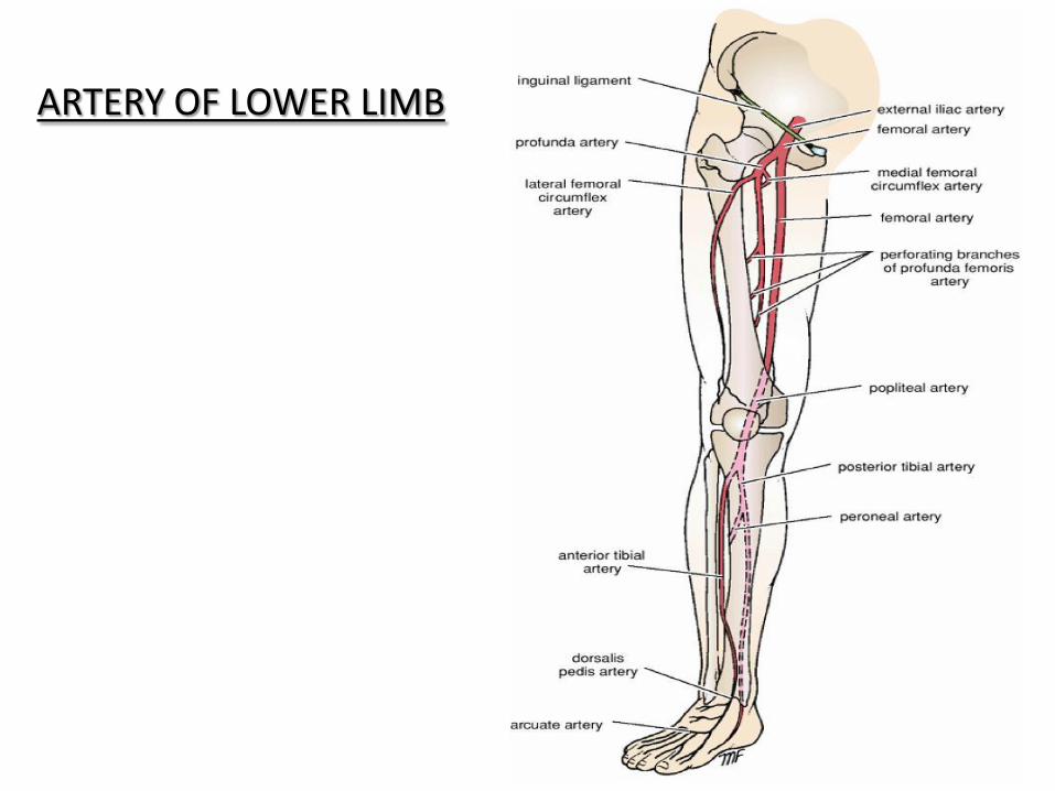
ARTERY OF LOWER LIMB

LUMBAR PLEXUS

LUMBAR PLEXUS L4- nervous furcalis

Muscles Of Gluteal Region


Q. In walking, the hip bone of the suspended leg is
raised by which of the following muscles acting
on the supported side of the body?
(a) Gluteus maximus
(b) Obturator internus
(c) Gluteus medius
(d) Obturator externus


Obturator externus


SCIATIC NERVE


HIP JOINT

Acetabular labrum
Capsule
Ligaments
• Iliofemoral (Bigelow) Ligament : inverted Y-
shaped
• Pubofemoral Ligament : triangular
• Ischiofemoral Ligament : spiral shaped
• Transverse Acetabular Ligament
• Ligament of the head of the femur
(ligamentum teres femoris ) : flat and triangular

KNEE JOINT


Ligaments
Extracapsular Ligaments • Ligamentum patellae
• Lateral collateral ligament
• Medial collateral ligament
• Oblique popliteal ligament : a tendinous expansion derived from semimembranosus muscle
Intracapsular Ligaments • Anterior Cruciate Ligament : limits anterior
displacement of tibia
• Posterior Cruciate Ligament : : limits posterior displacement of tibia
• Medial Menisci : semilunar in shape
• Lateral Menisci : 4/5th of a circle

• Menisco-femoral ligament : connect posterior horn of lateral
meniscus to medial condyle of femur
ligament of Humphery (anterior to PCL)
ligament of Wrisberg (posterior to PCL)

Bursae around knee
• In front : 4
• Laterally : 4
• Medially : 4
House maid knee : prepatellar bursitis
Clergyman’s knee : subcutaneous infra-patellar bursitis

Popliteal fossa

The popliteal fossa is a mostly fat-filled diamond-shaped space posterior
to the knee
The popliteal fossa is bound
Superolaterally by the biceps femoris (superolateral border).
Superomedially by the semimembranosus, lateral to which is the
semitendinosus (superomedial border).
Inferolaterally and inferomedially by the lateral and medial
heads of the gastrocnemius, respectively (inferolateral and inferomedial
borders).
Posteriorly by skin and popliteal fascia (roof).
Anteriorly by the popliteal surface of the femur, posterior capsule of
the knee joint, and the popliteus fascia covering the popliteus muscle
(floor).

The contents of the popliteal fossa include the
Tibial and common fibular nerves..
Termination of the small saphenous vein.
Popliteal artery and vein
Posterior cutaneous nerve of the thigh
Popliteal lymph nodes and lymphatic vessels.

Ankle Joint
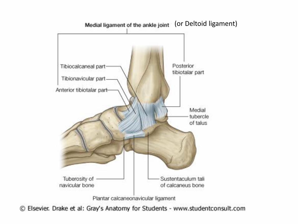
(or Deltoid ligament)