Embed Size (px)
Citation preview
ORIGINAL PAPER
Loss of vesicular dopamine release precedes tauopathyin degenerative dopaminergic neurons in a Drosophilamodel expressing human tau
Ting-Han Wu • Yu-Ning Lu • Chia-Lung Chuang • Chia-Lin Wu •
Ann-Shyn Chiang • David E. Krantz • Hui-Yun Chang
Received: 13 October 2012 / Revised: 7 February 2013 / Accepted: 2 March 2013 / Published online: 15 March 2013
� The Author(s) 2013. This article is published with open access at Springerlink.com
Abstract While a number of genome-wide association
studies have identified microtubule-associated protein tau
as a strong risk factor for Parkinson’s disease (PD), little is
known about the mechanism through which human tau can
predispose an individual to this disease. Here, we demon-
strate that expression of human wild-type tau is sufficient
to disrupt the survival of dopaminergic neurons in a Dro-
sophila model. Tau triggers a synaptic pathology visualized
by vesicular monoamine transporter-pHGFP that precedes
both the age-dependent formation of tau-containing neu-
rofibrillary tangle-like pathology and the progressive loss
of DA neurons, thereby recapitulating the pathological
hallmarks of PD. Flies overexpressing tau also exhibit
progressive impairments of both motor and learning
behaviors. Surprisingly, contrary to common belief that
hyperphosphorylated tau could aggravate toxicity, DA
neuron degeneration is alleviated by expressing the modi-
fied, hyperphosphorylated tauE14. Together, these results
show that impairment of VMAT-containing synaptic ves-
icle, released to synapses before overt tauopathy may be
the underlying mechanism of tau-associated PD and sug-
gest that correction or prevention of this deficit may be
appropriate targets for early therapeutic intervention.
Keywords Parkinson’s disease � Dopamine � Tau �MAPT � Neuron degeneration � Tauopathy
Introduction
Parkinson’s disease (PD) is the second most common
neurodegenerative disorder, affecting 1–2 % of people
over the age 60, and is characterized by clinical manifes-
tations of bradykinesia, rigidity, resting tremor, and
postural instability. These motor defects are thought to
result from degeneration of pigmented dopaminergic (DA)
neurons in the basal ganglia, particularly those projecting
from substantia nigra pars compacta to the striatum [13, 21,
45]. Deposition of Lewy bodies or formation of Lewy
neurites, composed of fibrillar aggregates of a-synuclein,
are the classical pathological hallmark of PD [48]. How-
ever, tau-containing neurofibrillary tangles (NFTs) are also
found in the brains of PD patients [60]. Although the
inherited forms of PD are rare, genetic variation in PD
Electronic supplementary material The online version of thisarticle (doi:10.1007/s00401-013-1105-x) contains supplementarymaterial, which is available to authorized users.
T.-H. Wu � Y.-N. Lu � C.-L. Chuang � H.-Y. Chang (&)
Department of Medical Science, Institute of Systems
Neuroscience, National Tsing Hua University,
101, Section 2 Kuang-Fu Road, Hsinchu 30013, Taiwan
e-mail: [email protected]
A.-S. Chiang
Department of Life Science, Institute of Biotechnology,
National Tsing Hua University, 101, Section 2 Kuang-Fu Road,
Hsinchu 30013, Taiwan
T.-H. Wu � Y.-N. Lu � C.-L. Chuang � A.-S. Chiang �H.-Y. Chang
Brain Research Center, National Tsing Hua University, 101,
Section 2 Kuang-Fu Road, Hsinchu 30013, Taiwan
C.-L. Wu
Department of Biochemistry, Graduate Institute of Biomedical
Sciences, College of Medicine, Chang Gung University,
Tao-Yuan, Kwei-Shan 333, Taiwan
D. E. Krantz
Department of Psychiatry and Biobehavioral Science, Gonda
Center for Neuroscience and Genetics Research, David Geffen
School of Medicine at University of California,
Los Angeles, CA 90095, USA
123
Acta Neuropathol (2013) 125:711–725
DOI 10.1007/s00401-013-1105-x

susceptibility genes may play a significant role, through
gene–environment interactions, in the development of
sporadic PD [45].
Microtubule-associated protein tau (MAPT), leucine-rich
repeat kinase 2 (LRRK2), and a-synuclein (SNCA) have been
identified as the top three PD susceptibility genes [46]. Both
SNCA and LRRK2 have been extensively studied in vitro and
in vivo; however, the molecular and pathological role of
MAPT in PD remains poorly understood [57]. The link
between MAPT and disease, originally identified for inherited
autosomal dominant frontotemporal dementia and Parkin-
sonism, is linked to chromosome 17 (FTDP-17). A similar
association was later found in several rare atypical Parkin-
sonism syndromes, including progressive supranuclear palsy
(PSP) and corticobasal degeneration (CB) [20, 22, 28]. More
recently, a number of studies have suggested that genetic
variants in MAPT can also predispose an individual to the
development of sporadic and familiar forms of PD [45].
Interestingly, most mutations of MAPT associated with
FTDP-17 are located in protein coding sequences for micro-
tubule-binding repeats and splicing sites [22], whereas the
mutations identified in allelic variants of MAPT associated
with PD are located in upstream regulatory elements [17].
The relationship between human tau and PD is illus-
trated by a number of genome-wide association studies
(GWAS) [16, 32, 38, 46]. The MAPT H1 haplotype is
associated with an increased risk of PD [17]. In addition,
individuals homozygous for MAPT H1/H1 have an
increased susceptibility to develop PD compared to those
bearing the heterozygous H1/H2 genotype [59]. Further-
more, some of the MAPT variants appear to increase
overall Tau expression [25]. Together, these studies sug-
gest that elevated expression of tau may increase the risk of
PD via a genetically gain-of-function mechanism, similar
to the duplication of the amyloid precursor proteins gene
(APP gene) [43] or duplication or triplication of SNCA
(encodes a-synuclein) [47]. However, unlike aberrant copy
numbers of APP and SNCA, which have been clearly
identified in hereditary AD and PD pedigrees, respec-
tively, evidence for a relationship between MAPT gene
dosage and either PD or tauopathies remains primarily
circumstantial.
In the present study, we explored whether expression of
wild-type MAPT affects DA neurons, the most vulnerable
cells in PD, by using a Drosophila animal model. We
found that expression of wild-type human tau (htauWT)
produces the progressive degeneration of DA neurons. In
addition, we observed age-dependent formation of tau-
positive, tangle-like pathology in the soma of the DA
neurons that was similar to NFTs seen in transgenic mice
and AD brains. Flies expressing tau also exhibited motor
and learning deficits that coincided with progressive neu-
rodegeneration. Surprisingly, we observed that expression
of htauWT caused molecular pathogenesis in synapses,
visualized by VMAT-pHluorin localization to nerve ter-
minals that preceded identified pathological events such as
tangle formation and overt cell loss. Our results suggest a
potential molecular pathological mechanism through which
tau may increase the risk of PD.
Materials and methods
Fly strains and genetics
Flies were cultured in standard corn meal–yeast–agar
media at 25 �C, and 75 % relative humidity with a light/
dark = 12/12 h cycle. Ddc-Gal4 and 996TPH-GAL4 were
kindly provided from Jay Hirsh (University of Virginia).
UAS-G-CaMP was obtained from Richard Axel (Depart-
ment of Biochemistry and Molecular Biophysics, Columbia
University). UAS-mCD8-GFP and GMR-GAL4 were
obtained from Larry Zipursky (University of California, Los
Angeles). Fly strains TH-GAL4, DVGLUT-GAL4, GAD-
GAL4, NPF-GAL4, RH1-GAL4, UAS-actinGFP and
tubGAL80ts were used in this study. UAS-htau.1 (II), UAS-
htau.2 (III) and UAS-VMAT- pHluorin (III) were character-
ized in this study. UAS-tauAP, UAS-tauE14 and UAS-tauWT
were described previously [49, 50].
Molecular cloning and transgenic flies
For generating human tau transgenes, the longest isoform
of human tau cDNA, encoding amino acids 1–441, (Ori-
gene Technologies, Rockville, MD), was cloned into
pUAST vector using EcoRI and NotI restriction sites. Site-
directed mutagenesis was used to generate htau G272V and
htau R406W with forward and reverse primer pairs of 50-GA
AGCACCAGCCGGGAGTC-30 and 50-CTTCCCGACTC
CTG-30 for G272V, and 50-GACACGTCTCCATGGCAT
CTC-30 and 50-GAGATGCCATGGAGACGTGTC-30 for
R406W, respectively. The mutations of htau G272V and htauR406W were confirmed by DNA sequencing and subcloned
into pUAST expression vector. Germ line transformation
and standard fly-balancer crosses were performed. We
obtained multiple UAS-htauWT, UAS-htauG272V and UAS-
htauR406W transgenic lines. Those with comparable
expression level were selected for this study. PHluorin was
inserted into the first luminal loop of VMAT and cloned
into pUAST vector.
Behavioral analysis
The startle-induced negative geotactic climbing assays
were performed as previously described [2, 44]. For loco-
motion assay, 4-week-old flies (4 weeks after eclosion)
712 Acta Neuropathol (2013) 125:711–725
123
were placed in chambers marked with grids and individual
were recorded (videotaped) for periods of 10 min. Loco-
motion (locomotion index) was manually scored by
counting the number of grids individuals crossed per
minute. Olfactory associative learning was measured by
training 3-week-old adult flies in a T-maze with the Pav-
lovian conditioning procedure as previously described [52].
Briefly, a group of 100 flies were first exposed to an odor (a
conditioned stimulus, CS?) paired with 12 1.5 s pluses of
75 V DC electric shock (an unconditioned stimulus, US).
For conditioning order stimulus, we used 3-octanol and
4-methyl-cyclohexanol. This was sequentially followed by
presentation of a second odor (CS-) without reinforce-
ment. Performance of conditioned avoidance responses
was immediately measured with a choice between CS?
and CS- odors in a T-maze for 2 min after training. A
performance index (PI) of aversive learning was calculated
as (the number of flies in CS- arm) - (the number of flies
in CS? arm)/(the total number of flies) 9 100.
Confocal and electron microscopy
Fly brains and eyes were fixed, stained and mounted in
Vectashield (Vector Laboratories, Burlingame, CA) as
previously described [9]. Congo-philic stain was performed
as previously describe [3]. Confocal microscopic observa-
tions of fly brains were performed using a Zeiss LSM 510
laser scanning confocal microscope (Carl-Zeiss, Germany).
For characterization of different clusters of DA neurons,
Z-stack confocal images that span 50 lm depth were col-
lected from posterior to anterior direction to ensure all
posterior clusters are covered. 3D images for each brain are
used for examining neuron structure. For presenting pur-
pose, Z-stack images are projected into a single 2D image.
For live brain imaging, fly brains were dissected in 19
PBS and G-CaMP was visualized immediately following
brain dissection by confocal microscopy [54]. For VMAT-
pHluorin experiments, released sites of VMAT-pHluorin of
DA neurons were visualized as previous described [40].
Primary antibodies included mouse tau monoclonal anti-
bodies AT8, AT100, and AT180 (Thermo Fisher Scientific,
Waltham, MA), rabbit tau polyclonal antibody (Dako,
Denmark), and mouse anti-TH (Immunostar, Hudson, WI).
Secondary antibodies FITC and Cy3 conjugated anti-
mouse or anti-rabbit (Jackson ImmunoResearch Laborato-
ries, West Grove, PA) were used. NIH imageJ64 was used
for quantification of G-CaMP and GFP using monochrome
images, by measuring pixel intensity, as relative fluores-
cence with fluorescence-background calibration. For
presenting G-CaMP activity, signal intensity was trans-
formed to the pseudocolored thermal scale. Scanning
electron microscopy was used to analyze fly eye mor-
phology as previously described [10]. For pair-helical
filament examination, transmission electron microscopy
(TEM) was used as previously described [23], with the
following modifications. Briefly, a resuspended sarkosyl
insolubility pellet was placed on a carbon-coated copper
grid (75 mesh) and stained with 1 % uranyl acetate.
Samples were examined using HT7700 TEM (Hitachi).
Western blots and sarkosyl insolubility assay
To detect the protein expression level, aged fly brains were
homogenized in sample buffer using a glass micro-tissue
grinder. To enrich tangles, sarkosyl-insoluble fractions of
tau were purified from 40 homogenized fly heads as pre-
viously described [14] and used for western blot analysis
and TEM observation. Standard western blotting proce-
dures were used as previously described [8]. Primary
antibodies included mouse cytochrome c (Gene Tex,
Irvine, CA), and rabbit cleaved caspase 3 (Cell signaling
technology, Danvers, MA). ECL reagents were used for
antibody detection (Millipore, Billerica, MA) and imaging
was performed using ImageQuant 350 (GE Healthcare).
Results
MAPT overexpression results in the progressive loss
of DA neurons
Numerous mutations in MAPT have been linked to FTDP-
17, including an increased ratio of the longest 4R Tau
isoform [8]. In addition, several GWAS suggest tau vari-
ants as a risk factor for PD, and an increased incidence of
PD is associated with elevated MAPT levels [16, 25, 32,
36, 38, 46, 55]. These observations suggest that misregu-
lation of tau expression might contribute to the death of
dopaminergic neurons. To investigate the effects of human
tau overexpression in DA cells, we chose to test the effects
of the largest htauWT isoform (2N4R), because an increase
of this largest isoform may predispose the susceptibility to
PD or some FTDP-17.
Since the most affected neurons in PD are the DA
neurons, we used the TH-GAL4 driver to overexpress UAS-
htauWT and co-express UAS-mCD8-GFP, to fluorescently
label the plasma membrane of the cell body and neurite
processes (TH::htauWT, mCD8-GFP). We then tested
whether htauWT expression resulted in degeneration of
these DA neurons. In young adults (1-week-old), the
number of GFP-marked DA neurons in htauWT transgenic
brains was comparable to control brains (TH::mCD8-GFP).
At 2 weeks after eclosion, limited degeneration was
observed in a small subset of PPL1 neurons expressing
htauWT compared to age-matched controls. Remarkably, at
4 weeks after eclosion, we observed a significant loss of
Acta Neuropathol (2013) 125:711–725 713
123

DA neurons in transgenic flies relative to controls (Fig. 1).
This loss occurred in two clusters of DA neurons: PPL1 and
PPM3 (Fig. 1). Other FTDP-17 associated two tau variants
of G272V and R406W, also showed similar effects (Fig.
S1). To validate these htauWT-expressing cells may have
undergone progressive degeneration, we characterized two
components of the cell death signaling: cytochrome c and
activated caspase 3-like caspase [53] for these htauWT-
expressing cells and their GFP expressing controls in
Drosophila. As expected, the expression of htauWT pro-
duced an abnormal increase in the expression of
cytochrome c, before a surge of caspase 3-like caspase
activation in 3-week-old flies expressing htauWT, relative to
controls, suggesting the activation of cell death signaling in
htauWT-expressing brains in Drosophila (Fig. S2). These
results indicate a progressive degeneration of DA neurons
in the brains of Drosophila expressing htauWT. To further
confirm the effect of htauWT, we employed a second GAL4
driver, DDC-GAL4, which targets htauWT in both DA and
serotonergic neurons. We confirmed that htauWT does
induce the age-dependent DA neuron degeneration (Fig.
S3). Together, these results show that expression of htauWT
can induce progressive loss of DA neurons, manifesting the
pathological DA degeneration in PD.
MAPT overexpression produces age-dependent motor
and learning deficits
Similar to its role in mammals, the Drosophila nervous
system utilizes many neurotransmitters to operate motor
and cognitive behaviors. In the dopaminergic system, fruit
fly use the same neurotransmitter as mammals to modulate
a number of intricate neuronal circuits, ranging from those
that control voluntary movement, mating, rewards, learn-
ing, and memory [41]. We tested whether expression of
htauWT would alter motor behaviors. Using a negative
geotaxis assay, we observed that TH::htauWT flies have a
dramatically age-dependent climbing deficit; they showed
significant loss of climbing ability compared to control flies
at 4–6 weeks post-eclosion (Fig. 2a). In addition, we
observed that flies expressing htauWT showed reduced
spontaneous locomotion; they appeared ‘‘sluggish’’ and
exhibited an increase in the number of ‘‘pauses’’ between
bouts of spontaneous locomotion and travelled shorter
distances during each bout (Fig. 2b).
Next, we performed short-term olfactory association
assays to test whether expression of htau in DA neurons
affects learning. To avoid the potential confound caused by
motor deficits, we tested 3-week-old cohorts, an age at
which the motor behavior of htauWT-expressing flies is
comparable to that of controls. As expected, we observed
modest but significant learning deficit in htauWT-expressing
flies, which scored 10 % lower than the age-matched
control groups (Fig. 2c). This result is consistent with
recent empirical findings indicating that dopaminergic
neurons projecting to the mushroom body are essential
mediators of olfactory associative learning and memory [1,
4, 7, 39, 42]. Together, these results show that flies
expressing htauWT in DA neurons manifest deficits in both
motor and learning behaviors, which corroborate the
observed age-dependent loss of DA neurons.
Aminergic and NPF neurons are relatively more
vulnerable to MAPT expression
To determine how expression of htauWT would affect neu-
rons using other types of neurotransmitters, we used
additional GAL4 drivers including DVGLUT-GAL4 for
glutamatergic neurons, GAD-GAL4 for GABAergic neu-
rons, TPH-GAL4 for a subset of serotonergic neurons and
NPF-GAL4 for a subset of peptidergic F neurons. We did not
observe loss of either GFP-marked glutamatergic or GAB-
Aergic cells (Fig. 3a–d, i); however, we observed that a
subset of putative serotonergic neurons that project to the
central complex and a subset of putative NPF neurons were
lost (Fig. 3e–i). These results are consistent with previous
studies that have shown human tau expression can cause
degeneration of several neuronal populations in Drosophila
[23, 33, 56]. Together, these results demonstrate that htauWT
can induce degeneration of DA neurons, serotonergic neu-
rons, and NPF neurons, whereas most glutamatergic and
GABAergic neurons are unaffected by htauWT expression
based on the fluorescent optical observation.
Formation of abnormal oligomeric tau and tangle-like
pathology in DA neurons
Hyperphosphorylated tau has been suggested to accumulate
as homogeneous or heterogeneous oligomers and subse-
quently form NFT [5]. To determine whether similar
pathogenesis occurs in DA neurons that express htauWT,
we used three common phosphoepitope antibodies (AT8,
AT100, and AT180) and a polyclonal anti-tau antibody that
have been widely utilized to characterize AD-like abnor-
mal phosphorylation of tau in the postmortem brains of AD
patients and animal models of tauopathies. Immunohisto-
chemical analysis of TH::htauWT brains at different ages
revealed that the morphology of DA neurons appeared
normal in the first week, with no detectable morphological
changes in the soma and neurites, suggesting that the
neuronal architecture was normal in young adults (Fig. S4).
At approximately 2 weeks post-eclosion, the staining
pattern of the soma in some DA neurons has changed
relative to the wild-type morphology, indicating that
the cytoskeletal networks had been perturbed or disinte-
grated by this time (Fig. S4). Strikingly, after 3 weeks, the
714 Acta Neuropathol (2013) 125:711–725
123

phosphoepitope antibodies and a polyclonal anti-tau anti-
body began to reveal abnormal tangle-like morphology,
resembling NFTs at the microscopic level in most DA
neurons including those in PPL1 and PPM3. This result
suggests that the appearance of abnormal NFTs in DA
neurons is an age-dependent process (Fig. S4). To explore
whether htau expression might also induce tangle pathol-
ogy in general, we investigated other types of neurons
expressing htauWT. Similar morphological change was also
detected in a small subset of 5HT neurons, but no obvious
alteration was detected in glutamatergic, GABAergic and
NPF neurons in 6-week-old flies (Fig. S4). DA neurons that
express htauWT exhibited largely pruned neurite branches
at the distal end in 4-week-old individuals (Fig. S4). The
few cells that survived until 6 weeks experienced soma
shrinkage, which was dramatically different from age-
matched, GFP-labeled DA neurons from control brains
(Fig. 4a, S4). Moreover, we confirmed that at 6 weeks,
these abnormal DA neurons expressing htauWT were
stained by Congo red, a synthetic dye which recognized the
Fig. 1 Expression of htauWT induces age-dependent DA neuron
demise. Representative confocal images show mCD8-GFP-marked
DA neurons in age-matched control fly brains (a–c TH::mCD8-GFP)
and htauWT-expressing brains (d–f TH::htauWT, mCD8-GFP) of one-
(a, d), two- (b, e), and four- (c, f) week-old individuals. Two clusters
of DA neurons, PPL1 (protocerebral posterior lateral 1, circles) and
PPM3 (protocerebral posterior medial 3, squares), are indicated.
Scale bar 100 lm. Cell counts of PPL1 (g) and PPM3 (h) DA neurons
from age-matched control and htauWT brains. Values shown represent
Mean ± SEM (unpaired t test, **P \ 0.001 at week 2 for g;
***P \ 0.0001 at week 4 for g and h; n = 12)
Acta Neuropathol (2013) 125:711–725 715
123

abnormal b-sheet conformation of htau aggregations
(Fig. 4b).
To determine whether these morphological changes
were associated with structural changes in htauWT, we
performed additional biochemical analyses on whole brain
extracts from aged flies expressing TH::htauWT. We
observed a gradual reduction of monomeric tau, and an
increase in high-molecular-weight species, of human tau
protein with increasing age (Fig. 4c). The appearance of
the putative tau oligomers ([400 kDa) seemed to coincide
with the onset of morphological change in DA neurons,
which is in agreement with the hypothesis that tau oligo-
mers may be toxic.
In order to more rigorously evaluate htau aggregates in
DA neurons, we performed ultrastructure examination on
sarkosyl-insoluble fraction extracted from htau transgenic
brains. TEM revealed abundant filamentous aggregates
formed in the extracts from fly brains of 4-week-old Dro-
sophila expressing htauWT (Fig. 4d), but not in identically
prepared sarkosyl-insoluble fraction from age-matched
nontransgenic control brains. These negatively stained fil-
aments were either helical (paired helical filaments, PHF)
or straight (paired straight filaments, SF), with diameters of
approximately 15–40 nm and the axial distance between
consecutive wider portions of approximately 80–100 nm
(Fig. 4d), resembling those PHFs found in AD [12].
Therefore, we suggest that the observed htau aggregates
with an NFT-like pathology may be composed by PHF or
SF.
To determine whether htauWT insolubility coincided
with the appearance of the tangle-like morphology in DA
neurons, we performed sarkosyl insolubility assays from
brains at different ages. Western blotting of anti-htau
revealed a modest reduction of insoluble tau monomer, and
an increase with age, of several insoluble high-molecular-
weight species between 75 and 400 kDa, including *90,
*160, and *240 kDa tau-positive bands (Fig. 4e). The
formation of these insoluble high-molecular-weight
aggregations apparently preceded the formation of tangles,
which is consistent with the hypothesis that insoluble tau-
containing aggregations may be responsible for the NFT-
like pathology.
Effects of tau phosphorylation on neuron degeneration
Previous studies have suggested that tau phosphorylation is
associated with AD and other tauopathies, although the role
of phosphorylated state in disease pathogenesis remains
controversial. To test whether the htauWT-induced DA
neuron degeneration in Drosophila is related to its phos-
phorylation state, we expressed htauWT and phosphorylation
site variants in DA neurons and compared their potential
toxicity in aged fly brains. We focused on SP/TP sites, for
which several proline-directed kinases such as glycogen
synthase kinase 3 beta (GSK-3b) have been shown to
modulate tau-mediated toxicity [23], and compared the
effects of htauWT, htauAP, and htauE14. The AP mutation
blocked proline-directed tau phosphorylation and the E14
mutation mimics phosphorylation of the endogenous 14
serine/threonine amino acids in htau. To confirm that htauWT
can undergo phosphorylation in Drosophila DA cells, we
performed western blots and observed a shift in the molec-
ular weight of htauWT, from 75 to 60 kDa, in response to
treatment with alkaline phosphatase (Fig. S5).
Fig. 2 Expression of htauWT in DA neurons causes motor and
learning deficits. Behavioral analyses of driver control (TH::mCD8-
GFP, blue), transgene control (UAS-htauWT, green), and htauWT
expression (TH::htauWT, mCD8-GFP, red) flies. a Negative geotaxis
assay was utilized for testing climbing activities of newly enclosed to
6 weeks age-matched adults. Accelerated decline of climbing activity
was observed in flies expressing htauWT in DA neurons (one-way
ANOVA with Bonferroni’s multiple comparison test, ***P \ 0.0001
compares each genotypes, n = 4). b Quantitative locomotion
behaviors of 4-week-old flies show reduced activity in htauWT
expression group as compared to that in control groups (one-way
ANOVA with Bonferroni’s multiple comparison test, **P \ 0.001,
n = 16). c Olfactory associative learning assay of 3-week-old flies
revealed that flies in which DA neurons expressed htauWT exhibited
defects of olfactory aversive associative learning as compared to the
control groups (one-way ANOVA with Bonferroni’s multiple com-
parison test, **P \ 0.001, n = 4)
716 Acta Neuropathol (2013) 125:711–725
123

An earlier study showed that the htauAP mutation is less
toxic than htauWT when expressed in Drosophila photore-
ceptors, but that the htauE14 mutation enhances neurotoxicity
in the retina [49, 50]. Our results, using GMR-GAL4 driver to
express these htau constructs in the retina, confirmed these
observations (Fig. 5a, b). Surprisingly, the htau constructs
appeared to have inverse effects when expressed in DA cells
using TH-GAL4. TH::htauAP brains showed an increase in
DA cell death, compared to flies expressing htauWT, and
htauE14 resulted in less neurodegeneration than tauWT
(Fig. 5c, d). Western blots validated that htauWT, htauAP, and
htauE14 were expressed at similar levels, indicating that the
phenotype is specific for the mutation in tau rather than
expression of the transgenes (Fig. S5).
Fig. 3 Expression of htauWT fails to manifest neuronal loss in most
glutamatergic and GABAergic neurons. a, b Representative confocal
images show glutamatergic neurons of 4-week-old fly brains marked
with mCD8-GFP. Several thousands of glutamatergic neurons with
comparable GFP signals in both control (a, DVGLUT::mCD8-GFP)
and htauWT brains (b, DVGLUT::htauWT, mCD8-GFP). c, d Repre-
sentative confocal images show mCD8-GFP-marked GABAergic
neurons in 6-week-old fly brain. Comparable GFP signals can be
detected in both control (c GAD::mCD8-GFP) and htauWT brains
(d GAD::htauWT, mCD8-GFP). e Representative confocal image
shows a group of putative serotonergic neurons (TPH996::mCD8-
GFP) projecting to central complex (arrows) at 4 weeks of age.
f Representative confocal image shows the absence of GFP signals in
neurons projecting to central complex in age-matched brain express-
ing htauWT (TPH996::htauWT, mCD8-GFP). g Representative
confocal image shows two pairs of putative NPF neurons
(NPF::mCD8-GFP) projecting to parietal central brains in control
(f). In contrast, the GFP signals in a pair of NPF neurons that are
located in the anterior region are absent in htauWT-expressing brains
(NPF::htauWT, mCD8-GFP) at 4 weeks of age. Scale bar 100 lm.
i Quantitative analyses show no significant differences between
control (black bars) and htauWT (white bars) brains in DVGLUT, and
GAD groups (unpaired t test, P = 0.54 for a and b; P = 0.60 for
c and d, n = 4). In TPH and NPF groups, both the cell measurements
(circles) between controls and htauWT are different (unpaired t test,
***P \ 0.0001 for both e and f, and for g and h, n = 4)
Acta Neuropathol (2013) 125:711–725 717
123

It is possible that the severe neuronal phenotype induced
by htauAP is a developmental, but not a degenerative event.
To circumvent potential developmental effects of htauAP
on DA neurons, we used a conditional expression system
by incorporating temperature-sensitive GAL80ts [27] in our
model to predominantly, if not exclusively, expressing
Fig. 4 Expression of htauWT produces AD-like abnormal tau phos-
phorylation and NFT-like pathology. a Single cell of PPL1 and PPM3
DA neurons from fly brains at 6 weeks of age. Compared to the age-
matched control DA neurons from PPL1 cluster labeled with GFP
(left column), immunostaining of three antibodies that recognize AD-
like hyperphosphorylated tau (AT8 top row, AT100 top second row,
AT180 bottom second row), and a polyclonal tau antibody (bottomrow) reveal tangle-like pathology in the degenerating DA neurons
from PPL1 (second column) and PPM3 (third column) clusters. b The
degenerating DA neurons from both PPL1 and PPM3 clusters are
stained with Congo red. The time course of pathological tangle-like
structure formation in htauWT brains during the aging process is
presented in Fig. S3. c Representative immunoblot of polyclonal anti-
htau shows that a substantial amount of htauWT proteins are converted
from monomer tau (75 kDa) to the oligomeric Tau species
([400 kDa). Anti-b-tubulin serves as a loading control. Protein
expression levels from four independent immunoblots with mono-
meric and oligomeric htau protein levels calibrated against the
loading control are shown as relative expression. Values shown
represent mean ± SEM. d Electron micrographs of abnormal
filaments extracted from tangle-rich preparation (sarkosyl-insoluble
pellets) of fly brains expressing htauWT. Images show several PHF-
like filaments are bundled (left panel), a single PHF (middle panels),
and a straight filament (right panel). Scale bar 100 nm. d Represen-
tative immunoblot from sarkosyl-insoluble pellets reveals monomer
tau ***(75 kD) and the htau-containing species with molecular
weights range between 75 and 400 kD (*90, *160 and *240 kD).
Anti-b-tubulin also serves as a loading control. e Monomeric and
polymeric htau protein levels are quantified by analyzing three
independent immunoblots and shown as relative expression. Values
represent mean ± SEM
718 Acta Neuropathol (2013) 125:711–725
123

htauAP in adult brains. By shifting temperature from 25 to
29 �C for newly enclosed adult flies. We induced transgene
expressing, and observed that adult onset of htauAP
expression (GAL80ts/UAS-htauAP; TH-GAL4, UAS-
CD8GFP/?) induced severe neurodegeneration, similar to
that of the continuously expression of htauAP (TH::htauAP,
Fig. 5 Different phosphorylation site mutations of tau exert contrary
effects on DA and histaminergic neurons. Representative SEM
(a) and confocal (b) images of fly compound eyes. Control (GMR-
GAL4) fly eyes show crystalline array of *800 hexagonal-shaped
ommatidia form the smooth external surface. Phalloidin staining
reveals the underlying organization of each ommatidium with actin-
rich rhabdomeres from seven photoreceptors being arranged in a
trapezoid format. Expression of three human tau transgenic alleles,
htauAP14 (GMR::htau-AP), htauWT (GMR::htau-WT), and htauE14
(GMR::htau-E14), produce different degrees of eye phenotypes and
the severity ranking of eye defect is htauE14 [ htauWT [ htauAP14
that is based on external eye size and roughness and the internal
organization of photoreceptor cells (a, b). Scale bars 10 lm.
c Representative confocal images of control DA neurons marked
with mCD8-GFP (TH::GFP), or co-expressing with either of the three
human tau alleles (htauAP14, TH::htau-AP; htauWT, TH::htau-WT; and
htauE14, TH::htau-E14) from 1-, 2-, and 4-week-old brains. Circlesindicate the PPL1 cluster. d Quantitative analysis presents DA neuron
numbers in PPL1 cluster at different ages from control and three tau
alleles. TH::GFP (purple), TH::htau-AP (green), TH::htau-WT (yel-low) and TH::htau-E14 (red). Values shown represent mean ± SEM
(unpaired t test compares individual tau allele to control in the age-
indicated groups; **P \ 0.001; ***P \ 0.0001, n = 12)
Acta Neuropathol (2013) 125:711–725 719
123

GFP), while the control flies (TH::GFP) exhibited normal
DA neurons (Fig. S6). Therefore, these data suggest the
effect of htauAP on DA neuron was an age-dependent
degeneration, but not a developmental defect.
Loss of vesicular dopamine release preceded tauopathy
and neurodegeneration
Previously, we have shown that vesicular monoamine
transporter (VMAT) reduced environmental toxins and
mutant human parkin-induced toxicity [26, 44]. We rea-
soned that the loss of DA neurons resulting from the
expression of human tau may be associated with a change
in the DA storage and release machinery. To investigate
this, we used the ecliptic VMAT-pHluorin, with a pH-
sensitive GFP conjugated in the first luminal loop of the 12
trans-membrane domains of VMAT to visualize the release
of VMAT-pHluorin containing synaptic vesicle released to
nerve terminals [34].
In wild-type fly brain, VMAT-pHluorin visualizes the
synaptic vesicle-releasing regions of DA neurons in the fly
brain. At one day after eclosion, VMAT was observed in
many brain regions, including mushroom bodies, the fan-
shaped body, and the protocerebral bridge, in wild-type
flies (Fig. 6a) [31]. The single neuron of PPL1 and PPM3
data has been deposited in the database of Drosophila
neurons (http://www.flycircuit.tw). In contrast, the locali-
zation of VMAT to the mushroom body and protocerebral
bridge was reduced in age-matched flies expressing htauWT
(Fig. 6b). This decrease was most pronounced in 3-week-
old fly brains compared to the wild-type fly brain (Fig. 6e,
f). Remarkably, the VMAT-pHluorin signals in the mush-
room body and protocerebral bridge were selectively
absent from the htauAP brains at one-day post-eclosion, but
remained prominent in controls (Fig. 6a, c). Interestingly,
some neurons that project to the fan-shaped body appeared
to be unaffected, as the VMAT-pHluorin signal appeared
normal in both control and three htau (htauWT, htauAP and
htauE14) expressing brains. Given that some TH-GAL4-
marked neurons are not dopaminergic, we suspect that the
unaffected VMAT-pHluorin labeling of the fan-shaped
body might be a result of expression in those cells. To
further investigate whether declines in VMAT-pHluorin
localization to nerve terminals were indeed related to
human tau-induced toxicity, DA neurons expressing of
htauWT, htauAP, or htauE14, were compared in different
ages (Fig. 6a–h). Surprisingly, the age-matched flies
expressing htauAP had earlier and more profound loss of
VMAT-pHluorin signals (Fig. 6c) than flies expressing
htauWT or htauE14 (Fig. 6b, d). Variation in the GFP signals
in brains co-expressing VMAT-pHluorin and htauWT or
htauAP were not a result of variation in levels of VMAT-
pHluorin expression, since levels of VMAT-pHluorin were
similar in both experimental and control groups (Fig. 6i).
It is possible that htau could cause a general dysfunction
of DA neurons resulting in multiple deficits in the DA
release machinery. To determine whether expression of
htauWT also disrupts calcium influx, we used a calcium-
sensitive fluorescent protein, G-CaMP, to monitor changes
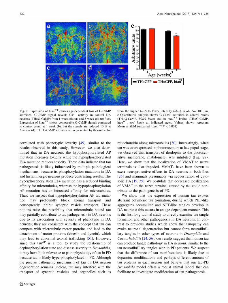
in calcium. By visualizing 1-week-old live brains, we
found that expression of htauWT did not alter the G-CaMP
activity, compared to controls (Fig. 7a, b, e), suggesting
that htauWT expression did not disrupt the general function
of DA neurons during neuronal activation. However, at
3 weeks, we found that DA neurons expressing htauWT
showed a minor but significant reduction of the G-CaMP
signal compared to controls (Fig. 7c–e), consistent with
overt DA neuronal degeneration around this age (Fig 7c–
e). In conclusion, these results suggest htauWT inhibits the
VMAT-containing synaptic vesicle release to neuronal
processes, before overt neuronal degeneration.
Discussion
In the present study, we used Drosophila DA neurons as a
model to examine the pathogenic mechanism of PD-linked
tauopathies. Expression of htauWT in these neurons resulted
in age-dependent, progressive neurodegeneration and the
deposition of abnormal polymeric tau protein aggregates.
Systematic characterizations of functional, pathological,
and behavioral readouts from these htauWT flies at different
ages demonstrate a plausible pathogenic link between tau
and vesicular DA release, as demonstrated by a fluorescent
VMAT-pHluorin marker in the synaptic vesicle-releasing
sites of DA neurons. This defect represents the earliest
pathological manifestation that precedes the robust patho-
physiological phenotypes and suggests that a functional
inhibition of DA neurotransmission is a prelude of the
hallmark lesions in our PD model.
The molecular components of DA signaling and
homeostasis in Drosophila are similar to those in humans;
both human and fly DA neurons express two types of
transporters: (1) a vesicular monoamine transporter for
transport of DA from the cytosol to the lumen of secretory
vesicles [29]; (2) a plasma membrane DA transporter
(DAT) for reuptake of exocytosed DA [18]. Therefore,
despite the anatomical differences, Drosophila models of
PD, as well as other human neurological disorders, can
help contribute to our understanding of the pathogenic
mechanism underlying these diseases [6]. This tau-PD
Drosophila model is the first animal model to demonstrate
that tau expression can cause degeneration of DA neurons,
a hallmark of Parkinson’s disease.
720 Acta Neuropathol (2013) 125:711–725
123

Most motor symptoms and mild cognitive impairment of
PD result primarily from progressive degeneration of DA
neurons in the substantia nigra, but the mechanism through
which pathogenic factors target DA neurons is not com-
pletely understood. While several toxic compounds are
known to produce PD symptoms in animal models, only
MPTP (1-methyl-4-phenyl-1,2,3,6-tetrahydropyridine) has
been firmly established as a cause of selective DA neuron
toxicity. The issue of neuronal vulnerability, in terms of
transporter or transmission mechanisms among individuals
with monogenic PD risk factors, has rarely been addressed
directly. One exception is a study that showed that the
expression of human a-synuclein in mice inhibited DA
transmission by reducing the recycling of synaptic vesicles
[37]. In addition, an association between DA metabolism
and the death of DA neurons in PD has been postulated,
and this association has been demonstrated in a a-synuclein
PD model. Oxidative metabolites of DA may conjugate
with a-synuclein to form an adduct of DA–a-synuclein,
which may stabilize the toxic form of a-synuclein through
covalent bound to DA quinone [11], while also promoting
selective neurotoxicity [58]. Tau protein is ubiquitous in
the brain, and has been shown to affect different types of
neurons in various cell and animal models. A previous
study showed that DA quinones, toxic metabolites that lead
to PD, could promote the assembly of tau into fibrillar
polymeric tau in vitro [15] which suggests a potential
interplay between DA and tau in vivo and may provide a
basis for the vulnerability of DA neurons.
One of the common themes of tau pathology is abnormal
hyperphosphorylation of tau, originally identified in AD
patients. However, the role of tau hyperphosphorylation in
the pathophysiology of disease remains debated. In Dro-
sophila photoreceptors, tau hyperphosphorylation is
Fig. 6 DA neurons expressing htauWT cause early impairment of
vesicular dopamine release to nerve terminals as visualized by
VMAT-pHluorin. a–f Confocal images of DA neurons from brains of
1-day-old (a–d) and 3-week-old (e–h) flies show VMAT-pHluorin
reporter (green). a DA neurons expressing VMAT-pHluorin
(TH::VMAT-pHluorin) reveal localization of VMAT-pHluorin to
presynaptic terminals in brain regions, and b age-matched DA
neurons co-expressed VMAT-pHluorin and htauWT (TH::VMAT-
pHluorin, UAS-htauWT) show decreased GFP signals in mushroom
bodies (arrowheads). e A representative confocal image shows the
VMAT-pHluorin signals to mushroom bodies and other brain
structure remains prominent (green) in 3-week-old control brain,
while the GFP signals localized to mushroom bodies is diminished in
DA neurons expressing htauWT (f, arrowheads). c, g Expression of
htauAP evokes severe loss of pHluorin signaling compared to age-
matched tauWT (b, f) and TauE14 (d, h). Rhodamine-phalloidin (red)
marks the brain structure. Scale bar 100 lm. i Representative western
blot shows the expression levels of VMAT-pHlurion of 1-week-old
fly brains in control and experiment groups. 1 TH-GAL4, 2TH::VMAT-pHluorin, 3 TH::VMAT-pHluorin, htauWT, 4TH::VMAT-pHluorin, htauAP, 5 TH::VMAT-pHluorin, htauE14.
Anti-b-tubulin served as a loading control
Acta Neuropathol (2013) 125:711–725 721
123
correlated with phenotypic severity [49], similar to the
results observed in this study. However, we also deter-
mined that in DA neurons, the hypophosphorylated AP
mutation increases toxicity while the hyperphosphorylated
E14 mutation reduces toxicity. These data indicate that tau
pathogenesis is likely influenced by multiple pathological
mechanisms, because its phosphorylation mutations in DA
and histaminergic neurons produce contrasting results. The
hyperphosphorylation E14 mutation has a reduced binding
affinity for microtubules, whereas the hypophosphorylation
AP mutation has an increased affinity for microtubules.
Thus, we suspect that hypophosphorylation AP tau muta-
tion may profoundly block axonal transport and
consequently inhibit synaptic vesicle transport. These
notions raise the possibility that microtubule bound tau
may partially contribute to tau pathogenesis in DA neurons
due to its association with severity of phenotype in DA
neurons; they are consistent with the concept that tau can
compete with microtubule motor proteins and lead to the
detachment of motor proteins (kinesin and dynein), which
may lead to abnormal axonal trafficking [51]. However,
since this tauAP is a tool to study the relationship of
dephosphorylation state and disease severity in Drosophila,
it may have little relevance to pathophysiology of tau in PD
because tau is likely hyperphosphorylated in PD. Although
the precise pathogenic mechanism of tau on DA neuron
degeneration remains unclear, tau may interfere with the
transport of synaptic vesicles and organelles such as
mitochondria along microtubules [30]. Interestingly, when
tau was overexpressed in photoreceptors at late pupal stage,
we observed that transport of rhodopsin to the photosen-
sitive membrane, rhabdomere, was inhibited (Fig. S7).
Here, we show that the localization of VMAT to nerve
terminals is also impeded. VMATs have been shown to
exert neuroprotective effects in DA neurons in both flies
[26] and mammals presumably via sequestration of cyto-
solic DA [19, 35]. We postulate that decreased localization
of VMAT to the nerve terminal caused by tau could con-
tribute to the pathogenesis of PD.
We show that the expression of human tau evokes
aberrant polymeric tau formation, during which PHF-like
aggregates accumulate and NFT-like tangles develop in
DA neurons; this occurs in an age-dependent manner. This
is the first longitudinal study to directly examine tau tangle
formation and other pathogenesis in DA neurons. In con-
trast to previous studies which show that tauopathy can
evoke neuronal degeneration but cannot form neurofibril-
lary tangles in other types of neurons in Drosophila and
Caenorhabditis [24, 56]; our results suggest that human tau
can produce tangle pathology in DA neurons, similar to the
tau neurofibrillary tangles seen in PD patients. We suspect
that the difference of tau manifestations is likely due to
dopamine modifications and perhaps different amount of
tau proteins in each neuron and believe that our tau-PD
Drosophila model offers a robust animal model that can
facilitate to investigate modification of tau pathogenesis.
Fig. 7 Expression of htauWT causes age-dependent loss of G-CaMP
activities. G-CaMP signal reveals Ca2? activity in control DA
neurons (TH::G-CaMP) from 1-week-old (a) and 3-week-old (c) flies.
Expression of htauWT shows comparable G-CaMP signals compared
to control group at 1 week (b), but the signals are reduced 10 % at
3 weeks (d). The G-CaMP activities are represented by thermal color
from the higher (red) to lower intensity (blue). Scale bar 100 lm.
e Quantitative analysis shows G-CaMP activities in control brains
(TH::G-CaMP, black bars) and in htauWT brains (TH::G-CaMP,
htauWT, red bars) at indicated ages. Values shown represent
Mean ± SEM (unpaired t test; **P \ 0.001)
722 Acta Neuropathol (2013) 125:711–725
123

Our neuropathological observations reveal at least two
distinct molecular mechanisms of tau pathogenesis in DA
neurons for PD. We show that failure of VMAT-containing
synaptic vesicle release to nerve terminals is an early
pathogenic manifestation, and the formation of neurofi-
brillary tangle-like pathology in the soma of DA neurons is
a late-stage of tauopathy, providing pathological evidence
for why MAPT may be a strong risk factor for idiopathic
PD. The correction or prevention of the deficit in vesicular
DA storage may be appropriate target for early therapeutic
intervention.
Acknowledgments We thank J. Hirsh, R. Axel, M. Feany, J.-L.
Juang, and Bloomington Drosophila Stock Center for fly strains. We
are grateful to Tzu-Kang Sang and Don Ready for their critical
comments and suggestions, and Horng-Dar Wang, Jui-Chou Hsu,
Kathy Sang for their suggestions on this manuscript. We thank the
support from Imaging Core of the Brain Research Center in National
Tsing Hua University. This work is supported by National Science
Council Grant 98-2311-B-007-014-MY3 and National Tsing Hua
University Grant N2612E1 (H.-Y. C).
Open Access This article is distributed under the terms of the
Creative Commons Attribution License which permits any use, dis-
tribution, and reproduction in any medium, provided the original
author(s) and the source are credited.
References
1. Aso Y, Herb A, Ogueta M, Siwanowicz I, Templier T, Friedrich
AB, Ito K, Scholz H, Tanimoto H (2012) Three dopamine
pathways induce aversive odor memories with different stability.
PLoS Genet 8:e1002768
2. Benzer S (1967) Behavioral mutants of Drosophila isolated by
countercurrent distribution. Proc Nat Acad Sci USA 58:1112–
1119
3. Berg I, Nilsson KP, Thor S, Hammarstrom P (2010) Efficient
imaging of amyloid deposits in Drosophila models of human
amyloidoses. Nat Protoc 5:935–944
4. Berry JA, Cervantes-Sandoval I, Nicholas EP, Davis RL (2012)
Dopamine is required for learning and forgetting in Drosophila.
Neuron 74:530–542
5. Binder LI, Guillozet-Bongaarts AL, Garcia-Sierra F, Berry RW
(2005) Tau, tangles, and Alzheimer’s disease. Biochim Biophys
Acta 1739:216–223
6. Bonini NM, Fortini ME (2003) Human neurodegenerative disease
modeling using Drosophila. Annu Rev Neurosci 26:627–656
7. Burke CJ, Huetteroth W, Owald D, Perisse E, Krashes MJ, Das
G, Gohl D, Sillies M, Certel S, Waddell S (2012) Layered reward
signalling through octopamine and dopamine in Drosophila.
Nature 492:433–437
8. Chang HY, Grygoruk A, Brooks ES, Ackerson LC, Maidment
NT, Bainton RL, Krantz DE (2006) Overexpression of the Dro-sophila vesicular monoamine transporter increases motor activity
and courtship but decreases the behavioral response to cocaine.
Mol Psychiatr 11:99–113
9. Chang HY, Ready DF (2000) Rescue of photoreceptor degener-
ation in rhodopsin-null Drosophila mutants by activated Rac1.
Science 290:1978–1980
10. Chang YC, Hung WT, Chang YC, Chang HC, Wu CL, Chiang
AS, Jackson GR, Sang TK (2011) Pathogenic VCP/TER94 alleles
are dominant actives and contribute to neurodegeneration by
altering cellular ATP level in a Drosophila IBMPFD model.
PLoS Genet 7:e1001288
11. Conway KA, Rochet JC, Bieganski RM, Lansbury PT Jr (2001)
Kinetic stabilization of the alpha-synuclein protofibril by a
dopamine-alpha-synuclein adduct. Science 294:1346–1349
12. Crowther RA (1991) Straight and paired helical filaments in
Alzheimer disease have a common structural unit. Proc Nat Acad
Sci USA 88:2288–2292
13. Dawson TM, Ko HS, Dawson VL (2010) Genetic animal models
of Parkinson’s disease. Neuron 66:646–661
14. de Calignon A, Polydoro M, Suarez-Calvet M, William C, Ad-
amowicz DH, Kopeikina KJ, Pitstick R, Sahara N, Ashe KH,
Carlson GA, Spires-Jones TL, Hyman BT (2012) Propagation of
tau pathology in a model of early Alzheimer’s disease. Neuron
73:685–697
15. Dixit R, Ross JL, Goldman YE, Holzbaur EL (2008) Differential
regulation of dynein and kinesin motor proteins by tau. Science
319:1086–1089
16. Edwards TL, Scott WK, Almonte C, Burt A, Powell EH,
Beecham GW, Wang L, Zuchner S, Konidari I, Wang G, Singer
C, Nahab F, Scott B, Stajich JM, Pericak-Vance M, Haines J,
Vance JM, Martin E (2010) Genome-wide association study
confirms SNPs in SNCA and the MAPT region as common risk
factors for Parkinson disease. Ann Hum Genet 74:97–109
17. Farrer M, Skipper L, Berg M, Bisceglio G, Hanson M, Hardy J,
Adam A, Gwinn-Hardy K, Aasly J (2002) The tau H1 haplotype
is associated with Parkinson’s disease in the Norwegian popula-
tion. Neurosci Lett 322:83–86
18. Giros B, Jaber M, Jones SR, Wightman RM, Caron MG
(1996) Hyperlocomotion and indifference to cocaine and
amphetamine in mice lacking the dopamine transporter. Nature
379:606–612
19. Guillot TS, Miller GW (2009) Protective actions of the vesicular
monoamine transporter 2 (VMAT2) in monoaminergic neurons.
Mol Neurobiol 39:149–170
20. Hoglinger GU, Melhem NM, Dickson DW, Sleiman PM, Wang
LS, Klei L, Rademakers R, de Silva R, Litvan I, Riley DE, van
Swieten JC, Wszolek ZK, Uitti RJ, Vandrovcova J, Hurtig HI,
Gross RG, Maetzler W, Goldwurm S, Tolosa E, Borroni B, Pastor
P, Cantwell LB, Han MR, Dillman A, van der Brug MP, Gibbs
JR, Cookson MR, Hernandez DG, Singleton AB, Farrer MJ, Yu
CE, Golbe LI, Revesz T, Hardy J, Lees AJ, Devlin B, Hakonarson
H, Muller U, Schellenberg GD (2011) Identification of common
variants influencing risk of the tauopathy progressive supranu-
clear palsy. Nat Genet 43:699–705
21. Hughes AJ, Daniel SE, Kilford L, Lees AJ (1992) Accuracy of
clinical diagnosis of idiopathic Parkinson’s disease: a clinico-
pathological study of 100 cases. J Neurol Neurosurg Psychiat
55:181–184
22. Hutton M, Lendon CL, Rizzu P, Baker M, Froelich S, Houlden H,
Pickering-Brown S, Chakraverty S, Isaacs A, Grover A, Hackett
J, Adamson J, Lincoln S, Dickson D, Davies P, Petersen RC,
Stevens M, de Graaff E, Wauters E, van Baren J, Hillebrand M,
Joosse M, Kwon JM, Nowotny P, Che LK, Norton J, Morris JC,
Reed LA, Trojanowski J, Basun H, Lannfelt L, Neystat M, Fahn
S, Dark F, Tannenberg T, Dodd PR, Hayward N, Kwok JB,
Schofield PR, Andreadis A, Snowden J, Craufurd D, Neary D,
Owen F, Oostra BA, Hardy J, Goate A, van Swieten J, Mann D,
Lynch T, Heutink P (1998) Association of missense and 50-splice-
site mutations in tau with the inherited dementia FTDP-17.
Nature 393:702–705
23. Jackson GR, Wiedau-Pazos M, Sang TK, Wagle N, Brown CA,
Massachi S, Geschwind DH (2002) Human wild-type tau inter-
acts with wingless pathway components and produces
neurofibrillary pathology in Drosophila. Neuron 34:509–519
Acta Neuropathol (2013) 125:711–725 723
123
24. Kraemer BC, Zhang B, Leverenz JB, Thomas JH, Trojanowski
JQ, Schellenberg GD (2003) Neurodegeneration and defective
neurotransmission in a Caenorhabditis elegans model of tauop-
athy. Proc Nat Acad Sci USA 100:9980–9985
25. Kwok JB, Teber ET, Loy C, Hallupp M, Nicholson G, Mellick
GD, Buchanan DD, Silburn PA, Schofield PR (2004) Tau hap-
lotypes regulate transcription and are associated with Parkinson’s
disease. Ann Neurol 55:329–334
26. Lawal HO, Chang HY, Terrell AN, Brooks ES, Pulido D, Simon
AF, Krantz DE (2010) The Drosophila vesicular monoamine
transporter reduces pesticide-induced loss of dopaminergic neu-
rons. Neurobiol Dis 40:102–112
27. Lee T, Luo L (2001) Mosaic analysis with a repressible cell
marker (MARCM) for Drosophila neural development. Trends
Neurosci 24:251–254
28. Lee VM, Goedert M, Trojanowski JQ (2001) Neurodegenerative
tauopathies. Annu Rev Neurosci 24:1121–1159
29. Liu Y, Edwards RH (1997) The role of vesicular transport pro-
teins in synaptic transmission and neural degeneration. Annu Rev
Neurosci 20:125–156
30. Mandelkow EM, Stamer K, Vogel R, Thies E, Mandelkow E
(2003) Clogging of axons by tau, inhibition of axonal traffic and
starvation of synapses. Neurobiol Aging 24:1079–1085
31. Mao Z, Davis RL (2009) Eight different types of dopaminergic
neurons innervate the Drosophila mushroom body neuropil:
anatomical and physiological heterogeneity. Front Neural Circ
3:5
32. Maraganore DM, de Andrade M, Lesnick TG, Strain KJ, Farrer
MJ, Rocca WA, Pant PV, Frazer KA, Cox DR, Ballinger DG
(2005) High-resolution whole-genome association study of Par-
kinson disease. Am J Hum Genet 77:685–693
33. Mershin A, Pavlopoulos E, Fitch O, Braden BC, Nanopoulos DV,
Skoulakis EM (2004) Learning and memory deficits upon TAU
accumulation in Drosophila mushroom body neurons. Learn
Mem 11:277–287
34. Miesenbock G, De Angelis DA, Rothman JE (1998) Visualizing
secretion and synaptic transmission with pH-sensitive green
fluorescent proteins. Nature 394:192–195
35. Mosharov EV, Larsen KE, Kanter E, Phillips KA, Wilson K,
Schmitz Y, Krantz DE, Kobayashi K, Edwards RH, Sulzer D
(2009) Interplay between cytosolic dopamine, calcium, and
alpha-synuclein causes selective death of substantia nigra neu-
rons. Neuron 62:218–229
36. Myers AJ, Pittman AM, Zhao AS, Rohrer K, Kaleem M, Mar-
lowe L, Lees A, Leung D, McKeith IG, Perry RH, Morris CM,
Trojanowski JQ, Clark C, Karlawish J, Arnold S, Forman MS,
Van Deerlin V, de Silva R, Hardy J (2007) The MAPT H1c risk
haplotype is associated with increased expression of tau and
especially of 4 repeat containing transcripts. Neurobiol Dis
25:561–570
37. Nemani VM, Lu W, Berge V, Nakamura K, Onoa B, Lee MK,
Chaudhry FA, Nicoll RA, Edwards RH (2010) Increased
expression of alpha-synuclein reduces neurotransmitter release by
inhibiting synaptic vesicle reclustering after endocytosis. Neuron
65:66–79
38. Pankratz N, Wilk JB, Latourelle JC, DeStefano AL, Halter C,
Pugh EW, Doheny KF, Gusella JF, Nichols WC, Foroud T,
Myers RH (2009) Genomewide association study for suscepti-
bility genes contributing to familial Parkinson disease. Hum
Genet 124:593–605
39. Qin H, Cressy M, Li W, Coravos JS, Izzi SA, Dubnau J (2012)
Gamma neurons mediate dopaminergic input during aversive
olfactory memory formation in Drosophila. Curr Biol 22:608–614
40. Reiff DF, Ihring A, Guerrero G, Isacoff EY, Joesch M, Nakai J,
Borst A (2005) In vivo performance of genetically encoded
indicators of neural activity in flies. J Neurosci 25:4766–4778
41. Riemensperger T, Isabel G, Coulom H, Neuser K, Seugnet L,
Kume K, Iche-Torres M, Cassar M, Strauss R, Preat T, Hirsh J,
Birman S (2011) Behavioral consequences of dopamine defi-
ciency in the Drosophila central nervous system. Proc Nat Acad
Sci USA 108:834–839
42. Riemensperger T, Voller T, Stock P, Buchner E, Fiala A (2005)
Punishment prediction by dopaminergic neurons in Drosophila.
Curr Biol 15:1953–1960
43. Rovelet-Lecrux A, Hannequin D, Raux G, Le Meur N, Laquer-
riere A, Vital A, Dumanchin C, Feuillette S, Brice A, Vercelletto
M, Dubas F, Frebourg T, Campion D (2006) APP locus dupli-
cation causes autosomal dominant early-onset Alzheimer disease
with cerebral amyloid angiopathy. Nat Genet 38:24–26
44. Sang TK, Chang HY, Lawless GM, Ratnaparkhi A, Mee L,
Ackerson LC, Maidment NT, Krantz DE, Jackson GR (2007) A
Drosophila model of mutant human parkin-induced toxicity
demonstrates selective loss of dopaminergic neurons and
dependence on cellular dopamine. J Neurosci 27:981–992
45. Shulman JM, De Jager PL, Feany MB (2011) Parkinson’s dis-
ease: genetics and pathogenesis. Annu Rev Pathol 6:193–222
46. Simon-Sanchez J, Schulte C, Bras JM, Sharma M, Gibbs JR, Berg
D, Paisan-Ruiz C, Lichtner P, Scholz SW, Hernandez DG, Kruger
R, Federoff M, Klein C, Goate A, Perlmutter J, Bonin M, Nalls MA,
Illig T, Gieger C, Houlden H, Steffens M, Okun MS, Racette BA,
Cookson MR, Foote KD, Fernandez HH, Schreiber S, Arepalli S,
Zonozi R, Gwinn K, van der Brug M, Lopez G, Chanock SJ,
Schatzkin A, Park Y, Hollenbeck A, Gao J, Huang X, Wood NW,
Lorenz D, Deuschl G, Chen H, Riess O, Hardy JA, Singleton AB,
Gasser T (2009) Genome-wide association study reveals genetic
risk underlying Parkinson’s disease. Nat Genet 41:1308–1312
47. Singleton AB, Farrer M, Johnson J, Singleton A, Hague S,
Kachergus J, Hulihan M, Peuralinna T, Dutra A, Nussbaum R,
Lincoln S, Crawley A, Hanson M, Maraganore D, Adler C,
Cookson MR, Muenter M, Baptista M, Miller D, Blancato J,
Hardy J, Gwinn-Hardy K (2003) alpha-Synuclein locus triplica-
tion causes Parkinson’s disease. Science 302:841
48. Spillantini MG, Schmidt ML, Lee VM, Trojanowski JQ, Jakes R,
Goedert M (1997) Alpha-synuclein in Lewy bodies. Nature
388:839–840
49. Steinhilb ML, Dias-Santagata D, Fulga TA, Felch DL, Feany MB
(2007) Tau phosphorylation sites work in concert to promote
neurotoxicity in vivo. Mol Biol Cell 18:5060–5068
50. Steinhilb ML, Dias-Santagata D, Mulkearns EE, Shulman JM,
Biernat J, Mandelkow EM, Feany MB (2007) S/P and T/P
phosphorylation is critical for tau neurotoxicity in Drosophila.
J Neurosci Res 85:1271–1278
51. Talmat-Amar Y, Arribat Y, Redt-Clouet C, Feuillette S, Bouge
AL, Lecourtois M, Parmentier ML (2011) Important neuronal
toxicity of microtubule-bound Tau in vivo in Drosophila. Hum
Mol Genet 20:3738–3745
52. Tully T, Quinn WG (1985) Classical conditioning and retention
in normal and mutant Drosophila melanogaster. J Comp Physiol
A Sens Neural Behav Physiol 157:263–277
53. Varkey J, Chen P, Jemmerson R, Abrams JM (1999) Altered
cytochrome c display precedes apoptotic cell death in Drosoph-ila. J Cell Biol 144:701–710
54. Wang JW, Wong AM, Flores J, Vosshall LB, Axel R (2003)
Two-photon calcium imaging reveals an odor-evoked map of
activity in the fly brain. Cell 112:271–282
55. Williams-Gray CH, Evans JR, Goris A, Foltynie T, Ban M,
Robbins TW, Brayne C, Kolachana BS, Weinberger DR, Sawcer
SJ, Barker RA (2009) The distinct cognitive syndromes of Par-
kinson’s disease: 5 year follow-up of the Campaign cohort. Brain
132:2958–2969
56. Wittmann CW, Wszolek MF, Shulman JM, Salvaterra PM, Lewis
J, Hutton M, Feany MB (2001) Tauopathy in Drosophila:
724 Acta Neuropathol (2013) 125:711–725
123

neurodegeneration without neurofibrillary tangles. Science 293:
711–714
57. Wray S, Lewis PA (2010) A tangled web—tau and sporadic
Parkinson’s disease. Front Psychiat 1:150
58. Xu J, Kao SY, Lee FJ, Song W, Jin LW, Yankner BA (2002)
Dopamine-dependent neurotoxicity of alpha-synuclein: a mech-
anism for selective neurodegeneration in Parkinson disease. Nat
Med 8:600–606
59. Zabetian CP, Hutter CM, Factor SA, Nutt JG, Higgins DS,
Griffith A, Roberts JW, Leis BC, Kay DM, Yearout D,
Montimurro JS, Edwards KL, Samii A, Payami H (2007) Asso-
ciation analysis of MAPT H1 haplotype and subhaplotypes in
Parkinson’s disease. Ann Neurol 62:137–144
60. Zimprich A, Biskup S, Leitner P, Lichtner P, Farrer M, Lincoln S,
Kachergus J, Hulihan M, Uitti RJ, Calne DB, Stoessl AJ, Pfeiffer
RF, Patenge N, Carbajal IC, Vieregge P, Asmus F, Muller-My-
hsok B, Dickson DW, Meitinger T, Strom TM, Wszolek ZK,
Gasser T (2004) Mutations in LRRK2 cause autosomal-dominant
parkinsonism with pleomorphic pathology. Neuron 44:601–607
Acta Neuropathol (2013) 125:711–725 725
123