Embed Size (px)
Citation preview

Linköping University Post Print
Loss of ICAM-1 signaling induces psoriasin
(S100A7) and MUC1 in mammary epithelial
cells
Stina Petersson, E. Shubbar, M. Yhr, A. Kovacs and Charlotta Enerbäck
N.B.: When citing this work, cite the original article.
The original publication is available at www.springerlink.com:
Stina Petersson, E. Shubbar, M. Yhr, A. Kovacs and Charlotta Enerbäck, Loss of ICAM-1
signaling induces psoriasin (S100A7) and MUC1 in mammary epithelial cells, 2011, Breast
Cancer Research and Treatment, (125), 1, 13-25.
http://dx.doi.org/10.1007/s10549-010-0820-4
Copyright: Springer Science Business Media
http://www.springerlink.com/
Postprint available at: Linköping University Electronic Press
http://urn.kb.se/resolve?urn=urn:nbn:se:liu:diva-63954
1
Loss of ICAM-1 signaling induces psoriasin (S100A7) and
MUC1 in mammary epithelial cells
Petersson S1, Shubbar E
1, Yhr M
1, Kovacs A
2 and Enerbäck C
3
Departments of 1Clinical Genetics and
2Pathology,
Sahlgrenska University Hospital, SE-413
45 Gothenburg, Sweden; 3Department of Clinical and Experimental Medicine, Division of
Cell Biology and Dermatology, Linköping University, SE-581 85 Linköping, Sweden
E-mail: [email protected]
E-mail: maria.yhr@ clingen.gu.se
E-mail: [email protected]
E-mail: [email protected]
Correspondence: Stina Petersson, Department of Clinical Genetics, Sahlgrenska University
Hospital, SE-413 45 Gothenburg, Sweden
E-mail: [email protected]
2
Abstract
Psoriasin (S100A7), a member of the S100 gene family, is highly expressed in high-grade
comedo ductal carcinoma in situ (DCIS), with a higher risk of local recurrence. Psoriasin is
therefore a potential biomarker for DCIS with a poor prognosis. High-grade DCIS is
characterized by a high proliferation rate and crowded cells consequently lose contact with the
extracellular matrix. The aim of the present work was therefore to elucidate the involvement
of adhesion signals in the regulation of psoriasin. Protein expression was evaluated by
Western blotting, flow cytometry and immunohistochemistry and using breast carcinoma
SAGE databases available from the CGAP website. Intercellular adhesion molecule 1
(ICAM-1) was down-regulated in MCF10A cells using short hairpin RNA. We found a
significant negative correlation between the expression of ICAM-1 and psoriasin and a
positive correlation between psoriasin and MUC1 in normal and DCIS SAGE libraries. In a
cluster analysis of 34 adhesion molecules and 20 S100 proteins, we showed that SAGE
libraries expressing the S100 proteins psoriasin, calgranulin-A and calgranulin-B clustered
together. Interestingly, the expression of all three proteins correlated strongly to the oncogenic
MUC1. We confirmed the negative correlation between ICAM-1 and psoriasin/MUC1, when
normal and breast cancer cells were cultured in suspension and on collagen respectively.
The down-regulation of ICAM-1 by short hairpin RNA in MCF10A cells led to the induction
of psoriasin, calgranulin-A, calgranulin-B and MUC1 and we demonstrated that these up-
regulations were not ROS dependent. By blocking the phospholipase C (PLC)-IP3 pathway in
these cells, we showed that the induction of psoriasin diminished. The results suggest that
psoriasin is an intracellular calcium-dependent target of the PLC pathway. Our findings
suggest that the down-regulation of ICAM-1 in mammary epithelial cells may contribute both
to the high expression of psoriasin seen in some high-grade DCIS tumors and to the induction
of MUC1.

3
Key words: Breast cancer, ductal carcinoma in situ, psoriasin, adhesion, ICAM-1, MUC1.
4
Introduction
Breast cancer is the leading cause of cancer-related death in women worldwide [1]. It has
become clear that breast cancer is highly heterogeneous at both the clinical and molecular
level and that each subtype of breast tumor has its own expression pattern and associated
clinical outcome [2]. The specific biological mechanisms that underlie breast cancer initiation
and progression remain poorly understood. Improved diagnostic tools have made it possible to
detect breast cancer, such as ductal carcinoma in situ (DCIS), in its early stages, but reliable
prognostic markers for this heterogeneous group of tumors are still lacking.
The S100 family is a group of calcium-binding proteins that display a change in expression in
different carcinomas, including breast cancer. S100 proteins have both intracellular and
extracellular functions and have been seen to play a role in various cellular processes
including proliferation, differentiation and cell shape [3-5]. We and others have identified
psoriasin (S100A7) as one of the few proteins that are highly and more frequently expressed
in high-grade DCIS than in invasive breast carcinomas. Interestingly, the continuous
expression of psoriasin in invasive breast cancer has been associated with a poor clinical
outcome [6-9]. These findings indicate that psoriasin may play a role in breast cancer
initiation and progression and could be a potential biomarker for DCIS with a poor prognosis.
We and others have demonstrated that psoriasin expression correlates to increased survival
and NFκB signaling in mammary epithelial cells [10, 11]. Psoriasin was found to interact with
JUN-activating binding protein1 (Jab1), which is involved in multiple signal transduction
pathways, including the regulation of JUN/AP1 transcription factors [12]. We showed that the
down-regulation of endogenous psoriasin expression in the MDA-MB-468 cell line by short
hairpin RNAs increased invasion in vitro but inhibited tumor growth in vivo. In accordance
with these findings, we demonstrated an up-regulation of matrix metalloproteinase 13
(MMP13) and a down-regulation of vascular endothelial growth factor (VEGF) in cells with

5
reduced psoriasin levels [13]. In addition, psoriasin protein is secreted and can function as a
chemotactic factor for CD4+
lymphocytes in the skin and, more recently, it has been
implicated in the antibacterial defense mechanism of the skin [14].
We have previously demonstrated the induction of psoriasin in mammary epithelial cells by
growth factor deprivation, prolonged cell confluency [8] and reactive oxygen species (ROS)
[10]. In addition, the most pronounced induction of psoriasin expression was observed by the
loss of attachment to the extracellular matrix (suspension culture). All of these conditions may
induce the expression of psoriasin in high-grade DCIS in vivo, thus may play a role in
tumorigenesis. The aim of the present work was therefore to investigate whether loss of cell
adhesion signaling may contribute to the high psoriasin expression seen in some high-grade
DCIS tumors.
Materials and methods
Cell lines and culture condition
The MDA-MB-468 (human breast cancer) and the MCF10A (immortalized normal breast
epithelium) cell lines were obtained from American Type Culture Collection (Manassas, VA,
USA) and were cultivated as previously described [15]. For suspension cultures, cells were
plated into poly-2-hydroxy-ethylmethacrylate (polyHEMA) (Sigma Aldrich, Stockholm,
Sweden) coated (10mg/cm2 in 95% ethanol) Petri dishes. Effects in confluence were analyzed
by maintaining the cells in confluent conditions for 5 days. To investigate the relationship
between psoriasin and extracellular signaling, cells were plated on collagen-1-treated Petri
dishes (Vitrogen; no. FXP-019).
To examine the effect of N-acetylcysteine (NAC), cells were incubated with 10mM of the
antioxidant for 1.5 hours. To investigate the role of phospholipase C (PLC), we tested U73122
(Sigma-Aldrich), a widely employed PLC inhibitor. We also tested 2-APB (Sigma-Aldrich),

6
an IP3-receptor inhibitor. The influence on psoriasin protein expression was investigated 3-48
hours after treatment. The phospholipase C activator, m-3M3FBS (Calbiochem), was
incubated for 24 hours. H2O2 (Sigma-Aldrich) was added to a final concentration of 0, 4 – 1, 2
mM and incubated for five minutes. After the indicated time, cells were harvested and
immediately frozen in liquid nitrogen or prepared for FACS analysis.
Generation of short hairpin RNA clones
For the generation of stable clones expressing short hairpin RNA (shRNA) for human ICAM-
1 (SuperArray, Bioscience Corporation), MCF10A cells were transfected using Lipofectamine
ltx with Plus reagent (Invitrogen). Stable clones were selected for two weeks in media
containing 3µg/ml puromycin.
Western blotting
Western blotting was performed as previously described [15]. Western blotting analyses of
cell lysate were produced with anti-psoriasin (mouse-Ab) (Imgenex, San Diego, CA, USA)
and anti-calgranulin-B (mouse-Ab) (Dianova GmbH, Hamburg, Germany). Moreover, anti-
calgranulin-A (mouse-Ab), anti-ICAM1 (mouse-Ab), anti-MUC1 (mouse-Ab) and anti-
GAPDH (rabbit-Ab) (Santa Cruz Biotechnology, CA, USA) were used. Anti-phospho-PLCγ1
(Tyr783) (rabbit-Ab) were purchased from Cell signaling Technology, USA.
Flow cytometry
Flow cytometry analyses were performed to examine the expression of ICAM-1 and MUC1
on the cell surface. Cells were trypsined and washed with 1× PBS. 1.5 to 3 x 10⁵ cells were
then re-suspended in 1× PBS. Antibodies were added and incubated for 30 minutes on ice.
The antibodies that were used were FITC Mouse anti-human CD227 (MUC1) (BD
7
Bioscience, CA, USA) at a dilution of 1:30 and FITC Mouse anti-CD54 (ICAM-1) (Diaclone,
Besancon Cedex, France) at a dilution of 1:50. After another washing, cells were re-
suspended in 1× PBS. Flow cytometry was performed using a FACSAria (BD
Immunocytometry Systems, Franklin Lakes, NJ).
Tissues
Tissue samples for immunohistochemical study were obtained from the files of the
Department of Pathology at Sahlgrenska University Hospital. From a set of cases (n= 11)
representing DCIS grade 1, 2 and 3, picked out at random, blocks of paraffin-embedded tissue
samples were obtained. The tissue blocks contained tumor tissue and adjacent normal breast
tissue. Histologically, the tumors showed some degree of structural heterogeneity. The
individual tumors contained a single or several components of papillar, cribriform, solid and
comedo-type structures. See Online Resource 1 for the original data.
Immunohistochemistry
A monoclonal antibody to psoriasin, commercially available from Imgenex (Imgenex, San
Diego, CA, USA), a monoclonal anti-MUC1 antibody and anti-ICAM-1 antibody (Santa Cruz
Biotechnology) were used. The psoriasin antibody was used at a dilution of 1:200, the MUC1
antibody at a dilution of 1:500 and ICAM-1 at a dilution of 1:400 (25 min at room
temperature). Antigen-antibody complexes were visualized with an ABC detection system
using diaminobenzidine (DAB) as the chromogen. The study was performed under conditions
approved by the Ethics Committee at the University of Gothenburg. All slides were evaluated
by an experienced breast pathologist (AK).

8
SAGE Genie informatics
SAGE Genie is a website for the analysis and presentation of SAGE data
(http://cgap.nci.nih.gov/SAGE) created by the Cancer Genome Anatomy Project (CGAP)
SAGE project. These informatics provide a quantitative view of the transcription levels of
selected genes in many different tissues [16]. The tumors used for SAGE analysis were six
normal mammary tissues (N1-N6), five high-grade comedo DCIS (D1, D3, D4, D5 and D6),
three intermediate-grade tumors with no necrosis (D2, D7 and D8), nine invasive tissues and
three metastatic tissues. The SAGE libraries analyzed in this paper have been previously
reported [17, 18].
Hierarchical clustering
Unsupervised hierarchical clustering was applied to data using the HCE 3.5 clustering
program with the aim of identifying subgroups of tumors with similar expression patterns. K-
mean clustering available in the HCE 3.5 clustering program was also applied. The output of a
cluster analysis is a dendrogram, which illustrates the grouping.
Nitro Blue Tetrazolium (NBT) assay for detection of ROS
The NBT assay was performed as described previously [19]. Briefly, NBT (Sigma) was added
at a final concentration of 1 mg/ml and the incubation were carried out at 37C° for 3h.
Intracellular blue formazan particles were dissolved in 960 µl of 2 M KOH (Sigma Aldrich)
and 1120 µl of DMSO and the OD was then measured at 630 nm using a multi-well plate
reader.
9
Statistical analysis
Correlation analysis was assessed by Spearman’s rank correlation test (r). To illustrate the
relationship between the expressions of two genes, a scatter plot was applied using SPSS 15.0.
Results
Synthetic RGD-containing peptides do not induce psoriasin
Integrin receptors are the main mediators of cell adhesion to the extracellular matrix (ECM).
They bind to their ligands by interacting with short amino acid sequences, such as the RGD
sequence (Arg-Gly-Asp). This sequence is recognized by many, but not all of the known
integrins. This sequence can be blocked by short synthetic peptides containing the RGD
sequence. Using this RGD-competitive ligand inhibitor for integrin binding, no induction of
psoriasin was observed (data not shown). Integrins recognizing this sequence do not therefore
appear to influence the regulation of psoriasin expression by ECM contact.
ICAM-1 is negatively correlated to psoriasin expression
We have previously shown that IFN-γ down-regulated psoriasin expression induced by
suspension culture in normal breast epithelial cells [15]. This finding led to the hypothesis that
IFN-γ may interfere with the psoriasin-regulating adhesion signaling that is lost in suspension
cultures.
IFN-γ is a multifunctional cytokine that activates the transcription of many genes. Using
microarray analysis, de Veer et al. identified > 300 interferon-stimulated genes, ISG [20].
Using the ISG database available on their website, 13 adhesion-related molecules stimulated
by IFN-γ were selected (Table 1). Utilizing the SAGE database available from the CGAP
website, the expression level of these IFN-γ-stimulated adhesion molecules and psoriasin
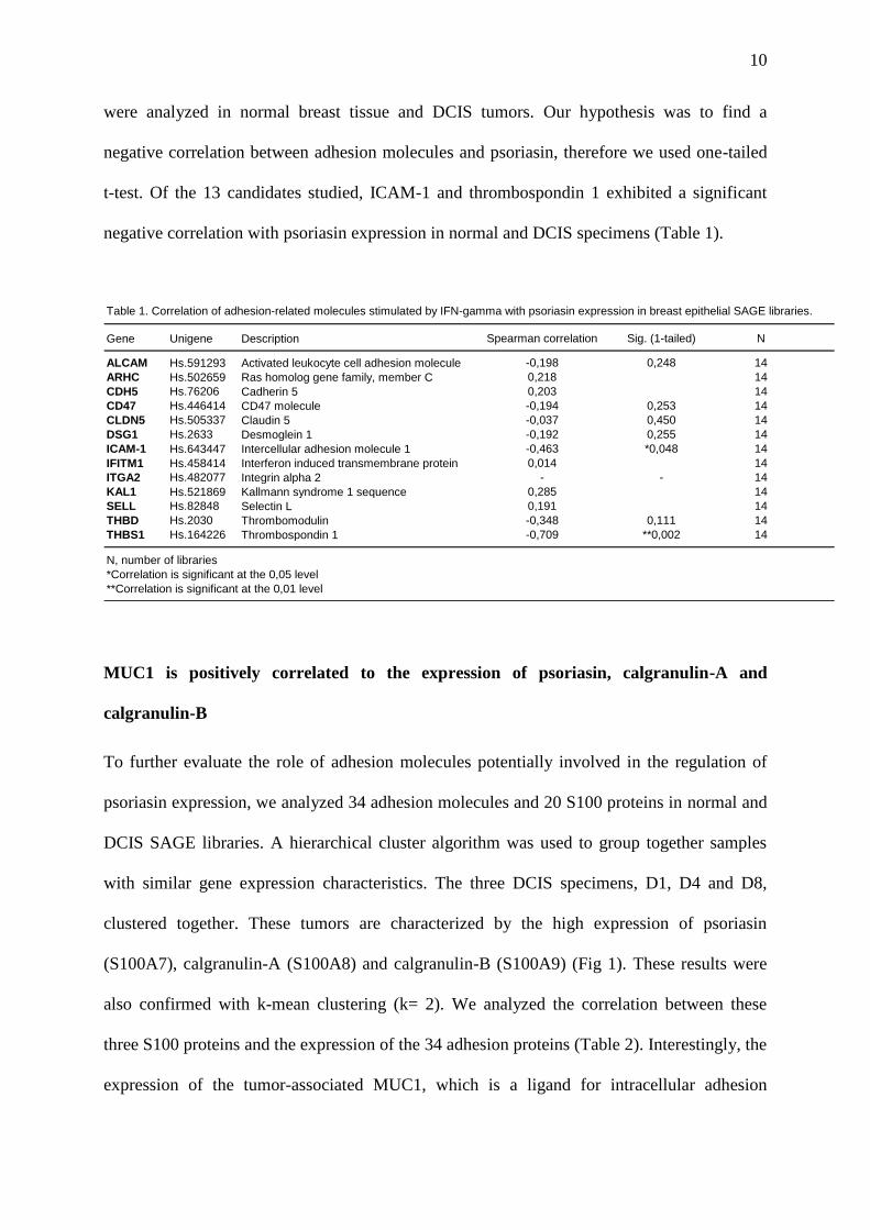
10
were analyzed in normal breast tissue and DCIS tumors. Our hypothesis was to find a
negative correlation between adhesion molecules and psoriasin, therefore we used one-tailed
t-test. Of the 13 candidates studied, ICAM-1 and thrombospondin 1 exhibited a significant
negative correlation with psoriasin expression in normal and DCIS specimens (Table 1).
Table 1. Correlation of adhesion-related molecules stimulated by IFN-gamma with psoriasin expression in breast epithelial SAGE libraries.
Gene Unigene Description Spearman correlation Sig. (1-tailed) N
ALCAM Hs.591293 Activated leukocyte cell adhesion molecule -0,198 0,248 14
ARHC Hs.502659 Ras homolog gene family, member C 0,218 14
CDH5 Hs.76206 Cadherin 5 0,203 14
CD47 Hs.446414 CD47 molecule -0,194 0,253 14
CLDN5 Hs.505337 Claudin 5 -0,037 0,450 14
DSG1 Hs.2633 Desmoglein 1 -0,192 0,255 14
ICAM-1 Hs.643447 Intercellular adhesion molecule 1 -0,463 *0,048 14
IFITM1 Hs.458414 Interferon induced transmembrane protein 0,014 14
ITGA2 Hs.482077 Integrin alpha 2 - - 14
KAL1 Hs.521869 Kallmann syndrome 1 sequence 0,285 14
SELL Hs.82848 Selectin L 0,191 14
THBD Hs.2030 Thrombomodulin -0,348 0,111 14
THBS1 Hs.164226 Thrombospondin 1 -0,709 **0,002 14
N, number of libraries
*Correlation is significant at the 0,05 level
**Correlation is significant at the 0,01 level
MUC1 is positively correlated to the expression of psoriasin, calgranulin-A and
calgranulin-B
To further evaluate the role of adhesion molecules potentially involved in the regulation of
psoriasin expression, we analyzed 34 adhesion molecules and 20 S100 proteins in normal and
DCIS SAGE libraries. A hierarchical cluster algorithm was used to group together samples
with similar gene expression characteristics. The three DCIS specimens, D1, D4 and D8,
clustered together. These tumors are characterized by the high expression of psoriasin
(S100A7), calgranulin-A (S100A8) and calgranulin-B (S100A9) (Fig 1). These results were
also confirmed with k-mean clustering (k= 2). We analyzed the correlation between these
three S100 proteins and the expression of the 34 adhesion proteins (Table 2). Interestingly, the
expression of the tumor-associated MUC1, which is a ligand for intracellular adhesion
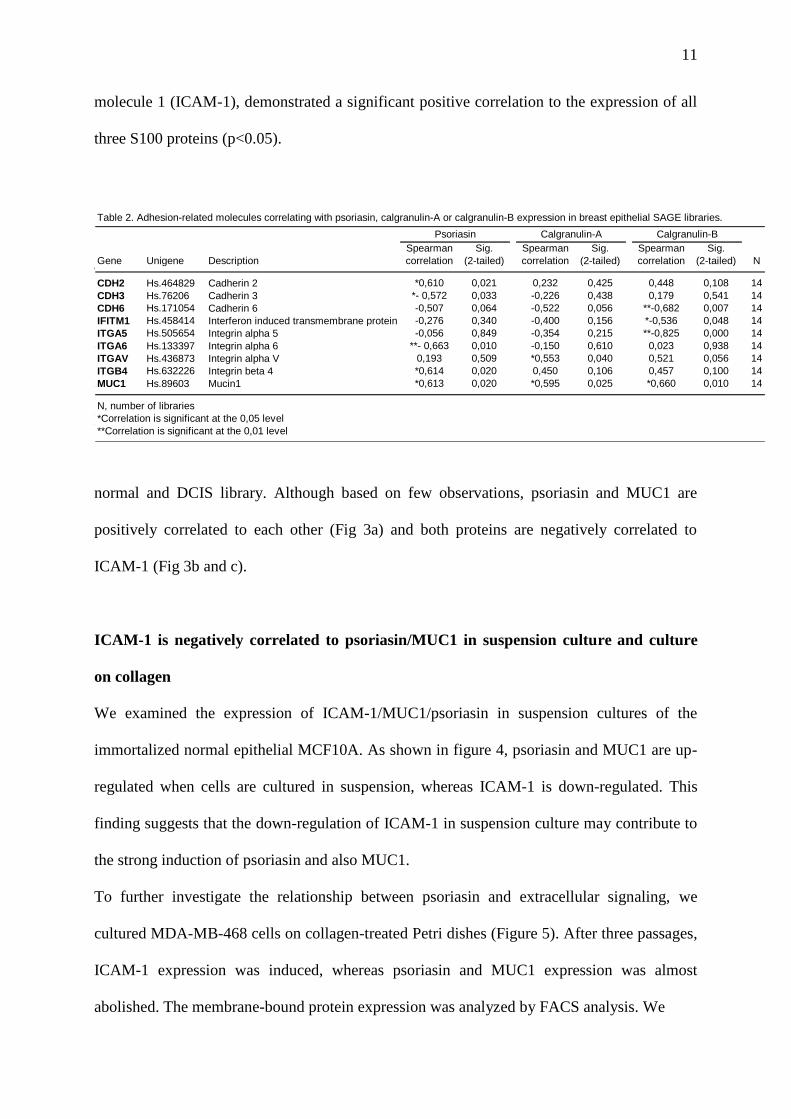
11
molecule 1 (ICAM-1), demonstrated a significant positive correlation to the expression of all
three S100 proteins (p<0.05).
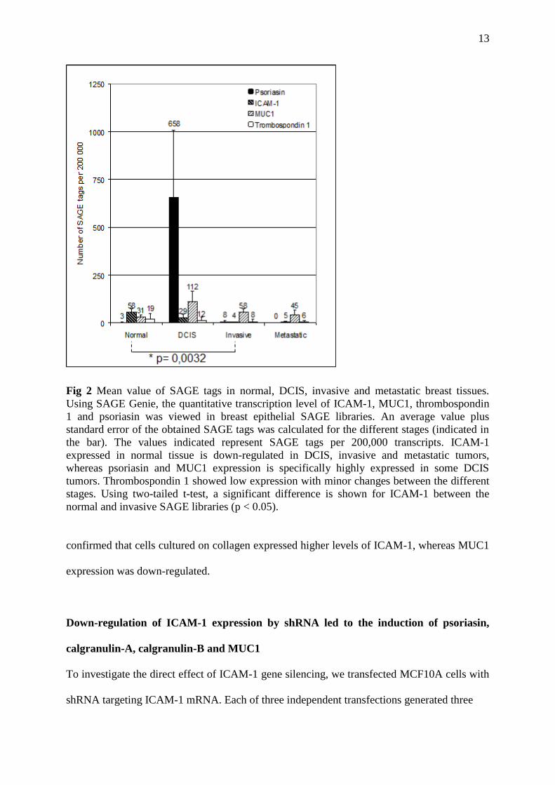
The expression level of psoriasin, ICAM-1, thrombospondin 1 and MUC1 is illustrated in
figure 2. Specifically, psoriasin, as well as MUC1, were highly up-regulated in DCIS
compared with normal breast tissue, while ICAM-1 was down-regulated. Thrombospondin 1
showed low expression levels.
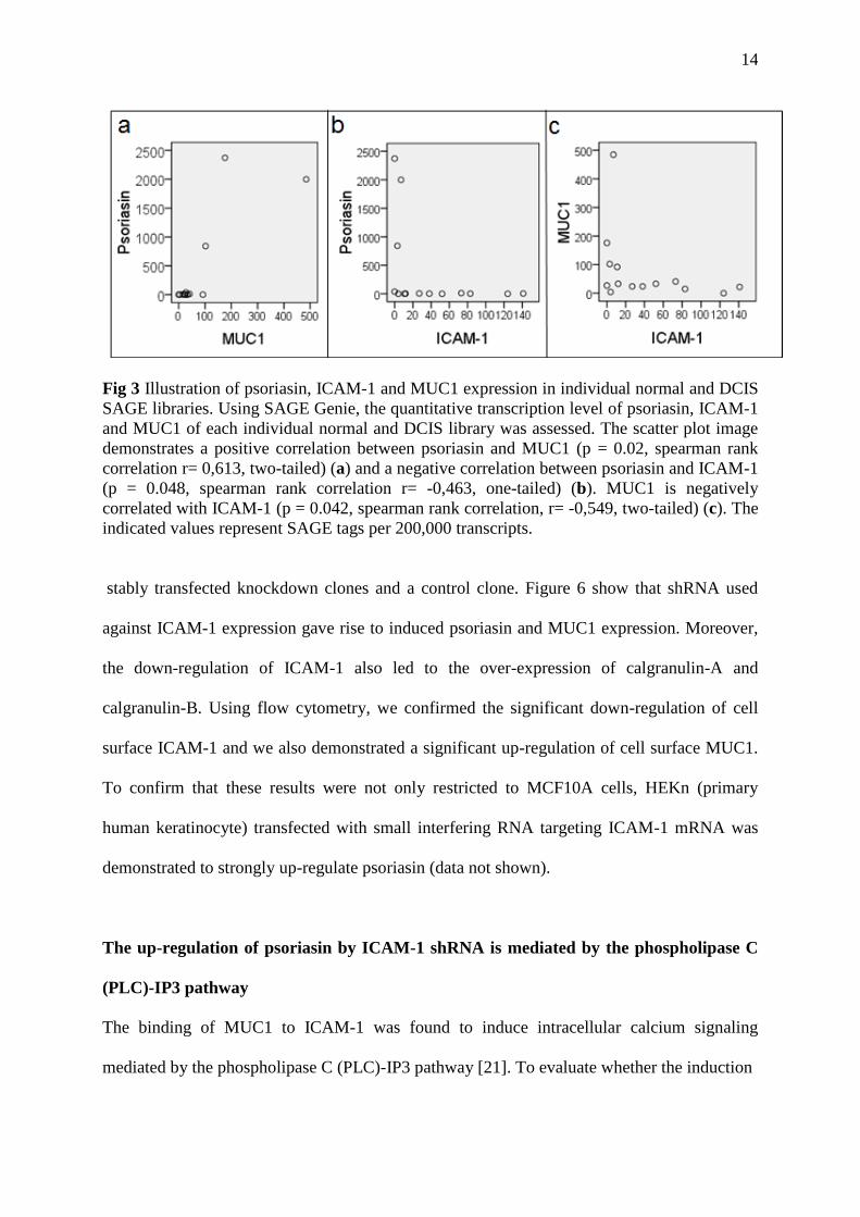
Next, we assessed the relative gene ratios of psoriasin, MUC1 and ICAM-1 in each individual
normal and DCIS library. Although based on few observations, psoriasin and MUC1 are
positively correlated to each other (Fig 3a) and both proteins are negatively correlated to
ICAM-1 (Fig 3b and c).
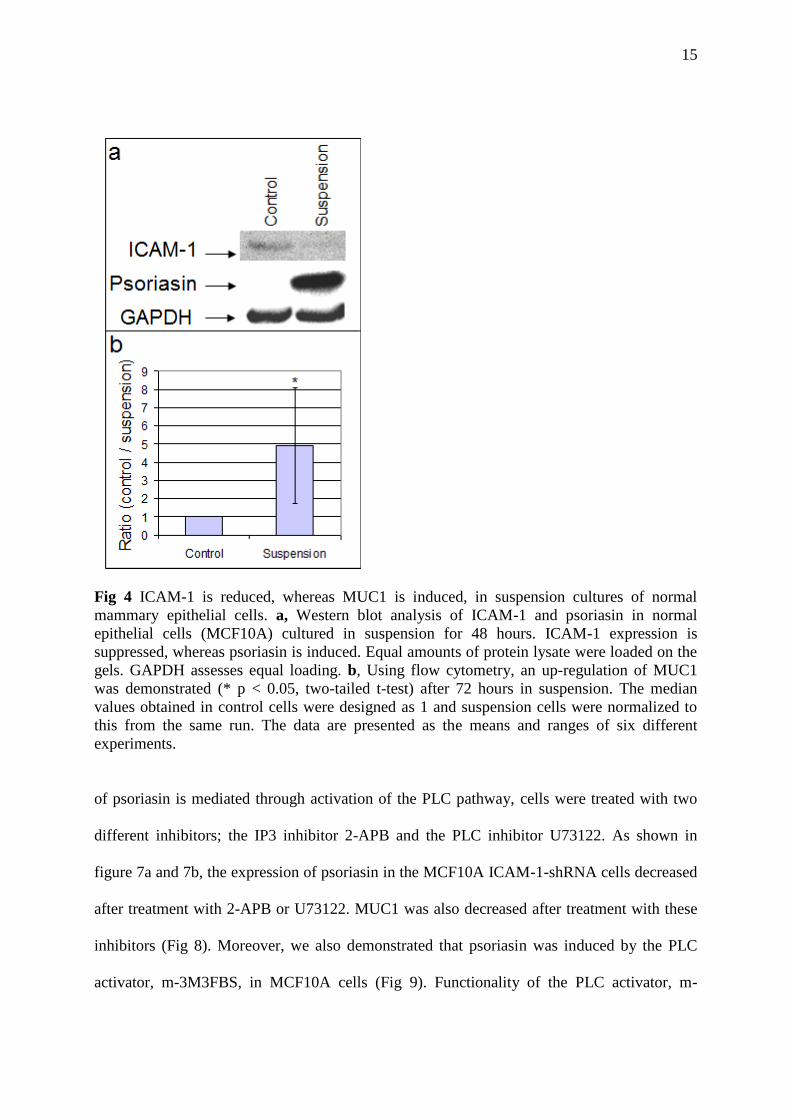
ICAM-1 is negatively correlated to psoriasin/MUC1 in suspension culture and culture
on collagen
We examined the expression of ICAM-1/MUC1/psoriasin in suspension cultures of the
immortalized normal epithelial MCF10A. As shown in figure 4, psoriasin and MUC1 are up-
regulated when cells are cultured in suspension, whereas ICAM-1 is down-regulated. This
finding suggests that the down-regulation of ICAM-1 in suspension culture may contribute to
the strong induction of psoriasin and also MUC1.
To further investigate the relationship between psoriasin and extracellular signaling, we
cultured MDA-MB-468 cells on collagen-treated Petri dishes (Figure 5). After three passages,
ICAM-1 expression was induced, whereas psoriasin and MUC1 expression was almost
abolished. The membrane-bound protein expression was analyzed by FACS analysis. We
Table 2. Adhesion-related molecules correlating with psoriasin, calgranulin-A or calgranulin-B expression in breast epithelial SAGE libraries.
Gene Unigene Description
Spearman
correlation
Sig.
(2-tailed)
Spearman
correlation
Sig.
(2-tailed)
Spearman
correlation
Sig.
(2-tailed) N
CDH2 Hs.464829 Cadherin 2 *0,610 0,021 0,232 0,425 0,448 0,108 14
CDH3 Hs.76206 Cadherin 3 *- 0,572 0,033 -0,226 0,438 0,179 0,541 14
CDH6 Hs.171054 Cadherin 6 -0,507 0,064 -0,522 0,056 **-0,682 0,007 14
IFITM1 Hs.458414 Interferon induced transmembrane protein -0,276 0,340 -0,400 0,156 *-0,536 0,048 14
ITGA5 Hs.505654 Integrin alpha 5 -0,056 0,849 -0,354 0,215 **-0,825 0,000 14
ITGA6 Hs.133397 Integrin alpha 6 **- 0,663 0,010 -0,150 0,610 0,023 0,938 14
ITGAV Hs.436873 Integrin alpha V 0,193 0,509 *0,553 0,040 0,521 0,056 14
ITGB4 Hs.632226 Integrin beta 4 *0,614 0,020 0,450 0,106 0,457 0,100 14
MUC1 Hs.89603 Mucin1 *0,613 0,020 *0,595 0,025 *0,660 0,010 14
N, number of libraries
*Correlation is significant at the 0,05 level
**Correlation is significant at the 0,01 level
Psoriasin Calgranulin-A Calgranulin-B

12
Fig 1 Hierarchical cluster analysis of adhesion-related molecules and S100 proteins in breast epithelial
SAGE libraries. Using SAGE Genie, the quantitative transcription level of S100 proteins and adhesion
molecules was viewed in breast epithelial SAGE libraries. The expression profiles of normal
epithelium and DCIS are compared using cluster analysis. a, Dendrogram image demonstrates the
degree of relatedness between the tissue samples. Each sample is represented by a single branch at the
bottom of the dendrogram. Samples displaying similar patterns of expression are grouped together on
closely connected branches of the dendrogram. b, Image demonstrates the level of gene expression
and is represented by the intensity of green color (low expression), black color (medium expression)
and red color (high expression). Each column represents a tissue sample and each row represents a
gene. The three DCIS libraries, D1, D4 and D8, clustered together. These tumors are characterized by
the high concomitant expression of psoriasin (S100A7), calgranulin-A (S100A8), calgranulin-B
(S100A9) and MUC1.
13
Fig 2 Mean value of SAGE tags in normal, DCIS, invasive and metastatic breast tissues.
Using SAGE Genie, the quantitative transcription level of ICAM-1, MUC1, thrombospondin
1 and psoriasin was viewed in breast epithelial SAGE libraries. An average value plus
standard error of the obtained SAGE tags was calculated for the different stages (indicated in
the bar). The values indicated represent SAGE tags per 200,000 transcripts. ICAM-1
expressed in normal tissue is down-regulated in DCIS, invasive and metastatic tumors,
whereas psoriasin and MUC1 expression is specifically highly expressed in some DCIS
tumors. Thrombospondin 1 showed low expression with minor changes between the different
stages. Using two-tailed t-test, a significant difference is shown for ICAM-1 between the
normal and invasive SAGE libraries (p < 0.05).
confirmed that cells cultured on collagen expressed higher levels of ICAM-1, whereas MUC1
expression was down-regulated.
Down-regulation of ICAM-1 expression by shRNA led to the induction of psoriasin,
calgranulin-A, calgranulin-B and MUC1
To investigate the direct effect of ICAM-1 gene silencing, we transfected MCF10A cells with
shRNA targeting ICAM-1 mRNA. Each of three independent transfections generated three
14
Fig 3 Illustration of psoriasin, ICAM-1 and MUC1 expression in individual normal and DCIS
SAGE libraries. Using SAGE Genie, the quantitative transcription level of psoriasin, ICAM-1
and MUC1 of each individual normal and DCIS library was assessed. The scatter plot image
demonstrates a positive correlation between psoriasin and MUC1 (p = 0.02, spearman rank
correlation r= 0,613, two-tailed) (a) and a negative correlation between psoriasin and ICAM-1
(p = 0.048, spearman rank correlation r= -0,463, one-tailed) (b). MUC1 is negatively
correlated with ICAM-1 (p = 0.042, spearman rank correlation, r= -0,549, two-tailed) (c). The
indicated values represent SAGE tags per 200,000 transcripts.
stably transfected knockdown clones and a control clone. Figure 6 show that shRNA used
against ICAM-1 expression gave rise to induced psoriasin and MUC1 expression. Moreover,
the down-regulation of ICAM-1 also led to the over-expression of calgranulin-A and
calgranulin-B. Using flow cytometry, we confirmed the significant down-regulation of cell
surface ICAM-1 and we also demonstrated a significant up-regulation of cell surface MUC1.
To confirm that these results were not only restricted to MCF10A cells, HEKn (primary
human keratinocyte) transfected with small interfering RNA targeting ICAM-1 mRNA was
demonstrated to strongly up-regulate psoriasin (data not shown).
The up-regulation of psoriasin by ICAM-1 shRNA is mediated by the phospholipase C
(PLC)-IP3 pathway
The binding of MUC1 to ICAM-1 was found to induce intracellular calcium signaling
mediated by the phospholipase C (PLC)-IP3 pathway [21]. To evaluate whether the induction
15
Fig 4 ICAM-1 is reduced, whereas MUC1 is induced, in suspension cultures of normal
mammary epithelial cells. a, Western blot analysis of ICAM-1 and psoriasin in normal
epithelial cells (MCF10A) cultured in suspension for 48 hours. ICAM-1 expression is
suppressed, whereas psoriasin is induced. Equal amounts of protein lysate were loaded on the
gels. GAPDH assesses equal loading. b, Using flow cytometry, an up-regulation of MUC1
was demonstrated (* p < 0.05, two-tailed t-test) after 72 hours in suspension. The median
values obtained in control cells were designed as 1 and suspension cells were normalized to
this from the same run. The data are presented as the means and ranges of six different
experiments.
of psoriasin is mediated through activation of the PLC pathway, cells were treated with two
different inhibitors; the IP3 inhibitor 2-APB and the PLC inhibitor U73122. As shown in
figure 7a and 7b, the expression of psoriasin in the MCF10A ICAM-1-shRNA cells decreased
after treatment with 2-APB or U73122. MUC1 was also decreased after treatment with these
inhibitors (Fig 8). Moreover, we also demonstrated that psoriasin was induced by the PLC
activator, m-3M3FBS, in MCF10A cells (Fig 9). Functionality of the PLC activator, m-

16
3M3FBS, and the inhibitor U73122 was demonstrated by phosphorylation of PLCγ1, which
confirms an active signaling pathway (Fig 10).
Fig 5 Type 1 collagen reduces psoriasin and MUC1 expression and induces ICAM-1
expression in MDA-MB-468 cells. a, Using Western blot analysis, endogenous psoriasin and
MUC1 in MDA-MB-468 cells are reduced when cells are cultured on collagen-1-treated Petri
dishes, whereas ICAM-1 is induced. 100 µg of protein lysate were loaded on the gel. GAPDH
assesses equal loading. b, Flow cytometry analysis confirms the up-regulation of ICAM-1 and
down-regulation of MUC1 on collagen-cultured cells (* p < 0.05, one-tailed t-test). The
median values obtained in control cells were designed as 1 and collagen-cultured cells were
normalized to this from the same run. The data are presented as the means and ranges of three
independent experiments.
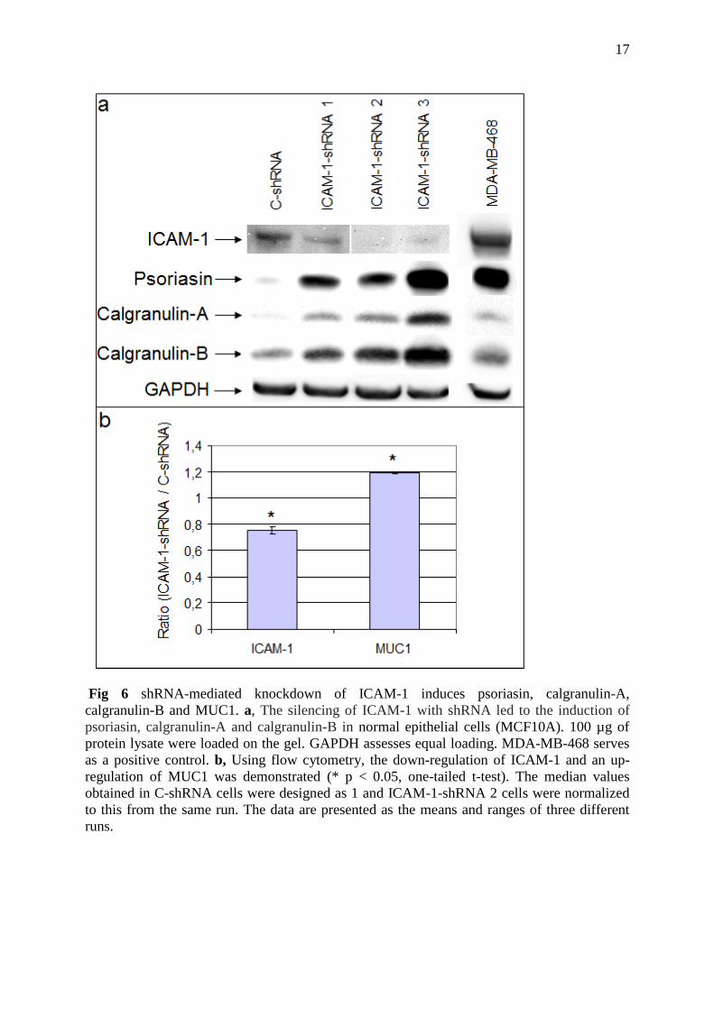
17
Fig 6 shRNA-mediated knockdown of ICAM-1 induces psoriasin, calgranulin-A,
calgranulin-B and MUC1. a, The silencing of ICAM-1 with shRNA led to the induction of
psoriasin, calgranulin-A and calgranulin-B in normal epithelial cells (MCF10A). 100 µg of
protein lysate were loaded on the gel. GAPDH assesses equal loading. MDA-MB-468 serves
as a positive control. b, Using flow cytometry, the down-regulation of ICAM-1 and an up-
regulation of MUC1 was demonstrated (* p < 0.05, one-tailed t-test). The median values
obtained in C-shRNA cells were designed as 1 and ICAM-1-shRNA 2 cells were normalized
to this from the same run. The data are presented as the means and ranges of three different
runs.

18
Fig 7 Psoriasin induction, by ICAM-1 silencing, is suppressed by phospholipase C (PLC)-IP3 inhibitors. The
inhibition of phospholipase C (PLC)-IP3 using 2-APB (a) and U73122 (b) led to reduced psoriasin expression in
MCF10A ICAM-1-shRNA cells. Psoriasin decreased after 2-APB treatment (100µM). Treatment with the PLC
inhibitor U73122 (10µM) for 3, 6 and 12 hours reduced psoriasin expression. Equal amounts of protein lysate
were loaded on the gels. GAPDH assesses equal loading. Psoriasin expression was quantified by the
AlphaEaseFCTM
software. Quantified results were adjusted with their own GAPDH level and control cells were
designed as 1 and treated cells were normalized to this.
Fig 8 MUC1 induction, by ICAM-1 silencing, is suppressed by phospholipase C (PLC)-IP3 inhibitors. The
inhibition of phospholipase C (PLC)-IP3 using 2-APB (100µM) and U73122 (10µM) led to reduced MUC1
expression in MCF10A ICAM-1-shRNA cells. Equal amounts of protein lysate were loaded on the gel. GAPDH
assesses equal loading.

19
Fig 9 Phospholipase C activation induces psoriasin in normal mammary epithelial cells.
Treatment with the phospholipase C activator, m-3M3FBS, induced psoriasin expression in
the MCF10A cell line (24 hours). Control cells were incubated with dimethylsulfoxide
(DMSO) alone (concentration equivalent to that used for 100µM m-3M3FBS). 90 µg of
protein lysate were loaded on the gel. GAPDH assesses equal loading. Psoriasin expression
was quantified by the AlphaEaseFCTM
software. Quantified results were adjusted with their
own GAPDH level and control cells were designed as 1 and treated cells were normalized to
this
We have previously demonstrated that psoriasin is induced by ROS and down-regulated by
Bcl-2 and other antioxidants like NAC [10]. We therefore used NAC in MCF10A ICAM-1-
shRNA cells and demonstrated a prominent expression of psoriasin after the treatment (Fig
11a). We also applied NBT assay to measure ROS production in MCF10A ICAM-1-shRNA
cells with upregulated psoriasin, compared to control-shRNA cells. We found no difference in
intracellular ROS levels (Fig 11b). Therefore, ROS cannot explain the upregulation of
psoriasin by ICAM-1 downregulation.
We have previously shown that psoriasin is induced by confluent conditions [8]. Moreover,
psoriasin is highly expressed in the MDA-MB-468 breast cancer cell line. To investigate the

20
Fig 10 Functionality of the phospholipase C activator, m-3M3FBS, and the inhibitor U73122. a,
Treatment with 100µM of m-3M3FBS stimulated the phosphorylation of PLCγ1 (pPLCγ1) in
MCF10A, which confirms an active signaling pathway. Control cells were incubated with
dimethylsulfoxide (DMSO) alone (concentration equivalent to that used for 100µM m-3M3FBS). b,
Phosphorylation of PLCγ1 in MCF10A cells, using H2O2, is reduced using the PLC inhibitor U73122.
MCF10A cells were treated with U73122. After three hours, cells were incubated with 0. 4- 1.2 mM
H2O2 for five minutes. Control cells were incubated with ethanol alone (concentration equivalent to
that used for 1.2 mM U73122). Quantified results were adjusted with their own GAPDH level and
control cells were designed as 1 and treated cells were normalized to this. Equal amounts of protein
lysate were loaded on the gels. GAPDH assesses equal loading.

21
Fig 11 Induction of psoriasin, by ICAM-1 silencing, is not explained by ROS production. a,
Cells were incubated with 10mM of the antioxidant NAC for 1.5 hours. There was still a
prominent expression of psoriasin after the treatments. 60 µg of protein lysate were loaded on
the gel. GAPDH assesses equal loading. b, We applied NBT assay to measure ROS
production in MCF10A ICAM-1-shRNA cells and in MCF10A control-shRNA cells. We
found no difference in intracellular ROS levels (p = 0.27, one-tailed t-test). The values are an
average of two independent experiments performed in triplicates.
involvement of the PLC pathway in the induction of psoriasin in these conditions, we treated
confluent MCF10A cells and MDA-MB-468 cells with the IP3-receptor inhibitor 2-APB and
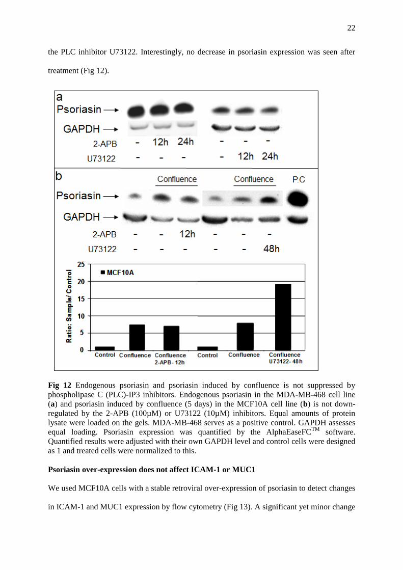
22
the PLC inhibitor U73122. Interestingly, no decrease in psoriasin expression was seen after
treatment (Fig 12).
Fig 12 Endogenous psoriasin and psoriasin induced by confluence is not suppressed by
phospholipase C (PLC)-IP3 inhibitors. Endogenous psoriasin in the MDA-MB-468 cell line
(a) and psoriasin induced by confluence (5 days) in the MCF10A cell line (b) is not down-
regulated by the 2-APB (100µM) or U73122 (10µM) inhibitors. Equal amounts of protein
lysate were loaded on the gels. MDA-MB-468 serves as a positive control. GAPDH assesses
equal loading. Psoriasin expression was quantified by the AlphaEaseFCTM
software.
Quantified results were adjusted with their own GAPDH level and control cells were designed
as 1 and treated cells were normalized to this.
Psoriasin over-expression does not affect ICAM-1 or MUC1
We used MCF10A cells with a stable retroviral over-expression of psoriasin to detect changes
in ICAM-1 and MUC1 expression by flow cytometry (Fig 13). A significant yet minor change
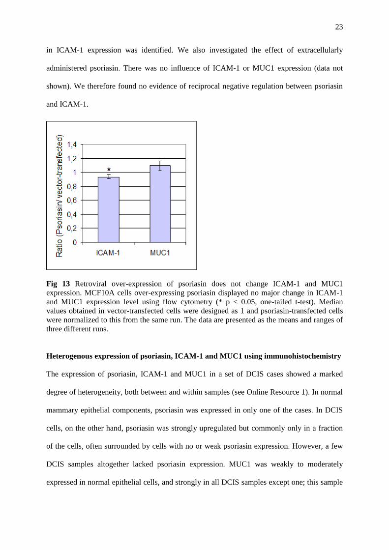
23
in ICAM-1 expression was identified. We also investigated the effect of extracellularly
administered psoriasin. There was no influence of ICAM-1 or MUC1 expression (data not
shown). We therefore found no evidence of reciprocal negative regulation between psoriasin
and ICAM-1.
Fig 13 Retroviral over-expression of psoriasin does not change ICAM-1 and MUC1
expression. MCF10A cells over-expressing psoriasin displayed no major change in ICAM-1
and MUC1 expression level using flow cytometry (* p < 0.05, one-tailed t-test). Median
values obtained in vector-transfected cells were designed as 1 and psoriasin-transfected cells
were normalized to this from the same run. The data are presented as the means and ranges of
three different runs.
Heterogenous expression of psoriasin, ICAM-1 and MUC1 using immunohistochemistry
The expression of psoriasin, ICAM-1 and MUC1 in a set of DCIS cases showed a marked
degree of heterogeneity, both between and within samples (see Online Resource 1). In normal
mammary epithelial components, psoriasin was expressed in only one of the cases. In DCIS
cells, on the other hand, psoriasin was strongly upregulated but commonly only in a fraction
of the cells, often surrounded by cells with no or weak psoriasin expression. However, a few
DCIS samples altogether lacked psoriasin expression. MUC1 was weakly to moderately
expressed in normal epithelial cells, and strongly in all DCIS samples except one; this sample

24
also lacking psoriasin expression. Also ICAM-1 showed a heterogenous expression, both in
normal cells and in the DCIS components. Thus, both normal mammary epithelial cells and
DCIS cells showed weak or even negative, to moderate staining. Because of this complex
picture, a grading of expression of the different proteins in individual tumors was not found
meaningful. Interestingly, in apocrine metaplasia representing differentiated normal epithelial
cells, ICAM-1 was downregulated whereas psoriasin was upregulated. Endothelial cells and
inflammatory cells stained strongly for ICAM-1 both in the tumor stroma and in the stroma of
normal parts of the samples. The staining was strong without the variation in intensity
observed in mammary epithelial cells.
Discussion
Adhesion plays a central role in cell survival. Cell-cell interaction and the anchorage of cells
to components of the extracellular matrix (ECM) are mediated primarily by integrins and
other adhesion molecules [22]. During the initiation of breast cancer, epithelial cells hyper-
proliferate, which leads to changes in adhesion to the basal membrane. In high-grade DCIS,
which is an early form of breast cancer, psoriasin is one of the few proteins that are very
highly expressed. We have observed that psoriasin is induced in vitro in suspension cultures
of normal mammary epithelial cells (MCF10A). IFN-γ treatment specifically down-regulate
psoriasin expression in suspension cultures [15]. Among the adhesion molecules implicated in
cell-cell interactions, CD54 or ICAM-1 have previously been identified to be up-regulated in
response to IFN-γ at the transcriptional level [22]. ICAM-1 is an important member of the
immunoglobulin superfamily (IgSF) of proteins that is essentially involved in the recruitment
and trafficking of leukocytes [23]. In contrast to the constitutive expression of ICAM-1 in the
endothelium, human epithelial cells normally have a low expression. In the normal and DCIS
SAGE libraries, we confirmed a significant negative correlation between the expression of
25
psoriasin and ICAM-1 levels. To confirm a connection between psoriasin induction and the
loss of ICAM-1 expression in normal breast epithelial cells, we demonstrated that ICAM-1
was in fact down-regulated in suspension, making this protein an interesting candidate for
further analysis.
We performed a cluster analysis of 34 adhesion molecules and 20 S100 proteins and report a
strong association between the expression of psoriasin, calgranulin-A and calgranulin-B. Like
psoriasin, we have demonstrated that calgranulin-A and calgranulin-B are induced by ROS,
confluence and suspension culture, which supports the assumption that they share common
signaling pathways in breast cancer. Their co-expression may suggest that the combined up-
regulation of these three proteins reflects a group of breast cancer with a poor prognosis. In
line with this, the cluster analysis revealed that all three proteins correlated strongly to the
oncogenic MUC1. MUC1, which is a transmembrane glycoprotein, is an established tumor
marker in breast cancer and is implicated in metastatic spread. In addition to psoriasin,
calgranulin-A and calgranulin-B, MUC1 was also induced in suspension cultures of normal
breast epithelial cells.
When looking into the relative gene ratios of psoriasin, ICAM-1 and MUC1 in each
individual normal and DCIS library in our study, we found a significant negative correlation
between ICAM-1 and psoriasin and a corresponding positive correlation between MUC1 and
psoriasin. We confirmed the negative correlation between ICAM-1 and psoriasin/MUC1,
when cells were cultured on collagen.
The direct regulatory effect was suggested by the use of shRNA targeting ICAM-1 mRNA in
MCF10A cells. The down-regulation of ICAM-1 expression by shRNA thus led to the
induction of psoriasin, calgranulin-A, calgranulin-B and MUC1.
Next, we focused on the mechanism for the up-regulation of psoriasin in the MCF10A cells
with reduced expression of ICAM-1. We still detected an expression of psoriasin after

26
treatment with the antioxidant NAC. Moreover, there was no increase in intracellular ROS in
cells with reduced expression of ICAM-1. These findings suggest that signals other than ROS
production contribute to the regulation of psoriasin in this context. ICAM-1 is the only known
ligand of the MUC1 extracellular domain [24]. Rahn et al. showed that MUC1 initiates a
calcium-based signal mediated by the phospholipase C (PLC)-IP3 pathway, involved in
intracellular calcium signaling [21]. Using the inhibitors U73122 and 2-APB for the PLC
pathway, the induction of psoriasin and MUC1 in these cells were reduced. Moreover, the
PLC activator m-3M3FBS was shown to increase the expression of psoriasin in normal
mammary epithelial cells. These finding supports the hypothesis that psoriasin is an
intracellular calcium-dependent target of the PLC pathway.
The immunohistochemical analysis from a set of DCIS cases emphasized the high degree of
heterogeneity within and between samples. The lack of complete concordance between the
results from the analysis of normal and DCIS SAGE libraries compared to the
immunohistochemistry may be explained by the fact that SAGE libraries are prepared from
fresh tissue and represent mRNA expression, whereas protein expression are assessed in
immunohistochemistry. Moreover, the normal mammary epithelial cells within and close to a
DCIS lesion may not be regarded as completely normal, since it is known that there is a
dramatic change in gene expression in stroma cells surrounding the malignant epithelial cells
[25].
MUC1 is implicated in many physiological processes such as adhesion, development and
differentiation. In addition, MUC1 is frequently over-expressed in many cancers including
breast cancer with a predominantly cytoplasmic expression [26]. The over-expression of
MUC1 is associated with a poorer prognosis and shorter survival in many cancers, including
breast cancer. The co-expression of psoriasin and MUC1 may contribute to the poor clinical
outcome characteristic for tumors over-expressing psoriasin. Interestingly, both MUC1 and

27
psoriasin correlate with increased survival in response to oxidative stress [10, 27]. Moreover,
both proteins have been shown to be regulated by the NFκB pathway [10, 28]. MUC1 affect
cancer cell migration by increasing E-cadherin/beta-catenin complex formation [29] and the
down-regulation of psoriasin has also been linked to increased beta-catenin signalling [30].
We now report their co-expression in response to ICAM-1 down-regulation and the positive
correlation between their expressions in breast SAGE libraries.
In conclusion, we have presented data suggesting that the loss of ICAM-1 expression on
normal mammary epithelial cells may contribute to the high expression of psoriasin in high-
grade DCIS. The upregulation of psoriasin by ICAM1 shRNA was mediated by the
phopholipase C (PLC)-IP3 pathway. Furthermore, the downregulation of ICAM-1 led to the
induction of calgranulin-A, calgranulin-B and MUC1.
Acknowledgments
We thank Dr Kornelia Polyak at DFCI, Boston, MA, for generous help and valuable
suggestions. We thank Katarina Junevik (Sahlgrenska University Hospital, Gothenburg) for
her help with flow cytometry analysis. We thank Maria Nethander at the Genomics Core
Facility (Gothenburg) for the help with hierarchical clustering analysis. This work was
supported by grants from the Swedish Cancer Society, the Swedish Psoriasis Association, the
Assar Gabrielsson Foundation, the Welander Foundation and the Tore Nilsson Foundation.
References
1. Kamangar F, Dores GM, Anderson WF (2006) Patterns of cancer incidence, mortality,
and prevalence across five continents: defining priorities to reduce cancer disparities
in different geographic regions of the world. J Clin Oncol 24:2137-2150.
2. Polyak K (2007) Breast cancer: origins and evolution. J Clin Invest 117:3155-3163.
3. Martinsson H, Yhr M, Enerback C (2005) Expression patterns of S100A7 (psoriasin)
and S100A9 (calgranulin-B) in keratinocyte differentiation. Exp Dermatol 14:161-
168.
28
4. Santamaria-Kisiel L, Rintala-Dempsey AC, Shaw GS (2006) Calcium-dependent and -
independent interactions of the S100 protein family. The Biochemical journal
396:201-214.
5. Donato R (2001) S100: a multigenic family of calcium-modulated proteins of the EF-
hand type with intracellular and extracellular functional roles. The international
journal of biochemistry & cell biology 33:637-668.
6. Leygue E, Snell L, Hiller T, Dotzlaw H, Hole K, Murphy LC, Watson PH (1996)
Differential expression of psoriasin messenger RNA between in situ and invasive
human breast carcinoma. Cancer Res 56:4606-4609.
7. Emberley ED, Alowami S, Snell L, Murphy LC, Watson PH (2004) S100A7
(psoriasin) expression is associated with aggressive features and alteration of Jab1 in
ductal carcinoma in situ of the breast. Breast Cancer Res 6:R308-315.
8. Enerback C, Porter DA, Seth P, Sgroi D, Gaudet J, Weremowicz S, Morton CC,
Schnitt S, Pitts RL, Stampl J, Barnhart K, Polyak K (2002) Psoriasin expression in
mammary epithelial cells in vitro and in vivo. Cancer Res 62:43-47.
9. Al-Haddad S, Zhang Z, Leygue E, Snell L, Huang A, Niu Y, Hiller-Hitchcock T, Hole
K, Murphy LC, Watson PH (1999) Psoriasin (S100A7) expression and invasive breast
cancer. The American journal of pathology 155:2057-2066.
10. Carlsson H, Yhr M, Petersson S, Collins N, Polyak K, Enerback C (2005) Psoriasin
(S100A7) and calgranulin-B (S100A9) induction is dependent on reactive oxygen
species and is downregulated by Bcl-2 and antioxidants. Cancer Biol Ther 4:998-1005.
11. Emberley ED, Niu Y, Curtis L, Troup S, Mandal SK, Myers JN, Gibson SB, Murphy
LC, Watson PH (2005) The S100A7-c-Jun activation domain binding protein 1
pathway enhances prosurvival pathways in breast cancer. Cancer Res 65:5696-5702.
12. Emberley ED, Niu Y, Leygue E, Tomes L, Gietz RD, Murphy LC, Watson PH (2003)
Psoriasin interacts with Jab1 and influences breast cancer progression. Cancer Res
63:1954-1961.
13. Krop I, Marz A, Carlsson H, Li X, Bloushtain-Qimron N, Hu M, Gelman R, Sabel
MS, Schnitt S, Ramaswamy S, Kleer CG, Enerback C, Polyak K (2005) A putative
role for psoriasin in breast tumor progression. Cancer Res 65:11326-11334.
14. Glaser R, Harder J, Lange H, Bartels J, Christophers E, Schroder JM (2005)
Antimicrobial psoriasin (S100A7) protects human skin from Escherichia coli
infection. Nat Immunol 6:57-64.
15. Petersson S, Bylander A, Yhr M, Enerback C (2007) S100A7 (Psoriasin), highly
expressed in ductal carcinoma in situ (DCIS), is regulated by IFN-gamma in
mammary epithelial cells. BMC cancer 7:205.
16. Boon K, Osorio EC, Greenhut SF, Schaefer CF, Shoemaker J, Polyak K, Morin PJ,
Buetow KH, Strausberg RL, De Souza SJ, Riggins GJ (2002) An anatomy of normal
and malignant gene expression. Proc Natl Acad Sci U S A 99:11287-11292.
17. Porter DA, Krop IE, Nasser S, Sgroi D, Kaelin CM, Marks JR, Riggins G, Polyak K
(2001) A SAGE (serial analysis of gene expression) view of breast tumor progression.
Cancer Res 61:5697-5702.
18. Porter D, Lahti-Domenici J, Keshaviah A, Bae YK, Argani P, Marks J, Richardson A,
Cooper A, Strausberg R, Riggins GJ, Schnitt S, Gabrielson E, Gelman R, Polyak K
(2003) Molecular markers in ductal carcinoma in situ of the breast. Mol Cancer Res
1:362-375.
19. Wadsworth TL, Bishop JA, Pappu AS, Woltjer RL, Quinn JF (2008) Evaluation of
coenzyme Q as an antioxidant strategy for Alzheimer's disease. J Alzheimers Dis
14:225-234.

29
20. de Veer MJ, Holko M, Frevel M, Walker E, Der S, Paranjape JM, Silverman RH,
Williams BR (2001) Functional classification of interferon-stimulated genes identified
using microarrays. Journal of leukocyte biology 69:912-920.
21. Rahn JJ, Shen Q, Mah BK, Hugh JC (2004) MUC1 initiates a calcium signal after
ligation by intercellular adhesion molecule-1. The Journal of biological chemistry
279:29386-29390.
22. Hou J, Baichwal V, Cao Z (1994) Regulatory elements and transcription factors
controlling basal and cytokine-induced expression of the gene encoding intercellular
adhesion molecule 1. Proc Natl Acad Sci U S A 91:11641-11645.
23. Reiss Y, Hoch G, Deutsch U, Engelhardt B (1998) T cell interaction with ICAM-1-
deficient endothelium in vitro: essential role for ICAM-1 and ICAM-2 in
transendothelial migration of T cells. European journal of immunology 28:3086-3099.
24. Hayashi T, Takahashi T, Motoya S, Ishida T, Itoh F, Adachi M, Hinoda Y, Imai K
(2001) MUC1 mucin core protein binds to the domain 1 of ICAM-1. Digestion 63
Suppl 1:87-92.
25. Allinen M, Beroukhim R, Cai L, Brennan C, Lahti-Domenici J, Huang H, Porter D,
Hu M, Chin L, Richardson A, Schnitt S, Sellers WR, Polyak K (2004) Molecular
characterization of the tumor microenvironment in breast cancer. Cancer cell 6:17-32.
26. Rahn JJ, Dabbagh L, Pasdar M, Hugh JC (2001) The importance of MUC1 cellular
localization in patients with breast carcinoma: an immunohistologic study of 71
patients and review of the literature. Cancer 91:1973-1982.
27. Yin L, Li Y, Ren J, Kuwahara H, Kufe D (2003) Human MUC1 carcinoma antigen
regulates intracellular oxidant levels and the apoptotic response to oxidative stress.
The Journal of biological chemistry 278:35458-35464.
28. Thompson EJ, Shanmugam K, Hattrup CL, Kotlarczyk KL, Gutierrez A, Bradley JM,
Mukherjee P, Gendler SJ (2006) Tyrosines in the MUC1 cytoplasmic tail modulate
transcription via the extracellular signal-regulated kinase 1/2 and nuclear factor-
kappaB pathways. Mol Cancer Res 4:489-497.
29. Yuan Z, Wong S, Borrelli A, Chung MA (2007) Down-regulation of MUC1 in cancer
cells inhibits cell migration by promoting E-cadherin/catenin complex formation.
Biochem Biophys Res Commun 362:740-746.
30. Zhou G, Xie TX, Zhao M, Jasser SA, Younes MN, Sano D, Lin J, Kupferman ME,
Santillan AA, Patel V, Gutkind JS, Ei-Naggar AK, Emberley ED, Watson PH,
Matsuzawa SI, Reed JC, Myers JN (2008) Reciprocal negative regulation between
S100A7/psoriasin and beta-catenin signaling plays an important role in tumor
progression of squamous cell carcinoma of oral cavity. Oncogene 27:3527-3538.
31. SAGE genie informatics [http://cgap.nci.nih.gov/SAGE]