Embed Size (px)
Citation preview

Available online at www.sciencedirect.com
15 (2008) 161–172www.elsevier.com/developmentalbiology
Developmental Biology 3
Long- and short-range signals control the dynamic expression of an animalhemisphere-specific gene in Xenopus
Adnan Mir a,b, Matthew Kofron a, Janet Heasman a, Melissa Mogle a, Stephanie Lang a,Bilge Birsoy a, Chris Wylie a,⁎
a Division of Developmental Biology, Cincinnati Children’s Hospital Research Foundation, 3333 Burnet Ave, Cincinnati, OH 45229, USAb Physician/Scientist Training Program, P. O. Box 670555, Cincinnati, OH 45267, USA
Received for publication 6 September 2007; revised 17 December 2007; accepted 17 December 2007Available online 27 December 2007
Abstract
Little is known of the control of gene expression in the animal hemisphere of the Xenopus embryo. Here we show that expression of FoxI1e, agene essential for normal ectoderm formation, is expressed regionally within the animal hemisphere, in a highly dynamic fashion. In situhybridization shows that FoxI1e is expressed in a wave-like fashion that is initiated on the dorsal side of the animal hemisphere, extends across tothe ventral side by the mid-gastrula stage, and is then turned off in the dorsal ectoderm, the neural plate, at the neurula stage. It is confined to theinner layers of cells in the animal cap, and is expressed in a mosaic fashion throughout. We show that this dynamic pattern of expression iscontrolled by both short- and long-range signals. Notch signaling controls both the mosaic, and dorsal/ventral changes in expression, and iscontrolled, in turn, by Vg1 signaling from the vegetal mass. FoxI1e expression is also regulated by nodal signaling downstream of VegT. CanonicalWnt signaling contributes only to late changes in the FoxI1e expression pattern.
These results provide new insights into the roles of vegetally localized mRNAs in controlling zygotic genes expressed in the animal hemisphereby long-range signaling. They also provide novel insights into the role of Notch signaling at the earliest stages of vertebrate development.© 2007 Elsevier Inc. All rights reserved.
Keywords: FoxI1e; Xenopus; Ectoderm; Animal gene expression
Introduction
The Xenopus blastula is conventionally divided into threeregions, with respect to both cell fate and gene expression. Cellsin the vegetal region give rise to the embryonic endoderm. Theyinherit the maternally encoded transcription factor VegT, whichactivates the synthesis of nodals. These, in turn, induce meso-derm in the adjacent marginal region. Cells in the animal regionform the ectoderm. It is not known how this fate is initiated. Inaddition to forming the germ layers, the three blastula regionseach become patterned to form the axes of the body, by theexpression of dorsal and ventral genes.
This picture of gene expression in the blastula tends to beregarded as dynamic with respect to time, but static with respectto each region of the blastula; a linear progression within each
⁎ Corresponding author.E-mail address: [email protected] (C. Wylie).
0012-1606/$ - see front matter © 2007 Elsevier Inc. All rights reserved.doi:10.1016/j.ydbio.2007.12.022
region of transcriptional initiation, leading to specification toenter a particular lineage, or to exhibit a particular type of cellbehavior. We show here that in fact, gene expression is highlydynamic within the animal region. The forkhead transcriptionfactor FoxI1e is expressed in the animal hemisphere, starting atthe blastula stage. It is required for the expression of genes inboth the early neural and ectodermal lineages, and for the laterdifferentiation of the epidermis (Mir et al., 2007). It is alsorequired to repress mesoderm specification in the animalhemisphere (Suri et al., 2005). Here we show that its expressionpattern changes rapidly in both time and space within the animalhemisphere. Expression is initiated dorsally at the blastulastage, then spreads to encompass the ventral region of the ani-mal hemisphere by the mid-gastrula stage. At the early neurulastage, expression is lost from the dorsal ectoderm cells that formthe neural plate. During this sequence, its expression domainalso changes with respect to the layers of the animal cap. First, itis restricted to the inner cells of the animal cap at the blastula

162 A. Mir et al. / Developmental Biology 315 (2008) 161–172
stage, and then to a layer of cells between the inner and outerlayers of the animal cap at the gastrula stage. Furthermore,throughout this temporal and spatial progression, FoxI1e ex-pression is always in a mosaic pattern, with positive cells inter-spersed with non-expressing cells.
These observations revealed a previously unsuspected re-gionalization of the animal hemisphere. The total expressiondomain of FoxI1e extends across the animal cap, excluding theouter cells at the blastula stage, and both inner and outer cells atthe gastrula stage, and all the time is restricted to only somecells, but not others, in this expression domain. During the lateblastula to mid-gastrula stages, the expression of FoxI1e movesacross this expression domain in a dorsal to ventral direction.This pattern is completely different from the expression patternsof other animally localized transcripts previously reported, suchas ectodermin (Dupont et al., 2005) and Xlim5 (Toyama et al.,1995; Houston and Wylie, 2003), which are not mosaic, nordynamic with respect to the dorsal–ventral axis. We thereforeset out to identify some of the factors that might control thesespatial and temporal changes.
We found that both long- and short-range signals controlthis pattern of expression. First, mosaic expression iscontrolled by Notch signaling. Both gain- and loss-of-functionexperiments showed that Notch signaling represses FoxI1eexpression, and that loss of Notch signaling causes expressionof FoxI1e in all cells of its total expression domain, andeliminates both the temporal and spatial progression ofexpression within this domain. This raised the issue of howNotch signaling is controlled in the animal hemisphere. Itcould be intrinsic, or controlled at long-range from the otherdeveloping germ layer regions. We show that control ofFoxI1e expression by Notch is, in turn, controlled by the TGF-β signal, Vg1, whose mRNA is maternally encoded, andinherited only by the vegetal cells of the embryo (Melton,1987). We also find that the level of FoxI1e, but not its mosaicdistribution, is controlled by nodal signaling downstream ofVegT, a vegetally localized, maternally encoded transcriptionfactor (Zhang and King, 1996).
These data reveal multiple levels of control of early zygoticgenes in Xenopus. First, long-range signals from the vegetalcells of the embryo, as well as short-range signals through theNotch pathway, combine to control both amount and position ofexpression of animal-specific genes during the blastula andgastrula stages. Second, none of these signals controls the “totalexpression domain” of FoxI1e, which remains confined to thegroup of cells that normally express it during its changingtemporal expression at the blastula and gastrula stages. Instead,they control the temporal and spatial sequence of expressionwithin this domain.
Materials and methods
Oocytes and embryos
Oocytes were generated for host transfer experiments by manualdefolliculation of surgically removed ovary as previously described (Heasmanet al., 1991). Culture and injections were carried out in L15-based oocyte culturemedium (OCM). The length of the culture period between injection and host
transfer varied by experiment. All mRNA injections were cultured overnight.VegT-depleted embryos were generated using a morpholino oligo [5′-CCCGACAGCAGTTTCTCATTCCACG-3′], and cultured for 3 days afterinjection. Vg1 depletions were carried out using the oligo Vg1c as previouslydescribed (Birsoy et al., 2006), with 4 days of culture after injection. Xotch wasdepleted using AS14MP [5′-GGAAGGGCTCAGCGCTAC-3′], with 3 days ofculture. For rescue experiments, mRNAwas injected on the last day of culture,before progesterone treatment, or at the 2-cell stage after fertilization. Eggs werecollected in high-salt solution, fertilized in vitro with isolated testis, andcultured in 0.1× MMR. Staging was according to Nieuwkoop and Faber (1967).Dissections were performed at stage 9 or stage 10 on agarose-coated dishes in1× MMR and then cultured in OCM. β-Catenin-depleted embryos weregenerated using a morpholino as previously described (Heasman et al., 2000).Synthetic mRNAs encoding β-catenin, BMP4, cmBMP7, β-galactosidase,NICD, Su(H)-DBM, and Vg1 were generated using Ambion mMessagemMachine kits.
Real-time RT–PCR
Total RNA was extracted as previously described (Zhang et al., 1998). Unlessotherwise indicated, input was 2 whole embryos, 3 marginal zones, or 5 vegetalmasses per sample. cDNA was synthesized using oligo dT primers, and semi-quantitative real-time RT–PCR was carried out using the LightCycler system asdescribed (Kofron et al., 2001). Ornithine decarboxylase (ODC) was used as aloading control, and all values were normalized to ODC levels. In all cases, water-only and reverse transcriptase-negative controls failed to produce specific products.Each experiment was repeated aminimum of three times in independent experimentsto verify reproducibility of results. Primer sequences were: Siamois: U: 5′-CTGTCCTACAAGAGACTCTG-3′, D: 5′-TGTTGACTGCAGACTGTTGA-3′.Xnr3: U: 5′-CTTCTGCACTAGATTCTG-3′, D: 5′-CAGCTTCTGGCCAAGACT-3′. FoxI1e: U-5′-GCACCTGCTGTGGTTCATAA-3′, D-5′-CACCACTG-TAGTGCGTCAGAA-3′. Xotch: U: 5′-AGTAACCCGTGCAAAAATGG-3′, D:5′-AGCTTCCGGTAAATCCAGGT-3′. ESR-1: U: 5′-TGGCAAAACTGGAA-CAGGAT-3′, D: 5′-TGGGATACAACAGGGAGCTT-3′.
Whole-mount in situ hybridization, membrane staining, and Red-Galstaining
In situ hybridizations were carried out as described by Harland (1991), witha probe concentration for FoxI1e of 5 μg/ml, and using BMB Purple (Roche) asthe alkaline phosphatase substrate. For membrane staining, embryos werestained for FoxI1e, sectioned, and stained with Alexa-488-conjugated WheatGerm Agglutinin (Molecular Probes) at 0.01 μg/μl in PBS+0.1% Tween-20 for30 min. For lineage labeling, embryos injected with 50 pg of nuclear β-galactosidase mRNA in the A1 blastomere were fixed for 1 h at room tem-perature in MEMFA, stained with Red-Gal (Research Organics) for 20 min at37 °C, and fixed for another hour at room temperature before in situ hybrid-ization for FoxI1e.
Results
FoxI1e is expressed in a dorsal to ventral wave, in a subset ofcells in the animal cap
In previous work, we found that FoxI1e expression wasmosaic in the early embryo (Mir et al., 2007). This could be dueeither to an asynchronous onset of expression, leading to a morestable homogeneous expression pattern, or it could be that geneexpression in the animal hemisphere is not uniform, but con-trolled in more complex ways than previously thought. To dis-tinguish between these possibilities, we carried out in situhybridization on embryos from the late blastula stage (stage 9)to the mid-gastrula stage (stage 11). We found that expression inall embryos began in a localized manner above the equator on

163A. Mir et al. / Developmental Biology 315 (2008) 161–172
one side of the embryo, and then spread across the animal cap(Fig. 1A). The site of onset of expression was identified as thedorsal side by injection of β-galactosidase mRNA into the 2dorsal animal cells at the 8-cell stage. The β-Gal signal con-sistently co-localized with FoxI1e expression (Fig. 1B, 92%,n=71), showing that FoxI1e expression begins on the dorsalside of the embryo, and spreads ventrally. To confirm thisfinding, we dissected early gastrulae into dorsal and ventralhalves, and used real-time RT–PCR to compare the amount ofFoxI1e expression. We found that FoxI1e mRNA was indeedexpressed at higher levels in dorsal halves at the late blastulastage (Fig. 1C).
The animal cap consists of two cell layers—an epithelial, orouter layer, and an inner, sensorial layer (Chalmers et al., 2003).From late blastula to early gastrula stages, FoxI1e was ex-pressed only in the sensorial layer. By the mid-gastrula stage,however, when expression had spread to the ventral side of theembryo, there was no signal in either the inner or the outer celllayers of the cap. Instead, there was expression in between thetwo layers.
These observations raise the question of how such an un-expected pattern of gene expression in the animal hemisphere iscontrolled.
Fig. 1. FoxI1e is expressed in a dorsal to ventral wave in the blastula and gastrula stagside of the embryo, and then spreading across the embryo. Its expression is always mstage with 50 pg of β-Gal mRNA were stained with Red-Gal before in situ hybridEmbryos were dissected into dorsal and ventral halves at stage 10 and frozen for real-tnormalized to ODC expression levels. (D) Stage 11 embryos were stained for FoxI1eof FoxI1e-positive cells between the sensorial and epithelial layers of the ectoderm.
Late, but not early expression of FoxI1e is controlled byWnt-dependent dorsal axis specification
Since FoxI1e expression begins dorsally, we wanted to knowif its initial expression is controlled by the Wnt signal trans-duction pathway, which is known to activate dorsal axis form-ation in Xenopus. To test this, we either depleted β-catenin (β-Cat) on the dorsal side of the embryo [by injecting the twodorsal cells at the 4-cell stage with 20 ng morpholino oligo(Heasman et al., 2000)], or overexpressed β-Cat on the ventralside (by injection of 100 pg β-Cat mRNA into the two ventralcells at the 4-cell stage). Embryos were dissected at the earlygastrula stage into dorsal and ventral halves, and FoxI1emRNAlevels measured by real-time RT–PCR. In embryos injectedwith β-Cat MO, there was a reduction in the expression of thedirect targets Xnr3 and Siamois in dorsal halves, and in mRNA-injected embryos, Xnr3 and Siamois were upregulated in ven-tral halves, indicating that the manipulations affected the em-bryos as expected (Figs. 2A, B). However, we found thatregardless of the experimental treatment, FoxI1e was alwaysexpressed at higher levels in the dorsal halves than in the ventralhalves. Also, the overall level of FoxI1e expression in wholeembryos was not consistently affected by β-Cat MO or mRNA
es. (A) In situ hybridization for FoxI1e shows initial staining at stage 9.5 on oneosaic. (B) Embryos injected in the two dorsal, animal blastomeres at the 4-cell
ization, showing the initial expression is on the dorsal side of the embryo. (C)ime PCR. FoxI1e expression is enriched on the dorsal side at stage 10. Results areand sectioned. Staining with Wheat Germ Agglutinin defines a small populationScale bars represent 200 μm, unless otherwise noted.

164 A. Mir et al. / Developmental Biology 315 (2008) 161–172
(Figs. 2C–F). These results show that the spatial regulation ofFoxI1e expression at the blastula and gastrula stages is inde-pendent of canonical Wnt signaling.
One of the downstream effects of Wnt signaling on the dorsalside of the embryo is inhibition of BMP signaling (Baker et al.,1999). We therefore assayed the effects of altered BMP sig-
naling on FoxI1e expression. To test the effect of increasedBMP signaling on FoxI1e, 500 pg of mRNA encoding BMP4was injected into manually defolliculated oocytes, which werefertilized using the host transfer method after overnight culture.The effect of blocking BMP signaling was tested by injection ofmRNA encoding the endogenous BMP inhibitor Noggin, at

165A. Mir et al. / Developmental Biology 315 (2008) 161–172
doses between 10 and 500 pg (Zimmerman et al., 1996) intoembryos at the 2-cell stage. In both groups, the levels of FoxI1emRNAwere measured by real-time RT–PCR.
Fig. 2G shows that, at the late blastula stage (stage 9.5),neither increased nor decreased BMP signaling consistentlyaffected the level of FoxI1e expression, suggesting that theinitiation, and the initial level of FoxI1e expression on the dorsalside is not controlled by BMP signaling. At the early gastrulastage, loss of BMP signaling did not change FoxI1e expressionlevels, but increased BMP signaling dramatically upregulatedFoxI1e expression. At the neurula stage (stage 14), loss ofBMP signaling caused complete loss of FoxI1e expression, andincreased BMP signaling enhanced its expression (Fig. 2G).These data show that initiation of expression on the dorsal side,and the control of its initial level of expression, is not controlledby BMP inhibition. However, the subsequent increase in expres-sion on the ventral side, and loss on the dorsal side, in the neuralplate, is controlled by BMP signaling.
Next, we analyzed the spatial expression of FoxI1e in β-catenin-depleted embryos. Embryos were injected at the 2-cellstage with 40 ng β-catenin MO, and dorsal cells marked by theinjection of 50 pg of mRNA encoding nuclear β-galactosidase(β-gal) in a dorsal animal cell (cell A1) at the 32-cell stage. Atstage 14, real-time RT–PCR showed similar levels of expressionbetween β-catenin-depleted and control whole embryos. Locali-zation of FoxI1e expression was assayed by in situ hybridization,combined with Red-Gal staining for the β-gal marker of dorsalcells. In uninjected embryos, FoxI1e expression was visible in theepidermis, but absent from the presumptive neural plate. In β-CatMO-injected embryos, however, cells marked by Red-Gal stain-ing that would normally have become part of the CNS andswitched off FoxI1e still had positive FoxI1e staining (Fig. 2H).
Taken together, these data show that neurula stage FoxI1eexpression is dependent on dorsal–ventral embryonic patterninginitiated by canonicalWnt signaling, and propagated through theectoderm by modulation of BMP signaling, but that the initialdorsal expression of FoxI1e is independent of this pathway.
VegT and downstream Nodals, as well as the maternal TGF-βligand Vg1, control the level of FoxI1e expression in the animalhemisphere
FoxI1e was initially identified in this project by its upre-gulation in embryos depleted of VegT, in which a 12-fold
Fig. 2. Wnt-dependent dorsal axis formation controls late, but not early, FoxI1e expresSiamois and Xnr3 at stage 10 (A), and embryos injected with 50 pg β-Cat mRNA hadhalves than ventral halves (C). The total level and distribution of FoxI1e was unchangand 14 (compared to ODC mRNA levels at each stage) in embryos injected with eith(lower panels). Neither BMP4 overexpression, nor inhibition using Noggin, consistenthe early gastrula stage (stage 10), BMP4 overexpression increased FoxI1e expressi(stage 14) was increased by BMP4 overexpression and completely ablated by Nogginthe restriction of FoxI1e to the epidermis at neurulation is BMP-dependent. (H) Oveinjected with 40 ng β-Cat MO at the 2-cell stage were injected with β-Gal at the 32-cwere carried out at stage 14. The anterior (A), posterior (P), dorsal (D), and ventral (Vthe dorsal animal blastomere (A1), marked by the yellow arrowhead in the neural platcells (arrowed) mark the derivatives of the A1 blastomere. FoxI1e expression in β-Catof restriction of FoxI1e from the prospective CNS. The reddish cast toward the postebleaching. Scale bars represent 200 μm.
upregulation of expression was seen in the vegetal mass (Miret al., 2007). Although consistent, this upregulation comprisesonly a small fraction of the increase in overall expression in thewhole embryo, suggesting that VegT may play a greater role incontrolling expression in the animal hemisphere. To test this, wecarried out in situ hybridization for FoxI1e expression in em-bryos depleted of VegT using a morpholino oligo injected intothe oocyte. The increase in FoxI1e expression in VegT-depletedembryos was found to be much more dramatic in the animalthan in the vegetal hemisphere (Fig. 3A), supporting the hypo-thesis that a major role of VegT in the embryo is to repress thelevel of FoxI1e in its normal expression domain, rather thaninhibit expression elsewhere in the embryo.
This role for VegT must be non-cell-autonomous, since itstranscripts are vegetally localized. It has been shown previouslythat Nodal signaling is downstream of VegT, and activatesmesoderm gene expression in the adjacent marginal zone. Inaddition, Nodal signaling has previously been shown to repressFoxI1e expression, as indicated by an upregulation in embryosinjected with the Nodal inhibiting construct CerS (Agius et al.,2000). We therefore analyzed this effect more carefully bydissecting vegetal masses which had all potential mesodermalcontamination removed, from embryos injected with 1 ng ofCerS mRNA at the 2-cell stage. Real-time RT–PCR analysis atstage 10 confirmed that although there was an upregulation ofFoxI1e in the vegetal mass relative to controls, a much greaterincrease was found outside the vegetal mass (Fig. 3B). In situhybridizations for FoxI1e mirrored the pattern of upregulationcaused by loss of VegT, with a very large increase of expressionin the animal cap, but little in the vegetal mass. Interestingly, theincreased expression in the animal cap did not include the outerlayer of cells (Fig. 3A). These results indicate that the VegT–Nodal pathway regulates the level of FoxI1e expression withinits normal expression domain, rather than determining the ex-pression domain itself.
Since VegT and Nodal signaling do not seem to be the mainfactors involved in repression of FoxI1e in the vegetal mass, wenext analyzed the role of the maternally encoded, vegetallylocalized TGF-β ligand, Vg1. Vg1 has recently been shown tofunction as an essential endogenous mesoderm inducing signalin Xenopus (Birsoy et al., 2006), and so we wanted to know if italso controls FoxI1e expression. Vg1-depleted embryos weregenerated by injecting 4 ng of Vg1-specific thioate-modifiedDNA oligo (Vg1c) into oocytes as previously described, and
sion. Embryos injected with 40 ng β-Cat MO had reduced levels of direct targetsincreased levels (B). In control explants, the level of FoxI1e was higher in dorsaled by β-Cat MO or mRNA (D–F). (G) Levels of FoxI1e mRNA at stages 9.5, 10,er 100 pg BMP4 mRNA (upper panels), or 10, 40, or 160 pg of noggin mRNAtly affected the level of FoxI1e expression at the late blastula stage (stage 9.5). Byon, but Noggin still had little effect. Expression of FoxI1e in the early neurula. This indicates that the early expression of FoxI1e is BMP-independent, but thatrall level of FoxI1e expression at stage 14 is unaffected by β-Cat MO. Embryosell stage in the A1 blastomere. Red-gal staining, and FoxI1e in situ hybridization) regions of the embryo are marked in the control embryo, with the derivatives ofe. Embryos lacking β-Cat (right panel) lack axes altogether. The red-gal positive-depleted embryos persists in that clone of cells, indicating the β-Cat dependencerior end of the uninjected embryo is residual maternal pigment, not affected by

Fig. 3. VegTand Nodal signaling act at long-range to affect FoxI1e expression in the animal cap. (A) In situ hybridization in VegT-depleted and CerS-injected embryosshows FoxI1e is most upregulated in the animal cap rather than the vegetal mass at stage 10. Dorsal is the to the right in the bisected uninjected embryo. The VegT andCerS embryos did not dorsal axes. (B) Vegetal masses stripped of all mesoderm contamination dissected from control and CerS-injected embryos confirm that the vastmajority of increase in FoxI1e expression is derived from non-endodermal tissue. Scale bars represent 200 μm.
166 A. Mir et al. / Developmental Biology 315 (2008) 161–172
then fertilizing them by the host transfer procedure. Vegetalmasses were dissected from blastula-stage embryos, and frozenat stage 10. Real-time RT–PCR analysis showed a dramaticupregulation of FoxI1e expression in Vg1-depleted wholeembryos compared to controls, again, with a disproportionateupregulation in the animal hemisphere (Fig. 4A). In situ hybrid-ization reveals a massive upregulation of FoxI1ewithin the innerlayer of the animal cap, with all cells in the inner layer expressingFoxI1e in the most strongly affected embryos (Fig. 4B, 20%,n=55).
To address the possibility that Vg1 and VegT cooperate torepress FoxI1e in the vegetal mass, embryos lacking both Vg1and VegT were generated. Depletion of each mRNA alonecaused a modest increase in FoxI1e expression in culturedvegetal masses. Depletion of both mRNAs did not show anincreased effect (Fig. 4C).
Taken together, these data indicate that VegT and TGF-βsignaling from the vegetal mass act in a long-range, non-cell-autonomous manner to control FoxI1e expression in the animalcap. They also suggest the presence in the early embryo of eitheranimally localized activators of FoxI1e, or vegetally localizedrepressors.
Notch represses FoxI1e expression
To determine if the Notch signaling pathway is responsiblefor the mosaic expression of FoxI1e, we designed antisenseDNA oligonucleotides against the Xenopus Notch homolog(Xotch) coding sequence. These were tested for efficiency by
injection into full-grown oocytes. After 48 h in culture, the levelof depletion was analyzed by real-time RT–PCR. Of the oligostested, oligo 14 was most effective at reducing endogenouslevels of Xotch mRNA. To stabilize the oligo, we used aphosphorothioate-modified version, which was able to reduceXotch mRNA to 15–25% of wild type. Xotch-depleted embryoswere generated using the host transfer method, and Fig. 5Ashows that the mRNA remained depleted through the gastrulastage.
At the late blastula stage in Xotch-depleted embryos, therewas a reproducible increase in FoxI1e mRNA expression (Fig.5B). We further analyzed these embryos by in situ hybridiza-tion, and found that whereas in the controls, FoxI1e expressionwas restricted to a small number of cells on the dorsal side of theembryo, in Xotch-depleted embryos, expression of FoxI1eextended further across the animal cap towards the ventral side(Fig. 5C, 90%, n=46).
To test whether an increase in Notch signaling would havethe reciprocal effect, we injected oocytes with 500 pg of syn-thetic mRNA encoding the intracellular domain of Notch(NICD), which is constitutively translocated to the nucleus andactivates Notch signaling (Coffman et al., 1993; Deblandreet al., 1999). At the late blastula and early gastrula stages,FoxI1e mRNA expression was dramatically decreased in theNICD-injected embryos compared to uninjected controls (Fig.5D). The Notch target; enhancer of split-related-1 (ESR-1); wasdramatically upregulated in these embryos, indicating the ex-pected activity of the construct (Fig. 5F). These results showthat Notch signaling is responsible for the restriction of FoxI1e

Fig. 4. Vg1 is a long-range inhibitor of FoxI1e expression. (A, B) Depletion of Vg1 results in a 5-fold increase in FoxI1e expression at stage 10, resulting largely froman increase in expression in non-endodermal tissues. (C) Co-depletion of VegT and Vg1 does not increase the expression of FoxI1e in the vegetal mass over either onealone, indicating the presence of an unidentified inhibitor in the vegetal mass, or the absence of an activator. Dorsal is to the right in the bisected embryos shown. Scalebars represent 200 μm.
167A. Mir et al. / Developmental Biology 315 (2008) 161–172
expression to its mosaic pattern in the animal hemisphere, andfor its initial dorsal expression.
We next wanted to determine if the core Notch signalingpathway was involved in regulating FoxI1e expression. In Xe-nopus, the core pathway is mediated by the transcription factorSuppressor of Hairless [Su(H)]. We injected oocytes with 500 pgof mRNA encoding a mutated version of Su(H) that is missingthe DNA binding domain [Su(H)-DBM], and acts in a dominantnegative manner (Deblandre et al., 1999). Su(H)-DBM-injectedoocytes were fertilized by the host transfer method, as above,and analyzed by RT–PCR and in situ hybridization. Real-timeRT–PCR analysis revealed a reproducible upregulation ofFoxI1e (Fig. 5E). ESR-1 expression was downregulated in Su(H)-DBM-injected embryos, confirming the inhibition of Notchsignaling (Fig. 5F). In situ hybridization showed expression ofFoxI1e in all cells of the inner layer of the ectoderm (Fig. 5G,85%, n=52), rather than the dorsal, mosaic pattern seen incontrol embryos. The results are similar to the Xotch knock-down, confirming the requirement for Notch signaling to restrictFoxI1e to a salt-and-pepper expression pattern.
Maternal Vg1 activates Notch signaling
Depletions of Vg1 and Notch have similar effects of FoxI1eexpression. This could be because they act in parallel, orbecause they act in series in a single pathway to control FoxI1eexpression. It has been suggested previously that Activin canactivate Notch signaling in the blastula (Abe et al., 2004). Totest the possibility that Vg1 controls Notch signaling in theanimal hemisphere, cultured oocytes were depleted of Vg1mRNA by injection of 4 ng Vg1c oligo, and fertilized. Theywere then injected at the 2-cell stage with 50 to 500 pg NICD
mRNA. Embryos were harvested during the late blastula andearly gastrula stages for real-time RT–PCR analysis, and duringthe early gastrula stage for in situ hybridization. Introduction ofthe activated Notch construct rescued FoxI1e expression levelsin the Vg1-depleted embryos (Figs. 6A, B). This suggests thatmaternal Vg1 activates Notch signaling in the blastula. Toconfirm this, we showed that expression of the Notch targetgene ESR-1 is reduced by depletion of Vg1 in the early embryo(Fig. 6C), and that Vg1 overexpression could not rescue the
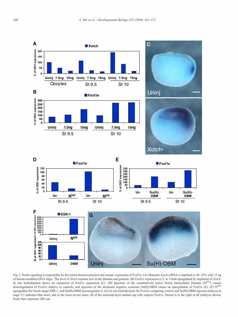
Fig. 5. Notch signaling is responsible for the initial dorsal restriction and mosaic expression of FoxI1e. (A) Maternal Xotch mRNA is depleted to 20–25% with 15 ngof thioate-modified DNA oligo. The level of Xotch remains low in the blastula and gastrula. (B) FoxI1e expression is 2- to 3-fold upregulated by depletion of Xotch.In situ hybridization shows an expansion of FoxI1e expression (C). (D) Injection of the constitutively active Notch Intracellular Domain (NICD) causesdownregulation of FoxI1e relative to controls, and injection of the dominant negative construct Su(H)-DBM causes an upregulation of FoxI1e (E). (F) NICD
upregulates the Notch target ESR-1, and Su(H)-DBM downregulates it. (G) In situ hybridization for FoxI1e comparing control and Su(H)-DBM injected embryos atstage 9.5 indicates that more, and in the most severe cases, all of the sensorial-layer animal cap cells express FoxI1e. Dorsal is to the right in all embryos shown.Scale bars represent 200 μm.
168 A. Mir et al. / Developmental Biology 315 (2008) 161–172

Fig. 6. Maternal Vg1 activates Notch signaling in the blastula to control FoxI1e expression. (A) Vg1-depleted embryos were injected with 50 or 500 pg of NICD mRNAat the 2-cell stage. NICD rescued the increase in FoxI1e expression caused by Vg1 depletion. (B) These results were confirmed by in situ hybridization for FoxI1e atstage 10, which shows an upregulation of FoxI1e in Vg1-depleted embryos, and a reversal of this upregulation by subsequent injection with NICD. The control embryois oriented with dorsal to the right. Depletion of Vg1 results in a delay of gastrulation, and so the orientations of both the Vg1-depleted and the NICD-rescued embryosare indeterminate. Scale bars represent 200 μm. (C) Real-time PCR at stage 10 shows that the Notch target ESR-1 is downregulated in Vg1-depleted embryos relativeto controls, indicating that Notch signaling depends on Vg1 at this stage. (D) 200 pg of Vg1 mRNAwas unable to rescue the increase in FoxI1e expression induced byloss of Notch signaling by injection of 500 pg Su(H)-DBM mRNA.
169A. Mir et al. / Developmental Biology 315 (2008) 161–172
FoxI1e overexpression caused by reduction of Notch signalingcaused by injection of 500 pg Su(H)-DBM mRNA (Fig. 6D).
Discussion
In this work, we have shown that expression of FoxI1e, agene expressed in the animal hemisphere, which controls ecto-derm formation, is subject to multiple levels of control. First isthe surprising observation that it is expressed in a mosaic fa-shion throughout its whole period of expression in the embryo.This is particularly interesting because no other genes have beenshown to be expressed in such a pattern at this early stage in
development. In a previous paper, we showed that expression ofgenes in both neural and epidermal branches of ectodermaldifferentiation are downregulated in FoxI1e-depleted embryos.These target genes are not expressed in mosaic fashions, indi-cating that FoxI1e-expressing cells in the blastula and gastrulaare probably controlling expression of downstream targets in anon-cell-autonomous manner.
The mosaic expression of FoxI1e could be accounted for in 3possible ways. First, it could be cell cycle-dependent, so that atany given time, only a subset of cells at a particular point in thecell cycle express it. The second possibility is that FoxI1e-expressing cells could originate from a few cells and then

170 A. Mir et al. / Developmental Biology 315 (2008) 161–172
disperse by migration across the animal cap. Finally, it could beactivated or repressed in a mosaic pattern by intercellular sig-naling. We show in this paper that blockade of Notch signalingabrogates the mosaic expression of FoxI1e, suggesting that thethird possibility is correct. This does not preclude the first two.However, it is unlikely that FoxI1emRNA is turned over duringpart of each cell cycle, and lineage analysis excludes that pos-sibility that there is large-scale migration of inner animal cellsfrom the dorsal to the ventral side of the embryo. This work alsoconfirms that Notch signaling is active in the blastula, a fact thatwas previously underappreciated.
Second, we show that the normal expression domain ofFoxI1e extends across the whole animal cap, but excludes themost superficial layer of cells throughout its expression period(Fig. 7). Cells in this expression domain turn on FoxI1e in atemporal sequence, from the dorsal to the ventral side, so that atthe gastrula stage, cells all across the expression domain areexpressing FoxI1e. Such a progression could be controlled bylocal factors in the animal cap, or at longer range by factors that
Fig. 7. Model of control of FoxI1e expression in the early embryo. (A) At the blastuladorsal side of the embryo within the subset of prospective ectodermal cells that are cexpression. The competent tissue, in yellow, is adjacent to the blastocoel in the senFoxI1e expression is unknown. It could be the inheritance of an intrinsic transcriphemisphere weakens, FoxI1e expression spreads to the ventral side of the embryo.restriction of animal cells to ectodermal lineages. The inner layer of cells is still comdependent. Activation of dorsal axis formation earlier in development leads to the reFoxI1e expression potential to the ventral ectoderm.
control dorsal/ventral patterning in the rest of the embryo. Weshow that Notch, and thus short-range signaling, is involved.However, we find that longer-range signaling, originating invegetal cells, also controls the expression pattern of FoxI1e. Weshow that the shifting expression of FoxI1e within its expres-sion domain results from different signaling pathways acting atdifferent times. For example, FoxI1e expression in the blastulaand gastrula is unaffected by manipulation of the Wnt signalingthat establishes the dorsal axis. However, the later down-regulation of FoxI1e in the neural plate requires the inhibitionof BMP signaling, which is downstream of Wnt signaling. Inthe late blastula and early gastrula ectoderm, vegetal pathwaysinitiated by VegT and Vg1 influence the spatial pattern ofFoxI1e expression. In the absence of either Vg1, VegT, or nodalsignaling, early expression of FoxI1e is initiated all across theanimal cap, instead of gradually spreading from dorsal toventral.
Finally, we have shown that Vg1 activates the Notch sig-naling pathway to restrict FoxI1e expression to a mosaic pat-
stage, signals from the vegetal hemisphere restrict the expression of FoxI1e to theompetent to express FoxI1e. Vg1 activates Notch signaling to maintain mosaicsorial layer of the animal cap. The molecular nature of this region that permitstion factor, or signaling from other tissues. (B) As signaling from the vegetalThe appearance and spread of FoxI1e expression coincides temporally with thepetent to express FoxI1e. (C) At the neurula stage, FoxI1e expression is BMP-lease of BMP inhibitors from the dorsal mesoderm, resulting in a restriction of

171A. Mir et al. / Developmental Biology 315 (2008) 161–172
tern. The mechanism by which Vg1 activates Notch signalingremains unclear, but the most likely possibilities are throughinteraction of phospho-smad2 with Notch or by upregulatingtranscription of zygotic components of Notch signaling. Ourattempts to identify these components have been unsuccessful,thus far. Though it has been shown that activin can induceDelta-1 and Delta-2 in animal caps (Abe et al., 2004), ouranalysis of their expression in Vg1-depleted embryos has notconfirmed this relationship. Previously, Vg1 has been shown tobe a maternal inducer of mesoderm (Birsoy et al., 2006). Theeffect of Vg1 on Notch could be downstream of mesoderminduction, or through release of signaling molecules into theblastocoel fluid, which would then act on the inner surface ofthe animal cap. The mechanisms by which vegetal pathwaysinfluence animal patterning require further study.
Previous studies have shown that FoxI1e is upregulated inVegT-depleted and in CerS-injected embryos. However, it hasnot, until now, been fully appreciated that the increase inexpression of FoxI1e and other ectodermal genes in the vegetalmass is minor compared to the increase in the animal half of theembryo. This strengthens the hypothesis that there is an ani-mally localized maternal activator of ectoderm formation. Add-itionally, there must be either an activator(s) specific to the deeplayer of the ectoderm, or a repressor(s) of FoxI1e in the super-ficial layer. Indeed, a number of differentially expressed trans-cription factors have been identified, and the candidate may beamong them (Chalmers et al., 2006).
This work represents the first description of regulation ofanimally expressed zygotic genes. Previous work has focusedprimarily on the exclusion of mesodermal gene expression fromthe ectoderm. FoxI1e has been shown to inhibit FGF-mediatedmesoderm induction (Suri et al., 2005), and the Smad4ubiquitin ligase ectodermin attenuates mesoderm induction inthe animal cap by inhibiting all TGF-β signaling, both activin-type and BMP-type (Dupont et al., 2005). Additionally, theMADS box transcription factor SRF disrupts the interaction ofSmad2 and FoxH1, thereby preventing activin-type TGF-βsignaling (Yun et al., 2007). In the absence of ectodermin, SRF,or FoxI1e, mesodermal gene expression expands animally. It isclear from these studies that the inhibition of mesoderminduction in the animal cap keeps the stage clear for ectodermspecification, and offers a mechanism to control its boundaries.However, they also provide evidence that signals originatingfrom vegetal hemisphere can reach the ectoderm. Although wehave not shown that the Vg1 pathway or nodal signalingdirectly affects FoxI1e expression, these data do allow for thispossibility.
FoxI1e is not the first zygotic gene identified that isexpressed in the entire early ectoderm. The transcriptionfactors Xlim5 (Toyama et al., 1995; Houston and Wylie, 2003)and AP-2 (Luo et al., 2002) are restricted to the CNS andepidermis, respectively, late during the gastrula stage, but areboth broadly expressed throughout the ectoderm before thisrestriction. However, this early expression is generally ignored.In an unpublished study, we have shown that both of thesegenes are upregulated in VegT-depleted embryos, indicatingoverlapping regulation. It will be important to analyze the
expression of these genes when testing animally localizedmaternal factors for their role in ectoderm specification.
We have begun to assemble a pathway from maternal con-trol, to intercellular signaling, to ectoderm patterning, but thequestion of why FoxI1e is expressed in a mosaic patternremains. Although it is not expressed in every ectodermal cell,and later becomes confined to the epidermis, FoxI1e is import-ant for the formation of the ectoderm germ layer, before itdivides into epidermis and CNS. There must be signalingevents downstream of FoxI1e that allow it to activate geneexpression in a non-cell-autonomous fashion. It has beenshown that Notch signaling prolongs mesodermal competencein the animal cap (Abe et al., 2004, 2005; Coffman et al.,1993). It has also been shown that ectoderm determination is agradual process that begins during the late blastula stage andcontinues through gastrulation (Heasman et al., 1984; Snape etal., 1987). It is possible that Notch signaling represses FoxI1eexpression in the animal cap in the mid-late blastula, but asanimal Notch signaling weakens, FoxI1e+ cells begin to appearin the ectoderm, forcing the cells around them to activate otherectodermal genes. The gradual activation of FoxI1e coincideswith the restriction of animal cap cells to ectodermal fates.This represents a model integrating Vg1, Notch, VegT, Nodals,and FoxI1e into the specification and patterning of the earlyectoderm.
Acknowledgments
The authors would like to thank Chris Kintner for his gene-rous gift of the NICD and Su(H)-DBM constructs, and for hisfeedback on this manuscript. The authors would also like toacknowledge the financial support of the Cincinnati Children’sHospital Research Foundation, and the National Institutes ofHealth (1R01HD045737).
References
Abe, T., Furue, M., Myoishi, Y., Okamoto, T., Kondow, A., Asashima, M., 2004.Activin-like signaling activates Notch signaling during mesodermalinduction. Int. J. Dev. Biol. 48, 327–332.
Abe, T., Furue, M., Kondow, A., Matsuzaki, K., Asashima, M., 2005. Notchsignaling modulates the nuclear localization of carboxy-terminal-phos-phorylated smad2 and controls the competence of ectodermal cells foractivin A. Mech. Dev. 122, 671–680.
Agius, E., Oelgeschlager, M., Wessely, O., Kemp, C., De Robertis, E.M., 2000.Endodermal Nodal-related signals and mesoderm induction in Xenopus.Development 127, 1173–1183.
Baker, J.C., Beddington, R.S., Harland, R.M., 1999. Wnt signaling in Xenopusembryos inhibits bmp4 expression and activates neural development. Genesand Development 13, 3159-59.
Birsoy, B., Kofron, M., Schaible, K., Wylie, C., Heasman, J., 2006. Vg 1 is anessential signaling molecule in Xenopus development. Development 133,15–20.
Chalmers, A.D., Strauss, B., Papalopulu, N., 2003. Oriented cell divisionsasymmetrically segregate αPKC and generate cell fate diversity in the earlyXenopus embryo. Development 130, 2657–2668.
Chalmers, A.D., Lachani, K., Shin, Y., Sherwood, V., Cho, K.W., Papalopulu,N., 2006. Grainyhead-like 3, a transcription factor identified in a microarrayscreen, promotes the specification of the superficial layer of the embryonicepidermis. Mech. Dev. 123, 702–718.
Coffman, C.R., Skoglund, P., Harris, W.A., Kintner, C.R., 1993. Expression of

172 A. Mir et al. / Developmental Biology 315 (2008) 161–172
an extracellular deletion of Xotch diverts cell fate in Xenopus embryos. Cell73, 659–671.
Deblandre, G.A., Wettstein, D.A., Koyano-Nakagawa, N., Kintner, C., 1999. Atwo-step mechanism generates the spacing pattern of the ciliated cells in theskin of Xenopus embryos. Development 126, 4715–4728.
Dupont, S., Zacchigna, L., Cordenonsi, M., Soligo, S., Adorno, M., Rugge, M.,Piccolo, S., 2005. Germ-layer specification and control of cell growth byEctodermin, a Smad4 ubiquitin ligase. Cell 121, 87–99.
Harland, R.M., 1991. In situ hybridization: an improved whole-mount methodfor Xenopus embryos. Methods Cell Biol. 36, 685–695.
Heasman, J., Wylie, C.C., Hausen, P., Smith, J.C., 1984. Fates and states ofdetermination of single vegetal pole blastomeres of X. laevis. Cell 37,185–194.
Heasman, J., Holwill, S., Wylie, C.C., 1991. Fertilization of cultured Xenopusoocytes and use in studies of maternally inherited molecules. Methods CellBiol. 36, 213–230.
Heasman, J., Kofron, M., Wylie, C., 2000. Beta-catenin signaling activitydissected in the early Xenopus embryo: a novel antisense approach. Dev.Biol. 222, 124–134.
Houston, D.W., Wylie, C., 2003. The Xenopus LIM-homeodomain proteinXlim5 regulates the differential adhesion properties of early ectoderm cells.Development 130, 2695–2704.
Kofron, M., Klein, P., Zhang, F., Houston, D.W., Schaible, K., Wylie, C.,Heasman, J., 2001. The role of maternal axin in patterning the Xenopusembryo. Dev. Biol. 237, 183–201.
Luo, T., Matsuo-Takasaki, M., Thomas, M.L., Weeks, D.L., Sargent, T.D., 2002.Transcription factor AP-2 is an essential and direct regulator of epidermaldevelopment in Xenopus. Dev. Biol. 245, 136–144.
Melton, D.A., 1987. Translocation of a localized maternal mRNA to the vegetalpole of Xenopus oocytes. Nature 328, 80–82.
Mir, A., Kofron, M., Zorn, A.M., Bajzer, M., Haque, M., Heasman, J., Wylie,C.C., 2007. FoxI1e activates ectoderm formation and controls cell positionin the Xenopus blastula. Development 134, 779–788.
Nieuwkoop, P.D., Faber, J., 1967. Normal Table of Xenopus laevis (Daudin).North Holland Publishing Co., Amsterdam, The Netherlands.
Snape, A., Wylie, C.C., Smith, J.C., Heasman, J., 1987. Changes in states ofcommitment of single animal pole blastomeres of Xenopus laevis. Dev. Biol.119, 503–510.
Suri, C., Haremaki, T., Weinstein, D.C., 2005. Xema, a foxi-class gene ex-pressed in the gastrula stage Xenopus ectoderm, is required for the suppres-sion of mesendoderm. Development 132, 2733–2742.
Toyama, R., Curtiss, P.E., Otani, H., Kimura, M., Dawid, I.B., Taira, M., 1995.The LIM class homeobox gene lim5: implied role in CNS patterning inXenopus and zebrafish. Dev. Biol. 170, 583–593.
Yun, C.H., Choi, S.C., Park, E., Kim, S.J., Chung, A.S., Lee, H.K., Lee, H.J.,Han, J.K., 2007. Negative regulation of Activin/Nodal signaling by SRFduring Xenopus gastrulation. Development 134, 769–777.
Zhang, J., King, M.L., 1996. Xenopus VegT RNA is localized to the vegetalcortex during oogenesis and encodes a novel T-box transcription factorinvolved in mesodermal patterning. Development 122, 4119–4129.
Zhang, J., Houston, D.W., King, M.L., Payne, C., Wylie, C., Heasman, J., 1998.The role of maternal VegT in establishing the primary germ layers in Xe-nopus embryos. Cell 94, 515–524.
Zimmerman, L.B., De Jesús-Escobar, J.M., Harland, R.M., 1996. The Spe-mann organizer signal noggin binds and inactivates bone morphogeneticprotein 4. Cell 86, 599–606.