Embed Size (px)
Citation preview

London Cancer
Skin Cancer Radiotherapy Guidelines
August 2013 Review: August 2014
Version: 1.0
Lead author: Girija Anand
2
Contents
1. General considerations .............................................................................................. 3
2. Non melanomatous skin cancer ................................................................................. 4
2.1. Indications for radiotherapy (RT), ............................................................................... 4
2.2. Contraindications for radiotherapy............................................................................. 4
2.3. Investigations .............................................................................................................. 4
2.4. Technique .................................................................................................................... 4
2.5. Target volume ............................................................................................................. 5
2.6. Energy / modality ........................................................................................................ 5
2.7. Dose prescriptions ....................................................................................................... 6
2.8. Dose fractionation regimes ......................................................................................... 6
2.9. Implementation & patient review .............................................................................. 7
3. Mycosis fungoides (Cutaneous T cell lymphoma) ........................................................ 8
4. Cutaneous angiosarcoma ........................................................................................... 8
5. Cutaneous lymphomas .............................................................................................. 8
6. Lentigo maligna ......................................................................................................... 9
7. Radiotherapy for melanoma ...................................................................................... 9
8. Merkel cell carcinoma ............................................................................................. 10
8.1. Staging ....................................................................................................................... 10
8.2. Treatment .................................................................................................................. 10
8.3. Radiotherapy technique ............................................................................................ 11
9. Treatment of Bowen’s disease ................................................................................. 11
10. Treatment of keloids ............................................................................................ 11
10.1. Irradiation to prevent recurrence of keloid scars after excision ........................... 11
10.2. Technique .............................................................................................................. 12
10.3. Energy .................................................................................................................... 12
10.4. Dose ....................................................................................................................... 12
10.5. Patient review ........................................................................................................ 12
11. Radiotherapy for Kaposi’s sarcoma .................................................................... 12
11.1. Dose ....................................................................................................................... 12
11.2. Follow up ............................................................................................................... 13
12. References ........................................................................................................... 14

3
1. General considerations
Either superficial X-rays or electrons can be used. The disadvantages of each are:
SXR o Increased absorption in bone or cartilage due to photoelectric effect so there
is a potential for an increase in necrosis if tumour overlies cartilage. o Less dose fall-off at depth so more dose delivered to deeper tissues (e.g.
brain in scalp tumours)
Electrons o Isodoses constrict at depth, especially with small fields so avoid in fields <3cm
diameter as dose homogeneity is compromised. o Dosimetry is less reliable with oblique angle of incidence to skin surface. o Bowing of isodoses due to lateral electron scatter means the modality is less
useful with tumours close to the eyes. o Dosimetry is less reliable over air cavities due to loss of electronic
equilibrium.
Energy: o The aim is to treat the tumour within the entrance at skin of 100% and exit at
90% isodoses. o The energy required for SXR is defined by tumour thickness + GTV-CTV
margin with reference to the appropriate Percentage Dose-Depth charts. o The energy required for Electron therapy is defined by the tumour thickness
+ GTV-CTV margin with reference to the appropriate Dose-depth charts. The presence or absence of bolus must also be taken into account and be defined on the prescription.
On Treatment Review: o No requirement for on-treatment imaging o Patients should be seen weekly on treatment by nurses and continue such
review until acute toxicities have settled.
Follow-Up: o Patients should be seen 4-8 weeks after treatment in clinic. o Under certain circumstances, follow-up may be coordinated more locally.

4
2. Non melanomatous skin cancer
2.1. Indications for radiotherapy (RT)1,2
Preferred for lesions that would require reconstructive surgery, where irradiation may be considered to produce better cosmesis, e.g. nose3, upper and lower eyelid4 (preferably avoid the middle third of the upper eyelid), the lip and lesions involving the commissure of mouth, the nasolabial sulcus, lesions arising in the pre and post auricular areas and the ear5.
a. Large superficial lesions b. Older patients >75 years c. Patients who refuse or are unfit for surgery (RT avoids anaesthesia) d. Post surgery- for incomplete/marginal excision, perineural invasion
2.2. Contraindications for radiotherapy
Radiotherapy is not normally used in the following circumstances as some skin sites tolerate radiotherapy poorly:
a. Sites of previous burns b. Sites of prior radiotherapy c. Fragile "tissue-paper" skin after steroids d. Areas of vascular insufficiency e. Middle third of the upper eyelid f. The skin of the back overlying the spine (this being an area of skin placed under
pressure, leading to compromised healing and late skin necrosis) g. The skin overlying the shin and malleoli of the lower leg (the skin is under tension
and there may be impaired healing) h. Patients with Gorlin’s syndrome, ataxia telangiectasia, xerodermapigmentosa. i. Carefully consider the use of RT in transplant recipients. Consider management
under the supervision of a dedicated immunosuppression clinic. Consideration of reducing immunosuppression due to increased risk of other skin cancers.
In patients with squamous cell or basal cell carcinoma of the skin, clinically uninvolved regional nodes are not irradiated electively.
2.3. Investigations
a. Histology/cytology (skin scrape, punch biopsy, excision biopsy) b. FNA of enlarged lymph nodes c. Imaging in high risk sites to delineate deep extent and bone or cartilage involvement d. Neck ultrasound or CT neck for high risk sites involving the head & neck region i.e.
ears, nasolabial fold, lips
2.4. Technique

5
Informed consent is obtained. Patients are treated in a position that best suits the location of the lesion. Immobilisation is achieved as appropriate. Low energy photons or electrons are used. Generally lesions around the eye, ear, nose and those with an irregular border will require a customised lead cut-out, necessitating a additional appointment for manufacture of the mould. All other areas are treated as ‘mark on set’ using standard lead cutouts to shield the non-target areas using the appropriate thickness of lead. The eye requires special protection when eyelids are treated. A spatula (if one eyelid involved) or a lead contact eye shield is inserted under local anaesthetic (LA) to protect the cornea, the lens and uninvolved eyelid (with spatula). Patients’ will require an eye pad for 2-3 hrs after the procedure until the LA effect is reversed & corneal reflex restored. Ensure that patient has an escort, as he/she may not be able to drive back. The nasal septum and gum are respectively shielded when lower nose and lip are treated. Wax or aluminium is used to cover the lead shield to absorb electron backscatter.
2.5. Target volume
All patients are examined under a bright light with a magnifying glass/ dermatoscope Squamous Cell Carcinoma [SCC]: Planning target volume (PTV)= Gross tumour volume (GTV) + 1cm margin all around laterally & 5mm or more deep according to clinician’s discretion. NB consider electrons for tumour thickness over 5mm and if the tumour is attached to bone or cartilage or is up against bone, discuss with physics. Basal cell carcinoma [BCC]: PTV = GTV + 0.5cm margin laterally (0.7cm-1cm if morphoeic, large, infiltrative or poorly defined) and 0.5 cm deep margin. If on mobile skin a 0.25cm deep margin is adequate. For electron therapy an additional 0.5cm-1cm margin is required laterally to account for penumbra of the electrons. Clinical photographs are taken for record, clearly indicating the site, extent of lesion and orientation of the photograph.
2.6. Energy / modality
Aim to cover PTV with the 90% isodose, both laterally and at depth. Low energy photons (60/120KV) or electrons are used depending on depth, site, size & availability. Avoid electrons near eyes (lateral scatter) and for field sizes <4cm. If the external auditory meatus is included in the electron field, a wax block should be used to avoid funnelling effect on the eardrum. Try and avoid superficial X-rays over cartilage & bone due to risk of radionecrosis (absorption by photoelectric effect).

6
When using electrons, discuss ‘build-up’ (bolus) requirements with physicist, to bring the skin dose to 95% or more, and encompass the target depth with the 90% isodose. Photons of low voltage deliver almost 100% to the skin, and so do not require ‘build-up’. The depth dose characteristics of the beam depend on energy and field size.
2.7. Dose prescriptions
Prescriptions are applied doses (Dmax) for photons, and to aim for a skin dose of 95-100%. (follow ICRU guidelines). Doses depend upon lesion size, histology and patient characteristics.
2.8. Dose fractionation regimes
Basal Cell Carcinoma – orthovoltage or electron doses:
18Gy/1# Small tumours where cosmesis is relatively unimportant
24Gy/2# Both fractions given five weeks apart, where cosmesis is less important and target <5cm (Sambrook split)
35Gy/5#/1-2 weeks For small lesions under 2 cm diameter ideally not when overly cartilage
40.5Gy/9#/2 weeks Target size up to 5 to 6cm diameter; this can be given as alternate day fractions over a 3 week period if patient access to hospital is limited, or in the elderly
50Gy/15#/3 weeks Target is between 4 and 6cm diameter and not in an area of poor radiation tolerance
55Gy/20#/4 weeks Target < 6cm, in areas of poor radiation tolerance
60Gy/30#/6 weeks Target > 6cm in areas of poor radiation tolerance
For field sizes <5cm can use dose fractionation regimes with radiobiological equivalent doses of 49Gy-50Gy in 2Gy/fraction in accordance with local protocols Squamous Cell Carcinoma doses:
18Gy/1# Small tumours where cosmesis is relatively unimportant
24Gy/2# Both fractions given five weeks apart, in elderly frail patients where cosmesis is less important and target <5cm
7
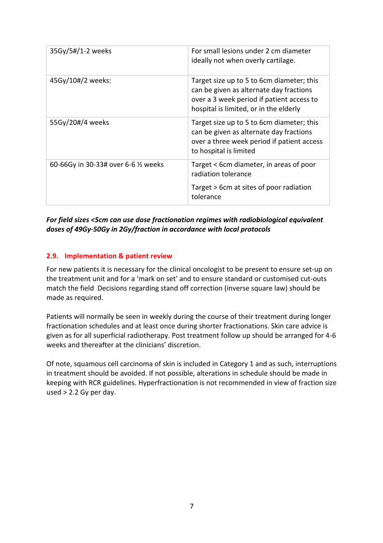
35Gy/5#/1-2 weeks For small lesions under 2 cm diameter ideally not when overly cartilage.
45Gy/10#/2 weeks:
Target size up to 5 to 6cm diameter; this can be given as alternate day fractions over a 3 week period if patient access to hospital is limited, or in the elderly
55Gy/20#/4 weeks Target size up to 5 to 6cm diameter; this can be given as alternate day fractions over a three week period if patient access to hospital is limited
60-66Gy in 30-33# over 6-6 ½ weeks
Target < 6cm diameter, in areas of poor radiation tolerance
Target > 6cm at sites of poor radiation tolerance
For field sizes <5cm can use dose fractionation regimes with radiobiological equivalent doses of 49Gy-50Gy in 2Gy/fraction in accordance with local protocols
2.9. Implementation & patient review
For new patients it is necessary for the clinical oncologist to be present to ensure set-up on the treatment unit and for a ‘mark on set’ and to ensure standard or customised cut-outs match the field Decisions regarding stand off correction (inverse square law) should be made as required. Patients will normally be seen in weekly during the course of their treatment during longer fractionation schedules and at least once during shorter fractionations. Skin care advice is given as for all superficial radiotherapy. Post treatment follow up should be arranged for 4-6 weeks and thereafter at the clinicians’ discretion. Of note, squamous cell carcinoma of skin is included in Category 1 and as such, interruptions in treatment should be avoided. If not possible, alterations in schedule should be made in keeping with RCR guidelines. Hyperfractionation is not recommended in view of fraction size used > 2.2 Gy per day.

8
3. Mycosis fungoides (Cutaneous T cell lymphoma)
For small localised plaques and tumours, treatment with low energy photons are adequate. PTV = GTV + 0.5 to 1 cm margins; margins tend to be smaller as these patients are often retreated. Dose: 8Gy in 2 fractions or 12Gy in 3 fractions treating daily For a larger or thicker tumour or collection of plaques, and particularly if the area is over a joint consider treatment with electrons: Dose: 20Gy in 5 fractions over 1 week or 30Gy in 10 fractions over 2 weeks.
4. Cutaneous angiosarcoma6
Cutaneous angiosarcoma is a rare and aggressive endothelial-derived sarcoma usually affecting the elderly. Combined modality approach is often required including surgery, radiotherapy and chemotherapy. All cases must be reviewed at network Sarcoma MDT. Radiotherapy technique
Photons or electrons are used as appropriate. A customised cut out may be required. A margin of at least 1-2 cm is required as sub microscopic disease can extend beyond clinical tumour margin.
Dose
55Gy in20 fractions over 4 weeks or 60Gy in 30 fractions over 6 weeks.
5. Cutaneous lymphomas
Indication
Biopsy proven lymphoma confined to the skin Investigations
Histopathology or cytology report with the Pathology having been reviewed by an appropriate MDT
Patients should have appropriate staging investigations, according to histopathological type. This may include PET-CT scanning and Bone Marrow Aspirate and Trephine.
Any case considered for a radical radiotherapy course for cutaneous lymphoma should have been assessed by the haematology MDT as having had a satisfactory level of pre-treatment staging. Radiation therapy is the treatment of choice for localized disease at presentation or on relapse. Polychemotherapy should be reserved for involvement of non-contiguous anatomic sites or those with extra cutaneous spread.

9
Patient Positioning
As appropriate to tumour site with reference to reproducibility and patient comfort and well as radiation physics requirements.
Where tumours overlie mucosal sites such as the buccal mucosa, nose or conjunctiva, consideration must be given to the provision of appropriate lead (Pb) shielding.
Pb shielding should have a wax covering facing the direction of the incident beam to reduce mucosal toxicity secondary to secondary electron backscatter.
Tumour Visualisation
Tumour edges must be demarcated with the use of a white, bright light and magnifying glass.
Tumour demarcation should take place with any Pb shielding in situ.
Fields- Generally treated with applied fields.
PTV = GTV (Palpable / visible tumour) + 10mm (consider a larger margin if the tumour is indistinct or larger than 5cm)
Dose
T cell Lymphoma:8Gy in 2 daily fractions over 2 days
Low-Grade B Cell Lymphoma: 15Gy in 5 daily over 5 days
High-Grade B Cell Lymphoma:30-40Gy in 15-20 daily fractions over 3-4 weeks See Lymphoma guidelines on Insight for further details.
6. Lentigo maligna
This is a non-invasive melanotic lesion generally occurring on the face. Treatment with radiotherapy is preferred in areas of cosmetic and functional importance and this can achieve local control. Although this is generally a flat lesion it has irregular borders with varying degrees of pigmentation and a customised cut out is usually required. Planning target volume = GTV + 1cm margin laterally and 0.5cm deep margin. Dose
45Gy in 10 fractions over 2 weeks with 60kv - 120kv photons.
7. Radiotherapy for melanoma
Treatment should be individualized to suit the patient and the treatment regimen should be discussed with at a specialist MDT. Treatment intent is usually palliative. Some commonly used schedules for cutaneous disease include the following:

10
Dose
30-36Gy in 6Gy per fraction once weekly over 5-6 weeks.
20Gy in 5 fractions treating daily over 1 week.
30Gy in 5 fractions with 2 fractions given each week.
8-10Gy single fraction especially in poor performance status patients where rapid control of symptoms such as bleeding is required.
Treatment can be offered in the adjuvant setting on a case by case basis following discussion at a specialist MDT. Radiotherapy may improve progression free survival, but has no impact on overall survival and may sometimes impact on quality of life due to morbidity from treatment7.
8. Merkel cell carcinoma8 9
These are rare neuroendocrine tumours arising from the mechanoreceptors of the basal epidermis that are particularly aggressive with a propensity for head and neck and the extremities. Causative factors include exposure to sunlight and immunosuppression. The tumour has many similarities to small-cell carcinoma of the lung, with intrinsic sensitivity to radiotherapy and chemotherapy and an aggressive metastatic potential.
8.1. Staging
IA Disease confined to skin and <2 cm in diameter IB Disease confined to skin and >2 cm in diameter II Involvement of regional lymph nodes III Metastatic disease
8.2. Treatment
Surgery is the initial treatment of choice. Most groups advocate a 2–3-cm tumour-free margin around the primary lesion when technically possible (no controlled trials comparing different margins), but this is often difficult to achieve in the head and neck region. Many authors recommend postoperative radiotherapy on the basis of retrospective comparison of small numbers of patients treated with surgery alone with those treated with surgery and PORT. The addition of radiotherapy reduced the local failure from 39% to 26% and the regional failure from 46% to 22%. Radiation volumes have included the GTV with generous margins to ensure that the dermal lymphatics surrounding the primary are treated to full dose. The doses used have varied from 45–60 Gy, with higher doses being applied to areas of bulky disease. Treatment of regional draining lymph nodes has been recommended, although prophylactic node dissection or radiotherapy has not shown to influence overall survival. Sentinel node biopsy has been proposed to identify risk of recurrence and minimise need for node dissection.

11
The presence of distant metastases carries a grave outlook, with median survival being only 9 months. Treatment intent is purely palliative. Radiation can be used to palliate bone and brain secondaries, and for advanced cutaneous deposits that are bleeding or fungating. The role of adjuvant chemotherapy remains undefined. Overall response rates to combination chemotherapy (usually platinum based) for surgically unresectable distant metastatic disease are generally high, although responses are transient.
8.3. Radiotherapy technique
Radical
Photons or electrons are used depending on depth, site and size of lesion to be treated. PTV=GTV + 3-5cm. Dose: 45-60Gy over 5-6 weeks (higher doses for residual or bulky disease).
Palliative
Phase 1: 20Gy in 5 fractions over 1 week. Or Phase 1: 20Gy/5#over one week split course (2 week gap) and
Phase 2: 20Gy/5# over one week10
9. Treatment of Bowen’s disease
This is also known as squamous cell carcinoma in situ and is generally treated by dermatologists. Cases may be referred for radiotherapy when non-responsive areas become troublesome to the patient and are associated with thickening, pain and bleeding. Guidelines from the British Association of Dermatologists suggest that Bowen’s disease of the lower limb does not have to be treated and can be observed. Dose
Treated as a squamous cell carcinoma to a dose of 45Gy in 10 fractions over 2 weeks.
10. Treatment of keloids11
As many of these patients are young, and will live long enough to experience potential late radiation toxicity, this must be discussed specifically when obtaining signed consent. The information given must be recorded in the case notes. No patients under 16 years old will be consented without the presence of a parent or guardian.
10.1. Irradiation to prevent recurrence of keloid scars after excision
These patients are treated jointly with the plastic surgery team. Radiotherapy is given to the operative site normally within 24 hours after operation. This requires close liaison: if there

12
are no free treatment spaces within 24 hours of the suggested operation time, inform the surgeons at once, so that operation dates can be altered.
10.2. Technique
Direct low voltage photon irradiation is normally used. The planning target volume (PTV) is the whole operative scar including suture sites, with 0.5 cm margins, and is treated with low kV energies. Remember that the treatment volume will need to be greater, due to fall-off in dose at field edges. This effect is more pronounced in long, narrow fields. Ask advice from Physics if you are unsure what extra margins to allow in positioning the lead shielding. All keloid scars are treated as ‘mark on set’. 2mm lead shielding is required to protect skin in non-target areas.
10.3. Energy
Low energy Kv photon or electrons.
10.4. Dose
10Gy applied dose/1#/1 day.
12Gy in 2 fractions over 7 days (fraction 2 post suture removal)
16Gy in 2 fractions over 7 days (BED >30 Gy)12
10.5. Patient review
It is necessary for a clinical oncologist to be present at time of treatment to see set up and correct for stand off and revisit consent process if any queries from patients or associated professionals. Patients are reviewed in clinic 6-8 weeks later and then discharged after a clinical photograph. Patients are not routinely followed but are advised to return to the Plastic surgery clinic if they are concerned about recurrence.
11. Radiotherapy for Kaposi’s sarcoma13 14 15
Radiotherapy continues to be a very useful mode of palliative treatment for all forms of Kaposi’s sarcoma. These are radiosensitive tumours and objective response rates vary between 70-95%. KS will often present as multicentric lesions. Cutaneous lesions can be treated with low energy photons or electrons depending on thickness, site and size of the lesion, with a margin of 0.5-1cm. (PTV=GTV + 0.5-1cm).
11.1. Dose
13
For single or small lesions < 4 cm Dose: 8 Gy as a single fraction treated with kv photons. If there is a cluster of lesions or a single lesion >4 cm Dose: 12 Gy in 3 fractions given on alternate days Alternatively for larger areas involving the extremities and other body sites: Use megavoltage photons or electrons Dose: 24 Gy in 6 fractions treating daily Or Dose: 36 Gy in 6 fractions given over six weeks, with each fraction given weekly, is an alternative, but the patient should be reviewed prior to each fraction to assess response. Should there be a rapid response the total dose received may be reduced. Or Dose: 60 Gy in 30 fractions for very large areas. Each fraction given daily. If the last two schedules are being used to treat extremities the principle of sparing a skin corridor should be adhered to. Kaposi sarcoma of the oral cavity presents a special problem as lesions radiated at this site are prone to severe mucositis especially in HIV positive patients. This site should always be treated with a fractionated regimen. 10Gy-20Gy in 2 Gy fractions with megavoltage parallel opposed photons.
11.2. Follow up
2-3 weeks after RT for mucosal sites. 3-4 months post RT for skin sites. For HIV patients, follow up in conjunction with Infection and Immunity Physicians.

14
12. References
1 Locke J, Karimpour S, Young G, Lockett MA, Perez CA. Radiotherapy for epithelial skin cancer. Int J
RadiatOncolBiolPhys 2001; 51(3): 748-55. 2 Bath FJ, Bong J, Perkins W, Williams HC Interventions for basal cell carcinoma of the skin. Cochrane
Database Syst Rev 2003;(2): CD003412 3 Caccialanza M, Piccinno R, Moretti D, et al. Radiotherapy of carcinomas of the skin overlying the
cartilage of the nose: results in 405 lesions. Eur J Derm 2003; 13(5): 462-5. 4 Schlienger P, Brunin F, Desjardins L, et al. External radiotherapy for carcinoma of the eyelid: Report
of 8 Lukawska J, Cottrill C, Bower M. The changing role of radiotherapy in AIDS-related malignancies. Clinical Oncology. 2003; 15: 2-6.50 cases treated. Int J RadiatOncolBiolPhys 1996; 34(2): 277-287
5 Caccialanza M, Piccinno R, Kolesnikova L, Gnecchi L. Radiotherapy of skin carcinomas of the pinna: a study of 115 lesions in 108 patients. Int J Dermatol 2005; 44(6): 513-7.
6 Morgan MB, Swann M, Somach S et al. Cutaneous angiosarcoma: a case series with prognostic correlation. J Am AcadDermatol. 2004; 50(6): 867-74.
7 Henderson MA et al., Adjuvant radiotherapy after lymphadenectomy in melanoma patients: Final results of an intergroup randomised trial (ANZMTG 01.02/TROG 02.01), J Clin Oncol 31, 2013 (suppl: abstr 9001)
8 Poulsen M. Merkel-cell carcinoma of the skin.Lancet Oncol. 2004;5(10):593-9 9 Suarez C, Rodrigo JP, Ferlito A, et al. Merkel cell carcinoma of the head and neck. Oral Oncol 2004;
40(8): 773-9. 10 Kancherla KN et al: The role of split course palliative hypofractionated radiotherapy in head & neck
cancers, Clinical Oncology (Royal College of Radiologists (Great Britain))[2011, 23(2):141-148] 11 Ragoowansi R, Cornes P, Moss AL, Glees JP. Treatment of Keloids by Surgical Excision & Immediate
Postoperative Single-Fraction Radiotherapy. PlastReconstrSurg 2003; 111(6): 1853-59. 12 Dose-effect relationships for recurrence of keloid and pterygium after surgery and radiotherapy”
Kal et al. Int J Radiat Oncol Biol Phys. 2009 May 1;74(1):245-51. 13 Lukawska J, Cottrill C, Bower M. The changing role of radiotherapy in AIDS-related malignancies.
Clinical Oncology. 2003; 15: 2-6. 14 Harrison M, Harrington KJ, Tomlinson DR, Stewart JS. Response and cosmetic outcome of two
fractionation regimens for AIDS-related Kaposi’s sarcoma. RadiotherOncol. 1998: 46(1): 23-8 15 Kirova YM, Belembaogo E, Frikha H, et al. Radiotherapy in the management of epidemic Kaposi’s
sarcoma: a retrospective study of 643 cases. RadiotherOncol. 1998; 46(1): 19-22.

![Inflammation and cancer: How hot is the link? · carcinoma [30], colon carcinoma, lung carcinoma, squamous cell carcinoma, pancreatic cancer [31,32], ovarian carcinoma biochemical](https://img.dokumen.tips/doc/110x75/5fcdd6c81c76a34db570e7e6/iniammation-and-cancer-how-hot-is-the-link-carcinoma-30-colon-carcinoma.jpg)