Embed Size (px)
Citation preview
[1]
LOCALIZATION OF NICOTINIC RECEPTORS ON HAIR CELLS
AND AFFERENTS OF THE TURTLE SEMICIRCULAR CANAL
A Major Qualifying Project
Submitted to the Faculty
Of the
WORCESTER POLYTECHNIC INSTITUTE
In partial fulfillment of the requirements for the
Degree of Bachelor of Science
By
_________________________
Akanksha Sharma
Date: October 19, 2007
Approved:
_______________________
Professor Jill Rulfs
Advisor
[2]
ABSTRACT
A distinct α9/α10 nicotinic acetylcholine receptor (nAChR) may mediate the inhibition of
type II hair cells in the turtle posterior crista. This project aims to localize these nAChRs
in the hair cell layer with the use of commercial antibodies and fluorophore-conjugated
nicotinic antagonists (α-bungarotoxin). An additional goal has been the optimization and
development of protocols for this specific purpose. Identification and visualization of
these nAChRs will provide greater insight into the complex neural pharmacology of the
inner ear.
[3]
ACKNOWLEDGMENTS
I would like to convey my sincere appreciation and heartfelt gratitude to a
wonderful researcher and mentor, Dr. Joseph C. Holt. This work was conducted under his
constant guidance and with his untiring support, in his laboratory at the Otolaryngology
Department at the University of Texas Medical Branch (UTMB) in Galveston, Texas.
I am grateful to Dr. Golda A. Leonard and Dr. Robert Leonard at UTMB who
were always available for me to call on for advice in my research, and whose expertise
has been a great help. I would also like to acknowledge Pat Gazzoli and Laura Teed, also
at UTMB, for their kind help and assistance in several ways. In addition, I would like to
thank Dr. Jill Rulfs for her role as my project advisor at WPI.
Last, but not the least, I thank my parents. Their unstinted support and constant
encouragement has played a significant role throughout my education.
[4]
TABLE OF CONTENTS
I. BACKGROUND ......................................................................................................... 6 A. Function ............................................................................................................... 6
B. Overall Structure .................................................................................................. 6 C. The Hair Cell...................................................................................................... 10 D. Mechanotransduction by Vestibular Hair Cells ................................................. 12 E. The Turtle Crista ................................................................................................ 13 F. Vestibular Afferents ........................................................................................... 14
G. Vestibular Efferents ........................................................................................... 15 H. Efferent Neurotransmitters of the Vestibular System ........................................ 15 I. Acetylcholine (ACh) .......................................................................................... 16 J. Muscarinic Acetylcholine Receptors (mAChRs) ............................................... 17
K. Nicotinic Acetylcholine Receptors (nAChRs) ................................................... 18 II. INTRODUCTION ................................................................................................. 20
A. The α9 Nicotinic Acetylcholine Receptor (α9 nAChR) .................................... 20 B. Immunohistochemistry ...................................................................................... 22
C. α-Bungarotoxin .................................................................................................. 23 D. Apamin ............................................................................................................... 24
III. SPECIFIC PURPOSE ............................................................................................ 26
IV. MATERIALS & METHODS ................................................................................ 29 A. Reagents, Primary and Secondary Antisera and Toxins .................................... 29
B. Tissue Preparation and Labeling ........................................................................ 31 V. RESULTS .............................................................................................................. 35
A. Afferent and Efferent Cell Markers ................................................................... 35
B. α9 and α4 nAChR Labeling ............................................................................... 39
C. α-Bungarotoxin Labeling ................................................................................... 41 D. Apamin Labeling for SK Channels .................................................................... 45
VI. DISCUSSION ........................................................................................................ 46
A. Characterizing the Crista.................................................................................... 46 B. α-Bungarotoxin Binding in Vestibular Hair Cells ............................................. 47
C. Apamin Binding in Vestibular Hair Cells .......................................................... 51 D. Future Outlook ................................................................................................... 53
VII. REFERENCES ...................................................................................................... 56

[5]
LIST OF FIGURES
Figure 1: Introduction to the preparation. ........................................................................... 7 Figure 2: Summary of efferent responses in the turtle posterior crista and their
pharmacology. ................................................................................................................... 27 Figure 3: Photograph of the Tissue Catcher ..................................................................... 32 Figure 4: Calretinin (CR) labeling in the turtle posterior crista ........................................ 36 Figure 5: Neurofilament-200 (NF-200) labeling in the turtle posterior crista .................. 37 Figure 6: Efferent markers in the turtle posterior canal hemicrista .................................. 38
Figure 7: Immunohistochemistry with antibodies against α9nAChR and α9nAChR
subunits and AlexaFluor-594 α-BTX in fixed tissue from turtle posterior canal crista. .. 40 Figure 8: Immunohistochemistry with Alexa Fluor 488 α-BTX in live, unfixed tissue
from the turtle posterior canal crista. ................................................................................ 42
Figure 9: Immunohistochemistry with Alexa Fluor 488 α-BTX in live, unfixed tissue
from the turtle posterior canal crista. ................................................................................ 43
Figure 10: Blocking α-BTX labeling with preincubation with methyllycaconitine. ........ 44 Figure 11: Labeling observed with application of Alexa Fluor 488 apamin to live, unfixed
tissue from the turtle posterior crista. ............................................................................... 45
[6]
I. BACKGROUND
A. Function
Organs of the vestibular nervous system, located in the inner ear, are primarily
responsible for providing input regarding our balance and movement within Earth‟s
gravitational field. Information on linear and rotational movement of the head, as well as
its static position, is tracked by these organs and relayed to the central nervous system
(CNS). This information can then be transmitted as instructions to muscles responsible
for posture and equilibrium and also to neural structures responsible for controlling eye
movements. Higher order processes such as spatial perception and navigation are also
dependent on the vestibular system.
The crucial functions of this system are best realized with a simple reflection at
what happens when it is stimulated in atypical ways or when it ceases to work correctly.
Inappropriate motion of or damage to the vestibular organs and/or its pathways can give
rise to sensations of vertigo, dizziness or a sense of imbalance, and can include feelings
of discomfort or nausea. Everyday examples include the experience of spinning in circles,
taking a long car ride on a bumpy road, a ride at an amusement park, the sense of
imbalance during an ear infection. Sudden falls, a major cause of injury and mortality in
the elderly, may also be attributed to a loss of vestibular sensitivity.
B. Overall Structure
The vestibular system is one of the more highly conserved organ systems, similar
both anatomically and physiologically from fish to mammals [36]. The vestibular system
starts in the ear with the membranous labyrinth, composed of a series of fluid-filled tubes
and sacs, housed within channels of the temporal bone referred to as the bony labyrinth
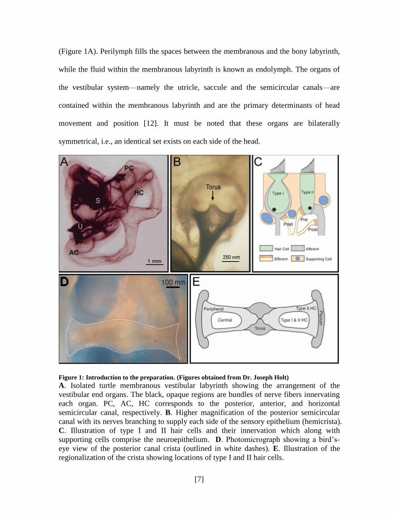
[7]
(Figure 1A). Perilymph fills the spaces between the membranous and the bony labyrinth,
while the fluid within the membranous labyrinth is known as endolymph. The organs of
the vestibular system—namely the utricle, saccule and the semicircular canals—are
contained within the membranous labyrinth and are the primary determinants of head
movement and position [12]. It must be noted that these organs are bilaterally
symmetrical, i.e., an identical set exists on each side of the head.
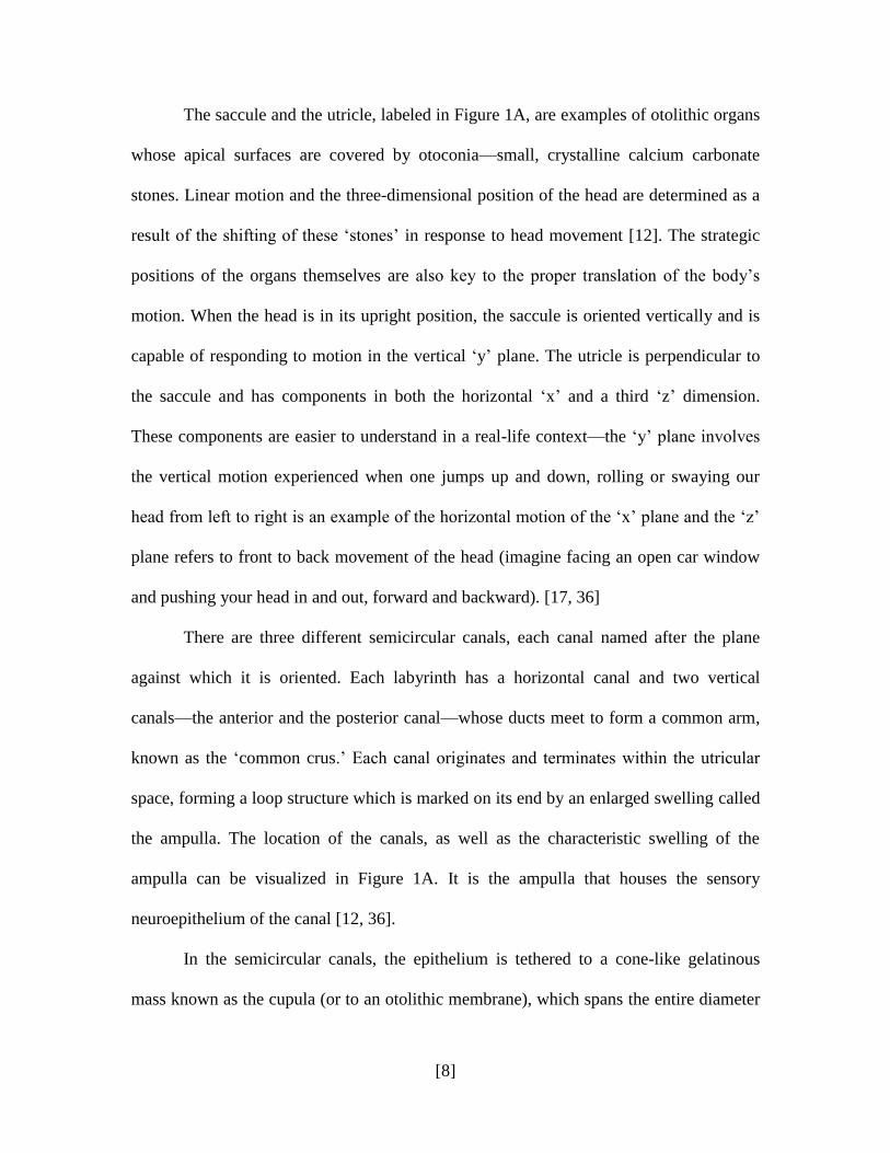
Figure 1: Introduction to the preparation. (Figures obtained from Dr. Joseph Holt)
A. Isolated turtle membranous vestibular labyrinth showing the arrangement of the
vestibular end organs. The black, opaque regions are bundles of nerve fibers innervating
each organ. PC, AC, HC corresponds to the posterior, anterior, and horizontal
semicircular canal, respectively. B. Higher magnification of the posterior semicircular
canal with its nerves branching to supply each side of the sensory epithelium (hemicrista).
C. Illustration of type I and II hair cells and their innervation which along with
supporting cells comprise the neuroepithelium. D. Photomicrograph showing a bird‟s-
eye view of the posterior canal crista (outlined in white dashes). E. Illustration of the
regionalization of the crista showing locations of type I and II hair cells.
[8]
The saccule and the utricle, labeled in Figure 1A, are examples of otolithic organs
whose apical surfaces are covered by otoconia—small, crystalline calcium carbonate
stones. Linear motion and the three-dimensional position of the head are determined as a
result of the shifting of these „stones‟ in response to head movement [12]. The strategic
positions of the organs themselves are also key to the proper translation of the body‟s
motion. When the head is in its upright position, the saccule is oriented vertically and is
capable of responding to motion in the vertical „y‟ plane. The utricle is perpendicular to
the saccule and has components in both the horizontal „x‟ and a third „z‟ dimension.
These components are easier to understand in a real-life context—the „y‟ plane involves
the vertical motion experienced when one jumps up and down, rolling or swaying our
head from left to right is an example of the horizontal motion of the „x‟ plane and the „z‟
plane refers to front to back movement of the head (imagine facing an open car window
and pushing your head in and out, forward and backward). [17, 36]
There are three different semicircular canals, each canal named after the plane
against which it is oriented. Each labyrinth has a horizontal canal and two vertical
canals—the anterior and the posterior canal—whose ducts meet to form a common arm,
known as the „common crus.‟ Each canal originates and terminates within the utricular
space, forming a loop structure which is marked on its end by an enlarged swelling called
the ampulla. The location of the canals, as well as the characteristic swelling of the
ampulla can be visualized in Figure 1A. It is the ampulla that houses the sensory
neuroepithelium of the canal [12, 36].
In the semicircular canals, the epithelium is tethered to a cone-like gelatinous
mass known as the cupula (or to an otolithic membrane), which spans the entire diameter
[9]
of the ampulla. When we shake our head to indicate yes or no, or turn to look behind us,
the canals instantaneously move with our head. The endolymph or fluid in the canals,
however, cannot move at the same speed and travels at a slower velocity. This inertial
motion results in a pressure upon the gel-like cupula, oppositely displacing its mass in a
mechanical movement which is then detected by the “tethered” neuroepithelium [12].
The specific steps involved in this mechanotransduction will be discussed in greater
detail at a later point.
Each vestibular organ has a layer of sensory epithelium. In the utricle and the
saccule, the epithelium forms a flat sheet like structure against a wall of the organ
(anterior for the utricle, ventrolateral for the saccule), which is referred to as the macula.
However, our studies are predominantly focused on the neuroepithelium of the
semicircular canals, which is known as the ampullary crest or the crista (plural: cristae).
A view of the crista as seen if looking straight down the canal is seen in Figure 1B, and a
bird‟s eye view (as seen from above) is provided as Figure 1D [12, 36].
The crista and the macula are mostly similar in terms of cellular makeup.
Essentially, the neuroepithelium is a matrix of hair cells, supporting cells, and the
synaptic connections of afferents and efferents which rests on a stroma composed of
connective tissue which is penetrated by blood vessels and nerve fibers (the central area
in Figure 1B and 1D) [18]. The epithelia‟s placement between the perilymph (sodium-
rich, potassium-deficient, typical extracellular fluid) and the endolymph (potassium-rich,
sodium-deficient, intracellular fluid) acts as an interface. This separation is necessary, of
course, to maintain the ionic gradient necessary for normal function of the system [12].
[10]
C. The Hair Cell
The most crucial component of the vestibular organs are the mechanically
sensitive hair cells, so named for the fingerlike projections protruding from the top of
each cell (Figure 1C). The apical pole of each individual hair cell is marked by a tuft of
sensory hairs, called stereocilia. These stereocilia are the primary transducers of
mechanical motion associated with the vestibular system, such as that caused by the
movement of the cupula or otoconia. The basal half of the hair cell is marked by synaptic
bodies, surrounded by vesicles and adjacent to attachments for the pre and post-synaptic
membrane specializations of the hair cell and its corresponding nerve terminal. [12, 36]
An individual stereocilium has a rigid actin core surrounded by plasmalemma.
Anywhere from 20-100 such stereocilia form rows on the tops of hair cells, usually in
descending order with the tallest immediately adjacent to a single kinocilium [23, 38].
The kinocilium is a microtubular structure which typically serves as the point of
attachment between the hair cell and the otolithic membrane or the cupula. The
arrangement of the stereocilia to the kinocilium is itself a critical characteristic of the
vestibular machinery. As mentioned above, the cupula is displaced in response to angular
acceleration, such as shaking or nodding the head. This movement in turn deflects
stereocilia and hair cells. Deflection of the stereociliary tuft (hair bundle) towards the
kinocilium results in depolarization (and therefore excitation) of the hair cell, while
deflection away from the kinocilium translates to a hyperpolarization (inhibition) of the
hair cell. This is the fundamental principle behind how the vestibular system detects and
translates head movements to a language that the central nervous system ultimately
understands. [13]

[11]
A brief note must be made here about stereocilia direction in the various organs:
in the semicircular canals, the hair bundles all face the same direction. Therefore,
displacement of the cupula itself translates into the depolarization (in one direction) or
the hyperpolarization (in the other direction) of the hair cell. In the saccule and the utricle,
however, the polarity of the hair bundles changes at specific points in the macula. A line,
known as the striola, may be drawn at this point to demarcate the change in polarity.
Secondly, the striola transects the macula such that as one moves from one side to the
other, hair cells change their orientation slightly to remain perpendicular to the striola.
This endows otolithic organs with many axes of sensitivity. [18, 29, 36]
In the crista and macula of reptiles, birds and mammals, there are two types of
vestibular hair cells. Type II hair cells (on the right, Figure 1C) are cylindrically shaped
and are innervated by a button-shaped, or bouton, afferent ending. Both efferents and
afferents make contact with the type II hair cell via bouton endings. Fish and amphibian
neuroepithelia only have type II hair cells, and consequently only bouton-type afferents.
Type I hair cells (on the left, Figure 1C) are flask-shaped with a rounded base and a short,
constricted neck. In contrast, with bouton afferents, the afferent innervation here engulfs
the entire type I hair cell and a large part of the neck, resulting in a so-called calyx ending
that can be easily distinguished in the dense hair cell layer. The structural nature of the
calyx afferent also prevents any efferent contact with the type I hair cell. [2]
Bouton and calyx afferents receive an electrical translation of the mechanical
movement of hair cells, and relay this information from the vestibular end-organs to the
brain. Efferents, on the other hand, carry directions received from the brain to target
organs. Efferents innervating the vestibular organs originate near the facial nuclei in the
[12]
brain stem, where they travel in nerve VIII and branch profusely to each vestibular organ.
They can be distinguished from afferents due to their highly vesiculated appearance at the
EM level. Efferents may end as boutons on calyx afferents, on afferents innervating type
II hair cells or on type II hair cells themselves (Figure 1C). [12]
Finally, supporting cells constitute a large part of the layer, wrapping around hair
cells and filling the spaces in-between. They are crowded with secretory-like granules
and organelles, and their basal portion is nucleated. Supporting cells are also important in
the synthesis and maintenance of the macula and cupula, in the removal of the
neurotransmitter from synaptic spaces and in the regulation of the ionic environment of
the perilymph and endolymph. [18]
D. Mechanotransduction by Vestibular Hair Cells
Mechanotransduction in the vestibular system may be defined simply as a
conversion of mechanical stimulus into electro-chemical activity. The slightest movement
of our head must be conveyed firstly to the vestibular system (which happens
mechanically) and then it must be transmitted to the brain chemically and then
electrically. When hair bundles are deflected by movement of the cupula,
mechanoelectric transducer (MET) channels either open or close, depending on the
direction of movement. Recall from the discussion of the epithelia that when stereocilia
are deflected towards the kinocilium, excitation (or depolarization) of the hair cell takes
place, and when deflected away from the kinocilium, inhibition (or hyperpolarization) of
the hair cell occurs. When MET channels open as a result of deflection towards the
kinocilium, a depolarizing transducer current is created as endolymphatic potassium ions
move into the hair cell. Voltage-sensitive calcium channels in the basolateral
[13]
plasmalemma of the hair cell open, leading to an influx of calcium ions and a subsequent
release of the afferent neurotransmitter, glutamate, from vesicles around the synaptic
ribbons onto the afferent synapse. Glutamate is packaged in vesicles that fuse with the
plasmalemma when cytosolic calcium is high, consequently releasing their contents. The
released glutamate then diffuses across the synapse, binds to the post-synaptic receptor,
and then depolarizes the afferent terminal. This depolarization is then translated into
action potentials, which are transmitted to the brain to be read as messages about the
position and movement of the head. [5, 10, 12]
If opening the MET channels leads to depolarizing and neurotransmitter release,
then it follows that the closing of MET channels leads to hyperpolarization and inhibition
of release. Instead of calcium channels opening, they close, resulting in a decrease in
neurotransmitter release. Less post-synaptic receptor activation results in less
depolarization of the afferent terminals, and therefore a decrease in action potentials. [5,
10, 12]
E. The Turtle Crista
The chosen animal model in this lab is the red-eared turtle (Trachemys scripta
elegans) which appropriately suits our research needs. Anatomically and physiologically
similar to the the mammalian vestibular system, the turtle inner ear is populated by both
type I and type II hair cells and a crista that is relatively easy to extract and manipulate.
All discussion from this point onwards, therefore, is focused specifically on the turtle
crista and its characteristics. The panels A-E are all representative of the turtle crista (A,
B and D were actually extracted from a red-eared turtle). Figure 1E characterizes the
turtle crista into the regions as described below.

[14]
Each vertical crista of the turtle is composed of two triangular shaped
„hemicristae,‟ The hemicrista is widest at the „planum‟ and tapers off towards the center
at the „torus,‟ a non-sensory region (labeled in Figure 1E and identifiable in 1D). The
horizontal crista resembles half of the vertical crista, consisting of a single hemicrista.
The crista has been demarcated into two zones—the central and the surrounding
peripheral zone—based on the cellular makeup of the regions. Type I hair cells are only
present in the central zone, which is also populated by type II hair cells. Type II hair cells
are also present in the peripheral zones, i.e., towards the torus and out at the ends of the
planum. [28, 34]
F. Vestibular Afferents
The central zone of the crista, rich in both type I and type II hair cells, is
innervated by either calyx, bouton or dimorphic afferents, while the type II-only
peripheral zone is rich in bouton fibers. Calyx afferents, as mentioned earlier, engulf the
entire type I hair cell, surrounding most of the body and neck and leaving mostly the
sensory hair bundle exposed on the apical end. Boutons are bud or button-shaped afferent
endings on type II hair cells. Dimorphic afferents have branches that extend out as
individual calyces on type I hair cells and as bouton endings on type II hair cells. (The
calyx and bouton afferents are the grey structures innervating the hair cells in Figure 1C.)
[2, 8, 9]
The bouton afferents can be further divided into three classes—boutons near the
planum (BP), near the torus (BT) and in the mid-central areas of the hemicrista (BM).
This is not simply a regional classification; electrophysiological characterization has
established varied response patterns to efferent and mechanical stimulation for each
[15]
group of afferents. Stimulation of the efferent component for the BT afferents has been
seen to produce a pronounced, long lasting inhibition. BM afferents display a shorter
inhibitory period, with a quick excitatory rebound. Calyx or CD afferents, in complete
contrast, exhibit a prolonged excitatory response to efferent stimulation. This varied
pattern of responses confirms that an impressive and complex machinery exists within
each hair cell and afferent; it also indicates that crucial differences in receptors and ion
channels can have a significant effect on efferent actions and ultimately neuronal
expression. (See Figure 2 for a visual characterization of this phenomenon.) [1, 9, 11, 12,
17]
G. Vestibular Efferents
As described previously, vestibular efferents may directly contact calyx afferents,
or end on afferents innervating type II hair cells and/or on type II hair cells themselves
(yellow structures in Figure 1C). The neuronal terminology used to describe the efferent-
afferent and efferent-hair cell connection has been modified to better characterize these
various connections of the vestibular system. The efferent component is termed as post-
synaptic when it connects to any afferent, be it calyx, bouton or dimorphic; however, an
efferent on the type II hair cell itself is referred to as a pre-synaptic component. It is
thought that a single efferent can provide both pre and post-synaptic terminations in the
hair cell layer. [8, 9, 12, 17]
H. Efferent Neurotransmitters of the Vestibular System
The predominant neurotransmitter of the efferent vestibular system is
acetylcholine (ACh). It is not, however, the only efferent neurotransmitter, although it is
the one that has been the most researched. Adenosine triphosphate (ATP), gamma
[16]
aminobutyric acid (GABA) and ACh have all been identified as neurotransmitters in this
vestibular efferent system. They may work through interactions with both ionotropic
receptors and G-protein coupled (i.e., metabotropic) receptors. Ionotropic receptors are
ligand-gated ion channels that are activated very quickly and are likely to underlie rapid
actions, while metabotropic receptors initiate signal transduction pathways and are
relatively slower in their neural modulation. Both are discussed in greater detail at a later
point.
GABA, as mentioned previously, has been implicated as both an efferent and an
afferent neurotransmitter. Preliminary studies have suggested that ATP may be co-
localized with ACh and perform in a similar fashion. In addition, calcitonin gene related
peptide (CGRP), enkephalin and substance P are some of several neuropeptides that have
also been proposed as participants in the system. There is strong evidence that CGRP,
already widely distributed in the central and peripheral system, is present in efferent
axons synapsing on afferents (i.e., the post-synaptic component) and is involved in the
slow excitation of hair cells. [12]
I. Acetylcholine (ACh)
ACh is perhaps best characterized for its role in stimulating skeletal muscle fibers
and its opposite role in inhibiting the cardiac muscle via the vagus nerve (i.e., slowing the
heart rate). Throughout the body, it is largely involved in the “involuntary” effects of the
nervous system. In the peripheral autonomic nervous system, ACh is the neurotransmitter
of all preganglionic autonomic fibers, all postganglionic parasympathetic fibers and some
postganglionic sympathetic fibers. These fibers are all labeled as „cholinergic.‟
Chemically, this choline ester exhibits behavior similar to both muscarine (an agonist of
[17]
muscarinic receptors, a metabotropic-type receptor) and nicotine (agonist of nicotinic or
ionotropic receptors). The two behaviors are important characteristics of ACh, resulting
in the formation of two distinct families of ACh receptors with a large variability in
pharmacology. [14]
In the peripheral vestibular system, efferent neurons are characterized as
cholinergic, based on the presence of the choline acetyltransferase enzyme (ChAT),
which is responsible for the synthesis of ACh. Acetylcholinesterase (AChE) is necessary
for the degradation of ACh in synaptic clefts and is also prolific among vestibular
efferents. Cholinergic (i.e., nicotinic and muscarinic) receptors regulate the action of the
ACh on their target structures. The two types of ACh receptors, briefly mentioned
previously, are the ionotropic nicotinic acetylcholine receptors (nAChRs) and the
metabotropic muscarinic acetylcholine receptors (mAChRs). Physiological experiments
with tubocurarine, a nAChR blocker, and atropine, a mAChR blocker, have demonstrated
the existence of both receptors in vestibular organs, since the proposed effects of ACh
released by efferent stimulation are significantly compromised with the application of
either drug. [14]
J. Muscarinic Acetylcholine Receptors (mAChRs)
mAChRs belong to the metabotropic or G-protein coupled receptor group. As
their name suggests, these receptors use G-proteins as a signal transducer. The pathway is
initiated by the binding of a ligand, molecules of ACh, to a specific region on the seven
transmembrane protein receptor. The shape or conformation of the receptor changes in
response to the binding of the ligand. A change in conformation activates the components
of the coupled G-protein—usually consisting of an α, β subunit and a γ subunit. All three

[18]
subunits activate other downstream proteins and ion channels within the membrane of the
cell. Eventually (within tens of milliseconds), this signaling cascade leads to neural
modulation, usually resulting in some inhibition or excitation. The complexity of these
signal transduction pathways means that the mAChRs operate relatively slowly (in
comparison to nicotinic receptors). mAChRs on the hair cells and afferents are likely to
responsible for some of ACh‟s slow excitatory actions. [14]
K. Nicotinic Acetylcholine Receptors (nAChRs)
nAChRs are ionotropic receptors that form ligand-gated ion channels in the
plasma membrane. These ion channels are permeable to sodium, potassium and calcium
ions. Activation by acetylcholine, therefore, rapidly increases the membrane permeability
to these cations. Typically within a couple of milliseconds (significantly faster than
mAChRs in the neural world), the influx of cations can lead to depolarizing currents
which may further activate other voltage gated ion channels thereby propagating the
effects. Depending on what nAChR and/or subsequent ion channel gets activated, the
final result may be rapid inhibitory and/or excitatory responses in hair cells and afferents,
occurring almost instantaneously after ACh binding. [14, 31]
Structurally, nAChRs are formed by the pentameric arrangement of five distinct
subunits surrounding an internal channel. nAChRs are pentameric, ligand-gated ion
channels whose pharmacology and physiology can be related to the subunits that
compose them. To date, 10 alpha subunits (α), 4 beta (β) subunits, and 1 each gamma (γ),
delta (δ), and epsilon (ε) subunit have been described. Functional nAChRs may be
comprised of single α, multiple α, single α and single β subunits, etc. Homomeric
receptors are composed of only a single type of α-subunit, while heteromeric receptors
[19]
are a combination of both α and β units (for example, α4β2) or 2 α subunits (e.g., α9/
α10). [3, 22, 31, 33]
Three pharmacological/physiological categories of nAChRs exist: 1) nAChRs of
the skeletal muscle and those that are present in fish electric organs, 2) neuronal nAChRs
that do not bind the snake toxin α-bungarotoxin (or α-BTX, discussed later) and 3)
neuronal nAChRs that do bind α-BTX [12]. Receptors that contain the α7 or α9 subunits
can be homomeric and have been pharmacologically characterized as binding to α-BTX.
The α9 nAChR is especially significant in that it is the only nAChR classified whose
presence has been absolutely confirmed in the vestibular hair cell organs, in both type I
and type II hair cells [4, 32, 39, 21, 30]. Its pharmacology and functional role in the
vestibular system is therefore a subject of great importance and interests to those studying
the efferent system.

[20]
II. INTRODUCTION
A. The α9 Nicotinic Acetylcholine Receptor (α9 nAChR)
As mentioned above, the α9 nAChR has been observed to be present in vestibular
hair cells. Luebke et al described its location in the rat: antibodies to the α9 nAChR
labeled afferents of either the calyx or the dimorphic type in the central zone with some
dimorphic afferents in the peripheral zone [30, 32]. In their experiments, the α9 nAChR
protein was not found in bouton-only afferents in any area of the rat crista. The
regionalization of this receptor protein in the turtle has not yet been well-defined or
confirmed, and is part of the work included in this study.
Pharmacologically, α9 containing nAChRs in hair cells display several
physiological and pharmacological characteristics that cannot be ascribed to the α9
nAChR alone. It is thus the general belief that in the vestibular system, the recently
described α10 subunit couples with the α9 nAChR in the ear, to result in its characteristic
pharmacological behavior [6, 39]. While this α9/α10 nAChR class has been described
only recently, it has been suggested that its evolutionary appearance may have preceded
that of any of the other nAChRs [21, 39, 40]. The α9/α10 nAChRs are functionally
unique in their behavior and pharmacological properties. α9, α9/α10 and the α7 nAChR
are all noted for their significant permeability to calcium, corroborating their importance
since this calcium is crucial to their physiological actions [3, 14, 22, 30, 32]. The α9/α10
nAChR response to ACh is antagonized competitively by nicotine; this nAChR is also
blocked by atropine (actually a muscarinic blocker), d-tubocuranine, curare and
strychnine [45]. The α7 and α9/α10 receptors: they are reversibly blocked by the snake
venom toxin α-bungarotoxin (α-BTX), and are the only nAChRs that display this
[21]
susceptibility [22, 40, 46]. Therefore, α-BTX has proven to be a useful tool, since α9/α10
nAChRs rich-regions.
Functionally, α9/α10 nAChRs have been implicated as mediators of hair cell
inhibition in vestibular organs [16, 17]. Efferent activation of these receptors on type II
hair cells therefore leads to an influx of calcium ions [17]. As the local levels of calcium
increase due to the calcium ion flowing into the cell, small-conductance, calcium-
dependent potassium channels (SK channels) are activated, resulting in a significant
efflux of potassium ions from the cell [25]. This phenomenon rapidly results in
membrane hyperpolarization and inhibition of neurotransmitter release in the type II hair
cells in the BM and BT afferent crista zones—decreased neurotransmitter release
translates into lesser action potentials, as illustrated in Figure 2A. A different, currently-
unidentified nAChR (hypothesized to be the α4 nAChR) has been suggested to play a
role in post-synaptic afferent excitation by efferents [1, 17].
Since the inhibitory function of the α9/α10 nAChR requires efferent stimulation,
the type II hair cell in the BM and BT zones, which makes direct contact with an efferent,
expresses this receptor. The type I hair cell is encapsulated by its afferent and therefore is
not likely to be innervated directly by efferents [13, 43]. However, it is intriguing to note
that several studies have found the α9 nAChR to be still present in these calyx hair cells.
At this point there is no explanation for this observation since the α9 receptor protein
likely fulfills no apparent function here; however, it is hypothesized its presence is
possibly a remnant of evolution or the organism‟s development [36]. At one point during
inner ear development, calyxes are not fully formed and efferents can obviously connect
directly to the type I hair cell: in this case, the α9 nAChR might be crucial in such a case
[22]
to facilitate hair cell inhibition before the afferent arrives. However, SK channels, which
are needed for inhibition, have not been positively identified in type I hair cells indicating
the need for further investigation [36, 44].
B. Immunohistochemistry
We are interested in localizing efferent nAChRs in the vestibular organs. One way
of localizing cellular proteins in tissue is to use immunohistochemistry (IHC). While the
basic principles of IHC are fairly straightforward, the actual process requires a significant
amount of optimization and trial-and-error. IHC protocols must be adapted for various
tissue types, antigens of interest, antibodies and detection systems used (fluorescence vs
peroxidase, live tissue vs fixed tissue, etc.). The antibody titer will determine the intensity
of the signal and must be adjusted to obtain the optimal image.
The primary need for any histological process is the preservation of the cells and
the tissues in as reproducible a state as possible. A fixative is usually used immediately
following extraction of tissue from the live organ of interest to essentially “freeze” or
“fix” the tissue in that state, usually by cross-linking molecules in the cellular membranes
However, fixation can potentially change protein structure and conformation, which will
duly have an affect on the antigenicity of the protein. [19, 37]
In a recent study by Moser et al which evaluated a wide variety of antibodies from
different manufacturers directed against key nAChRs (including several used in this
project). Through immunohistochemical experiments, the authors demonstrated that the
same immunoreactivity was observed in brain tissue from both wild-type and knock-out
mice for each nAChR-specific antibody [37]. This suggests that with conventional IHC

[23]
techniques—which involve the use of traditional fixatives—several popular commercial
antibodies are not suitable and do not provide accurate or reliable results.
To preserve staining quality and antigenicity, it is possible to use unfixed tissue
with a cryostat cutting technique. This is a more sensitive technique, since the tissue must
be treated gently and carefully without proper fixation, but often yields clearer, more
trustworthy results since the protein denaturing affects of fixation are not present. A third
alternate way, and one used in this study, is to carry out the ligand-labeling or antibody
staining steps of the protocol prior to fixation. This protocol provides the benefit of
preserving the antigen structure and sensitivity during treatment with a specific antibody
or label, and also prevents tissue disintegration or damage since fixation follows
incubation. A conventional primary and secondary antibody series is not necessarily
possible with this technique, however, since those require long periods of incubation
during which cell lysis in the dying tissue would build up excess artifact. This too can be
overcome by treating with a molecule that recognizes the antigen of interest directly
Examples of such molecules are receptor ligands and ion channel antagonists, and
include the two used in this study, α-bungarotoxin (α-BTX) and apamin.
C. α-Bungarotoxin
Exactly this type of substance has been used in this study to overcome the
difficulties of fixative based IHC and at the same time achieve clear, reproducible results.
Our focus, discussed previously, is on the α9 nAChR in the hair cell layer and α4 nAChR
on afferents. The α9 nAChR has been especially difficult to isolate and label in the hair
cell structure, and the turtle model adds to the complexity of the problem since antibodies
against the turtle are difficult to find. Our experience with IHC of the hair cells so far has
[24]
led us to strongly suspect that the paraformaldehyde-based fixative used in our studies is
compromising the antigenicity of the α9 nAChR. With experimentation of different
fixatives and varied antibody types, we have struggled to optimize the IHC protocols to
obtain some type of lucid and reproducible staining. To this end, we have found that α-
bungarotoxin (α-BTX) has initially proven to be an extremely useful tool.
The α-bungarotoxin toxin is a potent blocker of ACh. This 8kDa protein is present
in the venom of the banded krait, Bungarus multicinctus. It is a 74 amino acid
polypeptide, cross-linked by five disulfide bonds. Its amino acid sequence is homologous
to other toxins obtained from cobra and sea snakes. [41]
α-BTX binds to the nAChR, acting as a competitive antagonist of ACh. In
vestibular hair cells, it has seen to reversibly bind to α9 nAChR in several physiological
studies. In fact, the α9 inhibitory response in the turtle posterior crista was successfully
blocked by the application of α-BTX to the exposed vestibular epithelia [17]. Studies on
rat end organs have also confirmed α-BTX binding on type II vestibular hair cells and in
some type I chalices, which is consistent with the α9 nAChR localization described by
Luebke et al [30].
D. Apamin
Apamin is a known neurotoxin, purified from bee venom as a 10 amino acid
peptide with two disulphide bridges [26]. It plays an important role in neural
pharmacology as a blocker of calcium sensitive potassium channels, and specifically the
small-conductance potassium channels (SK channels) that are present on type II hair cells
(discussed earlier) [25]. In physiological experiments conducted in the turtle crista and
presented by Holt et al in 2006, apamin successfully converted the normal inhibitory
[25]
response to excitation, indicating that this toxin was binding to SK channels and actually
unmasking α9 nAChR mediated excitation [17]. Based on its pharmacological qualities,
one can anticipate that apamin could be a potentially useful tool in labeling the SK-
channel rich type II hair cells in the central and peripheral zones of the turtle posterior
crista. IHC experiments conducted in this study explore the potential of this compound
for this purpose.
[26]
III. SPECIFIC PURPOSE
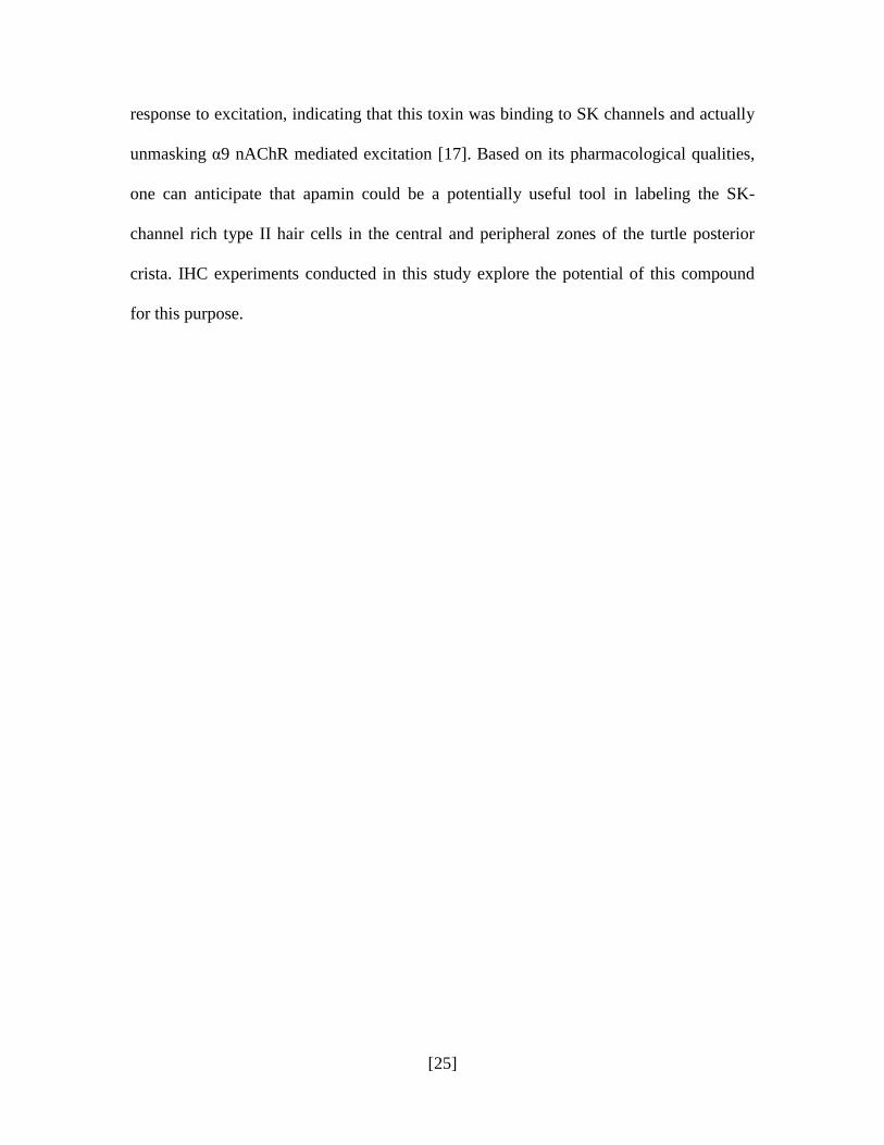
Previous studies in this laboratory have shown that afferents in the vestibular
periphery of the turtle crista produce varied responses to efferent stimulation. Stimulating
the efferent component for the bouton afferent near the torus (the BT units), results in a
pronounced, long lasting inhibition. Bouton afferents in the central zone, or BM units, are
also inhibited but for a shorter period with a quick excitatory rebound. Calyx or CD
afferents, on the other hand, exhibit a prolonged excitatory response to efferent
stimulation. [1, 11, 17, 34, 36]
The schematic shown in Figure 2 illustrates this phenomenon, separating the
efferent response into a bouton and a calyx category. The spikes represent action
potentials during the presentation of a single efferent shock train stimulation (at=0) for
the boutons and the calyx afferents in the hair cell layer. 20 shocks at 200/s at t=0 were
administered to the efferent fibers. It is obvious that the bouton response is inhibited,
indicating hyperpolarization, since the spikes completely disappear at this stimulation. In
the blue graph (calyx response), we observe the opposite to be true, with an immediate
increase in the number of spikes or action potentials, indicating depolarization and
excitation of the afferent since efferents do not synapse on the type I hair cell.
Preliminary studies designed to investigate the receptor behind each physiological
response used three known pharmacological blockers were used: strychnine, ICS and α-
BTX. The graphs in red represent data from bouton afferents, blue represent the calyx
response. Response after application of the blocker is represented by the black line. It is
observed that the bouton inhibitory response is blocked by all three substances.
Meanwhile, the calyx excitatory response remains almost unaffected. [1, 17]
[27]
A
B
Figure 2: Summary of efferent responses in the turtle posterior crista and their pharmacology. [1]
A. Schematic of a longitudinal section through the center of the turtle posterior crista
illustrating the relative location of hair cells and their respective afferents. Bouton
afferents (red) can be seen innervating type II hair cells near the torus (BT) and more
medially (BM). Bouton afferents near the planum (BP) are shown in black. Calyx and
Dimorphic afferents (CD) innervating type I hair cells in the central zone are shown in
blue. A typical efferent response for BT/BM afferents is also shown in red on the left
and that for the CD is shown in blue on the right. B. In the top row, efferent responses
in BT/BM afferents are shown before (red bars) and after (black line) the application of
various cholinergic antagonists. In the bottom row, efferent responses in CD afferents are
shown before (blue bars) and after (black line) the application of the same antagonists.
We can make two conclusions from this physiological data. Firstly, the
component responsible for the response in the bouton afferent and the type II hair cell is
pharmacologically different from the component on the calyx afferent (it is likely a
nAChR composed of a different nAChR subunits). Secondly, the inhibitory component in

[28]
the bouton afferent shares the pharmacologic and therefore is blocked by strychnine, ICS
and α-BTX. The α9/α10 nAChR fits this description.
The specific purpose of this study is to confirm, using various
immunohistochemical methods, the identity of this receptor and to localize its presence in
the hair cell layer of the turtle crista, and to compare with the nAChR on calyx afferents.

[29]
IV. MATERIALS & METHODS
A. Reagents, Primary and Secondary Antisera and Toxins
1. Reagents: All chemicals and reagents used in this study were purchased from the
Sigma Aldrich Chemical Company (St. Louis, MO).
2. Primary antibodies: The primary antibodies used in this study are listed below,
grouped by manufacturer. The specific dilution of each, as used in our experiments, is
also given in parenthesis.
2-1. Obtained from Chemicon International (Temecula, CA).
1. Rabbit IgG anti-rat, goat IgG anti-guinea pig against calretinin or CR (1:1000)
2. Goat IgG anti-human against choline acetyltransferase or ChAT (1:200)
3. Rabbit IgG anti-bovine directed against synapsin I (1:1000)
4. Rabbit IgG anti-rat directed against the vesicular acetylcholine transporter or
VAChT (1:200)
5. Rabbit IgG anti-guinea pig directed against the α4 nAChR (1:200)
2-2. Obtained from the laboratory of Dr. Miles Epstein (University of Wisconsin-
Madison Medical School) [24]
Rabbit IgG anti-chicken directed against ChAT (1:1000)
2-3. Obtained from the laboratory of Dr. Anna Lysakowski (University of Chicago) [30].
Rabbit IgG anti-turtle directed against the α9 nAChR (1:200)
2-4. Obtained from the Sigma-Aldrich Chemical Company (St Louis, MO).
Rabbit IgG anti-bovine directed against neurofilament-200 or NF-200 (1:200)

[30]
3. Secondary antibodies: All secondary antibodies used were fluorophore-conjugated
with either Alexa Fluor® 488 or 594 (both were used in different experiments). These
were obtained from Molecular Probes (Eugene, OR).
1. Donkey IgG anti-rabbit (1:500)
2. Chicken IgG anti-goat (1:500)
3. Goat IgG anti-rabbit (1:500)
4. Rabbit IgG anti-goat (1:500)
4. 4',6-diamidino-2-phenylindole (DAPI): DAPI was included in our mounting medium
(Vector Laboratories, Burlingame, CA) which was used as needed to coverslip slides.
5. Toxins:
1. α-Bungarotoxin or α-BTX: Fluorophore-conjugated with Alexa Fluor® 488
and 594 and obtained from Molecular Probes. The results provided were
obtained with a concentration between 10-20uM.
2. Apamin: Fluorophore-conjugated with Alexa Fluor® 488 and custom
synthesized by Molecular Probes. The results provided were obtained with a
1uM concentration of the substance.
3. α-Bungarotoxin: Unlabeled α-BTX for control experiments was obtained
from the Sigma Aldrich Chemical Company and from Biotium, Inc (Hayward,
CA). A 1mM concentration of the toxin was used.
4. Methyllycaconitine or MLA: This drug was obtained for control experiments
from Tocris, Inc (Ellisville, MD), and used at a concentration of 570uM.

[31]
B. Tissue Preparation and Labeling
1. α-Bungarotoxin Labeling:
Red-eared turtles (Trachemys scripta elegans) of both sexes and ranging in length
from 4 to 6 inches were purchased from Charles D. Sullivan Company Inc. (Nashville,
TN). They were housed according to the University of Texas Medical Branch (UTMB)
animal care guidelines. The experimental protocol was approved by the Institutional
Animal Care and Use Committee (IACUC) of UTMB.
Turtles were deeply anaesthetized with Nembutal (40 mg/kg, I.M.). Following
decapitation, the head was divided into two halves and the vestibular labyrinth was
exposed on each side. Each side was treated with 25µL of 10-20M α-bungarotoxin
conjugated to Alexa Fluor® 488 or 594. Each half was constantly oxygenated and
incubated in the dark for 30 minutes, at which time, a second dose of α-BTX 488/594
was applied and the half-head was further incubated for an additional 30 minutes. The
halves were then transferred to 4% paraformaldehyde (PF) in 0.1M phosphate buffer (PB,
pH 7.4) for 1-2 hours, following which the vestibular organs were dissected out and
placed in 30% sucrose overnight at 4°C. From this point onward, IHC processing was
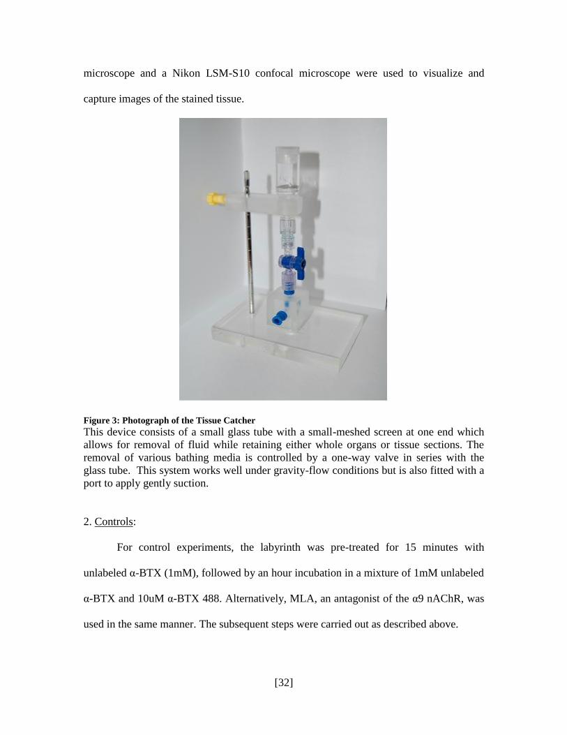
carried out in the tissue catcher, seen in Figure 3, a device designed in this laboratory
which is used to maximize convenience and efficiency with tissue processing. Post-
cryoprotection with sucrose, the organs were embedded in fresh sucrose-gelatin. 40-m
frozen sections of the semicircular canal cristae were cut and collected in a tissue catcher.
Subsequently, the gelatin was dissolved by warming the tissue catcher. The tissue
sections were then transferred to slides using an eyelash.
All slides were cover-slipped with Vectashield containing DAPI, which labels
DNA and is an excellent marker for cell nuclei. A standard Nikon fluorescence
[32]
microscope and a Nikon LSM-S10 confocal microscope were used to visualize and
capture images of the stained tissue.
Figure 3: Photograph of the Tissue Catcher
This device consists of a small glass tube with a small-meshed screen at one end which
allows for removal of fluid while retaining either whole organs or tissue sections. The
removal of various bathing media is controlled by a one-way valve in series with the
glass tube. This system works well under gravity-flow conditions but is also fitted with a
port to apply gently suction.
2. Controls:
For control experiments, the labyrinth was pre-treated for 15 minutes with
unlabeled α-BTX (1mM), followed by an hour incubation in a mixture of 1mM unlabeled
α-BTX and 10uM α-BTX 488. Alternatively, MLA, an antagonist of the α9 nAChR, was
used in the same manner. The subsequent steps were carried out as described above.
[33]
3. Double Labeling Experiments:
In double-labeling experiments, αBTX-treated organs were further processed
following fixation with 4% PF. The organs were first permeabilized with 4% Triton-0.1M
PB for 1 hour, followed by an incubation in blocking serum (0.5% fish gelatin, 1% BSA
and 0.5% Triton-X in 0.1M PB) to reduce non-specific background, for another hour.
Organs were then incubated with an antibody against the calcium-binding protein,
calretinin (CR) at RT for 16-24 hours. Double labeling with an antibody to the
neurofilament protein (NF-200) was also conducted in place of CR in some sets of tissue.
The following day, organs were transferred into the corresponding Alexa Fluor® 488 or
594 secondary antibody. Following the secondary incubation, organs were rinsed and
cryoprotected in 30% sucrose before being embedded and cut as described above. In
some experiments, the embedding and sectioning preceded the double-labeling procedure.
Immunohistochemical double-labeling of nicotinic receptor subunits with
antibodies against CR or NF-200 was performed in both whole organs and sections after
fixation with 4%PF.
4. Tissue Markers:
Several anatomical markers were used to demarcate and regionalize distinct
cellular components of the crista epithelium. DAPI, a DNA marker which fluoresces blue
under UV light, was used to label cell nuclei. CR, as previously mentioned, is a calcium-
binding protein and was used in our studies to label afferents and demarcate the crista
into the torus and planum zones based on the population of calyx and bouton afferents
[27]. NF-200 was used to label all neural fibers, both afferents and efferents, in the crista.
[34]
The use of these markers was helpful in creating a clearer image of the sensory
epithelium and the neural network, allowing better visualization of the staining of interest.
Three other antibodies used to demarcate efferent fibers and terminals were
synapsin I, ChAT and VAChT. Synapsin, a synaptic protein located in nerve terminals,
serves as a label for efferent terminals (it is believed that it is absent in hair cell and
afferent terminals) [7, 15, 42]. Choline acetyltransferase (ChAT) is responsible for the
synthesis of ACh and the vesicular acetylcholine transporter (VAChT) is a protein
necessary for the transport of acetylcholine into synaptic vesicles. Both the enzyme and
the transport protein are present in the efferent terminals, making antibodies directed
against the two ideal for marking efferents in the hair cell layer [21, 24]. Organs were
permeabilized with triton, blocked and then incubated with a primary antibody against
either synapsin, ChAT or VAChT for 16-24 hours. Usually these sets were also double-
labeled with calretinin. Subsequent steps were carried out as described for the double-
labeling experiments.
[35]
V. RESULTS
A. Afferent and Efferent Cell Markers
The use of cell markers was very beneficial to this study, allowing clearer
visualization of the protein of interest against a better defined and demarcated
background. Markers for afferent and efferent neurons clarified the cellular elements of
the sensory epithelium and allowed for a greater understanding of nAChR-specific
staining patterns by providing a „reference point‟ for better evaluation.
Figure 4 presents a longitudinal section of a posterior semicircular canal crista,
where antibodies to calretinin (CR, in green) label calyx afferents in the central zone,
some bouton afferents near the torus (BT), and type II hair cells near the planum (BP).
While nuclei of both hair cells and supporting cells are stained by DAPI, the intensity of
the labeling in supporting cells beneath the hair cells is noticeably greater. This is a result
of the dense layer of supporting cells around and between hair cells which forms a
„packing‟ with the nuclei of individual cells located much closer to each other. As a result,
a thick row of tightly packed supporting cell nuclei appears more dense with DAPI
staining than the hair cell layer they support. At higher magnifications (lower three
panels), calretinin clearly labels calyx afferents, bouton afferents near the torus and type
II hair cells near the planum.

[36]
Figure 4: Calretinin (CR) labeling in the turtle posterior crista
As seen in this longitudinal section of a posterior semicircular canal crista (top panel),
CR (green) is labeled in calyx afferents in the central zone, in bouton afferents near the
torus, and in type II hair cells near the planum (upper panel). The nuclei of supporting
cells are highlighted by the intense blue DAPI staining. At higher magnifications (lower
three panels), calretinin clearly labels calyx afferents (shown at 40X) in the central zone.
The outlines of type I hair cells are easily visualized. Bouton afferents near the torus
(shown at 20X) and type II hair cells near the planum (15X) are also labeled. The
boutons in the torus appear as small puncta along the base of type II hair cells.
Staining with polyclonal antibodies against NF-200 results in a „meshwork‟
labeling present throughout the crista, with a significantly stronger signal in the hair cell
layer (Figure 5). It is likely that both afferents and efferent neurons branching out into the
hair cell layers are labeled for this protein.
Double labeling with antibodies against both calretinin and NF-200 results in
some co-labeling in the central zone, while the peripheral zone is largely labeled for
neurofilament only. Calretinin, it can be inferred from this staining pattern, is only
present in a subset of afferent neurons in the hair cell layer. The entire region, however, is
rich with neurofilament labeling.

[37]
Figure 5: Neurofilament-200 (NF-200) labeling in the turtle posterior crista
Panels 5A-5C show longitudinal sections of posterior semicircular canal hemicristae with
calretinin and NF-200. A. Again, CR labels calyx afferents in the central zone, some BT
afferents, and type II hair cells near the planum. B. NF-200 labels all fibers including
boutons, calyx, and presumably efferent neurons. C. Composite of CR and NF-200
labeling revealing differences in staining patterns. D,E At higher magnifications (40X),
CR and NF-200 differentially label bouton afferents whereas it appears that all CD
afferents are labeled with both.
Labeling with efferent markers was equally successful in characterizing their
distribution in the hair cell layer. The enzyme ChAT is responsible for the synthesis of
ACh and is expected to be localized to efferent neurons. Figure 6A is an image of a hemi-
crista stained with antibodies to this enzyme, which labels as bright green punctae visible
from the torus to the planum. Synapsin is a key synaptic protein present in efferent nerve
terminals and is believed to be absent or have no association with hair cell synaptic
ribbons, making the protein mostly specific to efferents [7, 15, 42]. Antibodies to
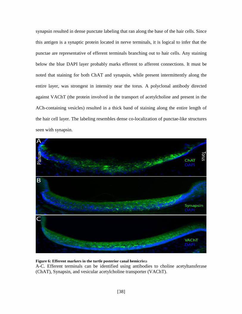
[38]
synapsin resulted in dense punctate labeling that ran along the base of the hair cells. Since
this antigen is a synaptic protein located in nerve terminals, it is logical to infer that the
punctae are representative of efferent terminals branching out to hair cells. Any staining
below the blue DAPI layer probably marks efferent to afferent connections. It must be
noted that staining for both ChAT and synapsin, while present intermittently along the
entire layer, was strongest in intensity near the torus. A polyclonal antibody directed
against VAChT (the protein involved in the transport of acetylcholine and present in the
ACh-containing vesicles) resulted in a thick band of staining along the entire length of
the hair cell layer. The labeling resembles dense co-localization of punctae-like structures
seen with synapsin.
Figure 6: Efferent markers in the turtle posterior canal hemicrista
A-C. Efferent terminals can be identified using antibodies to choline acetyltansferase
(ChAT), Synapsin, and vesicular acetylcholine transporter (VAChT).
[39]
It is important to note that the labeling patterns of ChAT and VAChT are not
necessarily identical to the staining seen for synapsin. Synapsin is likely present in all
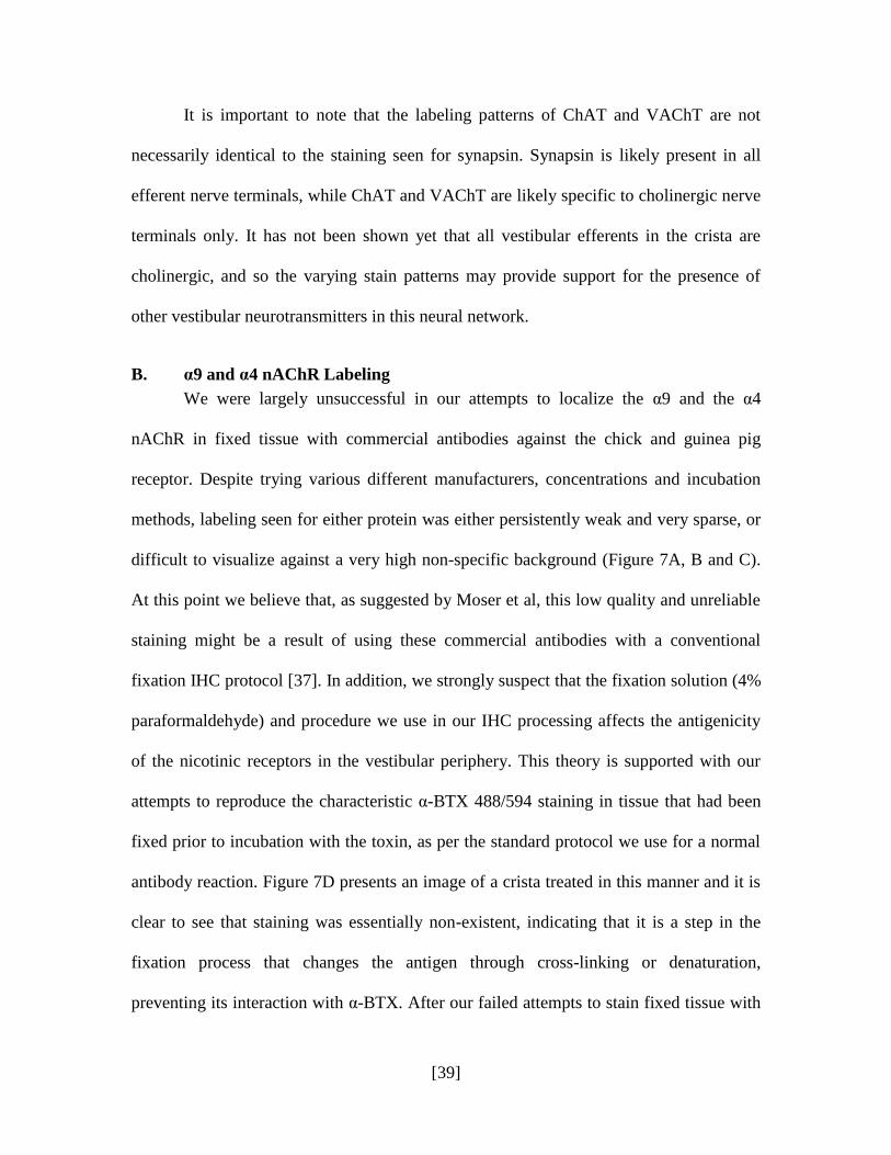
efferent nerve terminals, while ChAT and VAChT are likely specific to cholinergic nerve
terminals only. It has not been shown yet that all vestibular efferents in the crista are
cholinergic, and so the varying stain patterns may provide support for the presence of
other vestibular neurotransmitters in this neural network.
B. α9 and α4 nAChR Labeling
We were largely unsuccessful in our attempts to localize the α9 and the α4
nAChR in fixed tissue with commercial antibodies against the chick and guinea pig
receptor. Despite trying various different manufacturers, concentrations and incubation
methods, labeling seen for either protein was either persistently weak and very sparse, or
difficult to visualize against a very high non-specific background (Figure 7A, B and C).
At this point we believe that, as suggested by Moser et al, this low quality and unreliable
staining might be a result of using these commercial antibodies with a conventional
fixation IHC protocol [37]. In addition, we strongly suspect that the fixation solution (4%
paraformaldehyde) and procedure we use in our IHC processing affects the antigenicity
of the nicotinic receptors in the vestibular periphery. This theory is supported with our
attempts to reproduce the characteristic α-BTX 488/594 staining in tissue that had been
fixed prior to incubation with the toxin, as per the standard protocol we use for a normal
antibody reaction. Figure 7D presents an image of a crista treated in this manner and it is
clear to see that staining was essentially non-existent, indicating that it is a step in the
fixation process that changes the antigen through cross-linking or denaturation,
preventing its interaction with α-BTX. After our failed attempts to stain fixed tissue with

[40]
this conjugated α-BTX, we attempted to try the compound on live, unfixed tissue, with a
slightly modified protocol.
Figure 7: Immunohistochemistry with antibodies against α9nAChR and α9nAChR subunits and
AlexaFluor-594 α-BTX in fixed tissue from turtle posterior canal crista.
[41]
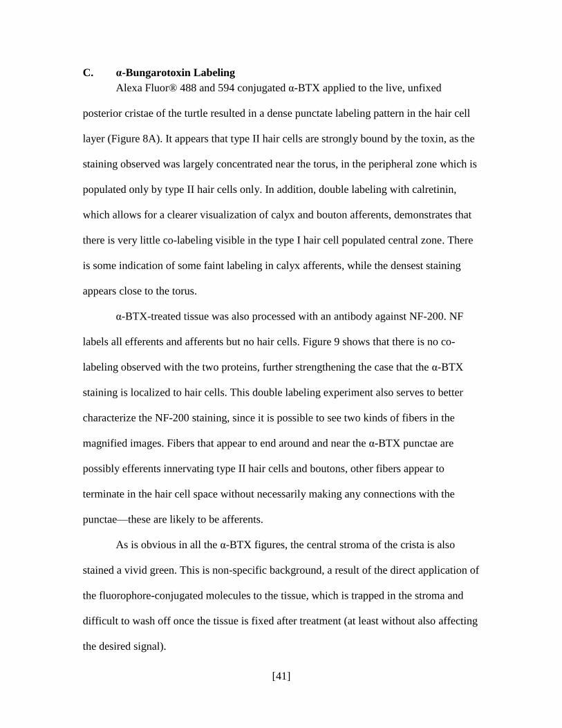
C. α-Bungarotoxin Labeling
Alexa Fluor® 488 and 594 conjugated α-BTX applied to the live, unfixed
posterior cristae of the turtle resulted in a dense punctate labeling pattern in the hair cell
layer (Figure 8A). It appears that type II hair cells are strongly bound by the toxin, as the
staining observed was largely concentrated near the torus, in the peripheral zone which is
populated only by type II hair cells only. In addition, double labeling with calretinin,
which allows for a clearer visualization of calyx and bouton afferents, demonstrates that
there is very little co-labeling visible in the type I hair cell populated central zone. There
is some indication of some faint labeling in calyx afferents, while the densest staining
appears close to the torus.
α-BTX-treated tissue was also processed with an antibody against NF-200. NF
labels all efferents and afferents but no hair cells. Figure 9 shows that there is no co-
labeling observed with the two proteins, further strengthening the case that the α-BTX
staining is localized to hair cells. This double labeling experiment also serves to better
characterize the NF-200 staining, since it is possible to see two kinds of fibers in the
magnified images. Fibers that appear to end around and near the α-BTX punctae are
possibly efferents innervating type II hair cells and boutons, other fibers appear to
terminate in the hair cell space without necessarily making any connections with the
punctae—these are likely to be afferents.
As is obvious in all the α-BTX figures, the central stroma of the crista is also
stained a vivid green. This is non-specific background, a result of the direct application of
the fluorophore-conjugated molecules to the tissue, which is trapped in the stroma and
difficult to wash off once the tissue is fixed after treatment (at least without also affecting
the desired signal).

[42]
Figure 8: Immunohistochemistry with Alexa Fluor 488 α-BTX in live, unfixed tissue from the turtle
posterior canal crista.
Panel A shows a longitudinal section of the crista labeled with the -BTX 488 alone.
Panel B is an image of the same section, labeled with an antibody against calretinin:
calyx and BT afferents have been labeled, along with type II cells near the planum. In
panel C, DAPI stains the nuclei of the hair cells and the supporting cells, and panel D is
simple a composite of A, B and C, demonstrating the localization of the -BTX staining
to the BT zone.

[43]
Figure 9: Immunohistochemistry with Alexa Fluor 488 α-BTX in live, unfixed tissue from the turtle
posterior canal crista.
Panel A shows a section of the crista obtained with confocal microscopy. The
characteristic punctate -BTX staining is seen in green against the background of the
neurofilament-200 labeling in red (10X). Panel B1 and B2 are confocal images taken at a
higher magnification (20X). Green punctae are in the hair cell layer, above the DAPI
stained supporting cell layer, and fibers stained with neurofilament end at or close to the
punctae at several points.
To investigate the reliability of the α-BTX staining observed, i.e., to confirm that
the punctae seen are indeed indicative of the location of the α9 nAChR, several controls
were carried out. Our first technique was to use antagonists of the α9 nAChR: if the α-
BTX fluorophore is binding to this receptor, then pre-incubating the tissue with a
significantly large amount of unlabeled antagonist should block any fluorescent labeling.
Methyllycaconitine (MLA) is a plant alkaloid that has been confirmed to bind to α-
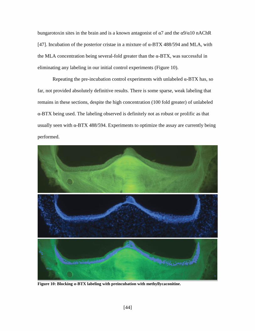
[44]
bungarotoxin sites in the brain and is a known antagonist of α7 and the α9/α10 nAChR
[47]. Incubation of the posterior cristae in a mixture of α-BTX 488/594 and MLA, with
the MLA concentration being several-fold greater than the α-BTX, was successful in
eliminating any labeling in our initial control experiments (Figure 10).
Repeating the pre-incubation control experiments with unlabeled α-BTX has, so
far, not provided absolutely definitive results. There is some sparse, weak labeling that
remains in these sections, despite the high concentration (100 fold greater) of unlabeled
α-BTX being used. The labeling observed is definitely not as robust or prolific as that
usually seen with α-BTX 488/594. Experiments to optimize the assay are currently being
performed.
Figure 10: Blocking α-BTX labeling with preincubation with methyllycaconitine.
[45]
D. Apamin Labeling for SK Channels
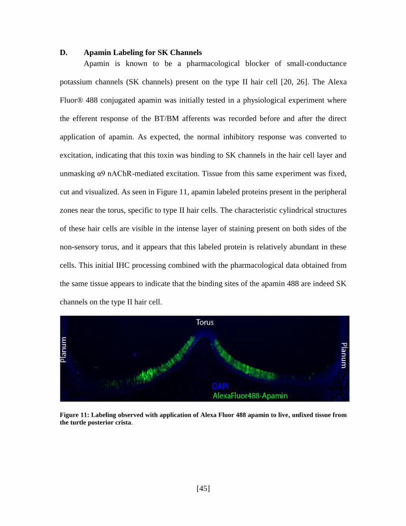
Apamin is known to be a pharmacological blocker of small-conductance
potassium channels (SK channels) present on the type II hair cell [20, 26]. The Alexa
Fluor® 488 conjugated apamin was initially tested in a physiological experiment where
the efferent response of the BT/BM afferents was recorded before and after the direct
application of apamin. As expected, the normal inhibitory response was converted to
excitation, indicating that this toxin was binding to SK channels in the hair cell layer and
unmasking α9 nAChR-mediated excitation. Tissue from this same experiment was fixed,
cut and visualized. As seen in Figure 11, apamin labeled proteins present in the peripheral
zones near the torus, specific to type II hair cells. The characteristic cylindrical structures
of these hair cells are visible in the intense layer of staining present on both sides of the
non-sensory torus, and it appears that this labeled protein is relatively abundant in these
cells. This initial IHC processing combined with the pharmacological data obtained from
the same tissue appears to indicate that the binding sites of the apamin 488 are indeed SK
channels on the type II hair cell.
Figure 11: Labeling observed with application of Alexa Fluor 488 apamin to live, unfixed tissue from
the turtle posterior crista.

[46]
VI. DISCUSSION
A. Characterizing the Crista
CR, NF-200, ChAT, VAChT, synapsin and DAPI all proved to be excellent
markers for the turtle crista, highlighting key cellular elements in the hair cell layer and
providing definition to the structural design. The immunoreactivity of each of these
markers in the turtle vestibular periphery has not been previously presented in terms of a
scientific publication, making the data presented here novel and a beginning for greater
exploration.
The staining for CR observed remained consistent in all our experiments and
resembles that seen in other organisms [27, 36]. Type I hair cells in the central zone are
very well outlined by CR-stained calyx afferents, bouton fibers are clearly visible (often
punctae-like) near the torus (BT), and type II hair cells near the planum (BP) are brightly
labeled with the use of a polyclonal anti-CR antibody. Afferents in the stroma are also
labeled as they travel up into the hair cell space. Calretinin is essentially our most
successful and reliable marker, allowing simple, efficient and clear demarcation of the
crista into its hair cell zones, and afferent fiber types.
Staining with antibodies against NF-200 has been similarly successful, though
with a somewhat higher non-specific background than seen with the CR staining. It
appears that antibodies to the neurofilament protein do not discriminate between efferent
and afferent fibers in the turtle crista. We infer this based on the observation that not all
fibers that are stained in the hair cell space seem identical in connectivity, as seen in
Figure 9. Double labeling with an efferent marker, such as antibodies against ChAT or
efferent synapse proteins, would provide a clearer answer regarding the NF-200 reactivity.

[47]
In any case, NF-200 has proved to be very useful in confirming that the α-BTX punctae is
most likely localized to the type II hair cell themselves and definitely not to the afferent
and efferent neural fibers in the hair cell space.
Antibodies to ChAT, VAChT and synapsin have all been effective in providing
information about the efferent network in the neuroepithelia of the turtle crista. Efferent
terminals have been highlighted with staining for all three proteins; however, the labeling
patterns are not necessarily identical, which points to other aspects of exploration. Not all
efferent fibers in the crista are believed to be cholinergic—the differences in staining
between ChAT, VAChT and synapsin may provide evidence for this conjecture. Since
synapsin is present in all efferent terminals (contacting both boutons and hair cells) and
ChAT and VAChT exists only in cholinergic efferent terminals, staining for synapsin
results in denser, more widespread labeling than the other two proteins. Further
exploration of their immunoreactivity might be one way of learning more about efferent
pharmacology in the vestibular periphery.
DAPI, a classic marker for cell nuclei, was used in all our IHC processing and
proved to be excellent for regionalizing the hair cell layer. The supporting cell layer is
stained intensely (as a result of the thickly packed nuclei) and there is a clear divide
between these supporting cells and where the less-intensely labeled hair cell layer begins.
B. α-Bungarotoxin Binding in Vestibular Hair Cells
Our IHC experiments with the Alexa Fluor® 488 and 594 conjugated α-BTX
result in a very characteristic staining pattern in the hair cell layer of the turtle posterior
crista, running along the type II hair cell populated peripheral zone, adjacent to the torus.
This staining is intense with α-BTX concentrations as low as 3uM, but is only present in
[48]
fresh, unfixed tissue. The toxin must be applied to freshly extracted tissue and incubated
in Ringer‟s solution, followed by fixation. If fixed tissue is incubated in a 3, 10 or 20uM
α-BTX solution, no obvious labeling is seen, suggesting that the fixation steps may have
a deleterious effect on the protein the α-BTX is interacting with.
Pharmacologically, as discussed previously, α-BTX appears to block the
inhibitory response the α9 nAChR is responsible for, as observed in turtle
electrophysiological preparations. α-BTX, therefore, clearly has an antagonistic
relationship with the α9 nAChR and must bind to the receptor to be able to block its hair
cell action. But based on this information, we might infer that the punctae observed in α-
BTX treated tissue are indeed labeling the α9 nAChR. This staining does not appear to be
non-specific, as it is has been repeatedly reproduced and is always seen in the same
regions with the same characteristics. Double labeling with other neural markers (namely
CR, NF-200 and DAPI) appears to confirm the localization of these punctae to vestibular
hair cells and not to efferents or afferents in the neural space. However, only preliminary
controls have been run at this point to absolutely authenticate the identity of these labeled
proteins, and so more testing is required to confirm the same.
We have carried out control studies with MLA, another cholinergic antagonist of
the α9 nAChR which has successfully blocked the inhibitory α9 response in
pharmacological studies. When tissue was treated with an excess of MLA and incubated
with α-BTX 488, no labeling was visible in the entire hair cell layer. This control was
repeated with an improved surgical technique that ensured proper accessibility of the
crista for the solutions. This second time, surprisingly, we observed sparse but obvious
labeling in the peripheral zones in most of the tissue sections. The second type of control
[49]
was conducted with the same protocol, using unlabeled α-BTX at a 100-fold greater
concentration than the α-BTX 488. While it was weak and sparse in each control set,
labeling was never completely blocked out as it was expected to, despite the literal flood
of unlabeled α-BTX that preceded the labeled protein.
We cannot ignore or reject the staining pattern—it is reproducibly, consistently,
and lucidly present in the type II hair cell region—and α-BTX has been characterized to
have a nAChR binding site in other organisms [21, 38] apart from its pharmacological
ability to block the α9 response in the turtle. On the other hand, at this preliminary stage,
we do not have the control data to support a conclusion regarding the localization of the
α9 receptor protein with this fluorophore toxin. Our current hypothesis is that the labeling
observed is real (i.e., there is an authentic protein interaction taking place between the
hair cell and the receptor protein), and we are considering multiple possibilities to gain
greater insight into what might be taking place at the molecular level.
Firstly, we evaluated the possibility that the unlabeled α-BTX might not be
effective, since the quality of the purified toxin can vary from manufacturer to
manufacturer. We tested its potency in a physiological prep and observed that the
inhibitory response was clearly blocked when a 3uM solution of α-BTX was applied to
the canals. The unlabeled α-BTX being used was therefore in proper working condition
pharmacologically. On the other hand, the quality of the α-BTX fluorescent toxin must
also be considered. The process of synthesis (it was obtained from Molecular Probes)
might have affected the toxin and its chemical properties: perhaps it is now binding to a
different protein than it should be? Our next step is therefore to test the fluorophore-
[50]
conjugated α-BTX pharmacologically and ensure that it is working to block the inhibitory
response, with about the same dose as the unlabeled toxin has been seen to do.
Another related theory to consider is that the conjugation of a fluorophore to the
purified α-BTX molecule, an artificial synthesis process, may have enhanced the potency
of the toxin and actually increased its affinity many times. This would boost its ability to
bind to the target protein, negatively affecting the competitive antagonism by unlabeled
α-BTX. One way to test this theory is to try both the fluorophore-conjugated and
unlabeled α-BTX in a physiological prep separately (using the same concentration for
each) and recording the speed at which the inhibitory response is blocked. To be
considered an appropriate antagonist for the other, each compound must display its effect
in relatively the same amount of time when used at the same concentration.
A third possibility is also related to the artificial attachment of the fluorophore to
α-BTX. Since we do not have detailed knowledge at this point of how the chemical
structure is manipulated to obtain a conjugated toxin, it is worthwhile to consider if this
created α-BTX is now involved in a different, abnormal, molecular mechanism. Instead
of binding at its target site on the hair cell, perhaps the 488/594 fluorophore is being
internalized by the hair cell itself and pulling in the α-BTX along with it , resulting in the
labeling pattern we observe. This internalization method need not be receptor-mediated,
and therefore application of the unlabeled α-BTX (which is specific to the receptor) does
not block the process. To investigate this further, it might be helpful to study the
fluorophore and investigate it qualities, perhaps obtaining it by itself and trying it with
the same IHC protocol. A physiological experiment, once again, could provide
[51]
information as to whether the compound is indeed interacting with the receptor or
participating elsewhere.
It might also be true that the concentration of α-BTX 488/594 being used in our
IHC experiments may be too high. In pharmacological studies conducted in this lab, a
1uM solution of α-BTX is sufficient and potent enough to block the response of the α9
nAChR. However, having failed to observe the same vivid labeling at a 1uM
concentration, we use a concentration that is usually in the range of 3uM to 20uM (10uM
was considered to be optimal after various trial and error experiments) in our IHC
experiments. Naturally, the two methods—of running through a solution directly on the
live, exposed organ in a physiological experiment versus incubating the organ in the
solution in a histochemical staining experiment—are very different and may influence the
dosage required. But, we believe that is likely that the 3-20uM concentration of
fluorophore toxin we use in the IHC experiment is in such excess that it is saturating the
binding sites on the hair cell layer, so that even a large amount of unlabeled α-BTX may
be unable to out-compete effectively. If the maximum response is reached with an excess
dose of the fluorophore toxin, the system is saturated and it is likely that even a 100-times
greater amount of the unlabeled toxin is not enough to appreciably change the labeling.
This can only be investigated by continuing to optimize the IHC protocol and
experimenting with surgical and incubation techniques that will allow us to improve our
staining with even lower concentrations of the labeled α-BTX.
C. Apamin Binding in Vestibular Hair Cells
The neurotoxin, apamin, has been characterized as a blocker of the small-conductance
potassium channels (SK channels) present on the type II hair cell [20, 26]. The SK
[52]
channels are key players in the inhibitory response that the α9 nAChR is responsible for,
since it is the efflux of K+ ions from these channels that leads to membrane
hyperpolarization and inhibition of neurotransmitter release [25]. Based on this role, it
follows that SK channels are likely to be located structurally close to the α9 nAChR
protein itself. Initial experiments with custom synthesized apamin conjugated to the
Alexa Fluor® 488 fluorophore have provided encouraging results. This conjugated toxin
was initially tested pharmacologically where, as expected, the normal inhibitory response
was blocked with the use of apamin, confirming that this toxin had successfully bound to
SK channels in the hair cell layer. Labeling was largely present in the peripheral zone
near the torus which is solely populated by type II hair cells. The pattern was not punctate
at this low magnification, but appeared to be dense and widespread in this region of the
hair cell space. The cylindrical structures of the hair cells are actually visible in the
intense layer of staining present on both sides of the non-sensory torus.
The appropriate pharmacological response described above combined with this
visual data on the physical location of this apamin-bound protein in the hair cell layer
(and more importantly in type II hair cells), strongly corroborates the identity of these
proteins as SK channels. In addition, the fact that this apamin labeling does not resemble
that seen with the conjugated bungarotoxin compound lends support to the authenticity of
the bungarotoxin staining pattern, since it cannot be attributed to the Alexa Fluor 488
fluorophore. If the presence of the fluorophore was compromising the IHC labeling, then
staining with apamin 488 would resemble staining with α-BTX 488, which we
demonstrate to not be the case.
[53]
Fluorophore-conjugated apamin, with further experimentation to reduce non-
specific background and obtaining a clearer signal, may be very useful in characterizing
the type II hair cell in terms of its physical structure and constituents. In addition,
combined staining with α-BTX 594 could prove to be an intriguing way of exploring both
the apamin and the inconclusive α-BTX labeling.
D. Future Outlook
Nicotinic acetylcholine receptors form a crucial part of the complex, intricate
machinery underlying the mechanisms driving excitation and inhibition in the vestibular
hair cells and afferents. To truly gain insight into how mechanical head movements can
be affected by efferent signals, the characterization of these nAChRs is important and
necessary. Localizing these nAChRs in the hair cell layer is essential to this process: a
large part of understanding how a receptor works is knowing where it exists and what
role that location plays in the larger picture. Additionally, our knowledge of the
vestibular hair cells and the communication between afferents, efferents and the two
types of hair cells remains inadequate, with many unidentified components and
unexplained characteristics.
With this study, our first step has been to structurally define and illuminate the
turtle crista and demonstrate its value as an animal model for immunohistochemical
studies of the vestibular nervous system. The vestibular organs of the turtle are not that
different from the mammalian system. The essential preservation of this system
evolutionarily—combined with factors such as the surgical ease in removing the
labyrinth, the presence of both types of hair cells and the success of various markers in
[54]
regionalizing its hair cell layer—all support the use of the turtle as an excellent and
convenient model for our nAChR studies.
Potentially, the techniques presented in this study involving the use of a
fluorophore-conjugated nicotinic antagonist could hold great value since it could provide
reliable, lucid results without the many negative aspects of commercial antibodies
(described by Moser et al and Huang et al among others) [19, 37]). Fixation is not
necessary, the question of serum compatibility or reactivity between species need not be
considered, concerns regarding the quality of the antibody (and the various additional
experiments to test the manufacturer‟s claims) as well as the many variables that
accompany primary and secondary antibody use do not come into play at all with this
technique. With appropriate optimization of the protocol, we are optimistic that this
method could be used with greater ease, efficiency and reliability, and prove to be an
excellent benefit to our own studies as well as others.
Identifying the α9 nAChR using a fluorophore-conjugated α-BTX antagonist was
the primary aim of this study. At this preliminary stage, our data remains inconclusive,
but we have made large strides in our knowledge of the receptor proteins and the current
immunohistochemical methods available to isolate them. We are in the midst of
designing various other physiological and anatomical experiments that could provide a
better explanation for our results as well as direct us to more definitive conclusions
regarding the α9 nAChR and its presence on the type II hair cell. As discussed in the
previous section, many possibilities exist and each must be explored to allow us to paint a
clearer picture of what is likely taking place at the molecular level. To this end, the use of

[55]
other neural markers (hair cell specific and additional efferent and afferent markers) and
other nicotinic and ion channel antagonists should also prove useful.
[56]
VII. REFERENCES
1. Brichta AM, Goldberg JM (2000) Morphological identification of physiologically
characterized afferents innervating the turtle posterior crista. J Neurophysiol.
83:1202-1223
2. Brichta AM, Peterson EH (1994) Functional architecture of vestibular primary
afferents from the posterior semicircular canal of a turtle, Pseudemys
(Trachemys) scripta elegans. J Comp Neurol. 344:481-507
3. Corringer PJ, Le Novère N, Changeux JP (2000) Nicotinic receptors at the amino
acid level. Annu Rev Pharmacol Toxicol. 40:431-458
4. Dailey SH, Wackym PA, Brichta AM, Gannon PJ, Popper P. (2000) Topographic
distribution of nicotinic acetylcholine receptors in the cristae of a turtle. Hear Res
141:51-56
5. Eatock RA (2000) Adaptation in hair cells. Annu Rev Neurosci. 23:285-314
6. Ellison M, Haberlandt C, Gomez-Casati ME, Watkins M, Elgoyhen AB,
McIntosh JM, Olivera BM (2006) Alpha-RgIA: a novel conotoxin that
specifically and potently blocks the alpha9alpha10 nAChR. Biochemistry
45:1511-1517
7. Favre D, Scarfone E, Di Gioia G, De Camilli P, Dememes D (1986) Presence of
synapsin I in afferent and efferent nerve endings of vestibular sensory epithelia.
Brain Res. 384:379-382
8. Fernández C, Baird RA, Goldberg JM. (1988) The vestibular nerve of the
chinchilla. I. Peripheral innervation patterns in the horizontal and superior
semicircular canals. J Neurophysiol. 60:167-181
[57]
9. Fernández C, Lysakowski A, Goldberg JM (1995) Hair-cell counts and afferent
innervation patterns in the cristae ampullares of the squirrel monkey with a
comparison to the chinchilla. J Neurophysiol. 73:1253-1269
10. Fettiplace R, Crawford AC (1980) The origin of tuning in turtle cochlear hair cells
Hear Res. 2:447-454
11. Goldberg JM, Brichta AM (1998) Evolutionary trends in the organization of the
vertebrate crista ampullaris. Otolaryngol Head Neck Surg 119:165-171
12. Goldberg JM, Brichta AM, Wackym PA (2000) Efferent vestibular system:
Anatomy, Physiology and Neurochemistry. Neurochemistry of the vestibular
system ed. A.J.H. and B.A.J. Boca Raton, FL: CRC press. 61-94
13. Guth PS, Perin P, Norris CH, Valli P (1998) The vestibular hair cells: post-
transductional signal processing. Prog Neurobiol. 54:193-247
14. Hoffman BB, Lefkowitz RJ, Taylor P (1996) Neurotransmission: The Autonomic
and Somatic Motor Nervous Systems. The Pharmacological Basis of Therapeutics,
ed. LS Goodman, LE Limbird, PB Milinoff, RW Ruddon, AG Gilman. 9th
Edition,
McGraw Hill. 105-141
15. Holstein GR, Martinelli GP, Nicolae RA, Rosenthal TM, Friedrich VL Jr. (2005)
Synapsin-like immunoreactivity is present in hair cells and efferent terminals of
the toadfish crista ampullaris. Exp Brain Res. 162:287-292
16. Holt JC, Lioudyno M, Guth PS (2003) A pharmacologically distinct nicotinic
ACh receptor is found in a subset of frog semicircular canal hair cells. J
Neurophysiol. 90:1526-1536
[58]
17. Holt JC, Lysakowski A, Goldberg JM (2006) Mechanisms of efferent-mediated
responses in the turtle posterior crista. J Neurosci. 26:13180-13193
18. Hunter-Duvar IM, Hinojosa R (1984) Vestibule: Sensory Epithelia.
Ultrastructural Atlas of the Inner Ear, ed. I. Friedmann and J. Ballantyne.
Butterworths & Co, London. 211-214
19. Huang LZ, Winzer-Serhan UH (2006) Effects of paraformaldehyde fixation on
nicotinic acetylcholine receptor binding in adult and developing rat brain sections.
J Neurosci Methods. 153:312-317
20. Ishii TM, Maylie J, Adelman JP (1997) Determinants of apamin and d-
tubocurarine block in SK potassium channels. J Biol Chem. 272:23195-231200
21. Ishiyama A, Lopez I, Wackym PA (1995) Distribution of efferent cholinergic
terminals and alpha-bungarotoxin binding to putative nicotinic acetylcholine
receptors in the human vestibular end-organs. Laryngoscope 105:1167-1172
22. Itier V, Bertrand D (2001) Neuronal nicotinic receptors: from protein structure to
function. FEBS Lett. 504:118-25
23. Jeffries DJ, Pickles JO, Osborne MP, Rhys-Evans PH, Comis SD (1986)
Crosslinks between stereocilia in hair cells of the human and guinea pig vestibular
labyrinth. J Laryngol Otol. 100:1367-1374
24. Johnson CD, Epstein ML (1986) Monoclonal antibodies and polyvalent antiserum
to chicken choline acetyltransferase. J Neurochem. 46:968-976
25. Kohler M, Hirschberg B, Bond CT, Kinzie JM, Marrion NV, Maylie J, Adelman
JP (1996) Small-conductance, calcium-activated potassium channels from
mammalian brain. Science 273:1709-1714
[59]
26. Lazdunski M, Fosset M, Hughes M, Mourre C, Romey G, Schmid-Antomarchi H
(1985) The apamin-sensitive Ca2+ dependent K+ channel molecular properties,
differentiation and endogenous ligands in mammalian brain. Biochem Soc Symp
50:131-142
27. Leonard RB, Kevetter GA (2002) Molecular probes of the vestibular nerve. I.
Peripheral termination patterns of calretinin, calbindin, and peripherin containing
fibers. Brain Res. 928:8-17
28. Lindeman HH (1969) Regional differences in structure of the vestibular sensory
regions. J Laryngol Otol. 83:1-17
29. Lindeman HH (1969) Regional differences in sensitivity of the vestibular sensory
epithelia to ototoxic antibiotics. Acta Otolaryngol. 67:177-189
30. Luebke AE, Maroni PD, Guth SM, Lysakowski A (2005) Alpha-9 nicotinic
acetylcholine receptor immunoreactivity in the rodent vestibular labyrinth. J
Comp Neurol. 492:323-333
31. Lukas RJ, Changeux JP, Le Novère N, Albuquerque EX, Balfour DJ, Berg DK,
Bertrand D, Chiappinelli VA, Clarke PB, Collins AC, Dani JA, Grady SR, Kellar
KJ, Lindstrom JM, Marks MJ, Quik M, Taylor PW, Wonnacott S (1999)
International Union of Pharmacology. XX. Current status of the nomenclature for
nicotinic acetylcholine receptors and their subunits. Pharmacol Rev. 51:397-401
32. Lustig LR, Hiel H, Fuchs PA (1999) Vestibular hair cells of the chick express the
nicotinic acetylcholine receptor subunit alpha 9. J Vestib Res. 9:359-367
33. Lustig LR (2006) Nicotinic acetylcholine receptor structure and function in the
efferent auditory system. Anat Rec A Discov Mol Cell Evol Biol. 288:424-434
[60]
34. Lysakowski A (1996) Synaptic organization of the crista ampullaris in vertebrates.
Ann NY Acad Sci. 781:658-662
35. Lysakowski A, Goldberg JM (1997) A regional ultrastructural analysis of the
cellular and synaptic architecture in the chinchilla cristae ampullares. J Comp
Neurol. 389:419-443
36. Lysakowski A, Goldberg JM (2003) Morphophysiology of the Vestibular
Periphery. The Vestibular System, ed. S Highstein, R.R. Fay, and A.N. Popper.
Vol. 19. New York: Springer-Verlag. 57-152
37. Moser N, Mechawar N, Jones I, Gochberg-Sarver A, Orr-Urtreger A, Plomann M,
Salas R, Molles B, Marubio L, Roth U, Maskos U, Winzer-Serhan U, Bourgeois
JP, Le Sourd AM, De Biasi M, Schröder H, Lindstrom J, Maelicke A, Changeux
JP, Wevers A (2007) Evaluating the suitability of nicotinic acetylcholine receptor
antibodies for standard immunodetection procedures. J Neurochem. 102:479-492.
38. Pickles JO (1984) Frequency threshold curves and simultaneous masking
functions in high-threshold, broadly-tuned, fibres of the guinea pig auditory nerve.
Hear Res. 16:91-99
39. Plazas PV, Katz E, Gomez-Casati ME, Bouzat C, Elgoyhen AB (2005)
Stoichiometry of the alpha9alpha10 nicotinic cholinergenic receptor. J Neurosci.
25:10905-10912
40. Plinkert PK, Zenner HP, Heilbronn E (1991) A nicotinic acetylcholine receptor-
like alpha-bungarotoxin-binding site on outer hair cells. Hear Res. 53:123-130
41. Rappoli R and Montecercco C (1997) Guidebook to Protein Toxins and Their Use
in Cell Biology. Oxford University Press. 192
[61]
42. Safieddine S, Wenthold RJ (1999) SNARE complex at the ribbon synapses of
cochlear hair cells: analysis of synaptic vesicle and synaptic membrane-associated
proteins. Eur J Neurosci. 11:803-812
43. Schessel DA, Ginzberg R, Highstein SM (1991) Morphophysiology of synaptic
transmission between type I hair cells and vestibular primary afferents. An
intracellular study employing horseradish peroxidase in the lizard, Calotes
versicolor. Brain Res. 544:1-16
44. Stocker M, Hirzel K, D‟hoedt D, Perdarzani P (2004) Matching molecules to
function: neuronal Ca2+ activated K+ channels and afterhyperpolarizations.
Toxicon. 43:933-949
45. Verbitsky M, Rothlin CV, Katz E, Elgoyhen AB (2000) Mixed nicotinic-
muscarinic properties of the α9 nicotinic cholinergic receptor.
Neuropharmacology 39:2515-2524
46. Wackym PA, Popper P, Lopez I, Ishiyama A, Micevych PE (1995) Expression of
alpha 4 and beta 2 nicotinic acetylcholine receptor subunit mRNA and
localization of alpha-bungarotoxin binding proteins in the rat vestibular periphery.
Cell Biol Int. 19:291-300
47. Yum L, Wolf KM, Chiappinelli VA (1996) Nicotinic acetylcholine receptors in
separate brain regions exhibit different affinities for methyllycaconitine.
Neuroscience 72(2):545-555