Embed Size (px)
Citation preview

4891
Introduction
Glioma is one of the most common and in-vasive tumors of the central nervous system. Glioma accounts for about 80% of primary ma-lignant brain tumors1. These tumors are divided into four histological subtypes: astrocytoma, oligodendroglioma, ependymoma and mixed tumors2-4. Treatment options include surgical resection, radiotherapy, and chemotherapy5, but the overall efficacy is still unsatisfactory. The average survival time of patients with glioma is only 4.5 months without treatment, increasing to 15 months upon temozolomide treatment6,7. Insufficient treatment efficacy prompts studies to discover new therapeutic targets in glioma.
Long non-coding RNAs (lncRNAs) are RNAs with the length of more than 200 nucleotides. Ln-cRNAs regulate gene expression at transcriptional, post-transcriptional, and epigenetic levels8. Recent studies demonstrated regulatory roles of lncRNAs in different tumors. This was shown with hepato-cellular carcinoma (lncRNA ATB9) gastric cancer (lncRNA GAPLINC10), and glioma (SPRY4-IT111). Furthermore, lncRNAs may regulate cancer ma-lignancy by interacting with signaling proteins, including the regulators of DNA demethylation TCF21 and GADD45A (lncRNA TARID12). As studies indicate the involvement of lncRNAs in the regulation of cancer malignancy, there is interest in evaluating lncRNAs as potential targets for cancer treatment. The involvement of lncRNAs in glioma remains largely unknown to date.
In the current study, we quantified expression of lncRNA H19 in 35 specimens of glioma or con-trol tissues and analyzed the relationship between
Abstract. – OBJECTIVE: Glioma is one of the most common and invasive tumors of the central nervous system. Long non-coding (lnc) RNAs are involved in many cancers, but their function and mechanism in glioma remain largely unknown. We wished to delineate the role of lncRNA H19 and its derivative miR-675 in this tumor.
PATIENTS AND METHODS: Using qPCR, we compared expression of lncRNA H19 in 35 specimens of glioma vs control tissue, and in two glioma cell lines U251 and U87 vs Normal Human Astrocyte (NHA) cells. Cell proliferation was evaluated after shRNA silencing of lncRNA H19 in glioma cell lines. The role of miR-675 was tested using antagomir and the mimic.
RESULTS: LncRNA H19 was overexpressed in glioma tissue and cell lines. In tissue, high-er expression levels were observed in more advanced stages of the tumor. Furthermore, lncRNA H19 was negatively associated with pa-tient survival time. In cell culture experiments, silencing of lncRNA H19 diminished prolifer-ation of glioma cell lines. These effects of ln-cRNA H19 appeared to be intermediated by miR-675. The latter was overexpressed in glio-ma tissue and was negatively associated with patient survival. Supporting the involvement of miR-675, its antagomir decreased prolifera-tion of glioma cell lines, whereas its mimic in-creased proliferation of NHA cells.
CONCLUSIONS: LncRNA H19 is overex-pressed in glioma tissue, and is positively as-sociated with the tumor grade and negatively associated with patient survival. In cell culture studies, lncRNA H19 promotes glioma cell pro-liferation. These tumor-promoting effects of ln-cRNA H19 appear to be mediated by miR-675.
Key Words: lncRNA H19, Glioma, miR-675, Proliferation.
European Review for Medical and Pharmacological Sciences 2016; 20: 4891-4897
T. ZHANG, Y.-R. WANG, F. ZENG, H.-Y. CAO, H.-D. ZHOU, Y.-J. WANG
The Department of Neurology, Institute of Surgery Research, Daping Hospital, Third Military Medical University, Chongqing, China
Corresponding Author: Yan-Jiang Wang, MD; e-mail: [email protected]
LncRNA H19 is overexpressed in glioma tissue, is negatively associated with patient survival,and promotes tumor growth through its derivative miR-675

T. Zhang, Y.-R. Wang, F. Zeng, H.-Y. Cao, H.-D. Zhou, Y.-J. Wang
4892
expression of this lncRNA and patient survival. In supporting cell studies, we demonstrated that lncRNA H19 promotes proliferation of glioma cells via its derivative miR-675. These observa-tions indicate that lncRNA H19 may be the thera-peutic target in glioma.
Patients and Methods
SpecimensThirty-five specimens of glioma and adjacent
normal brain tissue were obtained from patien-ts of the Department of Neurosurgery, Institute of Surgery Research, Daping Hospital, Third Military Medical University (Daping, China). Patient enrollment took place from June 2012 to January 2013. The study protocol was approved by the Institutional Review Board of Daping Hospital, Third Military Medical University, and informed consents were obtained from all patients.
The specimens were collected and kept in liquid nitrogen after surgery. Clinical data were procu-red, and patients were followed for 3 years. Ac-cording to the World Health Organization (WHO) classification, glioma specimens were classified into two groups: low-grade glioma (LGG, WHO I-II, 15 specimens), and high-grade glioma (HGG, WHO III-IV, 20 specimens).
Cell CultureHuman glioma cell lines U251 and U87 we-
re obtained from CAMS (Chinese Academy of Medical Sciences, Beijing, China). Normal Hu-man Astrocyte (NHA) cells were obtained from American Type Culture Collection (Manassas, VA, USA); these cells were used as control cells in our studies. Both glioma and NHA cells were cultured in Dulbecco’s modified Eagle medium (DMEM)/high glucose (Hyclone, Logan, UT, USA) supplemented with 10% fetal bovine serum (Gibco, Grand Island, NY, USA).
qPCRTotal RNA from glioma and normal brain
tissues were extracted using RNAiso Plus kit (TaKaRa, Otsu, Japan), after the specimens were ground to powder. Total RNA from cells was also extracted with RNAiso Plus kit. RNA concentration of both tissue specimens and cells was measured, and cDNA was synthesized using 1 µg of RNA (PrimeScript RT reagent kit with gDNA Eraser; TaKaRa). Expression of lncRNA
H19 was quantified by qPCR, using SYBR Pre-mix Ex Taq (TaKaRa). Expression of GAPDH was used as the reference gene. The primers were listed as follows: GAPDH primers: sense: TGTGGGCATCAATGGATTTGG; anti-sense: ACACCATGTATTCCGGGTCAAT; H19 pri-mers: sense: GGCTCTGGAAGCTAGAGGAA; anti-sense: CTGGGATGATGTGGTGGC.
shRNA SilencingThe short-hairpin (sh) RNA directed against ln-
cRNA H19 was constructed in pGPU6/GFP/Neo vector (shH19): CUUUCUGUCACAUUGAC-CACACCUG or UCUGAUUGCAGCAUCUU-CUUGAUUC. We further used antagomir against miR-675 (GenePharma, Shanghai, China) with the following sequence: TGAGCGGTGAGGGCATA-CAG13. The sequence of microRNA-675 mimic was as follows: CTGTATGCCCTCACCGCTCA13. Transfections were performed using Lipofectami-ne 2000 (Invitrogen, Carlsbad, CA, USA).
Cell Proliferation (CCK8) AssayCells were seeded in 96-well plates at the density
of 1500 cells per well, with five replicate wells per each outcome. After respective treatments, the wel-ls received 100 µl of culture medium and 10 µl of the reaction mixture from the CCK8 kit (Dojundo, Kumamoto-ken, Japan). The plate was incubated for 2 hours at 37º C. Then, cell proliferation was estimated by measuring absorbance at 450 nm.
Statistical AnalysisEach experiment was repeated at least three
times, and data were presented as means ± stan-dard deviation (SD). Descriptive and compara-tive analyses were done using GraphPad Prism software (GraphPad, La Jolla, CA, USA). The Student’s t-test or one-way ANOVA test was used to compare outcomes. Survival analysis was per-formed by using the log-rank test. The p < 0.05 was considered to be significant.
Results
lncRNA H19 is Overexpressed in Glioma Tissue and is Negatively Associated with Patient Survival Time
We first quantified lncRNA H19 expression in 35 specimens of glioma tissue and adjacent (control) tissue by qPCR. LncRNA H19 was overexpressed in glioma tissue (Figure 1A). Fur-thermore, the level of lncRNA H19 expression
LncRNA H19 is overexpressed in glioma tissue, is negatively associated with patient survival
4893
correlated with the tumor grade. In particular, specimens of grade III-IV glioma showed mar-kedly higher levels of lncRNA H19 expression than specimens of grade I-II glioma (Figure 1B). Following this, we analyzed the association between lncRNA H19 expression and patient survival. Higher levels of lncRNA H19 expres-sion were negatively associated with the overall survival, which was calculated using the log-rank test (Figure 1C).
LncRNA H19 is highly expressed in glioma cell lines and determines their proliferation
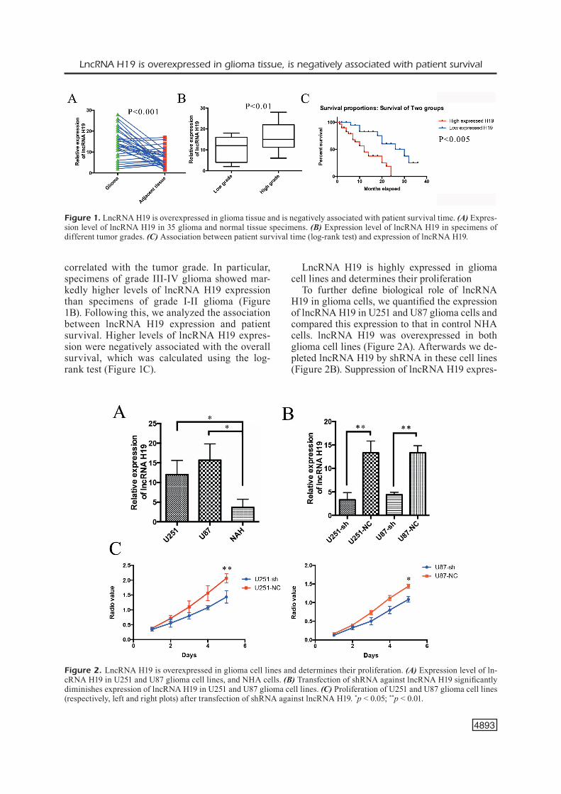
To further define biological role of lncRNA H19 in glioma cells, we quantified the expression of lncRNA H19 in U251 and U87 glioma cells and compared this expression to that in control NHA cells. lncRNA H19 was overexpressed in both glioma cell lines (Figure 2A). Afterwards we de-pleted lncRNA H19 by shRNA in these cell lines (Figure 2B). Suppression of lncRNA H19 expres-
Figure 1. LncRNA H19 is overexpressed in glioma tissue and is negatively associated with patient survival time. (A) Expres-sion level of lncRNA H19 in 35 glioma and normal tissue specimens. (B) Expression level of lncRNA H19 in specimens of different tumor grades. (C) Association between patient survival time (log-rank test) and expression of lncRNA H19.
Figure 2. LncRNA H19 is overexpressed in glioma cell lines and determines their proliferation. (A) Expression level of ln-cRNA H19 in U251 and U87 glioma cell lines, and NHA cells. (B) Transfection of shRNA against lncRNA H19 significantly diminishes expression of lncRNA H19 in U251 and U87 glioma cell lines. (C) Proliferation of U251 and U87 glioma cell lines (respectively, left and right plots) after transfection of shRNA against lncRNA H19. *p < 0.05; **p < 0.01.

T. Zhang, Y.-R. Wang, F. Zeng, H.-Y. Cao, H.-D. Zhou, Y.-J. Wang
4894
sion in U251 and U87 glioma cell lines led to a marked decline in their proliferation (Figure 2C). These observations are consistent with clinical findings (Table I).
The lncRNA H19 Derived miR-675 Promotes Proliferation of Glioma Cells
Recent studies demonstrated that miR-675, encoded by the exon 1 of lncRNA H19, is an important mediator of the effects of this lncR-NA in different types of cancer. We, therefore, wanted to verify whether the effects of lncRNA H19 observed in the aforementioned experiments were mediated by miR-675. We first studied the expression of miR-675 in U251 and U87 glioma cell lines. As expected, miR-675 was overexpres-sed in both cell lines (Figure 3A). To further ve-rify the role of this miRNA, we suppressed its expression by transfecting U251 and U87 cell li-nes with the respective antagomir. As expected, the proliferation of both glioma cell lines was markedly diminished when expression of miR-675 was suppressed (respectively, Figures 3B and 3C). To further confirm the role of miR-675, we transfected NHA cells with the mimic of miR-
675. This dramatically increased proliferation of control cells (Figure 3D).
miR-675 is overexpressed in glioma tissue and is negatively associated with patient survival time
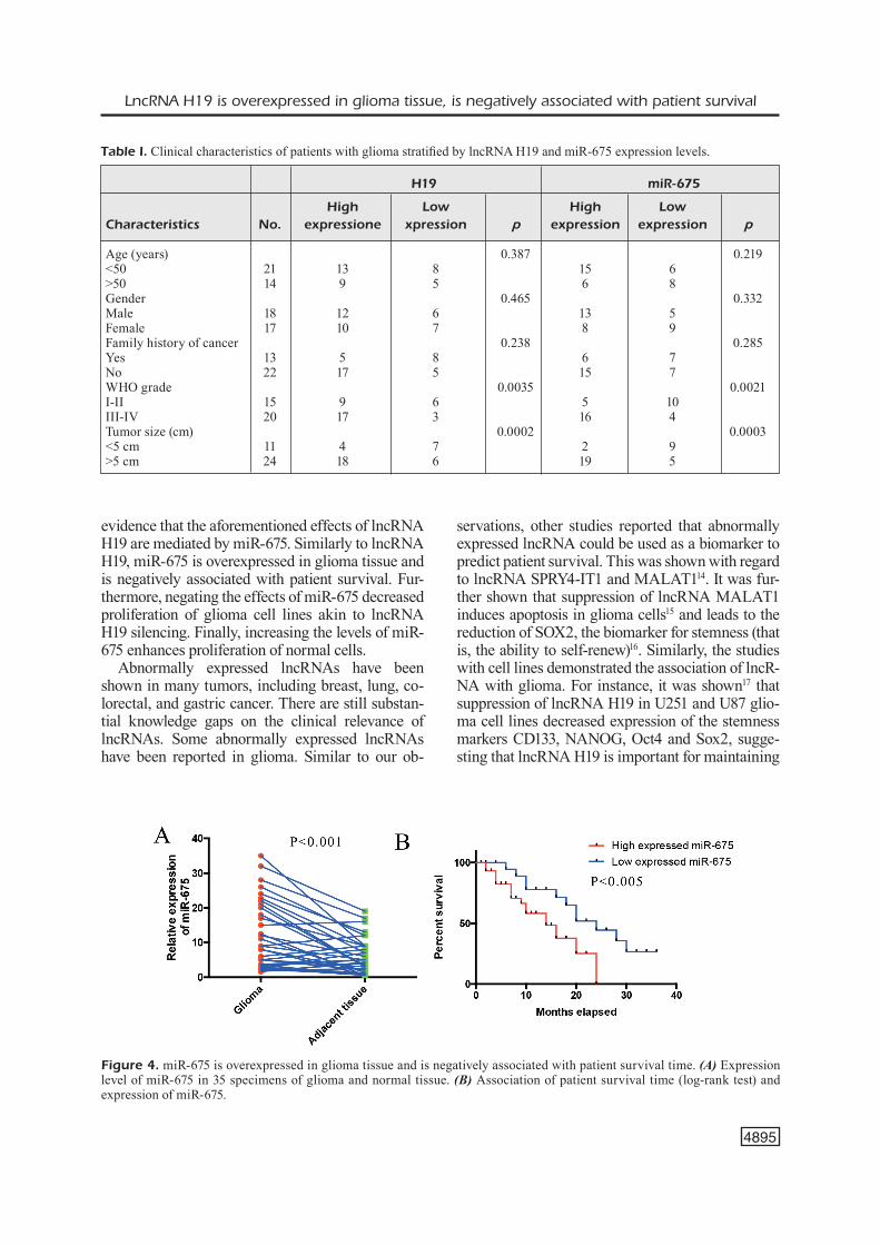
Following our findings in glioma cell lines, we retested 35 specimens of glioma and control tissue to quantify expression of miR-675. This asses-sment demonstrated overexpression of miR-675 in glioma tissue (Figure 4A). Similar to lncRNA H19, we observed a negative association between the expression level of miR-675 and patient survi-val (Figure 4B). Also, expression of miR-675 was closely associated with the tumor size in these glioma specimens (Table I).
Discussion
Our studies demonstrated that lncRNA H19 is overexpressed in glioma tissue, with higher expres-sion levels observed in more advanced stages of the tumor. Furthermore, lncRNA H19 is negatively as-sociated with the patient survival time. In cell cul-ture experiments, silencing of lncRNA H19 dimini-shed proliferation of glioma cells. We also provide
Figure 3. miR-675 is overexpressed in glioma cell lines and determines their proliferation. (A) Expression of miR-675 in U251 and U87 glioma cell lines, and NHA cell lines. (B and C) Respectively, proliferation of U251 and U87 glioma cell lines after transfection of antagomir of miR-675. (D) Proliferation of NHA cells after transfection of the mimic of miR-675. **p < 0.01; ***p < 0.001 .
LncRNA H19 is overexpressed in glioma tissue, is negatively associated with patient survival
4895
evidence that the aforementioned effects of lncRNA H19 are mediated by miR-675. Similarly to lncRNA H19, miR-675 is overexpressed in glioma tissue and is negatively associated with patient survival. Fur-thermore, negating the effects of miR-675 decreased proliferation of glioma cell lines akin to lncRNA H19 silencing. Finally, increasing the levels of miR-675 enhances proliferation of normal cells.
Abnormally expressed lncRNAs have been shown in many tumors, including breast, lung, co-lorectal, and gastric cancer. There are still substan-tial knowledge gaps on the clinical relevance of lncRNAs. Some abnormally expressed lncRNAs have been reported in glioma. Similar to our ob-
servations, other studies reported that abnormally expressed lncRNA could be used as a biomarker to predict patient survival. This was shown with regard to lncRNA SPRY4-IT1 and MALAT114. It was fur-ther shown that suppression of lncRNA MALAT1 induces apoptosis in glioma cells15 and leads to the reduction of SOX2, the biomarker for stemness (that is, the ability to self-renew)16. Similarly, the studies with cell lines demonstrated the association of lncR-NA with glioma. For instance, it was shown17 that suppression of lncRNA H19 in U251 and U87 glio-ma cell lines decreased expression of the stemness markers CD133, NANOG, Oct4 and Sox2, sugge-sting that lncRNA H19 is important for maintaining
Table I. Clinical characteristics of patients with glioma stratified by lncRNA H19 and miR-675 expression levels.
H19 miR-675
High Low High Low Characteristics No. expressione xpression p expression expression p
Age (years) 0.387 0.219<50 21 13 8 15 6 >50 14 9 5 6 8 Gender 0.465 0.332Male 18 12 6 13 5 Female 17 10 7 8 9 Family history of cancer 0.238 0.285Yes 13 5 8 6 7 No 22 17 5 15 7 WHO grade 0.0035 0.0021I-II 15 9 6 5 10 III-IV 20 17 3 16 4 Tumor size (cm) 0.0002 0.0003<5 cm 11 4 7 2 9 >5 cm 24 18 6 19 5
Figure 4. miR-675 is overexpressed in glioma tissue and is negatively associated with patient survival time. (A) Expression level of miR-675 in 35 specimens of glioma and normal tissue. (B) Association of patient survival time (log-rank test) and expression of miR-675.

T. Zhang, Y.-R. Wang, F. Zeng, H.-Y. Cao, H.-D. Zhou, Y.-J. Wang
4896
the stemness of glioma cells. Other researchers18 demonstrated that lncRNA H19 is involved in che-moresistance, indicating that this lncRNA may be a therapeutic target in temozolomide-resistant glioma.
Similar to us, other studies17,19 implicated lncRNA in cancer proliferation. In addition, some researchers demonstrated positive and negative interactions of lncRNA H19 with miRNAs. Thus, lncRNA H19 was shown to compete with miR-141 to promote proliferation of gastric cancer cells20. In other can-cers, lncRNA H19 was found to exert its effects by competitively binding to miR-17-5p21. Similarly, ln-cRNA H19 was found to suppress miR-630 in na-sopharyngeal carcinoma22. In our studies, lncRNA H19 was associated with miR-675. Specifically, the latter was the mediator of pro-cancer effects of ln-cRNA H19. While in previous studies lncRNA H19 simply interacted with miRNAs, the relationship with miR-675 is because of this miR is the derivati-ve of lncRNA H19.
Involvement of miR-675 in cancer-promoting effects of lncRNA H19 was confirmed by other studies as well. For instance, this was demonstra-ted with regard to bladder cancer23, breast can-cer24, and lung cancer25.
Our study had some limitations. While we were able to demonstrate that lncRNA H19, via miR-675, increases proliferation of glioma cells, the exact mechanism of this cancer-promoting effect remains unclear. The clinical relevance of this lncRNA will need to be further confirmed by subsequent studies.
Conclusions
lncRNA H19 is overexpressed in glioma tissue, and is positively associated with the tumor grade and negatively associated with patient survival. In cell culture studies, lncRNA H19 promoted glio-ma cell proliferation. These tumor-promoting ef-fects of lncRNA H19 were found to be mediated by miR-675.
Conflicts of interestThe authors declare no conflicts of interest.
References
1) Wang Q, Zhang J, Liu Y, Zhang W, Zhou J, Duan R, Pu P, Kang C, Han L. A novel cell cycle-asso-ciated lncRNA, HOXA11-AS, is transcribed from
the 5-prime end of the HOXA transcript and is a biomarker of progression in glioma. Cancer Lett 2016; 373: 251-259.
2) Cuddapah Va, RobeL S, WatkinS S, SontheimeR h. A neurocentric perspective on glioma invasion. Nat Rev Neurosci 2014; 15: 455-465.
3) houdek Z, CendeLin J, kuLda V, babuSka V, CedikoVa m, kRaLiCkoVa m, paCheRnik J, Stefano gb, VoZeh f. Intrace-rebellar application of P19-derived neuroprogenitor and naive stem cells to Lurcher mutant and wild type B6CBA mice. Med Sci Monit 2012; 18: BR174-180.
4) Zhang XQ, Leung gk. Long non-coding RNAs in glioma: functional roles and clinical perspectives. Neurochem Int 2014; 77: 78-85.
5) omuRo a, deangeLiS Lm. Glioblastoma and other malignant gliomas: a clinical review. JAMA 2013; 310: 1842-1850.
6) Wen pY, keSaRi S. Malignant gliomas in adults. N Engl J Med 2008; 359: 492-507.
7) Wang Y, Wang Y, Li J, Zhang Y, Yin h, han b. CR-NDE, a long-noncoding RNA, promotes glioma cell growth and invasion through mTOR signaling. Cancer Lett 2015; 367: 122-128.
8) meRCeR tR, dingeR me, mattiCk JS. Long non-co-ding RNAs: insights into functions. Nat Rev Genet 2009; 10: 155-159.
9) Yuan Jh, Yang f, Wang f, ma JZ, guo YJ, tao Qf, Liu f, pan W, Wang tt, Zhou CC, Wang Sb, Wang YZ, Yang Y, Yang n, Zhou Wp, Yang gS, Sun Sh. A long noncoding RNA activated by TGF-beta promotes the invasion-metastasis cascade in hepatocellu-lar carcinoma. Cancer Cell 2014; 25: 666-681.
10) hu Y, Wang J, Qian J, kong X, tang J, Wang Y, Chen h, hong J, Zou W, Chen Y, Xu J, fang JY. Long non-coding RNA GAPLINC regulates CD44-depen-dent cell invasiveness and associates with poor prognosis of gastric cancer. Cancer Res 2014; 74: 6890-6902.
11) Zhou Y, Wang dL, pang Q. Long noncoding RNA SPRY4-IT1 is a prognostic factor for poor overall survival and has an oncogenic role in glioma. Eur Rev Med Pharmacol Sci 2016; 20: 3035-3039.
12) aRab k, paRk YJ, LindRoth am, SChafeR a, oakeS C, WeiChenhan d, LukanoVa a, Lundin e, RiSCh a, mei-SteR m, dienemann h, dYCkhoff g, heRoLd-mende C, gRummt i, niehRS C, pLaSS C. Long noncoding RNA TARID directs demethylation and activation of the tumor suppressor TCF21 via GADD45A. Mol Cell 2014; 55: 604-614.
13) Li C, Lei b, huang S, Zheng m, Liu Z, Li Z, deng Y. H19 derived microRNA-675 regulates cell prolife-ration and migration through CDK6 in glioma. Am J Transl Res 2015; 7: 1747-1764.
14) ma kX, Wang hJ, Li XR, Li t, Su g, Yang p, Wu JW. Long noncoding RNA MALAT1 associates with the malignant status and poor prognosis in glio-ma. Tumour Biol 2015; 36: 3355-3359.
15) Xiang J, guo S, Jiang S, Xu Y, Li J, Li L, Xiang J. Si-lencing of long non-coding RNA MALAT1 promo-tes apoptosis of glioma cells. J Korean Med Sci 2016; 31: 688-694.

LncRNA H19 is overexpressed in glioma tissue, is negatively associated with patient survival
4897
16) han Y, Zhou L, Wu t, huang Y, Cheng Z, Li X, Sun t, Zhou Y, du Z. Downregulation of lncRNA-MALAT1 Affects Proliferation and the Expression of Stem-ness Markers in Glioma Stem Cell Line SHG139S. Cell Mol Neurobiol 2016; 36: 1097-1107.
17) Li W, Jiang p, Sun X, Xu S, ma X, Zhan R. Suppres-sing H19 modulates tumorigenicity and stemness in U251 and U87MG glioma cells. Cell Mol Neuro-biol 2016; 36: 1219-1227.
18) Jiang p, Wang p, Sun X, Yuan Z, Zhan R, ma X, Li W. Knockdown of long noncoding RNA H19 sensiti-zes human glioma cells to temozolomide therapy. Onco Targets Ther 2016; 9: 3501-3509.
19) tan d, Wu Y, hu L, he p, Xiong g, bai Y, Yang k. Long noncoding RNA H19 is up-regulated in esopha-geal squamous cell carcinoma and promotes cell proliferation and metastasis. Dis Esophagus 2016 Jun 1 [Epub ahead of print].
20) Zhou X, Ye f, Yin C, Zhuang Y, Yue g, Zhang g. The interaction between MiR-141 and lncRNA-H19 in regulating cell proliferation and migration in gastric cancer. Cell Physiol Biochem 2015; 36: 1440-1452.
21) Liu L, Yang J, Zhu X, Li d, LV Z, Zhang X. Long non-coding RNA H19 competitively binds miR-17-5p to regulate YES1 expression in thyroid cancer. FEBS J 2016; 283: 2326-2339.
22) Li X, Lin Y, Yang X, Wu X, he X. Long noncoding RNA H19 regulates EZH2 expression by inte-racting with miR-630 and promotes cell invasion in nasopharyngeal carcinoma. Biochem Biophys Res Commun 2016; 473: 913-919.
23) Liu C, Chen Z, fang J, Xu a, Zhang W, Wang Z. H19-derived miR-675 contributes to bladder can-cer cell proliferation by regulating p53 activation. Tumour Biol 2016; 37: 263-270.
24) Vennin C, SpRuYt n, dahmani f, JuLien S, beRtuCCi f, finetti p, ChaSSat t, bouRette Rp, Le bouRhiS X, adRia-enSSenS e. H19 non coding RNA-derived miR-675 enhances tumorigenesis and metastasis of breast cancer cells by downregulating c-Cbl and Cbl-b. Oncotarget 2015; 6: 29209-29223.
25) Wang J, Zhao YC, Lu Yd, ma Cp. Integrated bioin-formatics analyses identify dysregulated miRNAs in lung cancer. Eur Rev Med Pharmacol Sci 2014; 18: 2270-2274.