Embed Size (px)
Citation preview
VOLUME 93, JUNE 2014 297WWW.CUTIS.COM
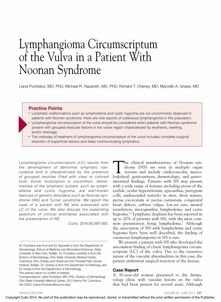
Lymphangioma circumscriptum (LC) results from the development of abnormal lymphatic vas-culature and is characterized by the presence of grouped vesicles filled with clear or colored fluid. Vulvar localization is uncommon. Abnor-malities of the lymphatic system, such as lymph-edema and cystic hygroma, are well-known features of genetic disorders such as Noonan syn- drome (NS) and Turner syndrome. We report the case of a patient with NS who presented with LC of the vulva. We also discuss the expanding spectrum of clinical anomalies associated with the presentation of NS.
Cutis. 2014;93:297-300.
The clinical manifestations of Noonan syn-drome (NS) are seen in multiple organ systems and include cardiovascular, muscu-
loskeletal, genitourinary, dermatologic, and gastro-intestinal findings. Patients with NS may present with a wide range of features including ptosis of the eyelids, ocular hypertelorism, epicanthus, pterygium colli, undescended testicles in men, short stature, pectus excavatum or pectus carinatum, congenital heart defects, cubitus valgus, low-set ears, mental retardation, micrognathia, lymphedema, and cystic hygroma.1,2 Lymphatic dysplasia has been reported in up to 20% of patients with NS, with the most com-mon presentation being lymphedema.1 Although the association of NS with lymphedema and cystic hygroma have been well described, the finding of cutaneous lymphangioma in NS is rare.
We present a patient with NS who developed the uncommon finding of classic lymphangioma circum-scriptum (LC) of the vulva. Due to the extensive nature of the vascular abnormalities in this case, the patient underwent surgical resection of the lesions.
Case ReportA 30-year-old woman presented to the derma-tology clinic with vascular lesions on the vulva that had been present for several years. Although
Lymphangioma Circumscriptum of the Vulva in a Patient With Noonan Syndrome Llana Pootrakul, MD, PhD; Michael R. Nazareth, MD, PhD; Richard T. Cheney, MD; Marcelle A. Grassi, MD
Dr. Pootrakul was from and Dr. Nazareth is from the Department of Dermatology, School of Medicine and Biomedical Sciences, State University of New York, Buffalo. Dr. Pootrakul currently is from the Division of Dermatology, Ohio State University Medical Center, Columbus. Drs. Cheney and Grassi are from Roswell Park Cancer Institute, Buffalo. Dr. Cheney is from the Department of Pathology and Dr. Grassi is from the Department of Dermatology. The authors report no conflict of interest. Correspondence: Llana Pootrakul, MD, PhD, Division of Dermatology, Ohio State University Medical Center, 2012 Kenny Rd, Columbus, OH 43221 ([email protected]).
Practice Points Lymphatic malformations such as lymphedema and cystic hygroma are not uncommonly observed in
patients with Noonan syndrome; there are rare reports of cutaneous lymphangioma in this population. Lymphangioma circumscriptum of the vulva should be considered when patients with Noonan syndrome
present with grouped vesicular lesions in the vulvar region characterized by erythema, swelling, and/or drainage.
The mainstay of treatment of lymphangioma circumscriptum of the vulva includes complete surgical resection of superficial lesions and deep communicating lymphatics.
Copyright Cutis 2014. No part of this publication may be reproduced, stored, or transmitted without the prior written permission of the Publisher.
CUTIS Do Not Copy

298 CUTIS®
Lymphangioma Circumscriptum
WWW.CUTIS.COM
the lesions remained unchanged in size, they had become increasingly bothersome due to persistent oozing and subsequently had negative effects on the patient’s quality of life. Her primary concerns were the appearance of the lesions, pruritus, and weep-ing discharge from the affected area. The patient previously was referred to the urology department and then laser therapy for the lesion was attempted with minimal benefit. She also had been evaluated by a gynecologist who proposed an initial diagno-sis of vulvar angiokeratoma and suggested surgi- cal treatment.
The patient’s medical history included NS, a heart murmur, and narrow-angle glaucoma. No
other concerns aside from cutaneous findings were elicited during a review of systems. Physical exami-nation revealed erythema and swelling with a pink mammillated surface on the bilateral labia majora accompanied by a clear, yellow, lymphlike discharge. The lesions did not extend to the introitus but dem-onstrated substantial involvement of the bilateral vulvar tissue.
The patient was referred to the gynecology department for diagnostic hysteroscopy, dilation and curettage, vaginoscopy, and bilateral vulvectomy. Two weeks following surgery, dramatic improvement of the vulvar lesions was noted, with some edema of the genital region as well as residual scattered red and clear vesicular lesions of LC (Figure 1). A microscopic analysis of surgical specimens performed by a board-certified pathologist (R.T.C.) revealed superficial and deep infiltrative growth of irregularly outlined lymphatic channels that did not closely border the overlying epithelial basement membrane, resulting in the presence of a prominent grenz zone (Figure 2). On immunohistochemical evaluation of sections of the left labium majus, lymphatic channels and endothelial vasculature stained positive with CD31 (platelet-endothelial cell adhesion molecule), which stained both lymphatic and blood vessel endothelium as well as a subset of tissue macrophages (Figure 3A). A number of vascular channels stained positive with D2-40, a marker that is relatively
Figure 2. A tissue section from the left labium majus revealed superficial and deep dilated vascular channels with increased density in the superficial dermis beneath a mildly papillomatous epidermis (A)(H&E, original magnifica-tion 2); ectatically dilated, endothelial-lined vascular spaces separated from an overlying basement membrane zone by an intervening grenz zone (B)(H&E, original magnification 10); and dilated endothelial-lined vascular channels with intraluminal eosinophilic lymph in the lumen. Intraluminal valves were seen in the centrally located lymphatic vessel (C)(H&E, original magnification 20).
Figure 1. Edema and scattered red and clear vesicular lesions 2 weeks following bilateral vulvectomy for treat-ment of lymphangioma circumscriptum of the vulva.
CBA
Copyright Cutis 2014. No part of this publication may be reproduced, stored, or transmitted without the prior written permission of the Publisher.
CUTIS Do Not Copy

VOLUME 93, JUNE 2014 299
Lymphangioma Circumscriptum
WWW.CUTIS.COM
specific for lymphatic endothelium. The endothelial lining of adjacent capillaries demonstrated negative staining for D2-40 (Figure 3B). Histopathologically, these results were consistent with the diagnosis of LC of the vulva.
CommentLymphangiomas previously have been classified as superficial and deep lesions, with LC comprising the superficial group and cavernous lymphangiomas and cystic hygromas classified in the deep group. Lymphangioma circumscriptum generally presents as cutaneous vesicles ranging in color from pink to black depending on the amount of blood intermixed with the clear lymph fluid.3 In 1970, Peachey et al4 reviewed and characterized lesions in 65 patients with cutaneous LC. The 2 most commonly described variants of cutaneous LC are classic and localized LC. Classic LC generally is characterized by exten-sive lesions with affected areas of skin greater than 1 cm². The lesions tend to be concentrated over the proximal regions of limbs and adjacent to limb girdles; however, they can be found on almost any area of the body.4 Localized LC presents as single discrete lesions of vesicles comprising an area of less than 1 cm². Lesions do not consistently present in any particular areas of the body but tend to be absent from regions where classic LC is seen.4
The differential diagnosis of LC of the vulva should include condyloma acuminatum, herpes zos-ter, molluscum contagiosum, angiomyofibroblastoma, and angiomyxoma. Lymphangioma circumscriptum of the vulva may be congenital or acquired and is
an uncommon finding. Vlastos et al5 reported only 11 cases of congenital LC of the vulva and 22 acquired cases. There have been additional reports of acquired LC of the vulva.6-13
Prior reports of lymphatic dysplasia associated with NS include the presence of cystic hygroma, lymphedema, intestinal lymphangiectasia, pulmo-nary lymphangiectasia, and facial cutaneous lym-phangioma in NS.14-16 Our case provides additional evidence that the characteristic phenotypes involv-ing lymphatic dysplasia in NS may be broadened to include cutaneous lymphangioma in addition to generalized lymphedema and cystic hygroma.
Management considerations for LC involve cos-metic effects, pain, swelling, persistent lymph fluid and blood leakage, and continued recurrence of infections.17 The mainstay of treatment of LC is sur-gical resection of the lesion with radical vulvectomy demonstrating the lowest recurrence rate following treatment of LC of the vulva.18 Complete surgi-cal resection also should include excision of deep communicating lymphatics. Additional therapeutic options include abrasive therapy, sclerotherapy, and electrocoagulation; however, these methods com-monly result in recurrence if the entire lesion and deep communicating system have not been destroyed by the treatment.18-20
ConclusionIt is not uncommon for patients with NS to dem-onstrate features of lymphatic abnormalities, with lymphedema and cystic hygroma most often observed.1 As demonstrated in this report, NS
Figure 3. Immunohistochemical analysis of a tissue section from the left labium majus demonstrated positive stain-ing for CD31 (platelet-endothelial cell adhesion molecule) and D2-40 in lymphatic channel endothelium. Numerous CD31 dilated vessels, both lymphatic and endothelial vascular channels (asterisk), were seen in the superficial dermis as well as D2-40 lymphatic channels (A)(original magnification 20). Adjacent capillaries (asterisk) showed negative staining for D2-40 (B)(original magnification 20).
A B
Copyright Cutis 2014. No part of this publication may be reproduced, stored, or transmitted without the prior written permission of the Publisher.
CUTIS Do Not Copy
300 CUTIS®
Lymphangioma Circumscriptum
WWW.CUTIS.COM
patients also may present with cutaneous LC. Although it generally is an uncommon finding, LC of the vulva should be considered when patients with NS present with vesicular lesions in the vulvar region characterized by erythema, swelling, and/or drainage. Appropriate management of LC includes complete surgical resection of superficial lesions and deep communicating lymphatics.
REFERENCES 1. Mendez HM, Opitz JM. Noonan syndrome: a review. Am J
Med Genet. 1985;21:493-506. 2. Zarabi M, Mieckowski GC, Mazer J. Cystic hygroma
associated with Noonan’s syndrome. J Clin Ultrasound. 1983;11:398-400.
3. Whimster IW. The pathology of lymphangioma circum-scriptum. Br J Dermatol. 1976;94:473-486.
4. Peachey RD, Lim CC, Whimster IW. Lymphangioma of skin. a review of 65 cases. Br J Dermatol. 1970;83:519-527.
5. Vlastos AT, Malpica A, Follen M. Lymphangioma circum-Lymphangioma circum-scriptum of the vulva: a review of the literature. Obstet Gynecol. 2003;101(5, pt 1):946-954.
6. Turan V, Ergenoglu M, Yeniel O, et al. Vulvar lym-phangioma circumscriptum. Int J Gynaecol Obstet. 2009;107:256-257.
7. Amouri M, Masmoudi A, Boudaya S, et al. Acquired lym-phangioma circumscriptum of the vulva. Dermatol Online J. 2007;13:10.
8. Stewart CJ, Chan T, Platten M. Acquired lymphangiecta-Acquired lymphangiecta-sia (‘lymphangioma circumscriptum’) of the vulva: a report of eight cases. Pathology. 2009;41:448-453.
9. Kokcu A, Yildiz L, Bildircin D, et al. Vulvar lymphangioma circumscriptum presenting periodic symptoms [published online ahead of print December 29, 2010]. BMJ Case Rep. doi:10.1136/bcr.06.2010.3056.
10. Ikeda M, Muramatsu T, Shida M, et al. Surgical manage-ment of vulvar lymphangioma circumscriptum: two case reports. Tokai J Exp Clin Med. 2011;36:17-20.
11. Bhat RM, Saldanha CS, Kambil SM, et al. Cutaneous lymphangiectasia of the vulva secondary to tuberculosis. Indian J Sex Transm Dis. 2012;33:35-37.
12. Shetty V, Venkatesh S. Acquired lymphangioma circum-scriptum of the vulva [published online ahead of print February 21, 2012]. Int J Gynaecol Obstet. 2012;117:190.
13. Uçmak D, Aytekin S, Sula B, et al. Acquired vulvar lymphangioma circumscriptum [published online ahead of print December 16, 2013]. Case Rep Dermatol Med. doi:10.1155/2013/967890.
14. Baltaxe HA, Lee JG, Ehlers KH, et al. Pulmonary lymphangectasia demonstrated by lymphangiogra-phy in 2 patients with Noonan’s syndrome. Radiology. 1975;115:149-153.
15. Vallet HL, Holtzapple PG, Eberlein WR, et al. Noonan syndrome with intestinal lymphangectasis. a metabolic and anatomic study. J Pediatr. 1972;80:269-274.
16. Evans DG, Lonsdale RN, Patton MA. Cutaneous lym-phangioma and amegakaryocytic thrombocytopenia in Noonan syndrome. Clin Genet. 1991;39:228-232.
17. Bond J, Basheer MH, Gordon D. Lymphangioma circum-scriptum: pitfalls and problems in definitive management. Dermatol Surg. 2008;34:271-275.
18. Roy KK, Agarwal R, Agarwal S, et al. Recurrent vulval con-Recurrent vulval con-genital lymphangioma circumscriptum—a case report and literature review. Int J Gynecol Cancer. 2006;16:930-934.
19. Lai CH, Hanson SG, Mallory SB. Lymphangioma circum-scriptum treated with pulsed dye laser. Pediatr Dermatol. 2001;18:509-510.
20. Huilgol SC, Neill S, Barlow RJ. CO2 laser therapy of vul-val lymphangiectasia and lymphangioma circumscriptum. Dermatol Surg. 2002;28:575-577.
Copyright Cutis 2014. No part of this publication may be reproduced, stored, or transmitted without the prior written permission of the Publisher.
CUTIS Do Not Copy