Embed Size (px)
Citation preview
LKB1 as the ghostwriter of crypt history
Marnix Jansen • Danielle Langeveld •
Wendy W. J. De Leng • Anya N. A. Milne •
Francis M. Giardiello • G. Johan A. Offerhaus
Published online: 30 July 2011
� The Author(s) 2011. This article is published with open access at Springerlink.com
Abstract Familial cancer syndromes present rare insights
into malignant tumor development. The molecular back-
ground of polyp formation and the cancer prone state in
Peutz-Jeghers syndrome remain enigmatic to this day.
Previously, we proposed that Peutz-Jeghers polyps are not
pre-malignant lesions, but an epiphenomenon to the
malignant condition. However, Peutz-Jeghers polyp for-
mation and the cancer-prone state must both be accounted
for by the same molecular mechanism. Our contribution
focuses on the histopathology of the characteristic Peutz-
Jeghers polyp and recent research on stem cell dynamics
and how these concepts relate to Peutz-Jeghers polyposis.
We discuss a protracted clonal evolution scenario in Peutz-
Jeghers syndrome due to a germline LKB1 mutation.
Peutz-Jeghers polyp formation and malignant transforma-
tion are separately mediated through the same molecular
mechanism played out on different timescales. Thus, a
single mechanism accounts for the development of benign
Peutz-Jeghers polyps and for malignant transformation in
Peutz-Jeghers syndrome.
Keywords LKB1 � Peutz-Jeghers syndrome � Hamartoma
� LGR5 � Clonal evolution � Polyp
Introduction
In his original monograph ‘On the remarkable familial
aggregation of mucosal polyposis of the gastrointestinal
and oropharyngeal tract and a pigmentation disorder’,
Dr. Jan Peutz explains how he first traced this constellation
of peculiar abnormalities in one family. His case descrip-
tion, published in 1921 in a monthly Dutch medical journal,
details the clinical vignette of an adolescent boy presenting
to the department of surgery of his hospital in The Hague
with symptoms of intestinal obstruction. Emergency lapa-
rotomy revealed an obstructing polyp in the small bowel
which was excised, after which the boy made a complete
recovery (Fig. 1). Previously, on routine physical exam, Dr.
Peutz had noticed perioral pigmentation in his young
patient. Moreover, on rigid rectoscopy numerous polyps,
akin to the one observed during emergency laparotomy, had
been found. The remarkable perioral pigmentation in his
index patient reminded Jan Peutz of a similar-looking pig-
mentation abnormality he had seen in a patient referred to
him 1 year previous. It turned out that this last patient was
in fact the younger brother of his index patient. In his case
description, Dr. Peutz recalls how it was the mother of his
index patient who pointed out that ‘these peculiar pig-
mentations only seemed to occur in the children that
resembled their father’. We can only assume that this
comment triggered dr Peutz to examine the other family
members. Subsequently, he describes how similar abnor-
malities were found in four siblings of his initial patient.
Ninety years after his original publication we know that
the patients described in his report were affected by a
cancer-prone polyposis syndrome that bears Peutz’s name.
We have continued to follow this original family [1].
Germ-line sequencing of members of this pedigree
revealed a T insertion in exon 1 at codon 66 (535insT) that
M. Jansen (&) � G. J. A. Offerhaus
Department of Pathology, Academic Medical Center,
PO Box 22660, 1100 DD Amsterdam, The Netherlands
e-mail: [email protected]
D. Langeveld � W. W. J. De Leng � A. N. A. Milne �G. J. A. Offerhaus
Department of Pathology, University Medical Center,
Utrecht, The Netherlands
F. M. Giardiello
Department of Medicine, Johns Hopkins University School
of Medicine, Baltimore, MD, USA
123
Familial Cancer (2011) 10:437–446
DOI 10.1007/s10689-011-9469-3

resulted in a frameshift producing a stopcodon in exon 4
(codon 162) of the LKB1 gene. Pedigree analysis reveals
that of the original family only 17 of 22 affected family
members survived into adulthood. The mean age of death
for affected family members was substantially younger
than for unaffected family members (38 vs. 69 years) and
causes of death included bowel obstruction, and intestinal
and extraintestinal cancers. This family testifies that Peutz-
Jeghers syndrome (PJS) is not a benign disorder and
encapsulates the major clinical areas of concern in a
nutshell.
Although much has been learned about Peutz-Jeghers
syndrome, many questions still remain. This issue brings
together many fascinating points of research all revolving
around Peutz-Jeghers and the germline genetic defect. My
contribution (as a surgical pathologist) deals with two
aspects, the histopathology of the characteristic Peutz-Je-
ghers polyps and, secondly, recent research on stem cell
dynamics and how this relates to Peutz-Jeghers polyposis.
Polyp histopathology: ‘naming and shaming’
Familial cancer syndromes present rare insights into
malignant tumor formation. Syndromes such as familial
adenomatous polyposis (FAP) are characterized clinically
by the development of large numbers of neoplastic pre-
cursor lesions some of which, due to their sheer number,
will progress to cancer. Whereas in syndromes such as
Lynch syndrome (previously, known as hereditary non-
polyposis colorectal cancer or HNPCC) only few precursor
lesions develop, most of which are guaranteed to progress to
cancer. PJS is characterized by the development of histo-
logically distinct gastrointestinal polyps. Since patients
with PJS are at an increased risk of developing cancer in the
gastrointestinal tract, it is important to establish whether
these polyps carry an inherent risk of neoplastic transfor-
mation. As both the polyps and the cancer-prone condition
are due to a germline mutation in the LKB1 tumor sup-
pressor gene, an understanding of the mechanism of polyp
formation should reveal important insight into the cancer-
prone condition of these patients; regardless of whether the
polyps per se are ultimately deemed to lack an inherent risk
of transformation.
The first sign of PJS is a distinctive pigmentation
observed around the lips, oral mucosa, genitalia or palmar
surfaces appearing early in childhood [2, 3]. However,
gastrointestinal polyps cause the main clinical symptoms.
These may develop throughout the gastrointestinal tract,
grow to large sizes, and cause obstruction, pain, and gas-
trointestinal bleeding with anaemia [4]. A polyp is defined
as any abnormality that protrudes above the mucosal sur-
face. Polyps are further subclassified by detailed micro-
scopic analysis of their mucosal lining and any associated
histological features. Consequently, polyposis syndromes
are primarily classified by the histological appearance of
the polyps and further subclassifications can be made by
analzying phenotypic expression, systemic manifestations
or the exact underlying genetic cause. For example, FAP
patients display adenomatous polyps which may harbour
varying grades of dysplasia or invasive carcinoma. The
large gastrointestinal polyps typically encountered in PJS,
are pedunculated polyps with non-dysplastic overlying
epithelium (Fig. 2). A characteristic feature of mature PJS
polyps is the prominent core of arborizing smooth muscle,
which extends into the head of the polyp. Smooth muscle
proliferation appears to accompany epithelial hyperprolif-
eration during the earliest stages of polyp development [5].
Of note, smooth muscle proliferation is not a histopathol-
ogical feature specific for the PJS polyp. For example, PJS-
like smooth muscle proliferation can also be observed in
conventional sporadic adenomatous polyps. In this setting,
the smooth muscle proliferation develops secondary to
mechanical insults (due to intestinal peristalsis) during
adenomatous polyp growth (Fig. 3) [5]. Furthermore, other
conditions unrelated to PJS such as mucosal prolapse
syndrome may present with histological features similar to
those observed in PJS polyps [5, 6]. Thus, it is unclear
whether the observed smooth muscle proliferation is
causally involved in polyp initiation or, alternatively,
develops only secondarily to accommodate epithelial
hyperproliferation. We return to this point below.
The potential for malignant degeneration of PJS polyps
and, specifically, whether cancer in PJS patients arises
from the non-dysplastic epithelium covering PJS polyps
remains a hotly debated subject. Analogous to the genetic
lessons learned from the stepwise tumor progression
Fig. 1 Depiction of the original small bowel resection specimen
containing the PJS polyp that led to an episode of small bowel
obstruction in the index patient first described by Jan Peutz in his
monograph on the pedigree
438 M. Jansen et al.
123

occurring during the adenoma-carcinoma sequence in FAP
patients (and, by extension, many sporadic colorectal
cancer patients), neoplastic transformation in PJS patients
is thought to model discrete genetic hurdles taken during
malignant tumor progression in sporadic cancer patients.
The gastrointestinal polyps in PJS constitute a prime sus-
pect for cancer initiation in PJS patients; but, do they really
harbour an underlying risk for malignant transformation in
spite of their benign appearance? Take the above men-
tioned case of a conventional adenomatous polyp display-
ing a prominent PJS-like core of arborizing smooth muscle
as an example. It would—on histopathological grounds
alone—not be possible to discern whether an adenomatous
polyp secondarily acquired a PJS-like smooth muscle stalk
Fig. 2 Overview of histological findings in a characteristic PJS polyp,
in this case resected from the duodenum. A H&E staining showing an
overview of the polyp. The epithelial lining demonstrates normal
gobletcel-bearing epithelium without signs of dysplasia. Reactive
epithelial changes can be noted at the surface of the polyp, which
shows edematous villi and active inflammation, presumably due to
surface erosion. Towards the center some crypts contain inspissated
mucin. b Smooth muscle actin (SMA) immunohistochemical staining
showing the arborizing core of smooth muscle (labeled red) that
characterizes PJS polyps. c Ki67 proliferation marker immunohisto-
chemical staining showing that proliferation is restricted to the crypt
bases indicating proper compartimentalization of proliferation and
differentiation. d Periodic-acid Schiff stain after diastase digestion
showing normal differentiation and maturation of the secretory goblet
cell lineage
Fig. 3 PJS-like smooth muscle proliferation in a conventional non-
PJS adenomatous polyp. a Gross macroscopy of a pedunculated
colonic polyp in a 67-year old patient not affected by PJS. A right
hemicolectomy was performed following the diagnosis of colon
cancer in the ascending colon in this patient; the polyp shown here
was found incidentally in the cecum. b Low-power photomicrograph
of the polyp shown in a. The polyp has a tubulovillous architecture
characterized by mucosal fronds emanating from the stalk of the
polyp. Note the slender extensions of the smooth muscle core into the
tips of the villous projections (indicated by asterisk in c). This
arborizing core of smooth muscle resembles the histology shown in
Fig. 2b for the polyp removed from the duodenum of a PJS patient.
c Unlike PJS polyps, which are characterized by a non-dysplastic
hyperproliferative epithelial lining, this polyp has a dysplastic
epithelial covering classified as low-grade dysplasia. The boxed areain b is shown at higher power in c
LKB1 as the ghostwriter of crypt history 439
123

or, alternatively, a PJS polyp has undergone adenomatous
transformation. Historically, case reports have described
dysplastic adenomatous transformation occurring in a
supposedly pre-existent hamartomatous PJS polyp [7].
However, dysplasia occurring in classical PJS polyps
appears to be a very uncommon phenomenon. In series
from the renowned familial polyposis registry at St Mark’s
hospital, a review of 491 PJS polyps showed no evidence
of dysplasia in any [8]. Moreover, follow-up over 45 years
of 48 patients at the Mayo Clinic failed to reveal dysplastic
change in any PJS polyp either [9]. More recently, we
proposed that the historical accounts of adenomatous
transformation occurring in PJS polyps might be explained
by a blurring of the order of events; that is, these lesions
likely arose from a conventional adenoma arising in a PJS
patient that secondarily developed a PJS-like smooth
muscle stalk, akin to the situation encountered in sporadic
patients, as described above [5].
Since the focus of cancer initiation in PJS patients
remains to be definitively settled, proper histopatholo-
gical classification of PJS polyps is an important issue.
Traditionally, mature PJS polyps have been classified as
hamartomas. A hamartoma is defined as a combined and
non-dysplastic overgrowth of all tissue layers native to the
site of origin of the lesion in equal proportion. However,
the ‘hamartoma’ designation for PJS polyps is presently not
based on an etiological rationale and purely descriptive
features such as the prominent core of arborizing smooth
muscle and epithelial hyperproliferation may lack a com-
mon causal relationship as stipulated by the hamartoma
designation. The hamartoma classification may, therefore,
be misleading (and may ultimately prove to be a misnomer)
because the actual etiological mechanism of polyp devel-
opment remains unclear at this point. This consideration is
also relevant to studies that have appeared in literature
linking the so-called ‘hamartoma syndromes’ PJS, tuberous
sclerosis and Cowden’s disease [10]. Clinically it appears
artificial to lump these syndromes together since they share
little medical overlap other than the ‘hamartoma’ desig-
nation. Cowden’s disease, for example, is characterized by
juvenile polyps. Using the designation ‘hamartoma’ as an
all-encompassing term for polyps that currently defy
proper etiological classification does not promote under-
standing of these syndromes.
Further research examining the histological architecture
of PJS polyps in closer detail is therefore warranted. For
example, the hamartoma designation mandates that the
epithelial covering of PJS polyps should display all lines of
differentiation normally observed at the site of origin of a
PJS polyp in a normal ratio. However, earlier studies noted
that regarding the epithelial covering of PJS polyps, some
differentiated epithelial lineages may be lost at the expense
of other lineages or in favour of a more immature
phenotype altogether [11]. Full-blown PJS polyps com-
monly reveal these alterations; however, the net shift that
occurs towards an immature phenotype is not to be inter-
preted as a sign of premalignant potential per se. This is
because the polyps often display many of these reactive
changes (such as decreased epithelial maturation and
increased proliferation) along with signs of significant
trauma, for example mucosal haemorrhage. In an interest-
ing recent study, epithelial maturation defects in Lkb1?/-
murine gastric mucosa was investigated before the onset of
frank polyposis [12]. Lina Udd and co-workers show that
foci of otherwise unremarkable gastric antral mucosa in
Lkb1?/- mice displayed subtle defects in epithelial matu-
ration. Curiously, these minute foci were often associated
with Ki67-positive gland bases [12], a point we will return
to shortly. Thus, it appears prudent to refer to the gastro-
intestinal lesions in PJS simply as ‘PJS polyps’ rather than
hamartomas, since the latter would imply an etiological
understanding currently not justified. Most importantly,
because the malignant potential of characteristic PJS
polyps (those polyps demonstrating a prominent smooth
muscle core with normal non-dysplastic overlying epithe-
lium) remains uncertain, PJS polyps should be separated
from (other) precursor lesions occurring in PJS patients or
engineered Lkb1 mouse models [13]. Accurate classifica-
tion of PJS polyps thus awaits an elucidation of the
sequence of events during PJS polyp initiation and the
pathophysiological mechanism responsible for PJS polyp
development.
To this point we have considered proper histopatholo-
gical classification of the polyps encountered in PJS
patients. Now we focus on recent research involving the
LKB1 tumor suppressor gene and how this might help
elucidate the mechanism of polyp development in PJS
patients. We will examine research, including our own
unpublished work that has investigated the role of LKB1 in
intestinal stem cell compartments.
Crypt dynamics in Peutz-Jeghers polyposis
Lineage competition in the gastrointestinal tract
Most epithelial tissues self-renew throughout adult life
owing to the presence of multipotent stem cells and/or
unipotent progenitor cells [14]. The cells of the intestinal
epithelium are arranged hierarchically, becoming progres-
sively more differentiated with age and passage along the
crypt-villus axis. The relatively straightforward histologi-
cal set-up and the high proliferative rate has made the
mammalian intestinal stem cell compartment one of the
most studied of all mammalian stem cell niches [15].
Recently, through the identification of LGR5 as a murine
440 M. Jansen et al.
123
intestinal stem cell marker, the direct analysis of mam-
malian intestinal stem cells has become possible [16].
These studies show that each intestinal crypt harbors
multiple stem cells that together give rise to all other post-
mitotic differentiated cells populating the villus. All stem
cells generate daughter progeny through continuous self-
renewal, and every individual stem cell and its clonal
daughter progeny can be considered to represent one
lineage in the crypt. Classic studies performed in chimeric
animals carrying a mixture of background marker alleles
show that stem cell lineages within the intestinal stem cell
crypt compartment undergo so-called lineage competition
[17]. That is, at birth every crypt in these animals displays a
mosaic (polyclonal) composition of marker alleles. How-
ever, within several weeks these crypts become monoclo-
nal, retaining just one of the marker alleles. This
mechanism of random loss of some lineages with
replacement by others is called lineage competition. Ele-
gant studies employing the aforementioned LGR5 stem cell
marker have re-affirmed this notion (vide infra). Lineage
competition is expected to continue even after the crypt has
reached a monoclonal status with respect to its chimeric
background. In other words, after one chimeric background
has attained dominance, all daughter lineages of this
mother lineage will now undergo lineage competition. In
time, as a result of random genetic drift, one daughter
lineage will again attain dominance, and its daughter lin-
eages will then undergo lineage competition. This is an
ongoing cycle of lineage competition within the crypt and
this lottery-like process of clonal evolution occurs at steady
state in each of the approximately 15 million crypts in the
adult human colon. The time between two successive stem
cell lineages attaining dominance in one crypt is called
crypt niche succession time. Importantly, new potential
markers such as genomic methylation changes or somatic
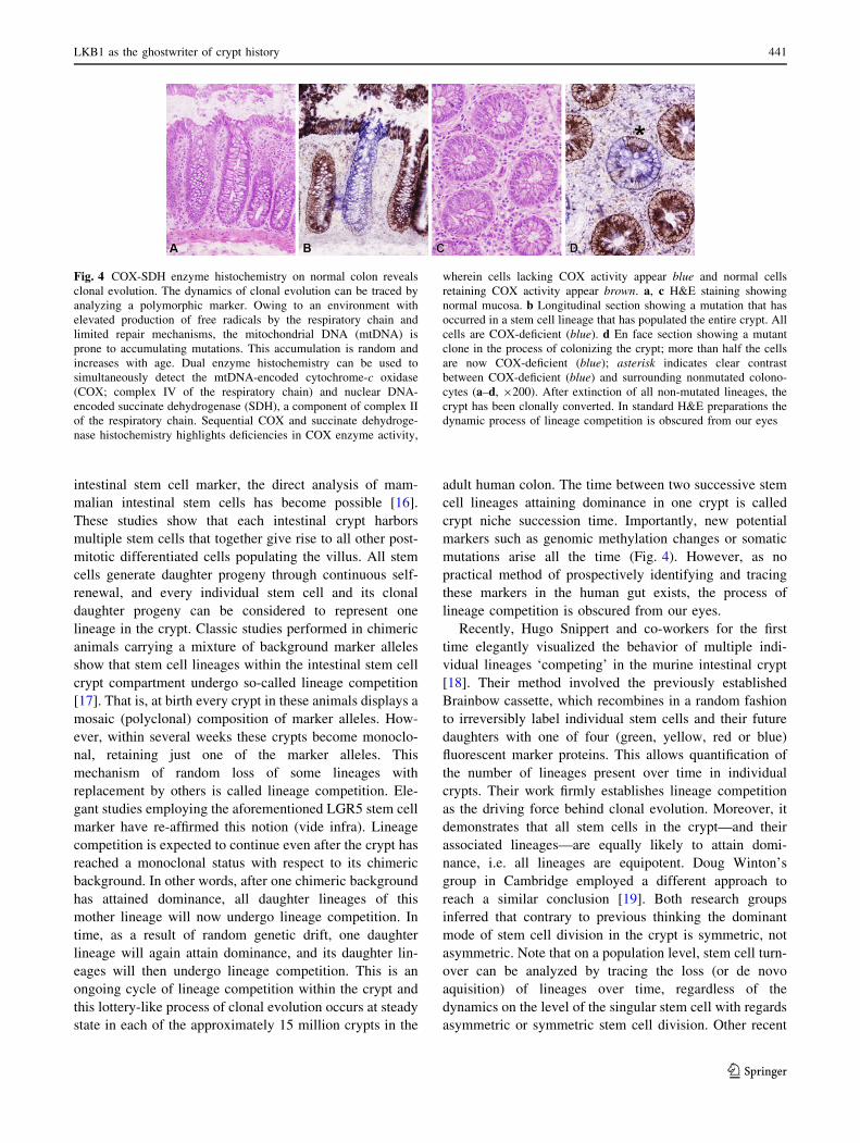
mutations arise all the time (Fig. 4). However, as no
practical method of prospectively identifying and tracing
these markers in the human gut exists, the process of
lineage competition is obscured from our eyes.
Recently, Hugo Snippert and co-workers for the first
time elegantly visualized the behavior of multiple indi-
vidual lineages ‘competing’ in the murine intestinal crypt
[18]. Their method involved the previously established
Brainbow cassette, which recombines in a random fashion
to irreversibly label individual stem cells and their future
daughters with one of four (green, yellow, red or blue)
fluorescent marker proteins. This allows quantification of
the number of lineages present over time in individual
crypts. Their work firmly establishes lineage competition
as the driving force behind clonal evolution. Moreover, it
demonstrates that all stem cells in the crypt—and their
associated lineages—are equally likely to attain domi-
nance, i.e. all lineages are equipotent. Doug Winton’s
group in Cambridge employed a different approach to
reach a similar conclusion [19]. Both research groups
inferred that contrary to previous thinking the dominant
mode of stem cell division in the crypt is symmetric, not
asymmetric. Note that on a population level, stem cell turn-
over can be analyzed by tracing the loss (or de novo
aquisition) of lineages over time, regardless of the
dynamics on the level of the singular stem cell with regards
asymmetric or symmetric stem cell division. Other recent
Fig. 4 COX-SDH enzyme histochemistry on normal colon reveals
clonal evolution. The dynamics of clonal evolution can be traced by
analyzing a polymorphic marker. Owing to an environment with
elevated production of free radicals by the respiratory chain and
limited repair mechanisms, the mitochondrial DNA (mtDNA) is
prone to accumulating mutations. This accumulation is random and
increases with age. Dual enzyme histochemistry can be used to
simultaneously detect the mtDNA-encoded cytochrome-c oxidase
(COX; complex IV of the respiratory chain) and nuclear DNA-
encoded succinate dehydrogenase (SDH), a component of complex II
of the respiratory chain. Sequential COX and succinate dehydroge-
nase histochemistry highlights deficiencies in COX enzyme activity,
wherein cells lacking COX activity appear blue and normal cells
retaining COX activity appear brown. a, c H&E staining showing
normal mucosa. b Longitudinal section showing a mutation that has
occurred in a stem cell lineage that has populated the entire crypt. All
cells are COX-deficient (blue). d En face section showing a mutant
clone in the process of colonizing the crypt; more than half the cells
are now COX-deficient (blue); asterisk indicates clear contrast
between COX-deficient (blue) and surrounding nonmutated colono-
cytes (a–d, 9200). After extinction of all non-mutated lineages, the
crypt has been clonally converted. In standard H&E preparations the
dynamic process of lineage competition is obscured from our eyes
LKB1 as the ghostwriter of crypt history 441
123

studies reached a similar conclusion for other stem cell
niches, such as the murine epidermal and germ line stem
cell compartment [20, 21]. This body of work indicates that
lineage competition in the crypt must be understood by
investigating clonal evolution from a population perspec-
tive. Fortuitously, previous work has paved the way for
investigating this complex matter at the human level.
A population genetics approach to lineage competition
Studies performed by Darryl Shibata and colleagues in
FAP patient material previously formulated a statistical
approach to crypt niche succession times [22, 23]. These
modeling studies are based on the retrospective analysis of
genomic differences within a population of intestinal crypt
progenitor cells through the concept of a molecular clock.
Molecular clocks, in paleontology, record time between a
last common ancestor and species divergence based on the
random accumulation of genetic changes according to a
pre-determined neutral mutation rate between related
genomes [24]. Analogously, it should be possible to reveal
phylogenies of progenitor lineages within individual crypts
based on the genetic changes accumulated over time since
progenitor lineages last went through crypt niche succes-
sion [25]. A marker under neutral selection is expected to
show an increased measure of diversity as lineages diverge
over time. Following this reasoning, Darryl Shibata and his
co-investigators analyzed the diversity in methylation
patterns of the CpG island of the CSX gene—which is not
expressed in human colon—within and between individual
crypts. These crypt heterogeneity patterns were signifi-
cantly greater in histologically normal FAP crypts com-
pared to crypts from control patients [23]. This indicates
that crypt niche succession times are expanded in FAP
crypts, since lineages that persist longer retain a greater
number of variants.
Prior to the discovery of the energy-sensing AMPK
module as a downstream target, LKB1 had been most
prominently linked to the regulation of cellular polarization
in model organisms. The alleles encoding the nematode
and fly counterpart of the LKB1 tumour suppressor were
both retrieved in genomic screens designed to pick mutants
defective in cellular polarity regulation [26, 27]. Not
withstanding the accumulated data on the LKB1-AMPK
module in cellular metabolism [28], these early studies
remain some of the strongest evidence linking LKB1 to
cellular polarization. Later studies in vertebrate model
systems provided additional evidence for a role of LKB1 in
cell polarity and directed cell division [29]. Consequently,
germline LKB1 mutations may upset stem cell turn-over in
PJS patients’ crypts as well. We, therefore, implemented
the technique outlined above involving the analysis of CSX
crypt diversity patterns on unaffected PJS mucosa and
age-matched control crypts. These analyses are difficult to
perform because of the relative scarcity of patient material.
Nonetheless, through this unbiased technique, we found
that PJS crypts retain a statistically increased number of
variants when compared to age-matched controls (unpub-
lished data). Thus, akin to FAP, crypt diversity is greater in
unaffected PJS crypts indicating that this physiological
process of clonal evolution is protracted in PJS. As dis-
cussed below, this protracted clonal evolution scenario
implicates a cancer-prone state in unaffected PJS mucosa at
normal background mutation rates.
Consequences of a protracted clonal evolution scenario
in PJS crypts
Stem cells accumulate mutations stochastically from birth
in phenotypically normal epithelia. During life, stem cell
populations and their pool of accumulated mutations con-
tinuously change as individual lineages become extinct or
attain niche dominance. Most mutations in stem cell lin-
eages will be lost, because only one current stem cell
lineage attains future dominance [17–19]. These mutations
need not evoke a selective advantage and may be initially
neutral. Individual mutations without selective advantage
may thus initially ‘hitchhike’ along with the inherent clonal
evolution of stem cell niches. The analysis of crypt
diversity patterns through neutral markers such as CSX
methylation tags is based on this hitchhiking pattern.
Eventually though, combinations of mutations confer a
visible tumor such as a tubular adenoma. Differences in
clonal evolution rate may allow crypt mutations to accu-
mulate at different frequencies even though these muta-
tions arise at a similar rate. This occurs because the rate at
which mutations are shed from the crypt depends on the
clonal evolution rate [22]. This was also shown in an ear-
lier study comparing crypt-restricted loss of O-acetyl-
transferase activity between normal colonic epithelia in
FAP and Lynch syndrome patients [30]. In this study
phenotypically normal FAP epithelium demonstrated more
crypts mutant for O-acetyltransferase activity than pheno-
typically normal age-matched Lynch syndrome epithelium.
These findings on O-acetyltransferase activity should
extend to other transcripts such as p53 or K-RAS. Thus, in
crypts undergoing a protracted clonal evolution scenario
such as in FAP [23] or PJS (our unpublished data) genetic
clonal diversity is greater.
Differences in the amount of accumulated mutations that
persist over time show that the rate of clonal evolution can be
protective (or anti-tumorigenic) because mutant stem cells
may be lost through lineage competition. Mathematical
modelling adapted from population genetics predicts that the
chance of loss of novel alleles within a population is given by
ploss = (2 N - 1)/2 N, where N is population size and the
442 M. Jansen et al.
123
chance of fixation of new alleles is pfix = 1 - ploss = 1/2 N.
In a similar fashion one can derive that the rate of drift
(loss or fixation of novel alleles under neutral selection) is
inversely proportional to population size (see Table 1 on
the dynamics of surnames in Korea). Therefore, popula-
tion size has a domineering effect on the rate of fixation
or loss of novel alleles and this effect becomes more
pronounced in smaller populations. Accordingly, with
larger population sizes, the time between population
bottlenecks or crypt niche succession increases propor-
tionally. A protracted clonal evolution or increased line-
age residence time, therefore, decreases the time to cancer
presentation by expanding the pool of selectable variants
retained over time. Or, put in other words, a protracted
clonal evolution will shorten the time to adenoma for-
mation through the increased likelihood of an accidental
acquisition of a biallelic gatekeeper mutation. Thus, by
definition, demonstration of an expanded progenitor pool
in a niche that undergoes lineage competition reflects a
cancer-prone state.
LKB1 hemizygosity expands the progenitor zone
in unaffected Peutz-Jeghers mucosa
We previously proposed that the ‘hamartomatous’ polyps
observed in PJS are an epiphenomenon to the cancer-prone
condition, and that its characteristic smooth muscle core is
indistinguishable from histopathological alterations seen in
forms of mucosal prolapse [5]. Analysis of murine PJS
polyps in Lkb1 hemizygous mice established independently
in several laboratories convincingly shows that the
wild-type allele need not be lost during polyp formation
[31–33]. Following our proposal that the polyps are an
epiphenomenon to the cancer-prone condition, we are
faced with the remarkable situation of a rare polyposis
syndrome where polyps and carcinomas occur simulta-
neously, yet these are not linked in a clinicopathological
sense. However, regardless of whether the polyps lack
premalignant potential, an understanding of their devel-
opment will reveal important insight into the co-existent
cancer-prone condition, as the molecular mechanism
responsible for polyp formation must, under the simplest
model, be responsible for the cancer-prone condition in PJS
as well.
We previously observed an expanded crypt progenitor
zone in normal intestinal mucosa in PJS patients by stan-
dard immunohistochemical labeling with the Ki67 prolif-
eration marker [34]. We inferred that germline mutation of
LKB1 leads to a defect in the regulation of stem and/or
transit amplifying cell turn-over in the mammalian intes-
tine. Importantly, in this study we observed an elongated
progenitor tract in the absence of an increased labeling
index (that is, the number of Ki67-labeled nuclei divided
by the total number of nuclei in the crypt was not signifi-
cantly different between PJS crypts and matched controls).
This shows that, besides the absolute size of the progenitor
zone, other basic characteristics of the crypt as a stem cell
niche (such as division rate, the pace of maturation and/or
differentiation, and niche exit) are unchanged [35]. The
observation of an expanded progenitor tract displaying a
normal labeling index argues for dysregulation at the level
of the intestinal stem cell. Our data derived from the
analysis of dynamic CSX methylation patterns in unaf-
fected PJS mucosa are in full agreement with this
hypothesis. Definitive analysis of the size of the dedicated
stem cell pool in PJS crypts awaits the availability of direct
stem cell markers in human tissue.
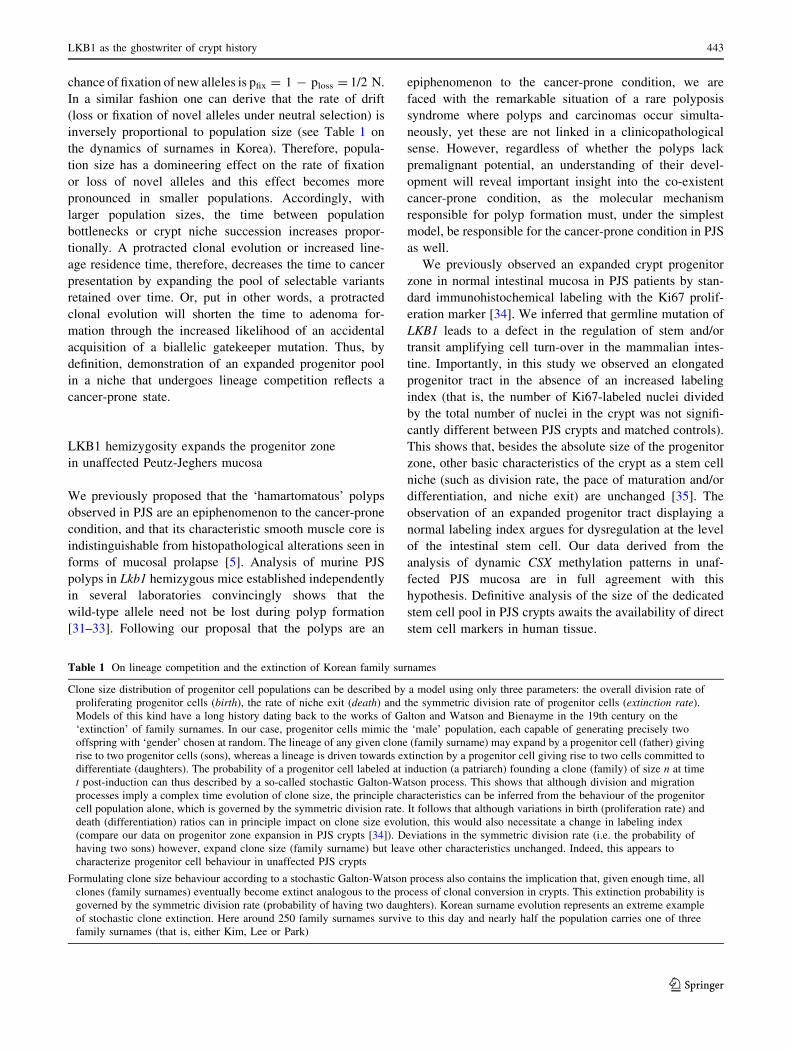
Table 1 On lineage competition and the extinction of Korean family surnames
Clone size distribution of progenitor cell populations can be described by a model using only three parameters: the overall division rate of
proliferating progenitor cells (birth), the rate of niche exit (death) and the symmetric division rate of progenitor cells (extinction rate).
Models of this kind have a long history dating back to the works of Galton and Watson and Bienayme in the 19th century on the
‘extinction’ of family surnames. In our case, progenitor cells mimic the ‘male’ population, each capable of generating precisely two
offspring with ‘gender’ chosen at random. The lineage of any given clone (family surname) may expand by a progenitor cell (father) giving
rise to two progenitor cells (sons), whereas a lineage is driven towards extinction by a progenitor cell giving rise to two cells committed to
differentiate (daughters). The probability of a progenitor cell labeled at induction (a patriarch) founding a clone (family) of size n at time
t post-induction can thus described by a so-called stochastic Galton-Watson process. This shows that although division and migration
processes imply a complex time evolution of clone size, the principle characteristics can be inferred from the behaviour of the progenitor
cell population alone, which is governed by the symmetric division rate. It follows that although variations in birth (proliferation rate) and
death (differentiation) ratios can in principle impact on clone size evolution, this would also necessitate a change in labeling index
(compare our data on progenitor zone expansion in PJS crypts [34]). Deviations in the symmetric division rate (i.e. the probability of
having two sons) however, expand clone size (family surname) but leave other characteristics unchanged. Indeed, this appears to
characterize progenitor cell behaviour in unaffected PJS crypts
Formulating clone size behaviour according to a stochastic Galton-Watson process also contains the implication that, given enough time, all
clones (family surnames) eventually become extinct analogous to the process of clonal conversion in crypts. This extinction probability is
governed by the symmetric division rate (probability of having two daughters). Korean surname evolution represents an extreme example
of stochastic clone extinction. Here around 250 family surnames survive to this day and nearly half the population carries one of three
family surnames (that is, either Kim, Lee or Park)
LKB1 as the ghostwriter of crypt history 443
123

The forming of a Peutz-Jeghers polyp
In PJS crypts the ratio of proliferative over non-prolifera-
tive cells remains essentially the same even though more
cells (in absolute numbers) are continuously fed onto the
villous epithelial carpet [34]. Incipient polyps might be
created by stochastic deviations from the normal equilib-
rium of stem cell divisions. In the study previously
discussed by Lina Udd et al. scattered foci of increased
Ki67-labeling in unaffected Lkb1?/- gastric mucosa were
observed [12]. These stochastic bursts of proliferative
activity may form small mucosal elevations which may
serve as a leadpoint upon which peristalsis can act. Clas-
sically, PJS polyps have been described as numbering from
a few to several dozens along the entire length of the
gastrointestinal tract. A recent study investigating COX-2
inhibition by sulindac analogues in a small cohort of PJS
patients necessitated repeated gastroscopies. This study
unexpectedly showed that the gastric epithelium in these
patients was in fact studded with ‘hundreds of small
hyperplastic lesions’ [36]. Potentially these lesions wax
and wane over time (small outpocketings may be extruded
or auto-amputated) and never come to clinical attention. In
fact, most larger lesions both in the murine system (typi-
cally at the level of the pylorus [2]) as well as in the human
system (at the level of the descending colon and rectum
[1]) occur at sites of greatest mechanical force. This sug-
gests that incipient polyps occur frequently and along the
gastrointestinal tract, but a literal tug of war between
mucosal elevations and mechanical force would favour a
small mucosal elevation to be moulded into a macroscopic
polyp. Indeed, an incipient polyp already shares features
with mucosal prolapse, in particular smooth muscle pro-
liferation. A small incipient polyp could serve as the
leadpoint for further outpouching due to mechanical forces
pulling on the nascent polyp. This would culminate in the
full-blown macroscopic cauliflower-like lesion with abun-
dant smooth muscle proliferation microscopically. Impor-
tantly, in this scenario smooth muscle proliferation is
secondary to, and accommodates for, epithelial hyperpro-
liferation. Our observations on epithelial turn-over
dynamics in PJS can explain the formation of incipient PJS
polyps as small hyperplastic lesions which lack an inher-
ently increased risk of neoplastic transformation.
Conclusion
Increased clonal diversity in normal PJS mucosa
Mutation and selection are the twin driving forces of
evolution. Epithelia may silently carry many potentially
oncogenic mutations, such as p53 mutations, that only
become exposed given the right context of conspiring
genomic hits. Evolutionary change, therefore, is critically
dependent on the presence of a range of heritable variants.
Time to visible tumor formation is shortened in a niche that
contains an increased number of progenitor cells [23]. An
expanded progenitor pool thus reflects a cancer-prone state
and predicts that pre-malignant lesions will arise at an
accelerated pace in comparison to the general population.
Our preliminary data on CSX crypt methylation patterns in
unaffected PJS mucosa are consistent with a protracted
clonal evolution scenario in PJS. Longer crypt niche suc-
cesion times increase the level of genetic clonal diversity
on which somatic evolution acting through natural selec-
tion depends (Fig. 5). Broadening the mutational repertoire
present in the crypt increases the odds of fortuitiously
incurring an accidental combination of mutations confer-
ring a tumor phenotype. In this scenario, patients with PJS
are expected to retain a greater number of mutations
incurred at a normal background mutation rate. This
increases genetic clonal diversity and accelerates somatic
evolution in PJS crypt stem cell niches. Similar consider-
ations concerning the neoplastic transformation in PJS
apply to other epithelia with dedicated stem cell compart-
ments (such as breast and pancreas), although we have
focused in this paper on stem cell turnover in the gastro-
intestinal crypt. This lottery-like process of accumulating
and selecting pathogenic hits in stem cell niches is played
out in each of the approximately 15 million crypts that
constitute the human colon.
Fig. 5 Increased clonal diversity in PJS crypts. This diagram
illustrates the consequences of differences in clonal evolution in
stem cell compartments such as the intestinal crypt. Over time stem
cell lineages accumulate mutations at a set background mutation rate.
By using a polymorphic marker such as COX enzyme activity we can
trace the appearance of individual lineages in the crypt (see Fig. 4).
We have observed an expanded crypt progenitor zone in unaffected
intestinal mucosa in PJS patients [34]. By comparing CSX crypt
heterogeneity patterns in unaffected PJS mucosa and age-matched
control crypts, we found that PJS crypts retain a greater number of
polymorphic marker mutations. This indicates that clonal evolution is
protracted in Peutz-Jeghers syndrome and predicts that pre-malignant
lesions will arise at an accelerated pace in comparison to the general
population (see text)
444 M. Jansen et al.
123
Definitively testing the impact of LKB1 hemizygosity in
lineage succession in the intestinal stem cell compartment
will require lineage tracing experiments where multiple
individual lineages can be traced competing over time in
individual crypts. With markers now available to unam-
biguously identify and genetically target progenitor cell
populations [18] this crucial part of early tumor history has
become open to scrutiny. In case LKB1 hemizygosity can
be shown to suffice for accelerated transformation then the
hamartomatous epithelium covering the polyps would
constitute the tip of the proverbial iceberg; it is the entire
epithelial lining that is at increased risk for malignant
degeneration. Demonstration of increased genetic clonal
diversity in phenotypically normal Peutz-Jeghers mucosa
would directly implicate a cancer-prone state at normal
background mutation rates. This would provide important
impetus to begin studying the impact of LKB1 hemizy-
gosity on lineage competition in stem cell niches. Thus,
LKB1 hemizygosity may independently mediate polyp
formation and the cancer-prone condition through one
molecular mechanism played out on different timescales.
Open Access This article is distributed under the terms of the
Creative Commons Attribution Noncommercial License which per-
mits any noncommercial use, distribution, and reproduction in any
medium, provided the original author(s) and source are credited.
References
1. Westerman AM, Entius MM, de Baar E, et al. (1999) Peutz-
Jeghers syndrome: 78-year follow-up of the original family.
Lancet 353(9160): 1211–1215
2. Katajisto P, Vallenius T, Vaahtomeri K et al (2007) The LKB1
tumor suppressor kinase in human disease. Biochim Biophys
Acta 1775(1):63–75
3. McGarrity TJ, Amos C (2006) Peutz-Jeghers syndrome: clin-
icopathology and molecular alterations. Cell Mol Life Sci
63(18):2135–2144
4. Gao H, van Lier MG, Poley JW, Kuipers EJ, van Leerdam ME,
Mensink PB (2010) Endoscopic therapy of small-bowel polyps
by double-balloon enteroscopy in patients with Peutz-Jeghers
syndrome. Gastrointest Endosc 71(4):768–773
5. Jansen M, de Leng WW, Baas AF et al (2006) Mucosal prolapse
in the pathogenesis of Peutz-Jeghers polyposis. Gut 55(1):1–5
6. Singh B, Mortensen NJ, Warren BF (2007) Histopathological
mimicry in mucosal prolapse. Histopathology 50(1):97–102
7. Miller LJ, Bartholomew LG, Dozois RR, Dahlin DC (1983)
Adenocarcinoma of the rectum arising in a hamartomatous polyp
in a patient with Peutz-Jeghers syndrome. Dig Dis Sci 28(11):
1047–1051
8. Shepherd NA, Bussey HJ, Jass JR (1987) Epithelial misplace-
ment in Peutz-Jeghers polyps. A diagnostic pitfall. Am J Surg
Pathol 11(10):743–749
9. Linos DA, Dozois RR, Dahlin DC, Bartholomew LG (1981) Does
Peutz-Jeghers syndrome predispose to gastrointestinal malig-
nancy? A later look. Arch Surg 116(9):1182–1184
10. Brugarolas J, Kaelin WG Jr (2004) Dysregulation of HIF and
VEGF is a unifying feature of the familial hamartoma syndromes.
Cancer Cell 6(1):7–10
11. Hemminki A, Tomlinson I, Markie D et al (1997) Localization of
a susceptibility locus for Peutz-Jeghers syndrome to 19p using
comparative genomic hybridization and targeted linkage analysis.
Nat Genet 15(1):87–90
12. Udd L, Katajisto P, Kyyronen M, Ristimaki AP, Makela TP.
Impaired gastric gland differentiation in Peutz-Jeghers syndrome.
Am J Surg Pathol 176(5):2467–2476
13. Huang X, Wullschleger S, Shpiro N, et al. (2008) Important role
of the LKB1-AMPK pathway in suppressing tumourigenesis in
PTEN deficient mice. Biochem J
14. Blanpain C, Horsley V, Fuchs E (2007) Epithelial stem cells:
turning over new leaves. Cell 128(3):445–458
15. Barker N, van de Wetering M, Clevers H (2008) The intestinal
stem cell. Genes Dev 22(14):1856–1864
16. Barker N, van Es JH, Kuipers J et al (2007) Identification of stem
cells in small intestine and colon by marker gene Lgr5. Nature
449(7165):1003–1007
17. Schmidt GH, Winton DJ, Ponder BA (1988) Development of the
pattern of cell renewal in the crypt-villus unit of chimaeric mouse
small intestine. Development (Cambridge, England) 103(4):785–790
18. Snippert HJ, van der Flier LG, Sato T, et al. (2010) Intestinal
crypt homeostasis results from neutral competition between
symmetrically dividing Lgr5 stem cells. Cell 143(1):134–144
19. Lopez-Garcia C, Klein AM, Simons BD, Winton DJ (2010)
Intestinal stem cell replacement follows a pattern of neutral drift.
Science 330(6005):822–825
20. Klein AM, Nakagawa T, Ichikawa R, Yoshida S, Simons BD
(2010) Mouse germ line stem cells undergo rapid and stochastic
turnover. Cell Stem Cell 7(2):214–224
21. Doupe DP, Klein AM, Simons BD, Jones PH (2010) The ordered
architecture of murine ear epidermis is maintained by progenitor
cells with random fate. Develop Cell 18(2):317–323
22. Calabrese P, Tavare S, Shibata D (2004) Pretumor progression:
clonal evolution of human stem cell populations. Am J Pathol
164(4):1337–1346
23. Kim KM, Calabrese P, Tavare S, Shibata D (2004) Enhanced
stem cell survival in familial adenomatous polyposis. Am J
Pathol 164(4):1369–1377
24. Raff RA (2007) Written in stone: fossils, genes and evo-devo. Nat
Rev Genet 8(12):911–920
25. Yatabe Y, Tavare S, Shibata D (2001) Investigating stem cells in
human colon by using methylation patterns. Proc Natl Acad Sci
USA 98(19):10839–10844
26. Watts JL, Morton DG, Bestman J, Kemphues KJ (2000) The C.
elegans par-4 gene encodes a putative serine-threonine kinase
required for establishing embryonic asymmetry. Development
(Cambridge, England) 127(7):1467–1475
27. Martin SG, St Johnston D (2003) A role for Drosophila LKB1 in
anterior-posterior axis formation and epithelial polarity. Nature
421(6921):379–384
28. Jansen M, Ten Klooster JP, Offerhaus GJ, Clevers H (2009)
LKB1 and AMPK family signaling: the intimate link between
cell polarity and energy metabolism. Physiol Rev 89(3):777–798
29. Barnes AP, Lilley BN, Pan YA et al (2007) LKB1 and SAD
kinases define a pathway required for the polarization of cortical
neurons. Cell 129(3):549–563
30. Campbell F, Geraghty JM, Appleton MA, Williams ED, Williams
GT (1998) Increased stem cell somatic mutation in the non-
neoplastic colorectal mucosa of patients with familial adenoma-
tous polyposis. Hum Pathol 29(12):1531–1535
31. Jishage K, Nezu J, Kawase Y et al (2002) Role of Lkb1, the
causative gene of Peutz-Jegher’s syndrome, in embryogenesis
and polyposis. Proc Natl Acad Sci USA 99(13):8903–8908
32. Bardeesy N, Sinha M, Hezel AF et al (2002) Loss of the Lkb1
tumour suppressor provokes intestinal polyposis but resistance to
transformation. Nature 419(6903):162–167
LKB1 as the ghostwriter of crypt history 445
123

33. Rossi DJ, Ylikorkala A, Korsisaari N et al (2002) Induction of
cyclooxygenase-2 in a mouse model of Peutz-Jeghers polyposis.
Proc Natl Acad Sci USA 99(19):12327–12332
34. de Leng WW, Jansen M, Keller JJ et al (2007) Peutz-Jeghers
syndrome polyps are polyclonal with expanded progenitor cell
compartment. Gut 56(10):1475–1476
35. Clayton E, Doupe DP, Klein AM, Winton DJ, Simons BD, Jones
PH (2007) A single type of progenitor cell maintains normal
epidermis. Nature 446(7132):185–189
36. Udd L, Katajisto P, Rossi DJ et al (2004) Suppression of Peutz-
Jeghers polyposis by inhibition of cyclooxygenase-2. Gastroen-
terology 127(4):1030–1037
446 M. Jansen et al.
123

![Crypt of Cthulhu #38 (1987.Cryptic)[CosmicJukebox] of Cthulhu/Misc/Crypt of Cthulhu/Crypt of... · Eastertide1986/5 foundinLovecraft'scellargallery: Alockedportfolio,boundintanned](https://img.dokumen.tips/doc/110x75/5b975f8609d3f27e758c8cfe/crypt-of-cthulhu-38-1987crypticcosmicjukebox-of-cthulhumisccrypt-of-cthulhucrypt.jpg)