Embed Size (px)
Citation preview
LIVER DISEASES AND ITS ANESTHETIC IMPLICATIONS

ANATOMY

MICROSTRUCTURE AND HISTOLOGY
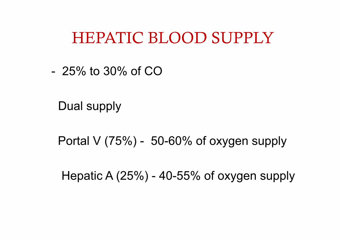
HEPATIC BLOOD SUPPLY
- 25% to 30% of CO
Dual supply
Portal V (75%) - 50-60% of oxygen supply
Hepatic A (25%) - 40-55% of oxygen supply
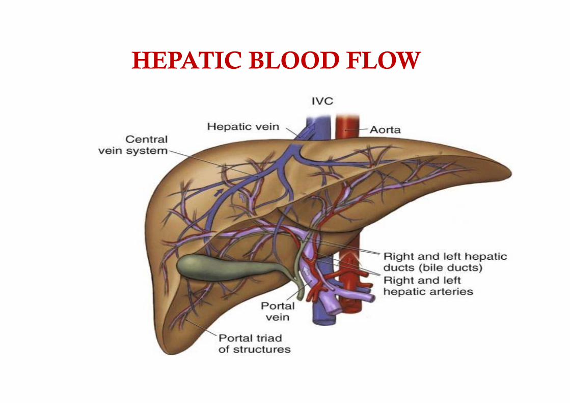
HEPATIC BLOOD FLOW

1)
CONTROL OF LIVER BLOOD FLOW
1) HEPATIC ARTERIAL BUFFER RESPONSE ‐most important intrinsic mechanism ‐ changes in portal venous flow cause reciprocal changes in hepatic arterial flow ‐mechanism involves the synthesis and washout of adenosine (i.e., a vasodilator) from periportal regions

2) AUTOREGULATION
‐Mechanism involves myogenic responses of vascular smooth muscle to stretch ‐ Only in postprandial state
3) METABOLIC CONTROL -Decrease oxygen tension or ph of portal
venous blood increase hepatic arterial flow whereas postprandial hyperosmolarity increase both hepatic and portal flow

• B.EXTRINSIC REGULATION • 1.NEURAL CONTROL • -Fibers of the vagus, phrenic, and splanchnic nerves (postganglionic
sympathetic fibers from T6 through T11) -When sympathetic tone ↓ : splanchnic reservoir volume increases. -Vagal stimulation : alters the tone of the presinusoidal sphincters -the net effect is a redistribution of intrahepatic blood flow without changing total hepatic blood flow.
• 2.HUMORAL CONTROL • - hepatic arterial bed has α1-, α2-, and β2-adrenergic receptors • - portal vein has only α-receptors • Glucagon induces relaxation of hepatic arterial smooth muscle. • angiotensin II constricts the hepatic arterial and portal venous beds. • Vasopressin elevates splanchnic arterial resistance, but it lowers
portal venous resistance.

••
•
SPECTRUM OF LIVER DISEASE
PARENCHYMAL
- Acute – infectious or non infectious ‐ Chronic Hepatitis – alcohol, autoimmune,drugs, inherited(wilson, alpha 1 antitrypsin),NASH , viral ‐ Hepatic Cirrhosis (+ portal hypertension)
CHOLESTATIC ‐ Intrahepatic
viral hepatitis drug induced
‐ Extrahepatic (Obstructive jaundice) Calculi, stricture, growth.

CIRRHOSIS OF LIVER
• A chronic progressive disease
• Extensive degeneration & destruction to the liver parenchymal cells
• Cell necrosis scar tissue nodular structure impedes blood flow hypoxia
Causes
• Chronic viral hepatitis • Metabolic: hemochromatosis, Wilson dis,
alfa‐1‐antitrypsin, NASH • Prolonged cholestasis (primary biliary
cirrhosis, primary sclerosing cholangitis) • Autoimmune diseases (autoimmune
hepatitis) • Drugs and toxins • Alcohol

Pathophysiology
• Alcoholic cirrhosis – accumulation of fat and scar formation in the liver cells
• Postnecrotic cirrhosis – broad bands of scar tissue resulted from viral, toxic, or autoimmune hepatitis
• Biliary cirrhosis – diffuse fibrosis with jaundice from chronic biliary obstruction
• Cardiac cirrhosis – from long‐standing right sided heart failure
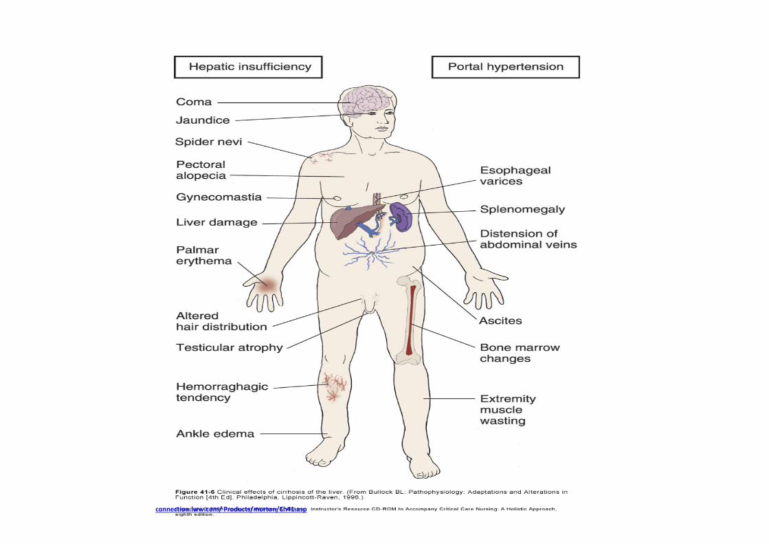
Clinical Manifestations
• Early – GI disturbances, dull pain in RUQ/epigastrium, fever, malaise, enlargement of liver & spleen
• Late – Jaundice, skin lesions (spider angiomas, palmar erythema), hematologic problems, endocrine disturbances, peripheral neuropathy
Complications 1. Portal Htn 2. Oesophagogastric varices 3. Ascites 4. Anemia & coagulopathy 5. SBP ( spontaneous bacterial peritonitis ) 6. Cardiomyopathy 7. Arterial hypoxemia & Hepatopulmonary syndrome 8. Hepatorenal syndrome 9. Hypoglycemia 10. Duodenal ulcer 11. Gallstones 12. Hepatic encephalopathy 13. Primary HCC
Pathophysiology of End Stage Liver Disease
• Predominant pathophysiological manifestation of liver disease is portal hypertension.
• Normal portal pressures are usually in the range of 5‐12 mmHg.
• Portal hypertension is generally defined when any 2 of the following 3 criteria are met: splenomegaly, ascites or bleeding esophageal varices.
• Portal pressures at this time are usually > 20 mmHg

Varices • Due to ↑portal hypertension
• Varicosities develop where collateral & systemic circulations communicate esophageal & gastric varices, caput medusae, & hemorrhoids
• most common ‐gastroesophageal varices • Painless massive haematemesis with or without melena & other features of PH.
• Endoscopy‐ best for evaluation

Collaterals
SITES : 1. Oesophagus
2. Gastric
3. Colo‐rectal
4. Portal hypertensive gastropathy

STANDARD TREATMENT OF PORTAL HYPERTENSION
1. Pre‐primary prophylaxis – EGD , no treatment for PH, treat cause of cirrhosis.
2. Primary prophylaxis ‐ non‐selective b‐blockers (propranolol, nadolol) are as effective as Endoscopic variceal ligation(EVL) depending upon risk
3. Controlling acute variceal hemorrhage ‐Safe vasoactive drugs are started as soon as possible, prior to diagnostic endoscopy .EVL is the procedure of choice if source confirmed, Sclerotherapy second line . TIPS recommended when everything fails
4. Secondary prophylaxis‐ if TIPS performed consider for transplant. If TIPS not performed combination of pharmacological (NSBB alone or NSBB + ISMN) plus EVL is associated with lower rebleeding rates than either therapy alone
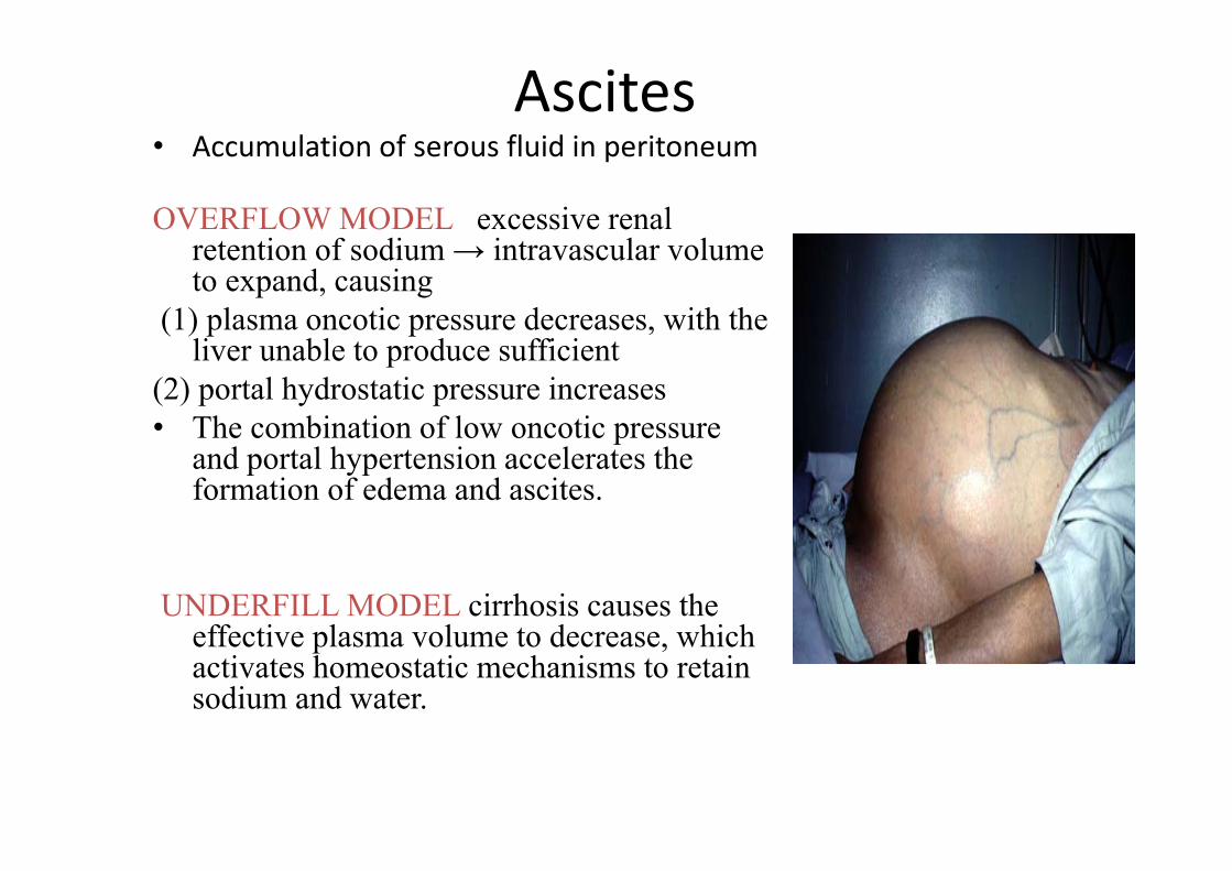
Ascites • Accumulation of serous fluid in peritoneum
OVERFLOW MODEL excessive renal retention of sodium → intravascular volume to expand, causing
(1) plasma oncotic pressure decreases, with the liver unable to produce sufficient
(2) portal hydrostatic pressure increases • The combination of low oncotic pressure
and portal hypertension accelerates the formation of edema and ascites.
UNDERFILL MODEL cirrhosis causes the effective plasma volume to decrease, which activates homeostatic mechanisms to retain sodium and water.

• Tense ascites may decrease functional residual capacity (FRC), adversely affect pulmonary gas exchange and increase risk of aspiration.
• Hydrothorax or pleural effusions may produce atelectasis.
• Secondary hyperaldosteronism may manifest as hypokalemic metabolic alkalosis.
• There is intra and extra‐pulmonary shunting, elevated mixed venous oxygen saturation (SvO2), altered lactate metabolism.
• Treatment‐ diagnostic paracentesis, salt restriction to 2000mg/day, diuretics( furosemide or spironolactone ), Large‐volume paracentesis,TIPS
COAGULOPATHY • All coagulation factors except for VIII are markedly reduced in patients
with liver disease
• CLD patients have thrombocytopenia due to splenomegaly and decreased thrombopoeitin
• Antithrombin‐III (AT‐III) levels fall due to reduced synthesis and/or increased consumption due to fibrinolysis
• Hemostatic changes associated with surgical bleeding are
1. thrombocytopenia, 2. platelet function defects, 3. inhibition of platelet aggregation and adhesion by nitric oxide and
prostacyclin, 4. decreased levels of coagulation factors: II, V, VII, IX, X, XI, quantitative
and qualitative abnormalities of fibrinogen, 5. low levels of α2‐antiplasmin, Factor XIII and thrombin activatable
fibrinolysis inhibitor, and elevated tPA.
Hemostatic changes associated with thrombosis : 1. Elevated vWF, decreased levels of ADAMTS‐13 (a
vWF cleaving protease), 2. Decreased levels of anti‐coagulants: ATIII, Protein
C and S, α2 macroglobulin, elevated levels of heparin cofactor II, elevated VIII, decreased levels of plasminogen, normalor increased PAI‐1.
3. Hypercoagulability can occur in patients with liver disease, especially those with cholestatic disease.

Portopulmonary hypertension (POPH) • Pulmonary hypertension syndrome with vascular obstruction and increased resistance to pulmonary arterial flow
• It occurs due to pulmonary endothelial/smooth muscle proliferation, vasoconstriction and in‐situ thrombosis.
• The development of POPH has not been demonstrated to correlate with the severity of liver disease
• The diagnostic criteria for POPH include a mean pulmonary artery pressure(mPAP) greater than 25 mmHg at rest and a pulmonary vascular resistance (PVR) of > 240 dynes.s.cm.
• A better measure is a transpulmonary gradient > 12 mmHg (mPAP‐PAOP) as this reflects the obstruction to flow (PVR)

• Female gender and autoimmune hepatitis have been reported to be risk factors.
• In cases confirmed by right‐sided heart catheterization, treatment with epoprostenol or bosentan may reduce pulmonary hypertension and thereby facilitate liver transplantation;
• Liver transplantation is contraindicated in patients with moderate to severe pulmonary hypertension (mean pulmonary pressure > 35 mm Hg).

Hepatopulmonary syndrome (HPS) • Characterized by arterial hypoxemia caused by intrapulmonary vascular dilatations.
• The clinical triad of • 1) portal hypertension; 2) hypoxemia; and 3) pulmonary vascular dilatations
• European Respiratory Society (ERS)/European Association for Study of the Liver(EASL) Task Force have certain set diagnostic criteria for hepatopulmonary syndrome(HPS). These include :
• Diagnosis of liver disease, • An A‐a oxygen gradient > 15 mmHg, • Pulmonary vascular dilatation documented by “positive" delayed, contrast‐enhanced echocardiography with left heart,
• Detection of microbubbles for > 4 cardiac cycles after right heart opacification of microbubbles
• Brain uptake > 6% following 99mTc macroaggregated albumin(MAA) lung perfusion scanning
TREATMENT • Medical therapy has been disappointing
• Experimentally, iv methylene blue, oral garlic powder, and oral norfloxacin may improve oxygenation by inhibiting nitric oxide‐induced vasodilation
• Pentoxifylline may prevent hepatopulmonary syndrome by inhibiting production of tumor necrosis factor‐.
• Long‐term oxygen therapy is recommended for severely hypoxemic patients
• The syndrome may reverse with liver transplantation, although postoperative mortality is increased in patients with a preoperative arterial oxygen tension < 50 mm Hg or with substantial intrapulmonary shunting.
• TIPS may provide palliation in patients with hepatopulmonary syndrome awaiting transplantation.

Hepatorenal syndrome • Pre‐renal acute kidney injury that occurs in decompensated cirrhosis.
• The syndrome is classified into two types: • Type 1 is characterized by a doubling of the serum creatinine level to greater than 2.5 mg/dl in less than 2 weeks
• Type 2 is characterized by a stable or slower progressive course of renal failure

• The International Ascites Club has suggested FIVE major criteria to confirm the diagnosis of HRS:
(1) chronic or acute liver disease with advanced hepatic failure and portal hypertension;
(2) a low GFR as assessed by serum creatinine >1.5 mg/dL or creatinine clearance below 40 mL/min;
(3) absence of shock, ongoing bacterial infection, fluid losses, or treatment with nephrotoxic drugs;
(4) no sustained improvement in renal function after oral diuretic withdrawal and plasma volume expansion; and
(5) less than 500 mg/day proteinuria with no ultrasonographic evidence of parenchymal renal disease or urinary obstruction

MANAGEMENT • IV infusion of albumin + vasoconstrictor regimens for 7–14days:
1. IV vasopressin or ornipressin (ischemic s/e); 2. IV ornipressin plus dopamine; 3. IV terlipressin (preferred agent); 4. IV norepinephrine; 5. oral midodrine, an ‐adrenergic drug, plus the somatostatin
analog octreotide, s/c or iv. • MARS(molecular adsorbents recirculating system ), a
modified dialysis method that selectively removes albumin‐bound substances. I
• TIPS • Liver transplantation is the treatment of choice.

Spontaneous bacterial peritonitis
• Consists of fever, leukocytosis, abdominal pain,and decreased bowel sounds
• ↑ gut wall permeability →growth of bacteria in peritoneal fluid
• Associated ↓macrophage func on • Risk factors‐ low protein in ascitic fluid, variceal bleeding
• Antibiotic prophylaxis in Pts with GI haemorrhageis recommended.
• High mortality ( 20‐50%)
Hepatic Encephalopathy
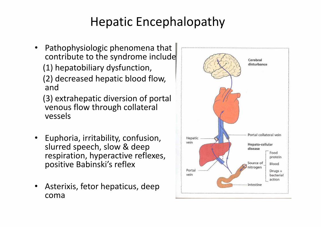
• Pathophysiologic phenomena thatcontribute to the syndrome include (1) hepatobiliary dysfunction, (2) decreased hepatic blood flow,and (3) extrahepatic diversion of portalvenous flow through collateralvessels
• Euphoria, irritability, confusion,slurred speech, slow & deeprespiration, hyperactive reflexes,positive Babinski’s reflex
• Asterixis, fetor hepaticus, deep coma

Factors That May Precipitate Hepatic Encephalopathy
• Excessive dietary protein • Constipation Increased ammonia • Gastrointestinal bleeding production • Infection • Azotemia
• Diarrhea and vomiting Dehydration with electrolyte and acid‐base • Diuretic therapy imbalance, increased ammonia generation, and • Paracentesis decreased hepatic perfusion
• Hypoxia • Hypotension
Adverse effect on liver and brain function • Anemia • Hypoglycemia
• Sedatives/hypnotics ‐‐Action at the GABAA/benzodiazepine receptor complex
• Creation of portal‐systemic shunt ‐‐Reduced hepatic metabolism
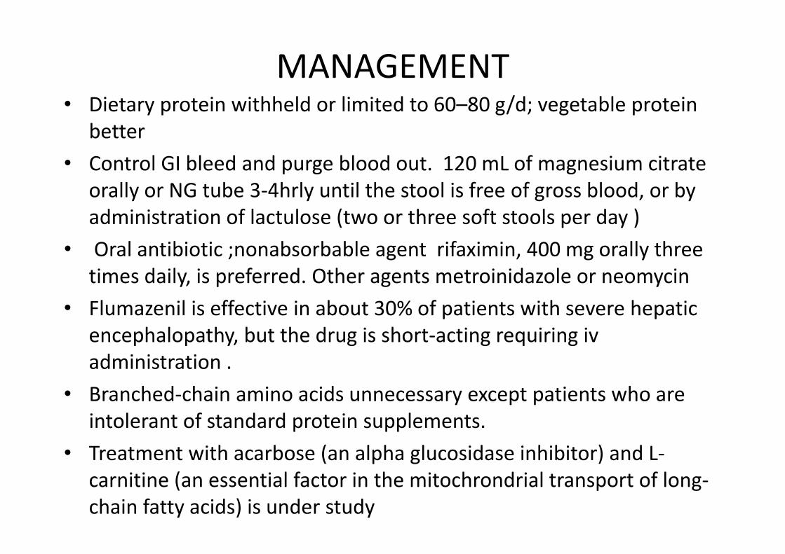
MANAGEMENT • Dietary protein withheld or limited to 60–80 g/d; vegetable protein
better • Control GI bleed and purge blood out. 120 mL of magnesium citrate
orally or NG tube 3‐4hrly until the stool is free of gross blood, or by administration of lactulose (two or three soft stools per day )
• Oral antibiotic ;nonabsorbable agent rifaximin, 400 mg orally three times daily, is preferred. Other agents metroinidazole or neomycin
• Flumazenil is effective in about 30% of patients with severe hepatic encephalopathy, but the drug is short‐acting requiring iv administration .
• Branched‐chain amino acids unnecessary except patients who are intolerant of standard protein supplements.
• Treatment with acarbose (an alpha glucosidase inhibitor) and L‐carnitine (an essential factor in the mitochrondrial transport of long‐chain fatty acids) is under study

PREOPERATIVE ASSESSMENT
OBJECTIVES 1. Assess the type and degree of liver dysfunction. 2. Type of surgery
3. Assess effect on other system. 4. To ensure – post operative facilities (High risk patient).
PREOPERATIVE ASSESSMENT
HISTORY -Dyspnoea, syncope, bleeding, delerium, effort
tolerance
CLINICAL EXAMINATION - Blood pressure, pulse, oxygenation, bruising,
ascites, orientation, jaundice
INVESTIGATIONS

•••••
••••
PREOPERATIVE INVESTIGATIONS
A)TO ASSESS GENERAL CONDITION OF PATIENT
1) Haematological Hb TLC, DLC Platelet Count Clotting factors (PT, PTTk)
2) Cardiorespiratory Chest X‐ray ECG Pulmonary.fn.tests Blood gases Echocardiography
3) Metabolic Serum proteins Serum glucose Electrolyte Urea / Creatinine

••••
B) TO KNOW THE PATTERN OF DISEASE
‐ S. Bilirubin
‐ SGOT, SGPT 90% predictive
‐ Alk. phosphatase
Single Marker Glutathione S transferase – drug induced Glutamyl transpeptidase – alcohol/drug induced

••••••
C) TO JUDGE THE SYNTHETIC ABILITY OF LIVER Serum albumin– < 2·5 gm% - severe damage Albumin/globulin ratio– reversed. Prothrombin time– > 1·5 sec. Over control
– INR - > 1.3
C) OTHER TESTS (DONE ONLY FOR MAJOR SURGERY) - liver biopsy - screening for hepatitis - α feto protein – Hepatocellular Carcinoma - Antinuclear antibodies – prim. biliary cirrhosis - Copper & Ceruloplasmin level – Wilson's disease ferritin and transferritin – Haemochromatosis

Cardiac assessment of End Stage Liver Disease (ESLD) patients
• May develop cirrhotic cardiomyopathy
• Increased CO and compromised ventricular response to stress leads to cardiac depression and repolarization abnormalities
• Low systemic vascular resistance and bradycardia
• Increase QT interval, electrical and mechanical dyschrony ,chronotropic incompetence
• Can develop CAD if cardiac risk factors present • Left ventricular outflow tract obstruction (LVOTO)

ROLE OF ECHO
• Preop echo
1. Ventricular function, size
2. Valvular function
3. Pulmonary artery pressure
4. Exclude any LVOTO or pericardial effusion
5. Pulmonary artery systolic pressure calculation

• TEE and/or pulmonary artery catheterization may be used intraoperatively to allow for real‐time hemodynamic monitoring and volume management.
• Stress testing of ESLD patients can be done to detect CAD.
• Coronary angiography is the gold standard for detecting CAD
• Rt heart catheterization role to measure PAP,PCWP and TPG

••
•––
•
GRADING OF SEVERITY OF DISEASE
Mild Hepatic dysfunction
‐ Cl. History + evidence of liver pathology
‐ normal plasma albumin, but enzymes
Moderate Hepatic dysfunction
‐ Limited impairment of synthetic function
PT not > 2∙5 sec. above normal Plasma albumin at least 3 gm%.
Severe hepatic dysfunction
‐More impairment of synthetic function.

Surgical Risk.
• Elective surgery is contraindicated when the patient has acute viral hepatitis, alcoholic hepatitis, fulminant hepatic failure, severe chronic hepatitis, is a Child Pugh C patient or has other manifestations of end stage liver disease.
• Two risk stratification schemes developed to assess theperioperative risk of patients with cirrhosis:
1. Modified Child‐ Turcotte ‐Pugh Scoring System
2.The Model of End‐Stage Liver Disease (MELD) score
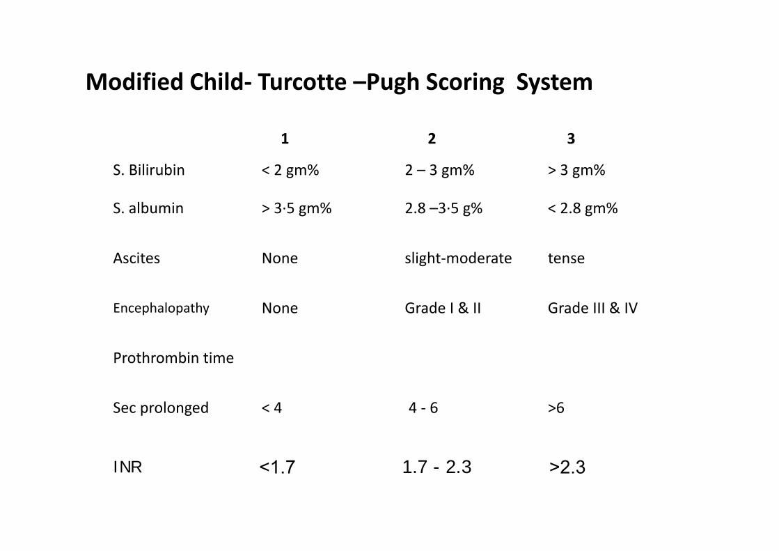
Modified Child‐ Turcotte –Pugh Scoring System
1 2 3
S. Bilirubin < 2 gm% 2 – 3 gm% > 3 gm%
S. albumin > 3∙5 gm% 2.8 –3∙5 g% < 2.8 gm%
Ascites None slight‐moderate tense
Encephalopathy None Grade I & II Grade III & IV
Prothrombin time
Sec prolonged < 4 4 ‐ 6 >6
INR <1.7 1.7 - 2.3 >2.3

Modified Child‐ Turcotte –Pugh Scoring System
CLASSES SCORE MORTALITY
A 5‐6 10%
B 7‐9 31%
C 10‐15 76%

Primarily used to select patients for liver transplant
MELD
• Objective assessment in predicting 3‐month mortality
0. 38 X ln (bilirubin mg/dl) + 1.12 X ln (INR) + 0.96 ln (creatinine mg/dl) + 0.64
Best outcomes : MELD score < 14.
• For patients with a MELD score of 15‐24 • Clinical judgment • Further discussion with the family and the patient

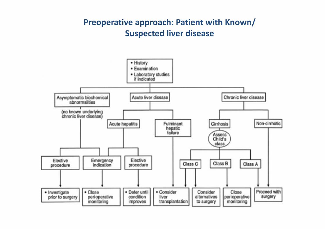
Preoperative approach: Patient with Known/ Suspected liver disease

PERIOPERATIVE MANAGEMENT
PREOPERATIVE PREPARATION (1) Childs Group
A – Elective Surgery recommended
B – acceptable after correction
C – only for emergency
(2) Assess hydration status.
(3) Correct Anemia / Coagulation / hypoalbuminemia
(4) Arrange appropriate blood / blood products.
(5) Inform – postoperative complications

PREMEDICATION
‐ If neuro. status normal –anxiolytic (oral) ‐ oral H2 antagonist ‐ Vit. K (Obst. J) – 10 mg B D X 3 day
‐ If Bilirubin > 8 mg% –
Mannitol ‐ 100 ml of 20% 2 hrs preop

–
ANAESTHETIC MANAGEMENT
GENERAL CONSIDERATIONS
Minimize physiological insult to liver & kidney
‐ Maintain O2 supply – demand relationship in liver. Adequate pulmonary ventilation and cvs fn.
‐Maintain renal perfusion
Avoid Hypotension, hypoproteinemia & hypoxia Meticulous fluid balance
Choose appropriate anaesthetic agent Metabolism of drugs + Effect on HBF
General anaesthesia
Induction agent
Thiopentone / propofol
Given in slow tirated dose
Avoid hypotension
Avoid sympathetic stimulation Propofol :
Highly lipid soluble
High extraction ratio
However kinetic profile similar to normal patients
Thiopentone :
Low extraction ratio
Elimination half life unaltered secondary to increased Vd

Muscle relaxants
Decreased S. Alb – Increased free drug concentration Drugs with hepatic clearance avoided
Vecuronium Rocuronium Pancuronium Mivacurium (infusion avoided)
Atracurium/Cis atracurium –Non specific ester hydrolysis
• Succinylcholine – For RSI • After screening for the usual contraindications
Prolonged immobility Critical illness Hyperkalemia
• Severe liver dysfunction ‐ decrease cholinesterase activity • May prolong the effect of succinylcholine somewhat • Rarely causes a clinical problem.

Morphine – Reduced metabolism
– Prolonged elimination half life
– Inc. Bioavailability
– Inc. Sedative and Respiratory depressant effects – Administration interval should be increased 1.5‐ to 2‐fold in these patients
Meperidine – 50% reduction in clearance
– Doubling of the half‐life
– In addition, clearance of normeperidine is reduced
– Patients with severe liver disease may experience neurotoxicity

Fentanyl and Sufentanil
No significant change in pharmacokinetics
Repeated administration or continuous infusions, accumulation may occur
and lead to prolonged effects
Alfentanil
Shows decrease in plasma clearance
Half‐life is almost doubled in patients with cirrhosis
Remifentanil
Elimination is unaltered in patients with severe liver disease

Spasm Of Sphincter Of Oddi
– Opioids can cause spasm of sphincter of oddi
Increase common bile duct pressures
– More with morphine, fentanyl, meperidine
– Avoided if intraoperative cholangiogram to be done
– Treatment ‐ Opioid antagonists (naloxone)
‐ Smooth ms.relaxant(nitroglycerine)
‐ Glucagon
Sedatives
• Midazolam : • Reduced protein binding and increased free fractions • Reduced clearance in patients with end‐stage liver disease • Produces prolonged elimination half‐lives • Enhanced sedative effect especially after multiple doses or prolonged infusions
• Dexmedetomidine • Primarily metabolized in the liver with minimal renal clearance. • Patients with hepatic failure of varying severity have
• Decreased clearance • Prolonged half‐lives • Lower bispectral index values
Hence dose adjustments indicated
Voltaile Anesthetics
• Useful & well tolerated • Can be entirely eliminated
Sevoflurane : Most effective in maintaining HBF Hepatic O2 delivery
Isoflurane /: Very good maintainance of Desflurane HBF
Hepatic O2 delivery O2 delivery to consumption ratio

Halothane Halothane (avoided)
Detrimental reductions in
• Hepatic oxygen delivery
• HBF by alterations in
Cardiac output
MAP
• Halothane hepatitis(rare)


Clinical Features of Halothane Hepatitis
Mild Form Fulminant Form Incidence, 1 : 5 Incidence, 1 : 10,000 Repeat exposure Multiple exposures not Mild elevation of Marked elevation of, ALT,AST ALT, AST,bilirubin, Focal necrosis Massive necrosis Self‐limited Mortality rate, 50%
Antibodies present

Xenon : • Considered to be an ideal inhaled anesthetic
• Nonexplosive and nonflammable
• Rapid induction and recovery profiles
• Cardiac stability
• It does not alter HABF
• Does not alter the results of liver function tests
• Animals exposed to xenon : Higher hepatic venous oxygen content levels
• Secondary to a possible reduction of plasma catecholamine levels
• Subsequent reduced hepatic metabolism
Xenon may prove to be an ideal anesthetic relative to hepatic perfusion.

Intraoperative considerations • IV access using large bore peripheral catheters as well as central
venous access catheters. • RSI in tense ascites pt –risk of aspiration
• Preventing circulatory cllapse by administration of IV colloid solutions because intravascular volume re‐equilibrium occurs 6 to 8 hrs after removal of larger volumes of ascitic fluid.
• Large volumes of colloids/crystalloids maybe given within a few minutes with the assistance of commercially available rapid infusion devices.
• Red cell salvage should be facilitated with use of Cell savers with/without leukocyte filters.
• Blood administration may be associated with hyperkalemia and hypocalcemia.

• Bleeding during liver surgery could be either surgical, due to previous or acquired coagulation disturbances, or both.
• The preoperative INR has no predictive value
• FFP role debatable
• Intraoperative hemostasis panels consisting of INR, fibrinogen and platelet count, and platelet function assays for both platelet count and function.
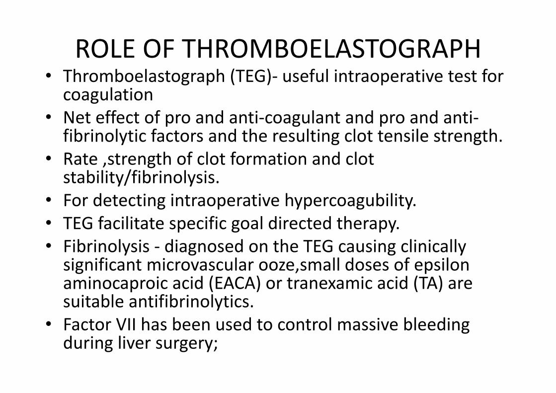
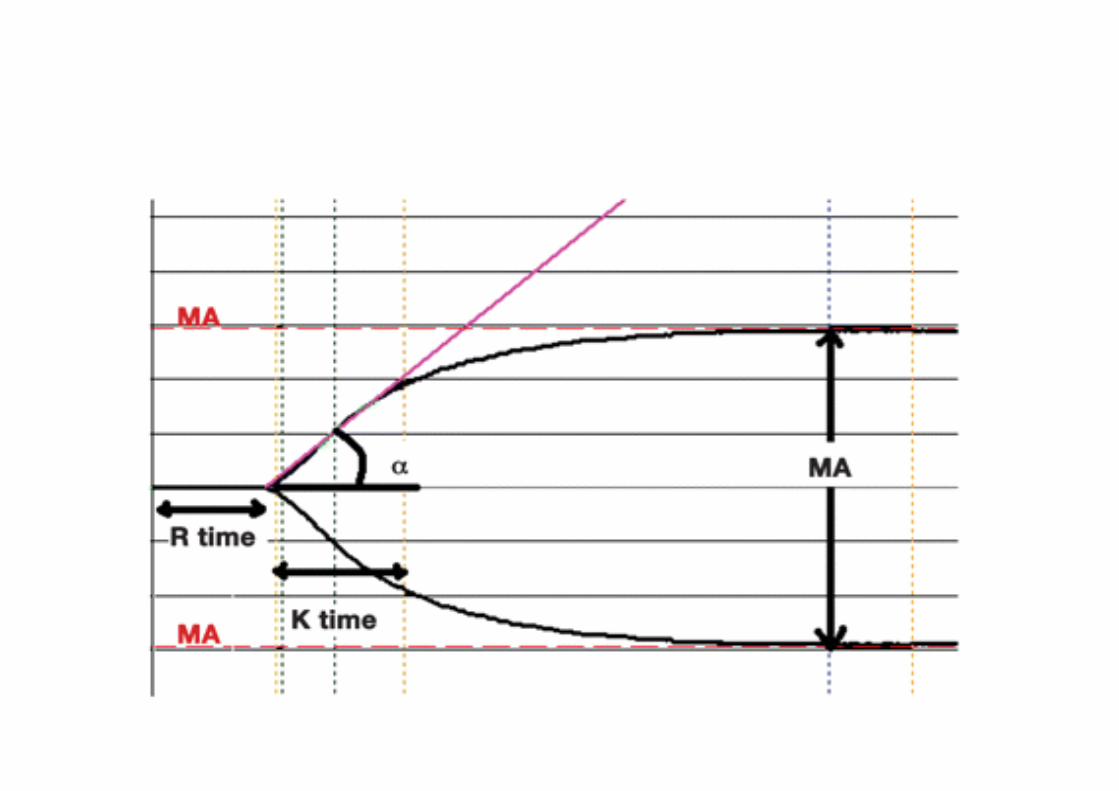
ROLE OF THROMBOELASTOGRAPH • Thromboelastograph (TEG)‐ useful intraoperative test for coagulation
• Net effect of pro and anti‐coagulant and pro and anti‐fibrinolytic factors and the resulting clot tensile strength.
• Rate ,strength of clot formation and clotstability/fibrinolysis.
• For detecting intraoperative hypercoagubility. • TEG facilitate specific goal directed therapy. • Fibrinolysis ‐ diagnosed on the TEG causing clinicallysignificant microvascular ooze,small doses of epsilonaminocaproic acid (EACA) or tranexamic acid (TA) aresuitable antifibrinolytics.
• Factor VII has been used to control massive bleedingduring liver surgery;

Intra Operative Monitoring
Routine
NIBP ECG Et CO2 SPO2 Urine output N/ms monitoring
Longer and extensive surgeries CVP ABG Invasive blood pressure monitoringS. Electrolyte , Blood sugarsTEG

POSTOPERATIVE MANAGEMENT
1)Minor Surgery or mild‐mod. liver dysfn. N/ms block reversed → Extubate
2)Major surgery / severe liver dysfn. ‐Continue IPPV in P.op. period
‐Fluid & Electrolyte imbalance corrected
‐CVS stability achieved
‐Hypothermia corrected
‐Urine Output 1 ml/kg/hr 3)Adequate analgesia (Small doses) 4)Blood / blood product replaced.

Postoperative pain relief • Thoracic epidural analgesia provides excellent analgesia forliver resections but restricted due to coagulation defects
• The catheter is usually inserted at the T6‐T9 space.Ropivacaine or bupivacaine are common local anestheticsused with or without the addition of small amounts of opioidssuch as fentanyl,sufentanil, hydromorphone or morphine.
• It also reduces the gastrointestinal paralysis compared with systemic opoids
• NSAIDS ‐ risk of GI bleeding, platelet dysfunction andnephrotoxicity ;avoided .
• Paracetamol is sometimes used • Fentanyl PCA is generally well tolerated • Morphine PCA can also be used but a lower bolus dose maybe needed, again to avoid accumulation.
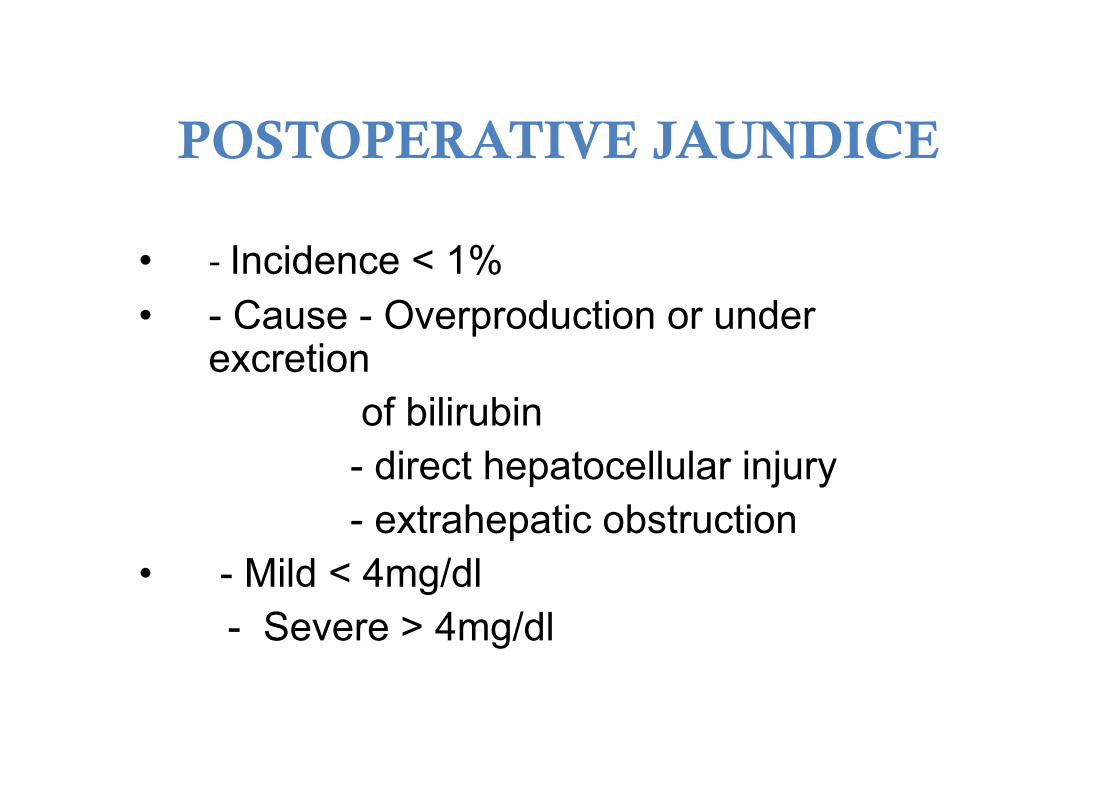
POSTOPERATIVE JAUNDICE
• ‐ Incidence < 1% • - Cause - Overproduction or under
excretion of bilirubin
- direct hepatocellular injury - extrahepatic obstruction
• - Mild < 4mg/dl - Severe > 4mg/dl

Causes Of Postoperative Liver Dysfunction

Agent Hepatocellular Steatosis Cholestasis
Acetaminophen * Alcohol *
Amiodarone *
Aspirin * *
Amoxi‐clav *
Isoniazid *
Ketoconazole *
Methotrexate * *
Phenytoin *
Anabolic steroids *
OCP *
Sulfonamides *
Valproic acid *
TPN *
Tetracycline * *
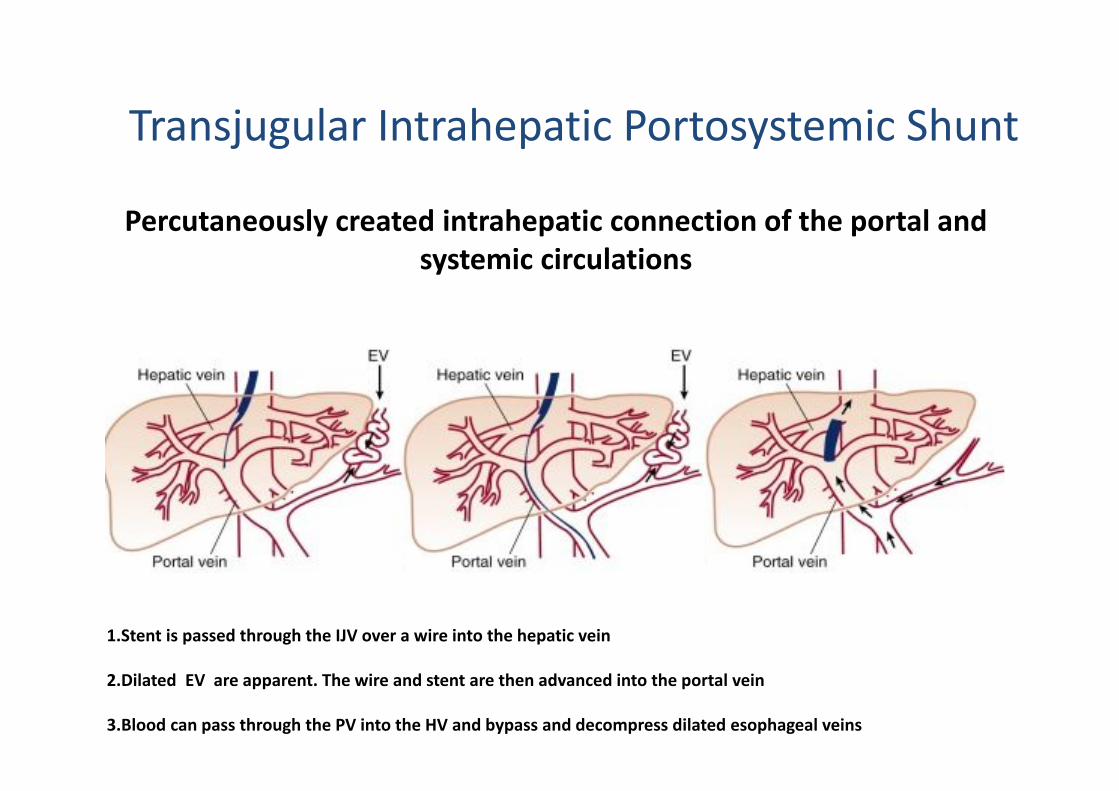
Transjugular Intrahepatic Portosystemic Shunt
Percutaneously created intrahepatic connection of the portal and systemic circulations
1.Stent is passed through the IJV over a wire into the hepatic vein
2.Dilated EV are apparent. The wire and stent are then advanced into the portal vein
3.Blood can pass through the PV into the HV and bypass and decompress dilated esophageal veins

Typically used in patients with end‐stage liver disease
• To decrease portal pressure
• Attenuate the complications • Variceal bleeding
• Refractory ascites
Complications
• Encephalopathy
• Stent stenosis and occlusion
Hepatic resection
• Preoperative considerations • Involve risk assessment : MELD classification.
• Severe thrombocytopenia or large varices : Major perioperative risk
• Fluid management : Controversial. • Liberal use : Goal of increasing intravascular volume as a buffer.
• Low central venous pressure : Minimize blood loss from Major veins
• Intravenous fluids • Supplemented sodium or potassium phosphate

Hepatic cryotherapy – Treat nonresectable malignant hepatic tumors
– Involves usage of subzero temperature
– Multiple‐lumen probes positioned under USG guidance
– Heat conservation instituted during the procedure
– With continual monitoring of core temperature.
– “Cryoshock syndrome” :
– Postoperative
• Pulmonary
• Renal
• Coagulation problems
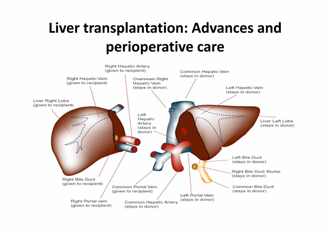
Liver transplantation: Advances and perioperative care

LIVER TRANSPLANT INDICATIONS CONTRAINDICATIONS • HEPATITIS SEPSIS • ALD CARDIOPULMONARY DISEASE • HEMOCHROMATOSIS EXTRA HEPATIC MALIGNANCY • PBC AIDS • PSC ANY SUBSTANCE ABUSE • CF UNFAV PSHYCHOSOCIAL • WILSON CIRCUMSTANCE • AMYLOIDOSIS • MALIGNANCY • BUDD CHIARI

STAGES
• Preanhepatic‐ from start of surgery to clamping of hepatic artery
• Anhepatic –from clamping to reperfusion of new liver
• Postanhepatic‐from reperfusion to end of case

Order of reconstruction • Standard method –suprahepatic ivc followed byinfrahepatic ivc anastomosis –PV anastomosis ‐arterial reconstruction – biliary drainage
• Piggy back method – only one ivc anastomosis
Test clamp maneuver • Used in standard method to assess resilience of circulatory system
• Suprahepatic ivc clamped – arterial pressure ,COdecrease
• If excessive circulatory depression proceedings delayed,reassess volume status ,cardiac performance,metabolic state.
• Venovenous bypass –if still circulatory depression

Intraoperative Monitoring and Management
• Haemodynamic monitoring‐standard cardiovascular monitors (electrocardiogram, pulseoximetry, invasive and non‐invasive blood pressure)
• Additionally requires CO monitoring • Pulmonary artery catheter (PAC) is the gold standard used in haemodynamic monitoring
• Monitoring of central venous oxygen saturation and mixed venous oxygen saturation during liver transplantation is of little value

Hemodynamic management • During the different stages of liver transplantation, i.e. pre‐anhepatic phase, anhepatic phase and neohepatic phase, there are rapid fluid shifts due to blood loss, inferior vena cava clamping and reperfusion.
• Decreasing central venous pressure (CVP) either by phlebotomy or avoiding plasma transfusion during the pre‐anhepatic phase have shown to reduce red cell transfusions

• Vasopressin is often added intraoperatively tomaintain the systemic vascular resistance.
• Vasopressin reduces portal blood flow byselective splanchnic vasoconstriction and hencemay be useful in reducing the intraoperativeblood loss.
• Methylene blue at a dose of 0.5 mg/kg bodyweight over 10 min rescue to treat hypotension due to vasopressor‐resistant vasoplegic shock.
• Phenylephrine is often used to tide over acutehypotensive episodes

Fluid management • Liver transplant surgery ‐massive fluid shifts both from intravascular volume depletion and large surgical blood loss.
• Albumin can be used as pts are hypoalbuminaemic and hypovolaemic ;cost factor limits it use
• Crystalloids use depends on their pH, electrolyte composition, osmolarity and metabolism

• No ideal crystalloid solution • 0.9% NS causes hyperchloremic acidosis while thelactate in Ringers Lactate (RL) requires livermetabolism for its elimination.
• RL is a hypotonic solution and may increase theintracellular fluid.
• Plasmalyte has a pH near normal, electrolyte andosmolarity similar to plasma and acetate, which ismetabolised extrahepatically to bicarbonate, butit is proinflammatory and potentially cardiotoxic.

Coagulation monitors • PT and APTT limited role • Thromboelastogram (TEG), rotational thromboelastometry (ROTEM) and Sonoclot provide a detailed assessment
Blood component management • Pre‐anhepatic phase is associated with blood loss • The aim at this stage is to avoid large volume of transfusion and dilutional coagulopathy
• Antifibrinolytics are used in the liver transplantation to prevent the hyperfibrinolytic state during the anhepatic and neohepatic phases
• The neohepatic phase is associated with a multifactorial coagulopathy of hyperfibrinolysis, dilutional coagulopathy, heparin‐like effect, platelet dysfunction, hypothermia and hypocalcaemia

Ischemia reperfusion injury
• Ischemia reperfusion injury (IRI) is associated with primary graft dysfunction and delayed graft function
• N‐acetyl cysteine (NAC) is being used in liver transplantation patients to prevent renal failure and IRI of the new liver. NAC, in addition to its direct antioxidant property, replenishes glutathione and acts as a free radical scavenger.
• Inhalational anaesthetics, especially sevoflurane, have been shown to offer protection against IRI in the myocardium in cardiac patients

• Early extubation in selected patients improves early graft function and reduce duration of stay in the Intensive Care Unit and nosocomial infections
• Selection of patients for early extubation depends on duration of the surgery, amount of blood and products transfused, patients' pre‐operative status (MELD score), ischaemia time and status of the graft.
• A safe operating room extubation after liver transplantation (SORELT) prediction rule may be used to select patients for early extubation, but requires validation

CONCLUSION
• Patients with liver disease are at increased risk for both perioperative morbidity and mortality.
• The multi‐system impact of liver failure means assessment and management of these patients often requires multi‐disciplinary discussion and critical care admission to optimise outcome

THANK YOU

ROLE OF THROMBOELASTOGRAPH • Parameter Interpretation Preferred therapy for
abnormal values R R is the time of latency FFP
placed in the TEG® analyzer until initial fibrin formation
α The α‐value measures rapidity of fibrin build up and cross linking Cryoprecipitate
K K time is a measure of the rapidity to reach certain level FFP of clot strength.
MA MA, or Maximum Amplitude, direct function of the maximum Platelets dynamic properties of fibrin and platelet bonding and represents the ultimate strength of fibrin clot.