Embed Size (px)
Citation preview

Clinical StudyLiver Transplantation for Hepatocellular Carcinoma: A SingleCenter Resume Overlooking Four Decades of Experience
Nikos Emmanouilidis,1 Rickmer Peters,1 Bastian P. Ringe,1
Zeynep Güner,1 Wolf Ramackers,1 Hüseyin Bektas,1 Frank Lehner,1
Michael Manns,2 Jürgen Klempnauer,1 and Harald Schrem1,3
1Department of General, Visceral and Transplant Surgery, Hannover Medical School, Carl Neuberg Strasse 1,30625 Hannover, Germany2Department of Gastroenterology, Hepatology and Endocrinology, Hannover Medical School, Carl Neuberg Strasse 1,30625 Hannover, Germany3IFB-TX Core Facility and HTA, Hannover Medical School, Carl Neuberg Strasse 1, 30625 Hannover, Germany
Correspondence should be addressed to Nikos Emmanouilidis; [email protected]
Received 29 September 2015; Accepted 3 December 2015
Academic Editor: Gaetano Ciancio
Copyright © 2016 Nikos Emmanouilidis et al. This is an open access article distributed under the Creative Commons AttributionLicense, which permits unrestricted use, distribution, and reproduction in any medium, provided the original work is properlycited.
Background. This is a single center oncological resume overlooking four decades of experience with liver transplantation (LT)for hepatocellular carcinoma (HCC). Methods. All 319 LT for HCC that were performed between 1975 and 2011 were included.Predictors for HCC recurrence (HCCR) and survival were identified by Cox regression, Kaplan-Meier analysis, Log Rank, and𝜒2-tests where appropriate. Results.HCCR was the single strongest hazard for survival (exp(𝐵) = 10.156). Hazards for HCCR were
tumor staging beyond the histologicMILAN (exp(𝐵) = 3.645), bilateral tumor spreading (exp(𝐵) = 14.505), tumor grading beyondG2 (exp(𝐵) = 8.668), and vascular infiltration of small or large vessels (exp(𝐵) = 11.612, exp(𝐵) = 18.324, resp.). Grading beyondG2 (exp(𝐵) = 10.498) as well as small and large vascular infiltrations (exp(𝐵) = 13.337, exp(𝐵) = 16.737, resp.) was associated withhigher hazard ratios for long-term survival as compared to liver transplantation beyond histological MILAN (exp(𝐵) = 4.533).Tumor dedifferentiation significantly correlated with vascular infiltration (𝜒2𝑝 = 0.006) and intrahepatic tumor spreading (𝜒2𝑝 =0.016). Conclusion. LT enables survival from HCC. HCC dedifferentiation is associated with vascular infiltration and intrahepatictumor spreading and is a strong hazard for HCCR and survival. Pretransplant tumor staging should include grading by biopsy,because grading is a reliable and easily accessible predictor of HCCR and survival. Detection of dedifferentiation should speed upthe allocation process.
1. Introduction
The repertoire of treatment strategies for hepatocellular car-cinoma (HCC) consists of liver resection (LR), chemother-apy (CTX), radio frequency ablation (RFA), transarterialchemoperfusion (TACP), selective internal radiation therapy(SIRT), transarterial chemoembolisation (TACE), percuta-neous ethanol instillation (PEI), monoclonal antibody ther-apy (mAB), and liver transplantation (LT).
The first elective liver resections were performed in thelate 19th century [1–3], but although Wendel [4] alreadyperformed a successful anatomic right hemihepatectomy for
a HCC in 1911, it took another 50 years and a better under-standing of the liver anatomy [5] before liver resections wereperformed on a larger scale by multiple centers worldwide[6–10]. The first liver transplantation for a “hepatoma” wasthe second LT that was published in the pioneering reportby Starzl et al. in 1963 [11]. A decade later Cyclosporin [12]was introduced as a new immunosuppressant and in thefollowing years larger series of liver transplantations wereaccumulated [13, 14]. The early survival analyses of LT forHCC though were rather disappointing [15] with 2-yearsurvival rates of 25–30% compared to 70% for benign diseases[16, 17]. Those disappointing results ignited the development
Hindawi Publishing CorporationJournal of TransplantationVolume 2016, Article ID 7895956, 22 pageshttp://dx.doi.org/10.1155/2016/7895956

2 Journal of Transplantation
n
25
20
15
10
5
0
Year1975 1980 1985 1990 1995 2000 2005 2010 2015
Underlying diseaseHepatitis B with DHepatitis BHepatitis CHepatitis C with B
AlcoholCryptogenicOther
(a)
n
25
20
15
10
5
0
Year1975 1980 1985 1990 1995 2000 2005 2010 2015
Tumor morphologyNo tumorUninodular
Multinodular, unilateralMultinodular, bilateral
(b)
n
25
20
15
10
5
0
Year1975 1980 1985 1990 1995 2000 2005 2010 2015
Neoadjuvant therapyNone (n = 146)PEI (n = 45)TACE (n = 39)PEI + TACE (n = 25)
Surgery (n = 22)CTX (n = 10)RFA (n = 6)Combined therapies (n = 26)
(c)
n
25
20
15
10
5
0
Year1975 1980 1985 1990 1995 2000 2005 2010 2015
UICC-7No or necroticIIIIIIA
IIIBIIICIVAIVB
(d)
Figure 1: Annual proportions of underlying diseases (a), neoadjuvant therapies (b), UICC-7 staging (c), and tumor morphologies (d). (a)There was no significant change in annual proportions of recipients underlying diseases over time. (b) Tumor morphologies of transplantedHCC changed over time in the favour of uninodular and unilateral tumors. (c)The overall rate for neoadjuvant therapy as well as the diversityof different treatment combinations increased over time. This effect was caused due to new therapies that were introduced consecutivelyfrom 1975 to 2010 (surgery (S), chemotherapy (CTX), transarterial chemoembolisation (TACE), percutaneous ethanol instillation (PEI), selectiveinternal radiation therapy (SIRT), and monoclonal antibodies (mAB)). (d) Between 1975 and 2010 the proportion of low graded UICC-7 stagedtumors increased significantly.
of nonsurgical treatment alternatives for HCC: starting withsystemic chemotherapy and transarterial chemoperfusion[18] on an experimental scale in the early 1980s. A decadelater SIRT [19], TACE [20, 21], and PEI [22] were introducedand another ten years later RFA [23] was added (Figure 1(c)).The latest development was the introduction of monoclonalantibody therapy in 2008 [24, 25].
Covariateswhich possibly affectHCC recurrence (HCCR)and survival after LT are underlying liver disease [26], tumorsize [27], grading [28], tumor multifocality, vascular invasion[26, 29], 𝛼-fetoprotein [30], and adjuvant or neoadjuvanttherapy [27, 31, 32]. But despite extensive and long experiencewith LT for HCC there are very few reports with follow-up data of more than a decade [13, 33–36]. Most long-termreports cover only 5 years of follow-up [27, 28, 32, 37–43].
Here we report our long-term single center experience ofmore than four decades with all consecutive patients (𝑛 =319) who received LT for HCC between 19th November1975 and 12th December 2010. The main focus of this study
was the oncological long-term aspects and the value of livertransplantation for the treatment of HCC.
2. Patients and Methods
2.1. Patients. Diagnosis ofHCCwas verified before LT and/orat the histological examination of the explanted liver (𝑛 =319). The mean follow-up was 6.4 years (median 4.8 years,range 0.2 to 30.9 years). Follow-up with respect to time fromlast contact to query in relation to time of LT to query wascompleted in 96% (median 100%, range 4 to 100%). Time spanof last contact to query in living patients was 0.5 to 29.4 years(median 5.9 years). Table 2 summarizes the clinical data ofthe investigated cohort.
2.2. ImmunosuppressiveTherapy. Early transplantations wereperformed under protection with Azathioprine and Cor-ticosteroids medication. Next step in immunosuppressiveevolution was the introduction of the Calcineurin-inhibitor

Journal of Transplantation 3
Cyclosporin A (CsA). Combinations of CsA with Corticos-teroids and even triple therapies with CsA, Azathioprine,and Corticosteroids were applied. Then FK-506—anotherCalcineurin-inhibitor—was introduced and added to theportfolio of immunosuppressants. The combination of FK-506 with Corticosteroids was a common replacement ther-apy for the standard protocol of CsA plus Corticosteroids.Azathioprine was only scarcely used, until it completelydisappeared as a standard medication in solid organ trans-plantation. Another significant improvement was the intro-duction of Mycophenolate Mofetil, which was mainly usedas a triple supplement in order to reduce the dosage ofCalcineurin-inhibitor medications, because it was realizedthat the Calcineurin-inhibitor nephrotoxicity was a signifi-cant problem in the long run. Other additional immunosup-pressants in recent years were the mTOR inhibitors sirolimus(Rapamycin) and everolimus (RAD-001) and the CTLA-4antibody belatacept (LEA29Y). The latter ones were appliedmainly as study drugs within multicenter trials and thuswere not commonly used. Overall, the high level of diversityin applied immunosuppressive therapies in this cohort ofpatients not only is caused by the number of differentimmunosuppressants and their combinations but is evenmore diversified due to different dosages and even therapychanges in individual patients during follow-up.
Today’s standard treatments in liver transplantation atour facility consist of Corticosteroids (prednisolone, methyl-prednisolone), basiliximab (only perioperatively), Mycophe-nolate Mofetil, and the Calcineurin-inhibitor FK-506.
2.3. TumorMorphology, UICC-7 Staging, and “Inside/Outside”hMILAN Categorization. All tumors were retrospectivelyrestaged according to the pathohistological examination ofthe explanted liver and following the 7th edition of the UICCclassification (UICC-7). For tumor morphology we also cat-egorized each tumor into either nondetectable, uninodular,multinodular/unilateral or multinodular/bilateral intrahep-atic tumor spreading. This categorization as well as thecategorization referring to MILAN criteria was done on thebasis of the histopathological reports in order to circum-vent the otherwise unavoidable bias by the technologicaldevelopment of imaging techniques during the last fortyyears. The retrospective classification either as “inside” oras “outside” MILAN was defined as histological MILAN(hMILAN). The preoperative MILAN classification, which isusually commonly applied for the listing of HCC patients andcarried out by imaging technologies, is renamed iMILAN fordiscrimination purposes.
2.4. Survival Data und HCC Recurrence (HCCR). HCCRand survival were checked in close cooperation with theGerman national cancer registry and the German nationaladdress registry and by continued follow-up in our outpatienttransplant clinics. Data were complemented by targetedinterviews of referring physicians if necessary. Descriptivestatistics related to HCC recurrence and HCC recurrencerelated deaths are summarized in Tables 3 and 4.
2.5. Statistical Analysis. Statistical analyses were performedusing SPSS v23 (PASW Statistics Inc., IBM, Somers, NY,USA). 𝑝 values and hazards for survival and HCC recur-rence (HCCR) were calculated by multi- or univariate Coxregression. Covariate hazards of survival were underlyingdisease, UICC-7 staging, hMILAN status, vascular infiltration,neoadjuvant therapy, and grading. HCCR as a hazard for sur-vival was included as a time-dependent covariate. Covariatehazards for HCCR were underlying disease, UICC-7 staging,hMILAN status, vascular infiltration, neoadjuvant therapy,and grading. 𝑝 values below 0.05 were defined as significant.Hazards (exp(𝐵)) > 1.0 indicated a higher risk and hazards(exp(𝐵)) < 1.0 indicated lower risk for HCCR or death.Survival data and HCCR data were graphically plotted usingKaplan-Meier statistics. Comparison of cohort identifiers wasperformed using a 𝜒2-test.
3. Results
3.1. Descriptive Statistics. Table 2 shows the descriptive statis-tics of the population of all 𝑁 = 319 patients that hadbeen transplanted with the diagnosis of HCC between 1975and 2010. Mean age at time of LT was 51.0 years (±SD 12.5)with a median of 54.1 and a male-to-female ratio of 3 : 1.Predominant underlying diseases were hepatitis C (𝑛 = 86;27%), hepatitis B (𝑛 = 85; 27%), hepatitis B with D (𝑛 = 15;5%), hepatitis C with B (𝑛 = 12; 4%), alcohol (𝑛 = 47; 15%),and cryptogenic cirrhosis (𝑛 = 50, 16%). Neither NAFLD(nonalcoholic fatty liver disease) nor NASH (nonalcoholicsteatohepatitis) was a standard terminology used for enlistingpatients for LT at our transplant center. But it can be assumedthat the group of cryptogenic cirrhosis also includes thoseforms of cirrhosis. Other underlying diseases or codiseases(𝑛 = 24; 8%) were juvenile hepatoblastoma, adenomato-sis, hypertyrosinemia, Wilson’s disease, hemochromatosis,𝛼1 antitrypsin deficiency, Budd Chiari syndrome, androgentherapy, biliary cirrhosis, autoimmune hepatitis, and chroniclead intoxication (Table 2).Therewas no significant change inthe category of underlying diseases over time (Figure 1(a)).Most HCC tumors had a multinodular morphology (𝑛 =166; 52%).This category of multinodular tumors was dividedinto multinodular/unilateral tumors (𝑛 = 79; 25%) andmultinodular/bilateral tumors (𝑛 = 87; 27%). UninodularHCCs were observed in 𝑛 = 133 (42%) patients. There wasalso a significant proportion of pretreated patients in whomno HCC could be detected at the histological examinationof the explanted recipients livers (𝑛 = 20; 6%). The largesttumor had a volume of 14137 cm3 and the smallest tumorhad a volume of 2 cm3 (mean = 320 cm3, median = 31.4 cm3).AFPmeasured before LT had a range from 0 to 214975 ng/mL(mean = 2513 ng/mL, median = 21 ng/mL). Living relatedtransplantations were performed in 𝑛 = 12 (4%) recipients.Split-liver transplantations were performed in 𝑛 = 19(6%) patients and partial/reduced size transplantations in𝑛 = 13 (4%) patients. Cold ischemic time ranged from100 to 1970 minutes (mean = 624 minutes, median = 611minutes). Twenty-nine patients (9.1%) received a second LTand one patient received an additional third LT. Two patients
4 Journal of Transplantation
3000
2500
2000
1500
1000
500
0
Tim
e fro
m d
iagn
osis
to L
T (d
ays)
HCC recurrenceNoYes
Year1975 1980 1985 1990 1995 2000 2005 2010 2015
(a)
Sens
itivi
ty
1.0
0.8
0.6
0.4
0.2
0.0
1.00.80.60.40.20.0
1 − specificity
(b)
Surv
ival
(day
s)
10000
1000
100
10
1
0
10001001010
Time from HCC diagnosis to LT (days)
(c)
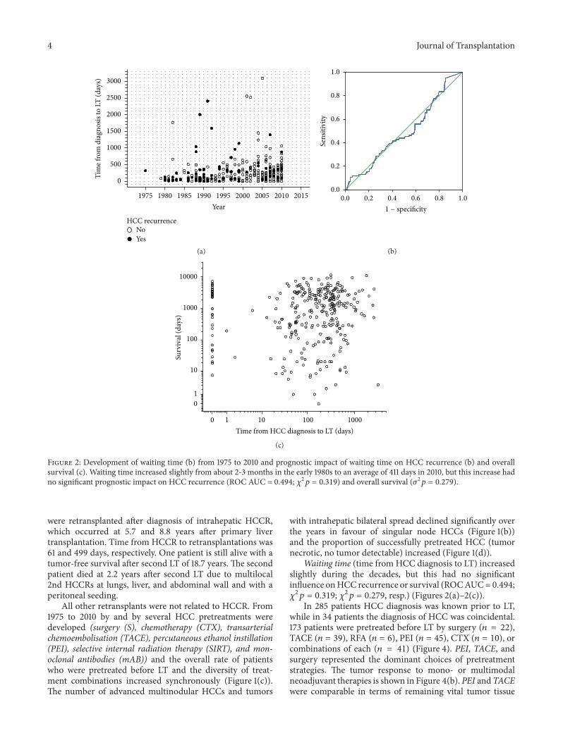
Figure 2: Development of waiting time (b) from 1975 to 2010 and prognostic impact of waiting time on HCC recurrence (b) and overallsurvival (c). Waiting time increased slightly from about 2-3 months in the early 1980s to an average of 411 days in 2010, but this increase hadno significant prognostic impact on HCC recurrence (ROC AUC = 0.494; 𝜒2𝑝 = 0.319) and overall survival (𝜎2𝑝 = 0.279).
were retransplanted after diagnosis of intrahepatic HCCR,which occurred at 5.7 and 8.8 years after primary livertransplantation. Time from HCCR to retransplantations was61 and 499 days, respectively. One patient is still alive with atumor-free survival after second LT of 18.7 years. The secondpatient died at 2.2 years after second LT due to multilocal2nd HCCRs at lungs, liver, and abdominal wall and with aperitoneal seeding.
All other retransplants were not related to HCCR. From1975 to 2010 by and by several HCC pretreatments weredeveloped (surgery (S), chemotherapy (CTX), transarterialchemoembolisation (TACE), percutaneous ethanol instillation(PEI), selective internal radiation therapy (SIRT), and mon-oclonal antibodies (mAB)) and the overall rate of patientswho were pretreated before LT and the diversity of treat-ment combinations increased synchronously (Figure 1(c)).The number of advanced multinodular HCCs and tumors
with intrahepatic bilateral spread declined significantly overthe years in favour of singular node HCCs (Figure 1(b))and the proportion of successfully pretreated HCC (tumornecrotic, no tumor detectable) increased (Figure 1(d)).
Waiting time (time from HCC diagnosis to LT) increasedslightly during the decades, but this had no significantinfluence onHCC recurrence or survival (ROCAUC=0.494;𝜒2𝑝 = 0.319; 𝜒2𝑝 = 0.279, resp.) (Figures 2(a)–2(c)).In 285 patients HCC diagnosis was known prior to LT,
while in 34 patients the diagnosis of HCC was coincidental.173 patients were pretreated before LT by surgery (𝑛 = 22),TACE (𝑛 = 39), RFA (𝑛 = 6), PEI (𝑛 = 45), CTX (𝑛 = 10), orcombinations of each (𝑛 = 41) (Figure 4). PEI, TACE, andsurgery represented the dominant choices of pretreatmentstrategies. The tumor response to mono- or multimodalneoadjuvant therapies is shown in Figure 4(b). PEI andTACEwere comparable in terms of remaining vital tumor tissue
Journal of Transplantation 5
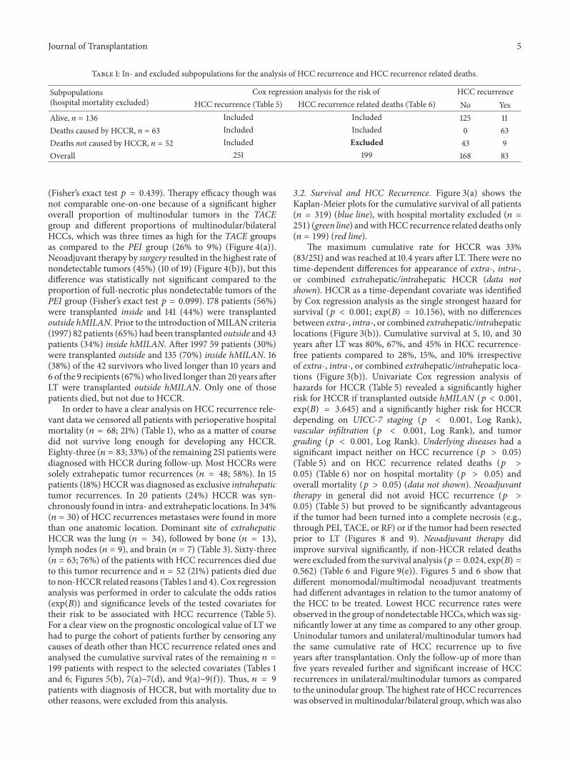
Table 1: In- and excluded subpopulations for the analysis of HCC recurrence and HCC recurrence related deaths.
Subpopulations(hospital mortality excluded)
Cox regression analysis for the risk of HCC recurrenceHCC recurrence (Table 5) HCC recurrence related deaths (Table 6) No Yes
Alive, 𝑛 = 136 Included Included 125 11Deaths caused by HCCR, 𝑛 = 63 Included Included 0 63Deaths not caused by HCCR, 𝑛 = 52 Included Excluded 43 9Overall 251 199 168 83
(Fisher’s exact test 𝑝 = 0.439). Therapy efficacy though wasnot comparable one-on-one because of a significant higheroverall proportion of multinodular tumors in the TACEgroup and different proportions of multinodular/bilateralHCCs, which was three times as high for the TACE groupsas compared to the PEI group (26% to 9%) (Figure 4(a)).Neoadjuvant therapy by surgery resulted in the highest rate ofnondetectable tumors (45%) (10 of 19) (Figure 4(b)), but thisdifference was statistically not significant compared to theproportion of full-necrotic plus nondetectable tumors of thePEI group (Fisher’s exact test 𝑝 = 0.099). 178 patients (56%)were transplanted inside and 141 (44%) were transplantedoutside hMILAN. Prior to the introduction ofMILAN criteria(1997) 82 patients (65%) had been transplanted outside and 43patients (34%) inside hMILAN. After 1997 59 patients (30%)were transplanted outside and 135 (70%) inside hMILAN. 16(38%) of the 42 survivors who lived longer than 10 years and6 of the 9 recipients (67%)who lived longer than 20 years afterLT were transplanted outside hMILAN. Only one of thosepatients died, but not due to HCCR.
In order to have a clear analysis on HCC recurrence rele-vant data we censored all patients with perioperative hospitalmortality (𝑛 = 68; 21%) (Table 1), who as a matter of coursedid not survive long enough for developing any HCCR.Eighty-three (𝑛 = 83; 33%) of the remaining 251 patients werediagnosed with HCCR during follow-up. Most HCCRs weresolely extrahepatic tumor recurrences (𝑛 = 48; 58%). In 15patients (18%) HCCRwas diagnosed as exclusive intrahepatictumor recurrences. In 20 patients (24%) HCCR was syn-chronously found in intra- and extrahepatic locations. In 34%(𝑛 = 30) of HCC recurrences metastases were found in morethan one anatomic location. Dominant site of extrahepaticHCCR was the lung (𝑛 = 34), followed by bone (𝑛 = 13),lymph nodes (𝑛 = 9), and brain (𝑛 = 7) (Table 3). Sixty-three(𝑛 = 63; 76%) of the patients with HCC recurrences died dueto this tumor recurrence and 𝑛 = 52 (21%) patients died dueto non-HCCR related reasons (Tables 1 and 4). Cox regressionanalysis was performed in order to calculate the odds ratios(exp(𝐵)) and significance levels of the tested covariates fortheir risk to be associated with HCC recurrence (Table 5).For a clear view on the prognostic oncological value of LT wehad to purge the cohort of patients further by censoring anycauses of death other than HCC recurrence related ones andanalysed the cumulative survival rates of the remaining 𝑛 =199 patients with respect to the selected covariates (Tables 1and 6; Figures 5(b), 7(a)–7(d), and 9(a)–9(f)). Thus, 𝑛 = 9patients with diagnosis of HCCR, but with mortality due toother reasons, were excluded from this analysis.
3.2. Survival and HCC Recurrence. Figure 3(a) shows theKaplan-Meier plots for the cumulative survival of all patients(𝑛 = 319) (blue line), with hospital mortality excluded (𝑛 =251) (green line) andwithHCCrecurrence related deaths only(𝑛 = 199) (red line).
The maximum cumulative rate for HCCR was 33%(83/251) and was reached at 10.4 years after LT.There were notime-dependent differences for appearance of extra-, intra-,or combined extrahepatic/intrahepatic HCCR (data notshown). HCCR as a time-dependant covariate was identifiedby Cox regression analysis as the single strongest hazard forsurvival (𝑝 < 0.001; exp(𝐵) = 10.156), with no differencesbetween extra-, intra-, or combined extrahepatic/intrahepaticlocations (Figure 3(b)). Cumulative survival at 5, 10, and 30years after LT was 80%, 67%, and 45% in HCC recurrence-free patients compared to 28%, 15%, and 10% irrespectiveof extra-, intra-, or combined extrahepatic/intrahepatic loca-tions (Figure 3(b)). Univariate Cox regression analysis ofhazards for HCCR (Table 5) revealed a significantly higherrisk for HCCR if transplanted outside hMILAN (𝑝 < 0.001,exp(𝐵) = 3.645) and a significantly higher risk for HCCRdepending on UICC-7 staging (𝑝 < 0.001, Log Rank),vascular infiltration (𝑝 < 0.001, Log Rank), and tumorgrading (𝑝 < 0.001, Log Rank). Underlying diseases had asignificant impact neither on HCC recurrence (𝑝 > 0.05)(Table 5) and on HCC recurrence related deaths (𝑝 >0.05) (Table 6) nor on hospital mortality (𝑝 > 0.05) andoverall mortality (𝑝 > 0.05) (data not shown). Neoadjuvanttherapy in general did not avoid HCC recurrence (𝑝 >0.05) (Table 5) but proved to be significantly advantageousif the tumor had been turned into a complete necrosis (e.g.,through PEI, TACE, or RF) or if the tumor had been resectedprior to LT (Figures 8 and 9). Neoadjuvant therapy didimprove survival significantly, if non-HCCR related deathswere excluded from the survival analysis (𝑝 = 0.024, exp(𝐵) =0.562) (Table 6 and Figure 9(e)). Figures 5 and 6 show thatdifferent monomodal/multimodal neoadjuvant treatmentshad different advantages in relation to the tumor anatomy ofthe HCC to be treated. Lowest HCC recurrence rates wereobserved in the group of nondetectableHCCs, whichwas sig-nificantly lower at any time as compared to any other group.Uninodular tumors and unilateral/multinodular tumors hadthe same cumulative rate of HCC recurrence up to fiveyears after transplantation. Only the follow-up of more thanfive years revealed further and significant increase of HCCrecurrences in unilateral/multinodular tumors as comparedto the uninodular group.Thehighest rate ofHCC recurrenceswas observed inmultinodular/bilateral group, whichwas also

6 Journal of Transplantation
Table2:Descriptiv
estatistics.
Und
erlyingdisease
Gender
Age
atLT
[year]
Tumor
morph
olog
yTu
mor
volume[cm3]
AFP
[ng/mL]
Grafttype
CIT[m
in.]
LT#
fm
Mean
Median
Max.
Min.
Notumordetectable
Uninodular
Multinodular,unilateral
Multinodular,bilateral
Mean
Median
Max.
Min.
Mean
Median
Max.
Min.
Fullsize
Partial
Split
Mean
Median
Max.
Min.
1st
2nd
3rd
Total
Deceased
Living
Deceased
Living
HepatitisB
with
D1
1450,6
50,3
61,6
38,6
19
23
868
978
—418
18,3
5571
113
—2
——
531
507
880
263
141
—15
HepatitisB
778
50,7
53,8
69,7
21,6
532
2325
236
424849
—2405
29176659
178
12
4—
638
635
1577
207
787
—85
HepatitisC
with
B2
1051,2
57,1
65,2
8,1
17
31
3013
202
—3478
25,5
3200
05
12—
——
—607
518
1054
388
111
—12
HepatitisC
2066
54,2
54,7
66,7
255
4025
1680
16116
4—
839
2156100
174
—4
62
584
571
1302
100
778
186
Hepatob
lasto
ma
31
14,8
14,1
17,8
13,3
—1
—3
432
375
905
74304
304
600
74
——
——
866
724
1740
275
4—
—4
Adenom
atosis
31
41,7
40,7
56,3
29,2
—1
21
276
934
4,75
57
24
——
——
638
579
1122
271
31
—4
Hypertyrosin
emia
—2
14,4
14,4
19,4
9,3—
2—
—34
3434
341276
1276
2431
120
2—
——
—420
420
480
359
2—
—2
Wilson’sdisease
1—
43,8
——
——
1—
—34
——
—5
——
—1
——
——
572
572
572
572
1—
—1
Hem
ochrom
atosis
—4
56,8
56,7
63,5
50,3
——
22
452
191768
2248
247
485
134
——
——
635
618
786
516
31
—4
𝛼1antitrypsin
def.
1—
56,8
——
——
1—
—17
——
—1
——
—1
——
——
588
588
588
588
1—
—1
Budd
Chiari
11
36,2
36,2
40,3
32,2
—1
1—
263
263
525
239
3973
52
——
——
665
665
773
557
11
—2
Alcoh
olabuse
641
55,4
55,4
68,7
264
1510
18534
3414137
—3679
12,3
109718
145
—1
1—
674
629
1970
187
425
—47
And
rogentherapy
—1
29,6
——
——
——
113
——
—5
——
—1
——
——
552
552
552
552
1—
—1
Biliary
cirrho
sis3
—24,9
17,7
54,1
3—
2—
1131
9382
339
39,3
39,3
391
—2
—64
4773
827
332
3—
—3
Cryptogenicc
irrho
sis23
2749
51,8
6718,6
421
916
856
938181
—6100
10,5
214975
144
11
4—
628
636
1180
227
464
—50
Autoim
mun
ehepatitis
—1
72—
——
——
1—
38—
——
485
——
—1
——
——
971
971
971
971
1—
—1
Chronicleadintox.
—1
44,7
——
——
—1
—1216
——
—540
——
——
1—
——
865
865
865
865
1—
—1
Journal of Transplantation 7
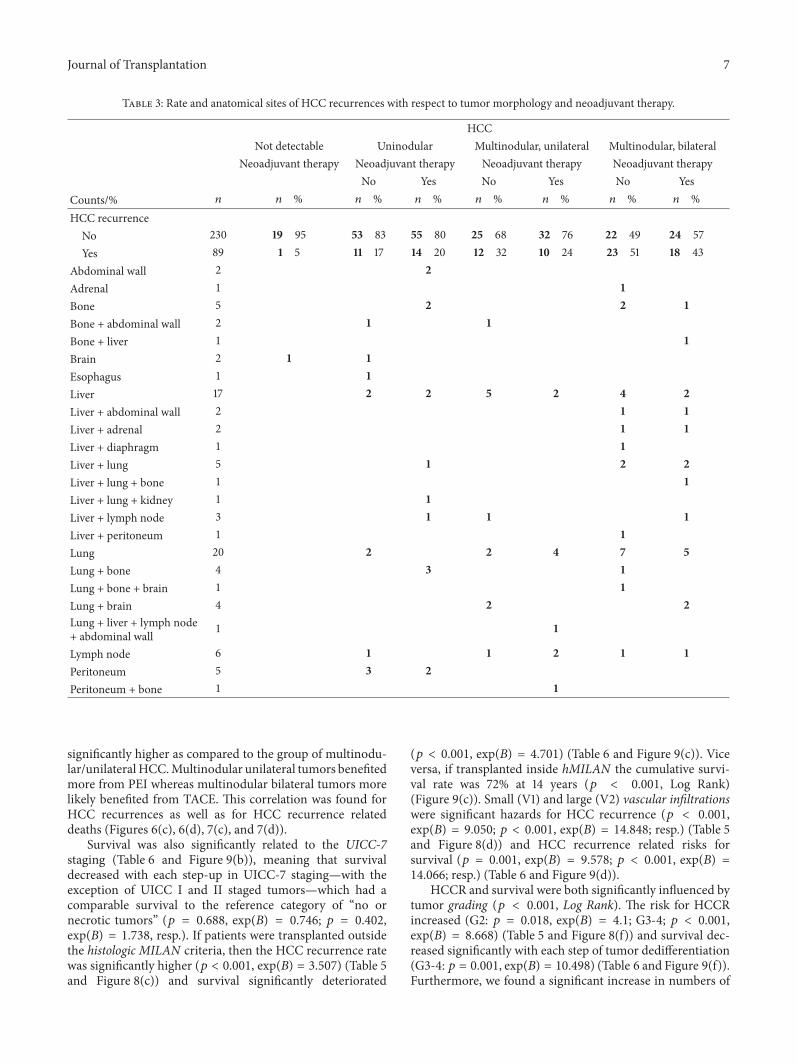
Table 3: Rate and anatomical sites of HCC recurrences with respect to tumor morphology and neoadjuvant therapy.
HCCNot detectable Uninodular Multinodular, unilateral Multinodular, bilateral
Neoadjuvant therapy Neoadjuvant therapy Neoadjuvant therapy Neoadjuvant therapyNo Yes No Yes No Yes
Counts/% 𝑛 𝑛 % 𝑛 % 𝑛 % 𝑛 % 𝑛 % 𝑛 % 𝑛 %HCC recurrence
No 230 19 95 53 83 55 80 25 68 32 76 22 49 24 57Yes 89 1 5 11 17 14 20 12 32 10 24 23 51 18 43
Abdominal wall 2 2Adrenal 1 1Bone 5 2 2 1Bone + abdominal wall 2 1 1Bone + liver 1 1Brain 2 1 1Esophagus 1 1Liver 17 2 2 5 2 4 2Liver + abdominal wall 2 1 1Liver + adrenal 2 1 1Liver + diaphragm 1 1Liver + lung 5 1 2 2Liver + lung + bone 1 1Liver + lung + kidney 1 1Liver + lymph node 3 1 1 1Liver + peritoneum 1 1Lung 20 2 2 4 7 5Lung + bone 4 3 1Lung + bone + brain 1 1Lung + brain 4 2 2Lung + liver + lymph node+ abdominal wall 1 1
Lymph node 6 1 1 2 1 1Peritoneum 5 3 2Peritoneum + bone 1 1
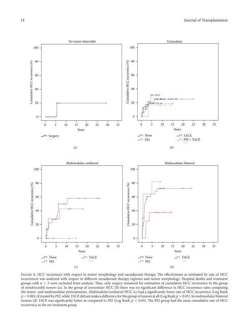
significantly higher as compared to the group of multinodu-lar/unilateralHCC.Multinodular unilateral tumors benefitedmore from PEI whereas multinodular bilateral tumors morelikely benefited from TACE. This correlation was found forHCC recurrences as well as for HCC recurrence relateddeaths (Figures 6(c), 6(d), 7(c), and 7(d)).
Survival was also significantly related to the UICC-7staging (Table 6 and Figure 9(b)), meaning that survivaldecreased with each step-up in UICC-7 staging—with theexception of UICC I and II staged tumors—which had acomparable survival to the reference category of “no ornecrotic tumors” (𝑝 = 0.688, exp(𝐵) = 0.746; 𝑝 = 0.402,exp(𝐵) = 1.738, resp.). If patients were transplanted outsidethe histologic MILAN criteria, then the HCC recurrence ratewas significantly higher (𝑝 < 0.001, exp(𝐵) = 3.507) (Table 5and Figure 8(c)) and survival significantly deteriorated
(𝑝 < 0.001, exp(𝐵) = 4.701) (Table 6 and Figure 9(c)). Viceversa, if transplanted inside hMILAN the cumulative survi-val rate was 72% at 14 years (𝑝 < 0.001, Log Rank)(Figure 9(c)). Small (V1) and large (V2) vascular infiltrationswere significant hazards for HCC recurrence (𝑝 < 0.001,exp(𝐵) = 9.050; 𝑝 < 0.001, exp(𝐵) = 14.848; resp.) (Table 5and Figure 8(d)) and HCC recurrence related risks forsurvival (𝑝 = 0.001, exp(𝐵) = 9.578; 𝑝 < 0.001, exp(𝐵) =14.066; resp.) (Table 6 and Figure 9(d)).
HCCR and survival were both significantly influenced bytumor grading (𝑝 < 0.001, Log Rank). The risk for HCCRincreased (G2: 𝑝 = 0.018, exp(𝐵) = 4.1; G3-4; 𝑝 < 0.001,exp(𝐵) = 8.668) (Table 5 and Figure 8(f)) and survival dec-reased significantly with each step of tumor dedifferentiation(G3-4: 𝑝 = 0.001, exp(𝐵) = 10.498) (Table 6 and Figure 9(f)).Furthermore, we found a significant increase in numbers of

8 Journal of Transplantation
Table4:Tu
mor
morph
ology,neoadjuvanttherapy,tum
orrespon
seto
neoadjuvanttherapy,H
CCrecurrence
rate,and
HCC
recurrence
relateddeaths.
Tumor
morph
olog
yNeoadjuvant
therapy
Respon
seHCC
recurrence
HCC
recurrence
relateddeath
Vitaltum
orremnants
Full-necrotic
Notumor
detectable
No
Yes
No
Yes
Not
specified
Notumor
PEI
——
2100%
2100%
—2
100%
——
TACE
——
2100%
2100%
—2
100%
——
Surgery
——
9100%
889%
111%
889%
111%
—Surgery+TA
CE—
—2
100%
2100%
—2
100%
——
Surgery+TA
CE+CT
X—
—1
100%
1100%
—1
100%
——
Overall
00
1615
115
10
Unino
dular
Non
e48
100%
——
3777%
1123%
3981%
817%
12%
PEI
2273%
827%
—25
83%
517%
2583%
413%
13%
TACE
1381%
319%
—12
75%
425%
1381%
319%
—PE
I+TA
CE6
75%
225%
—7
88%
113%
8100%
——
Surgery
375%
125%
——
4100%
250%
250%
—CT
X+TA
CE2
67%
133%
—3
100%
—3
100%
——
RFA
2100%
——
2100%
—2
100%
——
RFA+TA
CE—
1100%
—1
100%
—1
100%
——
PEI+
RFA
—1
100%
—1
100%
—1
100%
——
Overall
9617
088
2594
172
Multin
odular,unilateral
Non
e25
100%
——
1456%
1144
%17
68%
832%
—PE
I5
83%
117%
—6
100%
—6
100%
——
TACE
686%
114%
—4
57%
343%
571%
229%
—PE
I+TA
CE4
100%
——
4100%
—4
100%
——
Surgery
3100%
——
133%
267%
267%
133%
—CT
X4
100%
——
250%
250%
4100%
——
Surgery+TA
CE3
100%
——
133%
267%
133%
267%
—RF
A3
100%
——
267%
133%
267%
133%
—Surgery+TA
CE+CT
X1
100%
——
1100%
—1
100%
——
RFA+TA
CE1
100%
——
1100%
—1
100%
——
Overall
552
036
2143
140
Multin
odular,bilateral
Non
e34
100%
——
1235%
2265%
1544
%19
56%
—PE
I4
100%
——
4100%
—4
100%
——
TACE
7100%
——
229%
571%
229%
571%
—PE
I+TA
CE6
75%
225%
—5
63%
338%
675%
225%
—Surgery
2100%
——
—2
100%
—2
100%
—CT
X2
100%
——
—2
100%
150%
150%
—Surgery+TA
CE2
100%
——
150%
150%
150%
150%
—Surgery+TA
CE+CT
X3
100%
——
3100%
—3
100%
——
Surgery+CT
X1
100%
——
—1
100%
—1
100%
—PE
I+RF
A1
100%
——
—1
100%
1100%
——
Surgery+PE
I1
100%
——
—1
100%
1100%
——
Overall
632
027
3834
310
Total
214
2116
166
85186
632

Journal of Transplantation 9
Table 5: Identification of hazards for HCC recurrence by univariate Cox regression, 𝑛 = 251 (hospital deaths excluded).
Univariate Cox regressions for HCC recurrence 𝑛 𝑝 exp(𝐵)/hazard 95.0% CILower Upper
Underlying disease
Hepatitis B with D 12 0.176 Reference categoryHepatitis B 64 0.405 1.667 0.5 5.555Hepatitis C 75 0.847 1.126 0.336 3.777
Hepatitis C with B 9 0.419 0.394 0.041 3.784Alcohol 33 0.131 2.603 0.752 9.003
Cryptogenic cirrhosis 38 0.375 1.76 0.505 6.128Other 20 0.417 1.732 0.459 6.53
Tumor vitalityVital tumor 214 0.048 Reference category
Full-necrotic tumor 21 0.118 0.399 0.126 1.263No tumor detectable 16 0.053 0.142 0.02 1.022
Tumor morphology
No tumor detectable 16 <0.001 Reference categoryUninodular 113 0.192 3.789 0.513 27.971
Multinodular unilateral 57 0.073 6.251 0.840 46.513Multinodular bilateral 65 0.008 14.505 1.990 105.733
UICC-7
No or necrotic tumor 37 <0.001 Reference categoryUICC I 69 0.646 0.75 0.219 2.563UICC II 71 0.200 2.041 0.686 6.073UICC IIIA 25 <0.001 7.428 2.513 21.959UICC IIIB 32 <0.001 13.734 4.759 39.631UICC IIIC 7 0.001 9.808 2.627 36.611UICC IVA 7 <0.001 54.098 14.542 201.253UICC IVB 3 <0.001 180.683 34.823 937.506
hMILAN Inside 148 Reference categoryOutside 103 <0.001 3.507 2.237 5.496
Vascular infiltration
No or necrotic tumor 37 <0.001 Reference categoryV0 148 0.254 1.829 0.648 5.161V1 28 <0.001 9.05 3.042 26.92V2 37 <0.001 14.848 5.206 42.353
Missing data 1 0.977 0 0 1.65𝐸 + 245
Neoadj. therapy No 107 Reference categoryYes 144 0.071 0.676 0.441 1.035
Grading
No or necrotic tumor 37 <0.001 Reference categoryG1 35 0.272 1.937 0.596 6.299G2 130 0.023 3.282 1.179 9.138G3-4 43 <0.001 6.672 2.313 19.249
Missing data 6 0.008 6.550 1.636 26.214HCC = hepatocellular carcinoma.UICC-7 = 7th edition TNM classification of Unite International Contre Cancer.hMILAN = histologic MILAN classification.Vascular infiltration: V0 = none, V1 = small vessels, and V2 = large vessels.Tumor grading: G1 = low, G2 = intermediate, and G3-4 = high to anaplastic.
vascular infiltrating tumors and an increase of large vesselinfiltrations per step of tumor dedifferentiation (G1 →G2 → G3-4) (𝜒2𝑝 = 0.006) (Figure 10).
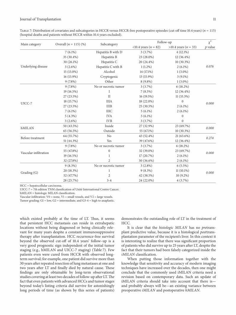
Because long-term survival was mainly limited by HCCR(𝑝 < 0.001, exp(𝐵) = 10.156; time-dependent Cox regres-sion) and HCCRs were diagnosed as late as 10 years after LT,but not later than 10.4 years after LT, we aimed to determinethe cohort identifiers with respect to this 10.4-year cut-off.
Therefore we analysed the database and compared thegroup of patients with HCCR occurrence (below 10.4 years)with the group of patients who had HCCR-free follow-up ofmore than 10.4 years after LT (hospital deaths censored). Wefound that hMILAN,UICC-7, vascular infiltration, and tumorgrading were highly significant prognostic parameters (𝜒2𝑝 <0.001, Table 7), while neoadjuvant therapy and underlyingdiseases remained nonsignificant.

10 Journal of Transplantation
Table 6: Identification of hazards for HCC recurrence related deaths by univariate Cox regression, 𝑛 = 199 (non-HCC recurrence relateddeaths excluded).
Univariate Cox regressions for HCC recurrence related deaths 𝑛 𝑝 exp(𝐵)/hazard 95.0% CILower Upper
Underlying disease
Hepatitis B with D 11 0.348 Reference categoryHepatitis B 50 0.368 1.964 0.451 8.553Hepatitis C 55 0.542 1.584 0.361 6.939
Hepatitis C with B 6 0.908 0.868 0.079 9.588Alcohol 30 0.162 2.919 0.65 13.109
Cryptogenic cirrhosis 30 0.13 3.151 0.714 13.896Other 17 0.433 1.93 0.373 9.979
Tumor vitalityVital tumor 168 0.083 Reference category
Full-necrotic tumor 17 0.147 0.352 0.086 1.442No tumor detectable 14 0.084 0.175 0.024 1.264
Tumor morphology
No tumor detectable 14 <0.001 Reference categoryUninodular 89 0.323 2.765 0.368 20.791
Multinodular unilateral 41 0.104 5.354 0.707 40.567Multinodular bilateral 55 0.019 10.898 1.488 79.801
UICC-7
No or necrotic tumor 31 <0.001 Reference categoryUICC I 54 0.688 0.746 0.178 3.124UICC II 51 0.402 1.738 0.477 6.327UICC IIIA 21 0.003 6.771 1.944 23.584UICC IIIB 26 <0.001 12.792 3.791 43.16UICC IIIC 6 0.006 8.066 1.800 36.142UICC IVA 7 <0.001 226.972 46.041 1118.915UICC IVB 3 <0.001 91.043 16.824 492.692
hMILAN Inside 112 Reference categoryOutside 87 <0.001 4.701 2.700 8.185
Vascular infiltration
No or necrotic tumor 31 <0.001 Reference categoryV0 117 0.371 1.733 0.52 5.779V1 21 <0.001 9.578 2.769 33.128V2 30 <0.001 14.066 4.221 46.866
Neoadj. therapy No 82 Reference categoryYes 117 0.010 0.525 0.321 0.859
Grading
No or necrotic tumor 31 <0.001 Reference categoryG1 26 0.26 2.179 0.562 8.442G2 103 0.061 3.098 0.948 10.124G3-4 36 0.001 7.909 2.357 26.542
Missing data 3 0.007 11.921 1.980 71.774HCC = hepatocellular carcinoma.UICC-7 = 7th edition TNM classification of Unite International Contre Cancer.hMILAN = histologic MILAN classification.Vascular infiltration: V0 = none, V1 = small vessels, and V2 = large vessels.Tumor grading: G1 = low, G2 = intermediate, and G3-4 = high to anaplastic.
4. Discussion
The results of this study containing the complete data of ourcenter since 1975 demonstrate that hepatocellular carcinomacan be cured by LT—even in advanced tumor stages. Asexpected, long-term survival was mainly limited by HCCrecurrence (HCCR) (𝑝 < 0.001, exp(𝐵) = 10.156; time-dependent Cox regression) and any covariate with highpotency for HCC recurrence therefore was a significant
negative predictor of survival as well. Vice versa, covariatesthat were not associated with a significantly higher rate ofHCC recurrences (e.g., underlying diseases) had no significantimpact on tumor-free survival. We were surprised thoughto find that not only intrahepatic HCCRs (some of whichmight have been de novoHCCs) but extrahepatic HCCR alsocan occur more than 10 years after LT—without synchronousintrahepatic HCC recurrences. We believe that these tumorsmust have been dormant metastatic HCC manifestations,
Journal of Transplantation 11
Table 7: Distribution of covariates and subcategories in HCCR versus HCCR-free postoperative episodes (cut-off time 10.4 years) (𝑛 = 115)(hospital deaths and patients without HCCR within 10.4 years excluded).
Main category Overall (𝑛 = 115) (%) Subcategory Follow-up 𝜒2
<10.4 years (𝑛 = 82) >10.4 years (𝑛 = 33) 𝑝 value
Underlying disease
7 (6.1%) Hepatitis B with D 3 (3.7%) 4 (12.1%)
0.076
35 (30.4%) Hepatitis B 23 (28.0%) 12 (36.4%)30 (26.1%) Hepatitis C 20 (24.4%) 10 (30.3%)3 (2.6%) Hepatitis C with B 1 (1.2%) 2 (6.1%)15 (13.0%) Alcohol 14 (17.1%) 1 (3.0%)16 (13.9%) Cryptogenic 13 (15.9%) 3 (9.1%)9 (7.8%) Other 8 (9.8%) 1 (3.0%)
UICC-7
9 (7.8%) No or necrotic tumor 3 (3.7%) 6 (18.2%)
0.000
19 (16.5%) I 7 (8.5%) 12 (36.4%)27 (23.5%) II 16 (19.5%) 11 (33.3%)18 (15.7%) IIIA 18 (22.0%) 027 (23.5%) IIIB 25 (30.5%) 2 (6.1%)7 (6.1%) IIIC 5 (6.1%) 2 (6.1%)5 (4.3%) IVA 5 (6.1%) 03 (2.6%) IVB 3 (3.7%) 0
hMILAN 50 (43.5%) Inside 27 (32.9%) 23 (69.7%) 0.00065 (56.5%) Outside 55 (67.1%) 10 (30.3%)
Before treatment 64 (55.7%) No 43 (52.4%) 21 (63.6%) 0.27451 (44.3%) Yes 39 (47.6%) 12 (36.4%)
Vascular infiltration
9 (7.8%) No or necrotic tumor 3 (3.7%) 6 (18.2%)
0.00055 (47.8%) 0 32 (39.0%) 23 (69.7%)19 (16.5%) 1 17 (20.7%) 2 (6.1%)32 (27.8%) 2 30 (36.6%) 2 (6.1%)
Grading (G)
9 (8.3%) No or necrotic tumor 3 (2.8%) 6 (5.5%)
0.00020 (18.3%) 1 9 (8.3%) 11 (10.1%)52 (47.7%) 2 42 (38.5%) 10 (9.2%)28 (25.7%) 3-4 24 (22.0%) 4 (3.7%)
HCC = hepatocellular carcinoma.UICC-7 = 7th edition TNM classification of Unite International Contre Cancer.hMILAN = histologic MILAN classification.Vascular infiltration: V0 = none, V1 = small vessels, and V2 = large vessels.Tumor grading: G1 = low, G2 = intermediate, and G3-4 = high to anaplastic.
which existed probably at the time of LT. Thus, it seemsthat persistent HCC metastasis can reside in extrahepaticlocations without being diagnosed or being clinically rele-vant for many years despite a constant immunosuppressivetherapy after transplantation. HCC recurrence-free survivalbeyond the observed cut-off of 10.4 years’ follow–up is avery good prognostic sign independent of the initial tumorstaging (e.g., hMILAN and UICC-7 staging) (Table 7). Fewpatients even were cured from HCCR with observed long-term survival; for example, one patient did survive more than30 years after repeated resection of lungmetastases at one andtwo years after LT and finally died by natural cause. Thesefindings are only obtainable by long-term observationalstudies covering at least twodecades of follow-up after LT.Thefact that even patients with advancedHCCs and tumor stagesbeyond today’s listing criteria did survive for astonishinglylong periods of time (as shown by this series of patients)
demonstrates the outstanding role of LT in the treatment ofHCC.
It is clear that the histologic MILAN has no pretrans-plant predictive value, because it is a histological posttrans-plantation parameter of the recipient’s liver. In this context itis interesting to realize that there was significant proportionof patients who did survive up to 25 years after LT, despite thefact that their tumors had been falsely categorized inside theiMILAN classification.
When putting those information together with theknowledge that sensitivity and accuracy of modern imagingtechniques have increased over the decades, then one mightconclude that the commonly used iMILAN criteria need arevision based on contemporary data. Such an update ofiMILAN criteria should take into account that there is—and probably always will be—an existing variance betweenpreoperative iMILAN and postoperative hMILAN.

12 Journal of Transplantation
Cum
ulat
ive s
urvi
val (
%)
100
80
60
40
20
0
Years0 5 10 15 20 25 30 35
All (n = 319)No hospital deaths (n = 251)With HCCR related deaths only (n = 199)
(a)
Years0 5 10 15 20 25 30 35
Location of HCCRNo HCCRExtrahepaticExtrahepatic/intrahepatic Intrahepatic
Cum
ulat
ive s
urvi
val (
%)
100
80
60
40
20
0
(b)
Figure 3: Survival with respect to hospital mortality and HCC recurrence. (a) Cumulative survival of all patients (𝑛 = 319, blue line),without hospital mortality (𝑛 = 251, green line) and with HCC recurrence related deaths only (𝑛 = 199, red line). (b) HCC recurrence-freesurvival (blue line, Cox regression analysis with HCC recurrence as time-dependent covariate) and with respect to extrahepatic (green line),intrahepatic (red line), or combined extrahepatic/intrahepatic HCC recurrences (orange line). HCC recurrence was highly significant hazardof survival (𝑝 < 0.001, exp(𝐵) = 10.156), but it made no difference to survival whether HCC recurrences were at intrahepatic, at extrahepatic,or at combined intrahepatic/extrahepatic locations (𝜎2𝑝 > 0.05).
Furthermore, for a more accurate assessment of the long-term prognosis, it could be beneficial not only to classifythe tumors according to size and numbers of tumors but toconsider also the bilateral distribution of tumors on both liverlobes as a prognostic relevant cofactor (Figure 5, Tables 5 and6).
Hence it is no surprise that several authors already havecast serious doubt [44, 45] on the concept of relying solely onthe commonly used iMILAN status for the listing of patientsand suggested the extension of the iMILAN criteria, whichhas already resulted in the definition of alternative listing cri-teria (e.g., the University of California San Francisco (UCSF)criteria) [46]. But these alternative allocation algorithms alsorely solely on pretransplant imaging technology and lacklong-term follow-up data that covers at least two decades afterLT.
Neoadjuvant therapy in general was only slightly advan-tageous with respect to HCC recurrence but neverthe-less did prolong survival significantly. Because the effectof different neoadjuvant treatment strategies in differentpatients by different specialists against different tumors ofdifferent numbers, sizes, gradings, and status of vascularinfiltration is variant, the extent of induced tumor necrosis
is completely variant as well. The bottom line is that lowestHCCR rates and best survival rates had been observedwhen all tumor mass was completely necrotic or miss-ing (e.g., after resection) (Figures 5, 8(b), 8(d), 8(f), 9(b),9(d), and 9(f)). In other words, the possibly advantageouseffect of a neoadjuvant therapy depends on whether alltumor mass is transferred into a complete necrosis ornot.
The data further demonstrate that tumor grading (G) iscurrently an underrated pretransplant prognostic parameter,which seems to be equally relevant for long-term prognosisafter LT as compared to allocation algorithms such as iMI-LAN, which are susceptible for the underrating of relevanthistological tumor parameters—for example, the status ofvascular infiltration.
Our data also demonstrates the existing close correlationof tumor dedifferentiation with intrahepatic tumor spreading(Figure 10(a)) and the potency of tumor cell differentiation(grading, G) to predict vascular infiltration (Figure 10(b)).As tumor grading and vascular infiltration have a significantprognostic impact on HCC recurrence and patient survival,these cofactors should be routinely utilized for a better timingof LT in HCC patients.

Journal of Transplantation 13
n
50
40
30
20
10
0
PEI
TACE
PEI +
TAC
ESu
rger
yCT
XSu
rger
y +
TACE RF
ASu
rger
y +
CTX
+ TA
CECT
X +
TACE
RFA
+ T
ACE
PEI +
RFA
Surg
ery
+ CT
XSu
rger
y +
PEI
Tumor morphologyNo tumor detectableUninodular
Multinodular, unilateralMultinodular, bilateral
(a)
n
50
40
30
20
10
0
PEI
TACE
PEI +
TAC
E
Surg
ery
CTX
Surg
ery
+ TA
CE RFA
Surg
ery
+ CT
X +
TACE
CTX
+ TA
CE
RFA
+ T
ACE
PEI +
RFA
Surg
ery
+ CT
X
Surg
ery
+ PE
I
Tumor responseNo tumorFull-necrotic tumor
Vital tumor
(b)
Figure 4: HCCmorphology per treatment group (a) and tumor response to pretreatments (b) asmeasured in numbers of nondetectable, full-necrotic, or vital tumors. Percutaneous ethanol instillation (PEI) (𝑛 = 45), transarterial chemoembolisation (TACE) (𝑛 = 39), and surgery(𝑛 = 22) were most frequently applied. Another major treatment group were patients that had been treated by a combination of PEI andTACE (𝑛 = 25). There were a significant higher number of uninodular tumors in the PEI group (71%) as compared to the TACE group(41%). The TACE group also had a significant higher proportion of multinodular tumors (52%) as compared to the PEI group (25%) and ahigher proportion of multinodular/bilateral tumors, which was three times as high as compared to the PEI groups (26% to 9%, resp.). Thepretreatment group surgery had the highest rate (45%) (10 of 22) of explanted livers without detectable tumor remnants, but this differencewas statistically not significant as compared to the proportion of full-necrotic and nondetectable tumors (𝑛 = 10+2) in the PEI group (Fisher’sexact test𝑝 = 0.099).The PEI group and TACE groupwere comparable in terms of remaining vital tumor tissue (Fisher’s exact test𝑝 = 0.439).
Years0 5 10 15 20 25 30 35
Tumor morphologyNo tumorUninodularMultinodular, unilateralMultinodular, bilateral
100
80
60
40
20
0
Cum
ulat
ive H
CC re
curr
ence
(%)
(a)
Years0 5 10 15 20 25 30 35
No tumor detectableUninodularMultinodular, unilateralMultinodular, bilateral
100
80
60
40
20
0
Cum
ulat
ive s
urvi
val (
%)
Tumor morphology
(b)
Figure 5: HCC recurrence (a) and survival (b) with respect to tumor morphology. HCC recurrence (hospital deaths excluded) (a) wassignificantly influenced by tumor morphology (Log Rank 𝑝 < 0.001). Survival (hospital deaths and non-HCC recurrence related deathsexcluded) (b) was significantly influenced by the intrahepatic tumor dissemination of the primary HCC (Log Rank 𝑝 < 0.001).
14 Journal of Transplantation
No tumor detectable
Surgery
Cum
ulat
ive H
CC re
curr
ence
(%)
100
80
60
40
20
0
Years0 5 10 15 20 25 30 35
· · ·
(a)
Uninodular
NonePEI
TACEPEI + TACE
Cum
ulat
ive H
CC re
curr
ence
(%)
100
80
60
40
20
0
Years0 5 10 15 20 25 30 35
· · ·
(b)
Multinodular, unilateral
NonePEI
TACE
Cum
ulat
ive H
CC re
curr
ence
(%)
100
80
60
40
20
0
Years0 5 10 15 20 25 30 35
· · ·
(c)
NonePEI
TACE
Multinodular, bilateral
Cum
ulat
ive H
CC re
curr
ence
(%)
100
80
60
40
20
0
Years0 5 10 15 20 25 30 35
· · ·
(d)
Figure 6: HCC recurrence with respect to tumor morphology and neoadjuvant therapy. The effectiveness as estimated by rate of HCCrecurrences was analysed with respect to different neoadjuvant therapy regimen and tumor morphology. Hospital deaths and treatmentgroups with 𝑛 < 5 were excluded from analysis. Thus, only surgery remained for estimation of cumulative HCC recurrence in the groupof nondetectable tumors (a). In the group of uninodular HCC (b) there was no significant difference in HCC recurrence rates comparingthe mono- and multimodular pretreatments. Multinodular/unilateral HCC (c) had a significantly lower rate of HCC recurrence (Log Rank𝑝 < 0.001) if treated byPEI, whileTACE did notmake a difference for this group of tumors at all (LogRank𝑝 > 0.05). Inmultinodular/bilateraltumors (d) TACE was significantly better as compared to PEI (Log Rank 𝑝 < 0.05). The PEI group had the same cumulative rate of HCCrecurrence as the no-treatment group.

Journal of Transplantation 15
100
80
60
40
20
0
Cum
ulat
ive s
urvi
val (
%)
Surgery
Years0 5 10 15 20 25 30 35
No tumor detectable
· · ·
(a)
100
80
60
40
20
0
Cum
ulat
ive s
urvi
val (
%)
NonePEI
TACEPEI + TACE
Years0 5 10 15 20 25 30 35
Uninodular
· · ·
(b)
100
80
60
40
20
0
Cum
ulat
ive s
urvi
val (
%)
NonePEI
TACE
Years0 5 10 15 20 25 30 35
Multinodular, unilateral
· · ·
(c)
100
80
60
40
20
0
Cum
ulat
ive s
urvi
val (
%)
NonePEI
TACE
Years0 5 10 15 20 25 30 35
Multinodular, bilateral
· · ·
(d)
Figure 7: Survival with respect to tumor morphology and neoadjuvant therapy. Hospital mortality and non-HCC recurrence related deathsas well as treatment groups with 𝑛 < 5 were excluded. In the category of nondetectable tumors only surgery remained with 𝑛 > 5. Thecumulative survival in this subcategory was 80% (a). In the category of uninodular HCC (b) there was no difference in survival comparingpatients that had been pretreated by PEI or TACE. For the combination of PEI and TACE a significantly better survival was observed (LogRank 𝑝 < 0.05) as compared to PEI or TACE alone. Formultinodular/unilateralHCC (c) TACE did not make a difference, while pretreatmentwith PEI achieved a significant better survival (Log Rank 𝑝 < 0.05). Inmultinodular/bilateral tumors (d) survival was significantly better forthe group of patients who were pretreated with TACE as compared to PEI or no pretreatment.

16 Journal of Transplantation
Cum
ulat
ive H
CC re
curr
ence
(%)
100
80
60
40
20
0
Years0 5 10 15 20 25 30 35
Hepatitis B and DHepatitis BHepatitis CHepatitis C and BAlcoholCryptogenic cirrhosisOther
Underlying disease
(a)
Cum
ulat
ive H
CC re
curr
ence
(%)
100
80
60
40
20
0
Years0 5 10 15 20 25 30 35
UICC-7
No or necrotic tumor
IIIIIIAIIIBIIICIVAIVB
(b)
Cum
ulat
ive H
CC re
curr
ence
(%)
100
80
60
40
20
0
Years0 5 10 15 20 25 30 35
hMILANInsideOutside
(c)
Cum
ulat
ive H
CC re
curr
ence
(%)
100
80
60
40
20
0
Years0 5 10 15 20 25 30 35
No or necrotic tumor
V0
V1
V2
Vascular infiltration
(d)
Figure 8: Continued.

Journal of Transplantation 17
Cum
ulat
ive H
CC re
curr
ence
(%)
100
80
60
40
20
0
Years0 5 10 15 20 25 30 35
Neoadjuvant therapyNoYes
(e)
Cum
ulat
ive H
CC re
curr
ence
(%)
100
80
60
40
20
0
Years0 5 10 15 20 25 30 35
No or necrotic tumor
GradingG1
G2
G3-4
(f)
Figure 8: Cumulative recurrence of HCC (hospital mortality excluded) (𝑛 = 251) (for statistics see Table 3). (a) Underlying disease had nosignificant impact on HCC recurrence. (b) UICC-7 staging had a significant impact on HCCR. Only UICC I and II staged tumors werecomparable to the reference category of no or necrotic tumors, while tumors of UICC-7 IIIA-IVB had significantly higher rates of HCCR. (c)The group of patients transplanted outside the histologicMILAN (hMILAN) had amaximum cumulative HCC recurrence rate of almost 70%at 10.4 years after LT, while patients transplanted inside hMILAN (reference category) only had a maximum cumulative HCC recurrence rateof about 25% at 7 years after LT. (d) Vascular infiltration was a highly significant predictor of HCC recurrence, while tumors without vascularinfiltration had a comparable HCC recurrence rate compared to the reference group of no or necrotic tumors. (e)Neoadjuvant therapy had nosignificant impact on HCC recurrence. (f) Tumor grading was a significant hazard for HCC recurrence. G1 staged tumors had a comparablerisk for HCC recurrence to the reference category (no or necrotic tumors), while G2 and G3-4 staged tumors were strong significant hazardsfor HCC recurrence.
5. Conclusion
Our retrospective data analysis demonstrates the histori-cal evolution in liver transplantation from the 1970s untiltoday. We clearly show that the diagnosis of hepatocellularcarcinoma can be survived for the long-term after livertransplantation (LT). Vascular infiltration is one decisivepredictor of HCCR and a major hazard for survival but is noteasily and reliably detectable before LT. Furthermore, the datashows that grading is closely related to vascular infiltrationand a multinodular and bilateral tumor spreading. Gradingcan be easily and reliably determined prior to LT by biopsy.We believe that this observation should be taken into accountin liver allocation and the timing of LT. Biopsies could bewell acquired synchronously during RFA or PEI bridginginterventions. Furthermore, due to the fact that needle tractseeding has a very low incidence of only 0.13% [47] and in faceof the potential benefits we believe that repeated fine needlebiopsies [48, 49] of HCC tumors should be considered whilethe patient is listed for LT. One thinkable scenario though
might be that a detected dedifferentiation would trigger adrop-out from thewaiting list due to expected poor prognosisand the implied ethical and judicial dilemma for patients whomay remove themselves from the liver transplant waiting listby agreeing to the consequences of liver biopsy cannot beeasily resolved. Vice versa, a consequence of more positivethinking could be a faster donor liver allocation process incase of detected progressive cellular dedifferentiation, hopingto perform LT before vascular infiltration and metastaticseeding of HCC have taken place. Of course, a single biopsyprovides no complete picture of the entire tumor, especiallynot if the tumor has a multinodular morphology withdifferent tumor gradings in each tumor nodule. However,our data show that every single detected dedifferentiationrepresents a significant risk increment for HCC recurrenceand therefore should be considered accordingly, not onlyduring the initial listing of patients, but also in patients whoare already listed and waiting for a donor organ.
Overall, we believe that an updated and refined liverallocation score for HCC patients could be developed to gain

18 Journal of Transplantation
100
80
60
40
20
0
Cum
ulat
ive s
urvi
val (
%)
Years0 5 10 15 20 25 30 35
Hepatitis B with DHepatitis BHepatitis CHepatitis C with BAlcoholCryptogenic cirrhosisOther
Underlying disease
(a)
100
80
60
40
20
0
Cum
ulat
ive s
urvi
val (
%)
Years0 5 10 15 20 25 30 35
UICC-7
No or necrotic tumor
IIIIIIAIIIBIIICIVAIVB
(b)
100
80
60
40
20
0
Cum
ulat
ive s
urvi
val (
%)
Years0 5 10 15 20 25 30 35
hMILANInsideOutside
(c)
100
80
60
40
20
0
Cum
ulat
ive s
urvi
val (
%)
Years0 5 10 15 20 25 30 35
No or necrotic tumor
V0
V1
V2
Vascular infiltration
(d)
Figure 9: Continued.

Journal of Transplantation 19
100
80
60
40
20
0
Cum
ulat
ive s
urvi
val (
%)
Years0 5 10 15 20 25 30 35
Neoadjuvant therapyNoYes
(e)
100
80
60
40
20
0
Cum
ulat
ive s
urvi
val (
%)
Years0 5 10 15 20 25 30 35
No or necrotic tumor
GradingG1
G2
G3-4
(f)
Figure 9: Cumulative survival after LT for HCC (HCC recurrence related deaths only) (𝑛 = 199) (for statistics see Table 4). (a) With theexception of a better survival comparing the hepatitis C versus cryptogenic cirrhosis subcategories there were no other significant differencesfor survival related to underlying diseases. (b) Survival forUICC I and II staged tumorswas comparable to the reference category (no or necrotictumors), while the risk for HCC recurrence death increased significantly and equivalently with each step of UICC-7 staging above IIIA. (c)Tumors outside the histologic MILAN were significant hazards for survival. Nevertheless, even in the group of patients transplanted outsidethe histologic MILAN (hMILAN) the cumulative survival was 30% at 25 years after liver transplantation. The cumulative survival of patientswho were transplanted inside the histologic MILAN (hMILAN) was 72% at 30 years after liver transplantation. (d) Small (V1) and large (V2)vascular infiltration were significant hazards for a HCC recurrence related death, while tumors without (V0) vascular infiltration were nosignificant hazards for survival compared to the reference category of no or necrotic tumors. (e) Neoadjuvant therapy in general decreasedthe HCC recurrence related death rate significantly. (f) Tumor grading was a significant predictor of survival. While G1 staged tumors hadno increased risk for HCC recurrence related death compared to the reference category (no or necrotic tumors), G2 and G3-4 graded tumorswere identified as significant hazards for HCC recurrence related deaths. The risk to die from HCC recurrence after liver transplantation wastwice as high for G3-4 tumors as compared to G2 graded tumors.
a higher predictive power compared to the usual iMILANclassification. Further refined biometrical studies on this issueare in progress.
Abbreviations
LT: Liver transplantationHCC: Hepatocellular carcinomaHCCR: Hepatocellular carcinoma recurrencehMILAN: Histology-based MILANiMILAN: Imaging-based MILAN.
Conflict of Interests
The work of the author Harald Schrem was supported bya grant from the German Federal Ministry of Educationand Research (reference number 01EO1302). Otherwise, this
research did not receive any specific grant from any fundingagency in the public, commercial, or nonprofit sector. Allauthors declare that there is no conflict of interests that couldbe perceived as prejudicing the impartiality of the researchreported.
Authors’ Contribution
Nikos Emmanouilidis and Rickmer Peters contributedequally. Nikos Emmanouilidis and Rickmer Peters partic-ipated in research design, participated in the writing ofpaper, participated in the performance of the research, andparticipated in data analysis. Bastian P. Ringe participatedin the writing of paper and participated in the performanceof the research. Zeynep Guner, Wolf Ramackers, HuseyinBektas, Frank Lehner, Michael Manns, and Jurgen Klemp-nauer participated in the performance of the research. Harald

20 Journal of Transplantation
Gra
ding
1
2
3-4
6 11 25
55 45 75
22 16 14
0 20 40 60 80 100
(%)
Tumor morphologyUninodularMultinodular, unilateralMultinodular, bilateral
(a)G
radi
ng
1
2
3-4
0 20 40 60 80 100
(%)
401 2
34 21 117
14 11 27
V0
V1
V2
Vascular infiltration
(b)
Figure 10: (a) Proportion of vascular infiltration with respect to tumor grading (G). The incidence of vascular infiltration and the proportionof small (V1) and large (V2) infiltrated vessels increased with each step of tumor dedifferentiation (G1 → G2 → G3-4) (𝜒2𝑝 = 0.006). Theoverall incidence of vascular infiltration was 7%, 31%, and 48% for G1, G2, and G3-4 graded tumors, respectively. The proportion of smallvascular infiltration (V1) was 5%, 12%, and 21% for G1, G2, and G3-4 graded tumors, respectively. The proportion of large vessel infiltration(V2) was 2%, 20%, and 27% for G1, G2, and G3-4 graded tumors, respectively. (b) Proportions of uninodular/multinodular tumors withunilateral/bilateral hepatic spreading in G1–4 graded tumors. There was a close correlation between tumor grading and intrahepatic tumorspreading. The proportion of multinodular and bilateral spread tumors increased with each step of tumor dedifferentiation (G1 → G2 →G3-4) (𝜒2𝑝 = 0.016). The proportion of uninodular tumors was 59%, 43%, and 27% for G1, G2, and G3-4 graded tumors, respectively. Theproportion of multinodular/unilateral tumors was 26%, 26%, and 31% for G1, G2, and G3-4 graded tumors, respectively. The proportion ofmultinodular/bilateral tumors was 14%, 31%, and 42% for G1, G2, and G3-4 graded tumors, respectively.
Schrem participated in the writing of paper, participated inthe performance of the research, and participated in dataanalysis.
Acknowledgment
The authors are grateful for the database retrieval enabled byKarlheinz Heiringhoff.
References
[1] C. Langenbuch, “Ein Fall von Resektion eines linksseitigenSchnurlappens der Leber,” Berliner klinische Wochenschrift, vol.25, no. 37, 1888.
[2] W. W. Keen IV, “Report of a case of resection of the liver forthe removal of a neoplasm, with a table of seventy-six cases ofresection of the liver for hepatic tumors,”Annals of Surgery, vol.30, pp. 267–283, 1899.
[3] A. Lius, “Di un adenoma del fegato,” Gazz Clini, vol. 23, p. 225,1903.
[4] W. Wendel, “Beitrage zur chirurgie der leber,” Archiv furKlinische Chirurgie, vol. 95, article 887, 1911.
[5] C. Couinaud, “Intrahepatic distribution of hepatic artery,” ActaAnatomica, vol. 22, no. 1, pp. 49–81, 1954.
[6] J. L. Lortat-Jacob, H. G. Robert, and C. Henry, “Excision ofthe right lobe of the liver for a malignant secondary tumor,”
Archives des Maladies de l’Appareil Digestif et des Maladies de LaNutrition, vol. 41, no. 6, p. 662, 1952.
[7] I. Honjo and C. Araki, “Total resection of the right lobe of theliver; report of a successful,” The Journal of the InternationalCollege of Surgeons, vol. 23, no. 1, part 1, pp. 23–28, 1955.
[8] A. Brunschwig and D. R. Morton, “Resection of abdominalcarcinomas involving the liver and spleen secondarily,” Annalsof Surgery, vol. 124, no. 4, pp. 746–754, 1946.
[9] S. C. Schiffman, C. E. Woodall, D. A. Kooby et al., “Factorsassociated with recurrence and survival following hepatectomyfor large hepatocellular carcinoma: a multicenter analysis,”Journal of Surgical Oncology, vol. 101, no. 2, pp. 105–110, 2010.
[10] H. Lau, S. T. Fan, I. O. L. Ng, and J. Wong, “Long termprognosis after hepatectomy for hepatocellular carcinoma: asurvival analysis of 204 consecutive patients,” Cancer, vol. 83,no. 11, pp. 2302–2311, 1998.
[11] T. E. Starzl, T. L. Marchioro, K. N. Vonkaulla, G. Hermann, R. S.Brittain, and W. R. Waddell, “Homotransplantation of the liverin humans,” Surgery, Gynecology — Obstetrics, vol. 117, pp. 659–676, 1963.
[12] R. Y. Calne, D. J. G. White, K. Rolles, D. P. Smith, and B. M.Herbertson, “Prolonged survival of pig orthotopic heart graftstreated with cyclosporin A,” The Lancet, vol. 311, no. 8075, pp.1183–1185, 1978.
[13] G. Tsoulfas, T. Kawai,N. Elias et al., “Long-term experiencewithliver transplantation for hepatocellular carcinoma,” Journal ofGastroenterology, vol. 46, no. 2, pp. 249–256, 2011.
Journal of Transplantation 21
[14] R. Y. Calne and R. Williams, “Orthotopic liver transplantation:the first 60 patients,” BritishMedical Journal, vol. 1, no. 6059, pp.471–476, 1977.
[15] J. G. O’Grady, R. J. Polson, K. Rolles, R. Y. Calne, and R.Williams, “Liver transplantation for malignant disease. Resultsin 93 consecutive patients,”Annals of Surgery, vol. 207, no. 4, pp.373–379, 1988.
[16] H. Bismuth, B. G. Ericzon, K. Rolles et al., “Hepatic transplan-tation in Europe. First report of the European liver transplantregistry,”The Lancet, vol. 2, no. 8560, pp. 674–676, 1987.
[17] B. Kremer, “Liver transplantation in tumors,” Langenbecks ArchChir Suppl II Verh Dtsch Ges Chir, vol. 283, 1989.
[18] B. I. Carr, A. Zajko, K. Bron, P. Orons, J. Sammon, and R. Baron,“Phase II study of Spherex (degradable starch microspheres)injected into the hepatic artery in conjunctionwith doxorubicinand cisplatin in the treatment of advanced-stage hepatocellularcarcinoma: interim analysis,” Seminars in Oncology, vol. 24, no.2, supplement 6, pp. S6–S6, 1997.
[19] W. Y. Lau, E. C. H. Lai, and T. W. T. Leung, “Current roleof selective internal irradiation with yttrium-90 microspheresin the management of hepatocellular carcinoma: a systematicreview,” International Journal of Radiation Oncology BiologyPhysics, vol. 81, no. 2, pp. 460–467, 2011.
[20] S. Kawai, J. Okamura, M. Ogawa et al., “Prospective andrandomized clinical trial for the treatment of hepatocellularcarcinoma—a comparison of lipiodol-transcatheter arterialembolization with and without Adriamycin (first cooperativestudy),” Cancer Chemotherapy and Pharmacology, vol. 31, no. 1,supplement, pp. S1–S6, 1992.
[21] A. Nicolini, P. Fasani, M. A. Manini et al., “Transarterialembolization with microspheres in the treatment of monofocalHCC,” Digestive and Liver Disease, vol. 41, no. 2, pp. 143–149,2009.
[22] F. Branco, C. Bru, R. Vilana, L. Bianchi, and A. A. de Mattos,“Percutaneous ethanol injection before liver transplantation inthe hepatocellular carcinoma,” Annals of Hepatology, vol. 8, no.3, pp. 220–227, 2009.
[23] T. Livraghi, S. N. Goldberg, S. Lazzaroni, F. Meloni, L. Solbiati,and G. S. Gazelle, “Small hepatocellular carcinoma: treatmentwith radio-frequency ablation versus ethanol injection,” Radi-ology, vol. 210, no. 3, pp. 655–661, 1999.
[24] G. K. Abou-Alfa, D. Amadori, A. Santoro et al., “Safety andefficacy of sorafenib in patients with Hepatocellular Carcinoma(HCC) and Child-Pugh A versus B cirrhosis,” GastrointestinalCancer Research, vol. 4, no. 2, pp. 40–44, 2011.
[25] W. Sun, “Treatment of inoperable HCC after sorafenib: wherewill the new paradigm take us?” Gastrointestinal CancerResearch, vol. 2, no. 1, article 49, 2008.
[26] A. Bozorgzadeh, M. Orloff, P. Abt et al., “Survival outcomes inliver transplantation for hepatocellular carcinoma, comparingimpact of hepatitis C versus other etiology of cirrhosis,” LiverTransplantation, vol. 13, no. 6, pp. 807–813, 2007.
[27] S. Roayaie, J. S. Frischer, S. H. Emre et al., “Long-term resultswith multimodal adjuvant therapy and liver transplantationfor the treatment of hepatocellular carcinomas larger than 5centimeters,” Annals of Surgery, vol. 235, no. 4, pp. 533–539,2002.
[28] M. A. Zimmerman, J. F. Trotter, M. Wachs et al., “Predictorsof long-term outcome following liver transplantation for hep-atocellular carcinoma: a single-center experience,” TransplantInternational, vol. 20, no. 9, pp. 747–753, 2007.
[29] S. Iwatsuki, J. W. Marsh, and T. E. Starzl, “Survival afterliver transplantation in patients with hepatocellular carcinoma,”Princess Takamatsu symposia, vol. 25, pp. 271–276, 1995.
[30] G. N. Ioannou, J. D. Perkins, and R. L. Carithers Jr., “Liver trans-plantation for hepatocellular carcinoma: impact of the MELDallocation system and predictors of survival,” Gastroenterology,vol. 134, no. 5, pp. 1342–1351, 2008.
[31] F. T. Lee Jr., “Treatment of hepatocellular carcinoma in cirrhosis:locoregional therapies for bridging to liver transplant,” LiverTransplantation, vol. 13, no. 11, pp. S24–S26, 2007.
[32] A. Bharat,D. B. Brown, J. S. Crippin et al., “Pre-liver transplanta-tion locoregional adjuvant therapy for hepatocellular carcinomaas a strategy to improve longterm survival,” Journal of theAmerican College of Surgeons, vol. 203, no. 4, pp. 411–420, 2006.
[33] D. Cherqui, A. Laurent, N. Mocellin et al., “Liver resectionfor transplantable hepatocellular carcinoma: long-term survivaland role of secondary liver transplantation,” Annals of Surgery,vol. 250, no. 5, pp. 738–745, 2009.
[34] R. T.-P. Poon, T. F. Sheung, M. L. Chung, L. L. Chi, and J.Wong,“Long-term survival and pattern of recurrence after resection ofsmall hepatocellular carcinoma in patients with preserved liverfunction: implications for a strategy of salvage transplantation,”Annals of Surgery, vol. 235, no. 3, pp. 373–382, 2002.
[35] C. Zavaglia, L. De Carlis, A. B. Alberti et al., “Predictors oflong-term survival after liver transplantation for hepatocellularcarcinoma,”The American Journal of Gastroenterology, vol. 100,no. 12, pp. 2708–2716, 2005.
[36] A. Vitale, E. Gringeri,M. Valmasoni et al., “Long-term results ofliver transplantation for hepatocellular carcinoma: an update ofthe University of Padova experience,” Transplantation Proceed-ings, vol. 39, no. 6, pp. 1892–1894, 2007.
[37] A. Noren, J. Urdzik, F. Duraj, C. E. Barbier, B.-M. Karl-son, and U. Haglund, “Longterm follow-up after transarterialchemotherapy for hepatocellular carcinoma in a Scandinaviancentre,” HPB, vol. 12, no. 9, pp. 637–643, 2010.
[38] C. J. Gannon, F. Izzo, T. A. Aloia et al., “Can hepatocellular can-cer screening increase the proportion of long-term survivors?”Hepato-Gastroenterology, vol. 56, no. 93, pp. 1152–1156, 2009.
[39] U. Baccarani, G. L. Adani, C. Avellini et al., “Comparisonof clinical and pathological staging and long-term results ofliver transplantation for hepatocellular carcinoma in a singletransplant center,”TransplantationProceedings, vol. 38, no. 4, pp.1111–1113, 2006.
[40] A. Lauterio, S. Di Sandro, A. Slim et al., “Hepatocellularcarcinoma in unrelated viral cirrhosis: long-term results afterliver transplantation,” Transplantation Proceedings, vol. 42, no.4, pp. 1212–1215, 2010.
[41] R. Cabrera, R. Dhanasekaran, J. Caridi et al., “Impact of transar-terial therapy in hepatitis C-related hepatocellular carcinomaon long-term outcomes after liver transplantation,” AmericanJournal of Clinical Oncology: Cancer Clinical Trials, vol. 35, no.4, pp. 345–350, 2012.
[42] G. Colella, G. F. Rondinara, L. De Carlis et al., “Liver trans-plantation for hepatocellular carcinoma: prognostic factorsassociated long-term survival,” Transplant International, vol. 9,supplement 1, pp. S109–S111, 1996.
[43] L. E. Munoz, H. Nanez, F. Rositas et al., “Long-term complica-tions and survival of patients after orthotopic liver transplanta-tion,” Transplantation Proceedings, vol. 42, no. 6, pp. 2381–2382,2010.

22 Journal of Transplantation
[44] D. Dubay, C. Sandroussi, L. Sandhu et al., “Liver transplantationfor advanced hepatocellular carcinoma using poor tumor differ-entiation on biopsy as an exclusion criterion,”Annals of Surgery,vol. 253, no. 1, pp. 166–172, 2011.
[45] J. P. Duffy, A. Vardanian, E. Benjamin et al., “Liver transplanta-tion criteria for hepatocellular carcinoma should be expanded:a 22-year experience with 467 patients at UCLA,” Annals ofSurgery, vol. 246, no. 3, pp. 502–511, 2007.
[46] N. Onaca, G. L. Davis, R. M. Goldstein, L. W. Jennings, andG. B. Klintmalm, “Expanded criteria for liver transplantationin patients with hepatocellular carcinoma: a report from theinternational registry of hepatic tumors in liver transplanta-tion,” Liver Transplantation, vol. 13, no. 3, pp. 391–399, 2007.
[47] W.-C. Tung, Y.-J. Huang, S.W. Leung et al., “Incidence of needletract seeding and responses of soft tissue metastasis by hepa-tocellular carcinoma postradiotherapy,” Liver International, vol.27, no. 2, pp. 192–200, 2007.
[48] M. Caselitz, N. Masche, J. S. Bleck et al., “Increasing sensi-tivity of morphological diagnosis in hepatocellular carcinoma(HCC) by combination of cytological and fine-needle histolog-ical examination after ultrasound guided fine needle biopsy,”Zeitschrift fur Gastroenterologie, vol. 41, no. 6, pp. 559–564,2003.
[49] J. Scholmerich, “Is ultrasound-guided fine-needle biopsy effec-tive for diagnosis of early HCC in liver cirrhosis?” NatureClinical Practice Gastroenterology and Hepatology, vol. 2, no. 1,pp. 16–17, 2005.