Embed Size (px)
Citation preview

Live imaging of Drosophila imaginal discdevelopmentSilvia Aldaz1, Luis M. Escudero1,2, and Matthew Freeman3
Medical Research Council, Laboratory of Molecular Biology, Cambridge CB2 0QH, United Kingdom
Communicated by A. Garcia-Bellido, Universidad Autonoma de Madrid, Madrid, Spain, June 25, 2010 (received for review March 19, 2010)
Live imaging has revolutionized the analysis of developmentalbiology over the last few years. The ability to track in real time thedynamic processes that occur at tissue and cellular levels givesa much clearer view of development, and allows greater temporalresolution, than is possible with fixed tissue. Drosophila imaginaldiscs are a particularly important model of many aspects of de-velopment, but their small size and location inside the larva andpupa has prevented live imaging techniques from extensively be-ing used in their study. Here, we introduce the use of viscousculture medium to enable high resolution imaging of imaginal discdevelopment. As a proof of principle, we have analyzed the trans-formation that occurs during metamorphosis of the wing imaginaldisc into the mature wing and report several previously unob-served stages of this model of organogenesis. These imagingmethods are especially useful to study the complex and dynamicchanges that occur during morphogenesis, but we show that theycan also be used to analyze other developmental and cellularevents. Moreover, our viscous medium creates a platform for fu-ture adaptation of other tissue culture conditions to allow imagingof a wide range of developmental events and systems.
wing | eversion | organogenesis | epithelia
Live imaging techniques in cell and developmental biology haverapidly evolved in the past few years. The study of dynamic
and complex developmental processes such as cell division, mi-gration, and morphogenesis have particularly benefited by theability to view living tissue by time-lapse microscopy. The natureof Drosophila development, together with the diversity of genetictools available, including mutants, mitotic clone techniques, anda wide range of fluorescently marked proteins, has made thisorganism an excellent subject for these approaches. Live imagingof the Drosophila embryo has been extensively used to follow,among many other processes, dorsal closure, germ band exten-sion, wound healing, myoblast fusion, and cell death (1–4).In larval and pupal stages, live imaging is more difficult, but
some processes, such as the migration of histoblasts in the ab-domen (5, 6), have been successfully filmed. Particularly difficultto image are imaginal discs, the epithelial precursors of mostadult organs, which are some of the most highly studied de-velopmental models: they are small and buried within the opa-que larval or pupal body. Reported live imaging of imaginal discshas been limited but includes analysis of rotation of the omma-tidia in the eye imaginal disc (7) and analysis of epithelial cellpacking, as well as glial migration in the pupal wing (8, 9). Be-yond embryos and imaginal discs, a few other organ culturesystems have been developed, including the migration of folliclecells in the oocyte and aspects of brain development (10, 11).During Drosophila metamorphosis, extensive and complex
remodeling of larval tissues sculpts the adult; specifically, theadult abdomen forms from the histoblasts and all other exo-skeleton structures derive from the imaginal discs. These eventsprovide a valuable model of organ formation, but the degree oftissue movement, and its location within the pupa, has mademetamorphosis difficult to study.Imaginal discs are formed by two contiguous epithelia, a co-
lumnar layer, the disc proper (DP), and a squamous layer, the
peripodial epithelium (PE), the latter contributing little to thefinal adult structure; together they form a bag-like double epi-thelium (Fig. 1A). During metamorphosis the imaginal discs arecompletely remodeled in an evagination process comprising twodiscrete stages: elongation, during which the columnar epithe-lium lengthens and changes shape; and eversion, when contrac-tion of the PE is thought to drive the appendages through thelarval epidermis (12) (reviewed in refs. 13, 14). The interactionbetween the PE and the larval epidermis has been described tobe essential (15), as is the partial invasion of the larval walls bythe stalk cells (15), but the difficulty of analyzing this dynamicprocess in fixed tissue has prevented a full analysis of the cellularchanges that occur. Fluorescence tomography has been used toimage evagination in vivo but has limited resolution (16).We have developed an ex vivo culture system that allows
direct confocal imaging of imaginal discs for up to about 24 h. Itextends organ culture studies pioneered by Milner in the 1970s(17, 18) and elaborates culture conditions to allow real time andhigh resolution confocal microscopy of disc development. Invariations of the method, we have cultured discs attached orunattached to the pupal epidermis. To validate these techniques,we here describe wing disc eversion, although they are adaptableto multiple developmental processes in all imaginal discs. In-deed, while imaging morphogenesis, we have observed processessuch as the formation of sensory organs, pupal cell divisions atthe dorsal/ventral boundary, patterned apoptosis, and woundhealing. Comparison of live processes ex vivo with fixed tissuedissected at different stages and with previously published data(15) gives us confidence that our system reproduces normalevents. Overall, this method brings together the advantages ofDrosophila imaginal discs as a model system with the power oflive imaging of development.
ResultsA Method to Image Imaginal Disc Development ex Vivo. Experimentsfrom the 1970s by Milner and colleagues (17–19) described cul-ture medium needed to allow ex vivo evagination of imaginaldiscs. This media, however, is not compatible with confocal imag-ing of the explanted disc. The lack of attachment of the specimen toa substrate prevents positioning of the tissue; moreover, the spec-imen easily drifts out of focus. We have revisited these nowsomewhat neglected approaches but, to eliminate the problemsassociated with high resolution imaging of development, we havedeveloped modifications of the media and designed simple sup-port chambers to allow imaging of discs attached or unattached tothe pupal case (Fig. 1).
Author contributions: S.A., L.M.E., and M.F. designed research; S.A. and L.M.E. performedresearch; S.A., L.M.E., and M.F. analyzed data; and S.A., L.M.E., and M.F. wrote the paper.
The authors declare no conflict of interest.1S.A. and L.M.E. contributed equally to this work.2Present address: Hospital Virgen Del Rocío/CSIC/Universidad de Sevilla, Instituto Biome-dicina Sevilla, Av. Manuel Siurot, Sevilla 41013, Spain.
3To whom correspondence should be addressed. E-mail: [email protected].
This article contains supporting information online at www.pnas.org/lookup/suppl/doi:10.1073/pnas.1008623107/-/DCSupplemental.
www.pnas.org/cgi/doi/10.1073/pnas.1008623107 PNAS | August 10, 2010 | vol. 107 | no. 32 | 14217–14222
DEV
ELOPM
ENTA
LBIOLO
GY
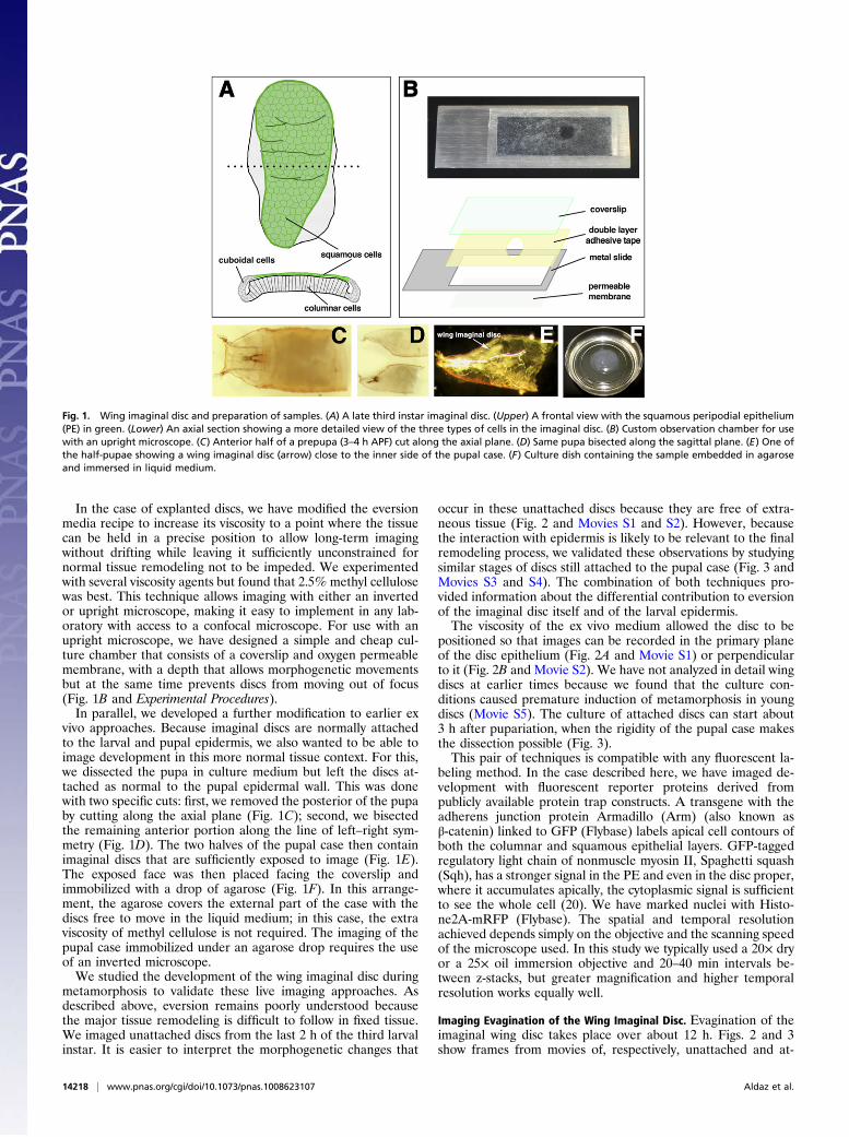
In the case of explanted discs, we have modified the eversionmedia recipe to increase its viscosity to a point where the tissuecan be held in a precise position to allow long-term imagingwithout drifting while leaving it sufficiently unconstrained fornormal tissue remodeling not to be impeded. We experimentedwith several viscosity agents but found that 2.5% methyl cellulosewas best. This technique allows imaging with either an invertedor upright microscope, making it easy to implement in any lab-oratory with access to a confocal microscope. For use with anupright microscope, we have designed a simple and cheap cul-ture chamber that consists of a coverslip and oxygen permeablemembrane, with a depth that allows morphogenetic movementsbut at the same time prevents discs from moving out of focus(Fig. 1B and Experimental Procedures).In parallel, we developed a further modification to earlier ex
vivo approaches. Because imaginal discs are normally attachedto the larval and pupal epidermis, we also wanted to be able toimage development in this more normal tissue context. For this,we dissected the pupa in culture medium but left the discs at-tached as normal to the pupal epidermal wall. This was donewith two specific cuts: first, we removed the posterior of the pupaby cutting along the axial plane (Fig. 1C); second, we bisectedthe remaining anterior portion along the line of left–right sym-metry (Fig. 1D). The two halves of the pupal case then containimaginal discs that are sufficiently exposed to image (Fig. 1E).The exposed face was then placed facing the coverslip andimmobilized with a drop of agarose (Fig. 1F). In this arrange-ment, the agarose covers the external part of the case with thediscs free to move in the liquid medium; in this case, the extraviscosity of methyl cellulose is not required. The imaging of thepupal case immobilized under an agarose drop requires the useof an inverted microscope.We studied the development of the wing imaginal disc during
metamorphosis to validate these live imaging approaches. Asdescribed above, eversion remains poorly understood becausethe major tissue remodeling is difficult to follow in fixed tissue.We imaged unattached discs from the last 2 h of the third larvalinstar. It is easier to interpret the morphogenetic changes that
occur in these unattached discs because they are free of extra-neous tissue (Fig. 2 and Movies S1 and S2). However, becausethe interaction with epidermis is likely to be relevant to the finalremodeling process, we validated these observations by studyingsimilar stages of discs still attached to the pupal case (Fig. 3 andMovies S3 and S4). The combination of both techniques pro-vided information about the differential contribution to eversionof the imaginal disc itself and of the larval epidermis.The viscosity of the ex vivo medium allowed the disc to be
positioned so that images can be recorded in the primary planeof the disc epithelium (Fig. 2A and Movie S1) or perpendicularto it (Fig. 2B and Movie S2). We have not analyzed in detail wingdiscs at earlier times because we found that the culture con-ditions caused premature induction of metamorphosis in youngdiscs (Movie S5). The culture of attached discs can start about3 h after pupariation, when the rigidity of the pupal case makesthe dissection possible (Fig. 3).This pair of techniques is compatible with any fluorescent la-
beling method. In the case described here, we have imaged de-velopment with fluorescent reporter proteins derived frompublicly available protein trap constructs. A transgene with theadherens junction protein Armadillo (Arm) (also known asβ-catenin) linked to GFP (Flybase) labels apical cell contours ofboth the columnar and squamous epithelial layers. GFP-taggedregulatory light chain of nonmuscle myosin II, Spaghetti squash(Sqh), has a stronger signal in the PE and even in the disc proper,where it accumulates apically, the cytoplasmic signal is sufficientto see the whole cell (20). We have marked nuclei with Histo-ne2A-mRFP (Flybase). The spatial and temporal resolutionachieved depends simply on the objective and the scanning speedof the microscope used. In this study we typically used a 20× dryor a 25× oil immersion objective and 20–40 min intervals be-tween z-stacks, but greater magnification and higher temporalresolution works equally well.
Imaging Evagination of the Wing Imaginal Disc. Evagination of theimaginal wing disc takes place over about 12 h. Figs. 2 and 3show frames from movies of, respectively, unattached and at-
Fig. 1. Wing imaginal disc and preparation of samples. (A) A late third instar imaginal disc. (Upper) A frontal view with the squamous peripodial epithelium(PE) in green. (Lower) An axial section showing a more detailed view of the three types of cells in the imaginal disc. (B) Custom observation chamber for usewith an upright microscope. (C) Anterior half of a prepupa (3–4 h APF) cut along the axial plane. (D) Same pupa bisected along the sagittal plane. (E) One ofthe half-pupae showing a wing imaginal disc (arrow) close to the inner side of the pupal case. (F) Culture dish containing the sample embedded in agaroseand immersed in liquid medium.
14218 | www.pnas.org/cgi/doi/10.1073/pnas.1008623107 Aldaz et al.

tached discs, and the processes we describe are shown di-agrammatically in Fig. 4 and Fig. S1. However, we encouragereaders to view the movies directly: it is very difficult to illustratedynamic 3D remodeling in still images, underscoring the needfor live imaging as a primary data source in these kinds of study.Combining the information obtained from more than 40 mov-
ies, we have divided the evagination process into two steps:folding and retraction. All of our analysis is derived from moviesof discs dissected at a similar stage and which were undamaged.Before folding starts, the presumptive wing is a relatively flat bi-layer epithelium (Figs. 2 A and B and 4A); the future dorsal andventral surfaces of the wing are adjacent to each other in the sameplane, separated by the wing margin. The wingmargin thenmovesso that the wing surfaces become apposed on opposite sides ofthe disc proper—the PE remains stretched over the disc proper asthese movements occur, expanding to cover the entire presumptivewing (Fig. 4 B and C). Once the wing margin reaches the borderof the disc, the disc folds up to 90° from its initial plane (Figs. 2 and4 A and C). Although the movement of the wing margin and ex-pansion of the PE are gradual processes, the folding happens quitefast. Fig. 4D, showing the attached disc, indicates how the foldingbrings the PE cells into close proximity with the larval epidermis (inred). The whole folding process can be seen in unattached cultureddiscs, but almost all of the attached discs already show some foldingby the time they are dissected (Fig. 3 and 4D). Furthermore, at-tached discs only fold to about 45° from their initial plane, pre-sumably because of constraints imposed by the associated pupalepidermis.The second step of evagination is peripodial retraction. This
step corresponds with eversion in vivo, the movement of the discthrough the pupal epidermis. In unattached discs, a hole opens at
the stalk region (Fig. 2B) and the PE rapidly moves over the DPfollowing a wave of retraction (Fig. 2 A and B and Movies S1 andS2). During this process the PE loses its epithelial morphology,and, by end of the retraction when the PE passes over the wingmargin, the cells are round and separated (Fig. 2A); in fact, mostof the squamous layer disintegrates, although some PE cells re-main attached to the thorax and appear mesenchymal. The re-traction is fast, being completed in 1 or 2 h (Fig. 2 A and B andMovies S1 and S2). After retraction, the disc expands, the foldsin the columnar epithelium disappear, and both thorax and wingregion expand. Particularly remarkable is the rapid expansion ofthe wing into an easily recognizable adult structure (Fig. 2 A andB). Importantly, both the final everted discs and the intermediatesteps are comparable to discs dissected from equivalent pupalstages and previous reports, suggesting that our explants re-semble the process in vivo.In the attached disc, retraction has different consequences,
owing to the substrate to which the disc is anchored. The contactbetween disc and epidermis starts at the stalk region and, asperipodial retraction progresses, contact extends from theproximal thorax to the most distal regions, the wing blade (Figs. 3and 4). The most distal tip of the wing never interacts with theepidermis. During the initial stages of retraction, clear separa-tion frequently occurs between the PE and the DP (Fig. 3B andMovie S4). It has previously been reported that the stalk cellsinvade the epidermis so that the opening of the stalk createsa hole through which the disc can emerge (15). Our observationsare consistent with this. We see that once the PE cells are closelyapposed to the pupal epidermis, there is a subsequent contrac-tion of the PE that corresponds to the retraction observed inmovies of unattached discs (Fig. 3B and Movie S4). We interpret
Fig. 2. Ex vivo imaginal disc eversion. (A) Frames from Movie S1 showing a prepupal imaginal disc during the different steps of the eversion process. The discis labeled with the Sqh-GFP transgene (green), and grunge-Gal4 driving UAS-mRFP (red); grunge-Gal4 is mainly expressed in peripodial cells but also in twobright spots in the region of the hinge of the columnar epithelium. This combination highlights the retraction process (white arrows mark the front of theretracting PE cells). The apoptotic bodies of the squamous cells accumulate at one side of the hinge region (orange arrow). The last images show the ex-pansion of the wing pouch and the formation of the precursors of the wing margin sensory organs (yellow arrowheads). (B) Frames from Movie S2 showinga prepupal imaginal disc mounted on its side showing the distribution of Sqh-GFP (green) and His2A-RFP (red). The first two frames show the folding of thewhole disc after the addition of the ecdysone. This lateral view allows observation of the opening of the hole in the stalk region and the start of retraction(white arrows).
Aldaz et al. PNAS | August 10, 2010 | vol. 107 | no. 32 | 14219
DEV
ELOPM
ENTA
LBIOLO
GY

this contraction to be responsible for driving the eversion of thedisc into the space between the larval epithelium and the outercuticle. We have observed that in the cases where, due to themounting process, the PE and the larval walls are not in prox-imity to each other, the disc does not evert properly, suggestingthat the interaction between them remains essential for eversion.Overall, the information coming from both culture systems, al-though showing some differences due to the constraints andlimitations of each approach, can be integrated into a coherentinterpretation of the dynamics of eversion in vivo.During retraction, PE cells transform from epithelial to a mass
of rounded cells, which appear to disintegrate. We used an ap-optotic marker, apoliner, which moves from plasma membraneto nucleus at the onset of apoptosis (21), to confirm that, indeed,a wave of programmed cell death sweeps across the PE, elimi-nating most, if not all, cells (Movie S6).
Further Applications of ex Vivo Culture and Imaging. We have fo-cusedonwing disc eversion as a proof of principle for disc culturingand imaging, but the use of viscous medium is also valuable forstudies of other developmental processes that occur in discs, in-cluding those that need to be followed at a cellular level with hightemporal resolution. For example, using a 100× objective, we areable to image at 10-min intervals on a spinning disc microscope(Fig. 5A and Movies S7 and S8); in fact, the timing can be muchfaster if a smaller field of view and shallower z-stack is specified.To illustrate the versatility of these approaches, we have imageda number of distinct developmental processes that we observedwhile studying eversion.Where appropriate genetically encoded markers exist, in-
tracellular events can be tracked. We show the reorganization ofintracellular myosin-II-rich fibers that occurs as cell shapeschange (Fig. 5A and Movie S7), but another powerful use wouldbe to follow signaling events in real time. An additional advan-tage of ex vivo culture is that it allows the simple application ofexogenous markers, compounds, and drugs. For example, fluo-
rescently labeled annexin-V (22) added to the medium providesa simple reporter of apoptosis without the need for a geneticmarker (Fig. 5B and Movie S9); we have also used FM lypophilicdyes as markers. Imaginal discs are particularly intensivelystudied models of cell proliferation, and this is also a readilyobserved phenomenon in culture, both at a whole tissue level
Fig. 4. Schematic representation of disc eversion. (A–C) Front view (Top in allpanels) and side view (Middle in all panels) of a third instar wing disc. Thesquamous cells of the PE are represented in green and the columnar (andcuboidal) epithelium in gray. Note that in reality there is no sharp distinctionbetween the squamous and columnar cells: a transition zone of cuboidal cellsseparates them. Here, we define the PE as the squamous cells alone. (Bottom)The image in each panel shows a side view of the disc, highlighting the dorsaland ventral compartments of the wing pouch of the imaginal disc and howthey move during eversion. The green dotted line illustrates the bending be-tween stages. (A) The third instar wing disc is flat, with ventral and dorsalcompartments in the same plane. (B) By late third instar, the squamous epi-thelium has expanded, and the dorsal and ventral compartments of the wingpouch become apposed (arrow). (C) At the prepupal stage the disc starts tobend. The apposition of the two wing surfaces is complete. The peripodial cellscover almost the complete apical surface of the columnar epithelium. (D)Representation of the disc attached to the pupal case. The cuticle and theepidermis are represented in red. The three steps show how the discapproaches the epidermis, the PE contracts, folding the whole disc, and thebasal part of the columnar epithelium protrudes (arrow).
Fig. 3. Interaction between the wing disc and the pupal epidermis. (A)Frames from Movie S3 a prepupal imaginal disc attached to the pupal case.The disc expresses Sqh-GFP (green) and grunge-Gal4 >UAS-RFP (red). The redlabel also marks the close opposed pupal case and epidermis (white arrow).In the first frames, the PE cells are weakly stained, but when the contractionstarts, intensity increases (orange arrows). At the same time the DP getscloser to the epidermis, folds, and protrudes its basal part (yellow arrows).(B) Frames from Movie S4, showing a His2A-mRFP (red), Sqh-GFP (green)wing disc. In this example, the separation between the disc proper and PE isvery clear (orange arrows). The yellow arrows highlight the squamous epi-thelium retracting. The columnar epithelium protrudes from inside.
14220 | www.pnas.org/cgi/doi/10.1073/pnas.1008623107 Aldaz et al.

(Fig. 5C and Movie S10) and, when appropriate markers areused, in individual cells (Fig. 5D and Movie S11). Wound healingis yet another process that can be imaged in ex vivo culture: Fig.5E and Movie S12 track the repair of a needle-stick injury toa wing disc, highlighting with GFP-tagged myosin II the re-organization of the actomyosin cytoskeleton. Finally, in this briefcatalog to emphasize the range of developmental processes thatcan be imaged using these culture conditions, we have followedthe patterned divisions and differentiation that leads to the de-velopment of sensory organs along the wing veins (Fig. 5F andMovie S13).
DiscussionDrosophila imaginal discs have provided great insight into mecha-nisms of development. Their growth and differentiation from simpleepithelial sheets, comprising a small number of cells, to complex 3Dorgans of thousands of cells, represents a model of many generaldevelopmental processes including growth, proliferation, differenti-ation, morphogenesis, programmed cell death, and epithelial reor-ganization. The extensive genetic techniques available for their studyinclude theuseofmitotic clones,Gal4/UAStargetedgeneexpression,and transposon mutagenesis. Here we introduce live imaging meth-ods that significantly extend the scope of imaginal discs as develop-mental models. To validate the use of live imaging to analyze mor-phogenesis in viscous medium, we have focused on metamorphosisas a model of epithelial remodeling and organogenesis—a processparticularly suited to live imaging—but the ability to track develop-ment in real time provides a potential resource for the analysis ofmost processes that occur in discs.Previous work describing metamorphosis of imaginal discs has
primarily relied on observing dissected, fixed tissue. This makes itdifficult to interpret the complex remodeling, does not allow hightemporal resolution of the sequence of events, and introducessignificant fixation artifacts (for example, the collapse onto thecolumnar epithelium of the adjacent squamous epithelium). Ourdescription of disc eversion agrees in many ways with previouslyreported data, which, combined with our complementary datafrom attached and unattached discs, give us confidence that the
culture system is faithfully reproducing in vivo development.Unsurprisingly, however, it has also revealed several events thathave not previously been reported, again emphasizing the extrainformation that is acquired by direct observation of living tissue.These include previously undescribed morphogenetic movementslike the 90° folding of the disc, the subsequent rapprochementwith the pupal epidermis, and the unexpected disintegration byapoptosis of most of the PE. Limited apoptosis in the stalk regionhas been described (15), but the coordinated death of the wholeperipodial layer has previously been unreported.Overall, the movements we describe here suggest that the
folding of the disc drives the approach of the whole disc to thepupal wall. It has previously been proposed that contraction ofthe PE is responsible for the forces needed during the eversion ofthe disc (13, 15). Our data support this and further suggest thatthe interaction between the PE and the pupal epidermis alsoparticipates in the mechanics of disc eversion.The particular features of the culture methods we have de-
veloped include the ability to follow disc development over longperiods (at least 24 h), viscous medium that supports discs in anyorientation and prevents drifting, the use of confocal imaging toreconstruct tissue development in 3D, and temporal and spatialresolution limited only by microscope technology and the size of thetissue under observation. Another major advantage is the simplicityof the technique: it does not rely on any unusual equipment orreagents. Finally, in addition to being applicable to all imaginaldiscs, viscous culture media could easily be adapted to facilitate liveimaging of other organ systems in Drosophila and other species.
MethodsEversion Media Composition. Shields and Shang M3 insectum medium fromSigma (S3652)was supplementedwith 2%FCSand0.5%penicillin-streptomycin(15140–122; Invitrogen). Ecdysone fromSigma, 20-hydroxyecdysoneH5142,wasstored in a stock solution of 500 μg/mL in 10% isopropanol at−20 °C, and addedat a final concentration of 0.1 μg/mL for unattached discs and 0.5 μg/mL for at-tacheddiscs. To culture unattacheddiscs,methyl-cellulose (M0387-100G; Sigma)was added at a concentration of 2.5% wt/vol. The methyl-cellulose was addedto themediawithecdysoneand stirreduntil dissolved, then leftovernightat4 °C
Fig. 5. Further applications of ex vivo culture. (A) Frames fromMovie S7 showing a wing disc labeled with Sqh-GFP. The red arrows point to the nuclei of twoperipodial cells. Using a 100× objective, intracellular structures like myosin II fibers can be tracked. (B) Frames fromMovie S9 showing a prepupal imaginal disclabeled with Sqh-GFP (green) and annexin-V-Cy3 (red), which was added to the medium after dissection, as a reporter of apoptosis during retraction of the PE(indicated by arrows). (C) Two frames from Movie S10 showing numerous dividing cells (several marked by arrows) in a wing disc labeled with Arm-GFP. (D)Frames from Movie S11 showing a wing disc labeled with His2A-mRFP (red) and Sqh-GFP (green). Yellow arrows indicated dividing cells in which the differentstages of mitosis can be tracked, both in the XY and XZ planes. (E) Initial (Upper) and final (Lower) frames, taken from Movie S12, showing the healing ofa wound (red arrow) in the notum of a wing disc labeled with Sqh-GFP. (F) Frames from Movie S13 showing a wing disc labeled with Arm-GFP and thedevelopment of two wing vein sensillae in the wing pouch (green and red arrows).
Aldaz et al. PNAS | August 10, 2010 | vol. 107 | no. 32 | 14221
DEV
ELOPM
ENTA
LBIOLO
GY

to eliminate bubbles; 1 mL aliquots were stored at −20 °C. The frozen mediaworks for up to 3 mo.
Fly Stocks and Genetics. Flies were grown under standard culture techniques.The following lines were used: sqh-GFP (20); arm-GFP, UAS-mRFP, His2A-mRFP1 (Flybase), UAS-E-cad-GFP (2), grunge-Gal4 (23), Ubx-Gal4LDN (24).Overexpression using the Gal4/UAS system (25) was performed at 25 °C.
Annexin-V Staining. We used the annexin-V-CY3 apoptosis kit from Biovision(catalog no. K103-25). Annexin-V was added to the media at a dilution 1/200.
Dissection and Mounting. Late third instar larvae were washed in sterile PBSand disinfected in 70% ethanol for 5 min. After rinsing in PBS, they weretransferred to eversion media, without methylcellulose, where the discs werecarefully dissected using forceps and needles. Wounded discs were discarded.Unharmed discs were placed in an observation chamber, when being imagedon an upright microscope, or in a 35-mm culture dish with glass bottom(Fluorodish FD35-100, World Precision Instruments, Sarasota, FL), when aninverted microscope was being used. After dissection, the media was re-placed with viscous media: approximately 30 μL in the observation chamberand 1 mL in culture dishes. The discs were then positioned carefully witha needle. When an observation chamber was used, it was covered with anoxygen semipermeable membrane, (catalog number 5793; YSI).
Attached discs were dissected in eversionmedia. The posterior tip of pupae3 h postpupariation was removed at the level of the spiracles with micro-dissection scissors. A second transverse cut in the medial region was thenmade, leaving approximately the anterior 50%of the pupa. Finally a third cutwas made longitudinally, leaving two mirror-image halves, each with a wingdisc attached to the pupal walls. Internal tissues obscuring the discs wereremoved carefully (Fig. 1 C–E). The dissected tissue was then placed ina culture dish, with the open surface facing the coverslip on the base (Fig.1).
A drop of 2% agarose, melted in PBS and allowed to cool until just before itset, was then placed over the cuticle. Once solid, approximately 2.5 mL offresh medium was added to the dish.
Observation Chamber Construction. The chamber was made with a doublelayer of double side tape (Sellotape acid free). The choice of the tape isimportant: some released adhesive into the medium, impairing the eversion.The two layers were perforated using a hole puncher, generating a hole ofapproximately 6 mm diameter. The tape was attached to a 22 × 50-mmcoverslip and trimmed to the same size. The coverslip was then attached toa metal slide with a cut-out panel. Finally, the chamber/hole was filled withmedium and the discs placed in it and then covered with the semipermeablemembrane as described.
Confocal Microscopy. We have used three different confocal systems: a ZeissLSM510 on an uprightmicroscope; a Zeiss LSM 710 on an invertedmicroscope;and a Perkin-Elmer spinning disc UltraVIEW ERS with an Orca ER CCD camera(Hamamatsu). Exact settings varied with experiment and system but werechosen to balance the need for a clear image with minimizing laser exposureto avoid tissue damage. Typically we took between 80 and 120 z sections at1-μm interval every 20 to 50 min. When imaging the whole discs, the intervalwas decreased to allow a full 3D reconstruction. The images were analyzedusing Adobe Photoshop CS2, Image J, and Volocity (Improvision) software.
ACKNOWLEDGMENTS. We thank Luis F. de Navas, Simon Bullock, and NickBarry for generously supplying stocks and support. We thank MatthewFreeman’s laboratory members, Thomas Lecuit, and Enrique Martín-Blancofor helpful discussions. Natalia Azpiazu and Antonio Baonza helped usgreatly with useful comments on the manuscript. This work was supportedin part by Marie Curie (to S.A. and L.M.E.) and European Molecular BiologyOrganization (to L.M.E.) fellowships.
1. Bertet C, Sulak L, Lecuit T (2004) Myosin-dependent junction remodelling controlsplanar cell intercalation and axis elongation. Nature 429:667–671.
2. Oda H, Tsukita S (1999) Dynamic features of adherens junctions during Drosophilaembryonic epithelial morphogenesis revealed by a Dalpha-catenin-GFP fusion pro-tein. Dev Genes Evol 209:218–225.
3. Wood W, et al. (2002) Wound healing recapitulates morphogenesis in Drosophilaembryos. Nat Cell Biol 4:907–912.
4. Richardson BE, Beckett K, Baylies MK (2008) Live imaging of Drosophila myoblastfusion. Methods Mol Biol 475:263–274.
5. Ninov N, Martín-Blanco E (2007) Live imaging of epidermal morphogenesis during thedevelopment of the adult abdominal epidermis of Drosophila. Nat Protoc 2:3074–3080.
6. Bischoff M, Cseresnyés Z (2009) Cell rearrangements, cell divisions and cell death ina migrating epithelial sheet in the abdomen of Drosophila. Development 136:2403–2411.
7. Escudero LM, Bischoff M, Freeman M (2007) Myosin II regulates complex cellulararrangement and epithelial architecture in Drosophila. Dev Cell 13:717–729.
8. Classen AK, Anderson KI, Marois E, Eaton S (2005) Hexagonal packing of Drosophilawing epithelial cells by the planar cell polarity pathway. Dev Cell 9:805–817.
9. Aigouy B, Van de Bor V, Boeglin M, Giangrande A (2004) Time-lapse and cell ablationreveal the role of cell interactions in fly glia migration and proliferation. Development131:5127–5138.
10. Bianco A, et al. (2007) Two distinct modes of guidance signalling during collectivemigration of border cells. Nature 448:362–365.
11. Ayaz D, et al. (2008) Axonal injury and regeneration in the adult brain of Drosophila.J Neurosci 28:6010–6021.
12. Fristom D, Fristom JW (1975) The mechanism of evagination of imaginal discs ofDrosophila melanogaster. 1. General considerations. Dev Biol 43:1–23.
13. Gibson MC, Schubiger G (2001) Drosophila peripodial cells, more than meets the eye?Bioessays 23:691–697.
14. Usui K, Simpson P (2000) Cellular basis of the dynamic behavior of the imaginalthoracic discs during Drosophila metamorphosis. Dev Biol 225:13–25.
15. Pastor-Pareja JC, Grawe F, Martín-Blanco E, García-Bellido A (2004) Invasive cellbehavior during Drosophila imaginal disc eversion is mediated by the JNK signalingcascade. Dev Cell 7:387–399.
16. Vinegoni C, Pitsouli C, Razansky D, Perrimon N, Ntziachristos V (2008) In vivo imagingof Drosophila melanogaster pupae with mesoscopic fluorescence tomography. NatMethods 5:45–47.
17. Milner MJ (1977) The eversion and differentiation of Drosophila melanogaster legand wing imaginal discs cultured in vitro with an optimal concentration of beta-ecdysone. J Embryol Exp Morphol 37:105–117.
18. Milner MJ, Sang JH (1974) Relative activities of alpha-ecdysone and beta-ecdysone forthe differentiation in vitro of Drosophila melanogaster imaginal discs. Cell 3:141–143.
19. Milner MJ, Muir J (1987) The cell biology of Drosophila wing metamorphosis in vitro.Rouxs Arch Dev Biol 196:191–201.
20. Royou A, Sullivan W, Karess R (2002) Cortical recruitment of nonmuscle myosin II inearly syncytial Drosophila embryos: Its role in nuclear axial expansion and itsregulation by Cdc2 activity. J Cell Biol 158:127–137.
21. Bardet PL, et al. (2008) A fluorescent reporter of caspase activity for live imaging. ProcNatl Acad Sci USA 105:13901–13905.
22. Martin SJ, et al. (1995) Early redistribution of plasma membrane phosphatidylserine isa general feature of apoptosis regardless of the initiating stimulus: Inhibition byoverexpression of Bcl-2 and Abl. J Exp Med 182:1545–1556.
23. Gibson MC, Lehman DA, Schubiger G (2002) Lumenal transmission of decapentaplegicin Drosophila imaginal discs. Dev Cell 3:451–460.
24. de Navas L, Foronda D, Suzanne M, Sánchez-Herrero E (2006) A simple and efficientmethod to identify replacements of P-lacZ by P-Gal4 lines allows obtaining Gal4insertions in the bithorax complex of Drosophila. Mech Dev 123:860–867.
25. Brand AH, Perrimon N (1993) Targeted gene expression as a means of altering cellfates and generating dominant phenotypes. Development 118:401–415.
14222 | www.pnas.org/cgi/doi/10.1073/pnas.1008623107 Aldaz et al.