Embed Size (px)
Citation preview

en 2 www.dov237.com en
List of Contents
Alphabetic Index 3 Introduction 5 Safety Precautions 6 Contraindications to Applying the Quantum Therapy Method 6 Design and Operation of the Device 7 Methodical Recommendations 10 1. Traumatology 11 2. Skin Diseases 36 3. ENT Diseases 40 4. Dentistry 48 5. Gynecology 51 6. General Medicine 54 Basic Principles of Affecting the Organism Tissues and Cells by Laser Radiation and Magnetic Fields 68 Literature 71
Appendix A: Short Dictionary of Physical Terms 72

en 3 www.dov237.com en
А
B
C
E
D
F G
Alphabetic Index
H
M
N
O
Acute and chronic bronchitis 54 Acute and chronic rhinitis 42 Acute pneumonia 54 Alveolysis 50 Arthritis, arthrosis of phalanges of hand and foot 18 Arthritis, arthrosis 11 Arthritis, gonarthrosis 12 Arthritis, hip joint arthrosis 14 Arthritis, wrist joint arthrosis 16 Axilla glandular inflammation 28 Bone fractures 26 Bronchial allergy 56 Calcaneal spur 20 Cervical erosion 51 Cholecystitis 62 Concussions 28 Decubitus 31 Dermatitis 36 Disruption and hydrops of mamilla 52 Dyskinesia of bile passages 62 Eczema 36 Endodontitis 48 Furunculosis 37 Gingivitis 50 Herpes 38 Humeroscapular parasynovitis 32 Muscles stretching treatment 34 Myositis 34 Neurodynia 24 Obliterating atherosclerosis 66 Obliterating endarteritis 66 Open wounds 30 Osteochondrosis 22 Otitis 46

en 4 www.dov237.com en
R
W
T
P
S
V
Pancreatitis 64 Peptic ulcer disease 60 Peritenditis 34 Phlebeurysm 66 Radiculitis 22 Sinuitis 44 Tonsillitis 40 Trophic ulcer 31 Ventricle peptic ulcer 58 Verrucas 37 Wrinkles 39

en 5 www.dov237.com en
Introduction
We are entering the 21st century being equipped by a substantial inventory of various medical devices and drug preparations, however, many of them are not al-ways of great help. Fortunately, the medical science does not stand still and offers new treatment methods. One of such methods is magnetic-laser therapy. This ther-apy is broadly used in the medical practice, and becomes more popular day after day.
Nowadays, many magnetic-laser therapy devices exist, and the "Vityas" Quantum Therapy Device, manufactured by the "Vityas" Republic's Unitary Pro-duction Enterprise, is a bright representative thereof. The Device generates several radiation types at the same time (laser pulse-modulated radiation of infrared wave-range, continuous laser radiation of the visible red light and radiation of permanent magnetic field), each of them being curative, and in combination, they provide a more expressed curative effect.
The "Vityas" Device has successfully underwent its clinical tests at the Scientific-Research Institute of Traumatology and Orthopedics, the Dermatovene-rologic Dispensary of Minsk, the Physiotherapy Division of the Belarusian Medi-cal Academy of Post-Diploma Training of the Ministry pf Public Health of the Re-public of Belarus, and has been recommended into medical practice because of its efficiency and operating safety.
The affection of the "Vityas" Device may be applied both independently, and combined with medication, which in a number of cases allows to drop the du-ration and volume of medication. Treatment with the "Vityas" Device radiation does not prevent application of any other traditional and non-traditional treatment methodologies.
Application of the "Vityas" Device is notable for absence of pain, operating safety, asepticity, a possibility to apply treatment in the doses, variable by duration and by radiation power. The advantageous features of the "Vityas" Device among other devices of its class are its reliability, compactness, ease and comfort of opera-tion, which enable to use it both at medicinal institutions, and at home.
The methods of using the quantum therapy provided in the present Methodi-cal Guide are broadly known and have been applied in clinical practice for many years. The Guide is built on the principles of clearness, simplicity, efficiency and "make no harm".

en 6 www.dov237.com en
Safety Precautions 1. Persons not younger than 18 years, who have studied the present Certificate
and the "Methodical Manual on Application of the "Vityas" Device", are allowed to work with the Device.
2. Start the radiation mode only after placement of the radiator onto the point (zone) of affection.
3. The aim of the treatment method, the therapeutic dozes, and the control of the results of treatment, shall be made by the doctor on laser therapy, or by the doctor-specialist according to the "Methodical Manual on Application of the "Vityas" Device".
IT IS FORBIDDEN: - To switch on the Device at a faulty power unit, damage of insulation of the
cord and the body; - To disassemble the Device and connect to the mains in the disassembled con-
dition; - To leave the switched on Device without supervision; - To direct the focused direct or reflected laser radiation into one's eyes; - To effect the area of the heart projection with the radiation of the Device; - To give the Device to children. Attention! Never allow any direct or reflected affection of laser
radiation on the organs of vision
Contraindications to Applying the Quantum Therapy Method
1. Pregnancy 2. Convulsive state 3. Mental disorders, and diseases at the background of the psycho-emotional
arousal 4. Agnogenic febrile state 5. Renal, hepatic, blood-circulation and respiratory insufficiencies 6. Heavy endocrine pathologies, for example, achrestic diabetes mellitus, etc. 7. Heavy blood diseases 8. Oncology 9. Vesical calcification and cholelithiasis (to treat under physician's observation)

en 7 www.dov237.com en
Design of the Device and Operation а) Design of the Device
1) Take the Device out of the packing, and without pressing any control buttons connect the power unit (1) of the Device into the mains 220 V, 50 Hz, and: • the Device will automatically pass through its operation self-checking mode; • the operation self-checking mode lasts for 5 seconds and terminates with indication of
the "mode number" accompanied by a sound signal. For example: O2
2) By buttons (3) or (4) set the required mode number of the Device af-fection, selected from the Section "Methodological Recommendations" for a particular disease. For example: O3 3) The Device is ready for operation.
Notes: 1. In case of a failure of the Device, the digital indicator will give a failure mes-sage. A failed Device is subject to repairs.
2. At switching the Device on with the control buttons depressed, the check-up modes are set, which may be quitted by a repeated switching on (subpoint 1).
b) Preparation to Operation
START STOP
Figure 1.
The general view of the Device is given in Fig. 1. The control buttons (3, 4, 5), indicators (6, 7), and laser radiators are installed in a single body (8). The power unit (1) is connected by the connecting cord (2). The bodies of the Device and the power unit are made from high impact plastic.
The face panel of the Device has controls for se-lecting the modes: buttons (3), (4), START/STOP (5), and indicators: digital (6) and single (7).
The opposite side (relative the face one) has the radiator. It consists of a constant magnet and a quantum radiator built on laser diodes of infrared and red spectrum of radiation. The radiation modes are listed in Table (page 9).
1- power unit 230/220 V, 50/60 Hz 2- connecting cord 3- button of reducing the number of the selected mode ( ) 4- button of increasing the number of the selected mode ( )5- button START/STOP 6- digital indicator 7- single indicator 8- body of the Device

en 8 www.dov237.com en
c) Operating the Device
1) Study the Methodical Recommendations on treating a particular disease.2) Take a comfortable posture. Place the radiator of the Device above the firstaffection point (according to the methodology of treating a particular disease),having attached in to the skin of the body or at a small distance from it. Start themode of laser radiation. 3) The start of the radiation mode is made by pressing the button START (5).
At this: a sound signal is given, the lasers and the unit indicator are switched on, thedigital indicator passes over into the timer mode, which indicates the remaining time of af-fecting the particular point. For example: 2 L, where: "2" means two minutes, and "L"means a symbol simulating the hands of the clock by blinking. 4) Go on affecting the selected point until the digital indicator reaches the modeof Automatic pause. For example: п. 2, where п. means the pause, and 2 is the numberof the next affection point, where the radiator should be put to continue the affection mode. At this: a sound signal is given, the unit indicator blinks, the quantum radiator of laser dio-des is switched off. The duration of the automatic pause is 10 s. 5) To continue the procedure, place the radiator of the Device during the pauseover the next affection point. The automatic pause will interrupt automatically,or it may be forced interrupted by a short-time pressing the button START. 6) Automatic mode stop is made by the software after the end of the affectiononto the last (tenth). At this: a sound signal is given, the unit indicator goes out, the laser radiators are switchedoff, and the digital indicator shows the number of the mode at which the affection has takenplace. 7) A forced pause is ensured by a short-time depressing of the STOP buttonduring the affection (at radiation and timer counting). It is used to stop the radia-tion mode for a long time. The sign of the pause is indicated and the number ofthe point, at which a stop has been made.
The start to continue the node is made by a repeated short-time depressing ofthe START button.
8) A forced stop of the mode is achieved by pressing the STOP button andkeeping it depressed for 3 seconds.
d) Switching the Device Off 1) Switch the Device off by disconnecting the power unit (1) from the mains
230/220 V, 50/60 Hz.

en 9 www.dov237.com en
Mod
e N
o.
Type
of l
aser
ra-
diat
or
Type
of r
adia
-tio
n (d
urin
g af
-fe
ctio
n)
Tim
e of
aff
ec-
tion
on a
poi
nt
(bot
h la
sers
are
O
N)
Tota
l aff
ectio
n tim
e pe
r one
pr
oced
ure
Tota
l ene
rgy
of ra
dia-
tion
(aff
ectio
n) p
er
one
proc
edur
e (1
0 c y
cles
)
Ener
gy o
f rad
ia-
tion
(a
ffect
ion
on a
po
int,)
Char
acte
rist
ics
of m
odes
of r
adia
tion
of t
he "V
itya
s" d
evic
e
Not
es:
1 Th
e tim
e of
aut
omat
ic p
ause
for m
ovin
g th
e de
vice
to th
e ne
xt a
ffec
tion
poin
t is 1
0 s.
2 A
cyc
le in
clud
es a
n af
fect
ion
mod
e an
d a
paus
e of
10
s.
3
All
the
mod
es h
ave
the
tota
l pow
er o
f las
er ra
diat
ion
of 1
0 m
W a
t the
spot
are
a at
the
outp
ut o
f rad
iato
r of a
bout
1 c
m2 .
4 Th
e le
vel o
f mag
netic
fiel
d, c
reat
ed b
y th
e pe
rman
ent m
agne
t, m
akes
from
5 to
50
mTl
.
O1
12.0
J 20
minu
tes
O3
27.0
J 45
minu
tes
O2
18.0
J 30
minu
tes
1.2 J
2.7 J
1.8 J
red λ =
620 –
700 n
m
infra
red
λ =
810 –
880 n
m
red λ =
620 –
700 n
m
infra
red
λ =
810 –
880 n
m
red λ =
620 –
700 n
m
infra
red
λ =
810 –
880 n
m
conti
nuou
s 12
,500
Hz
conti
nuou
s 12
,500
Hz
conti
nuou
s 12
,500
Hz
2 minu
tes
4.5
minu
tes
3 minu
tes

en 10 www.dov237.com en
Recommended Practice GENERAL RULES
Be sure to consult your physician on application of the Device for treating your disease.
Application of the Device is allowed only after establishing an exact diagno-sis by the physician.
It is allowed to use the Device in combination with the traditional medication. The Laser Device may be used with a concentrating orifice for irradiating
acupuncture points. In such version, the Device may be used by the specialists pro-ficient in acupuncture methodology.
After each affection session, a 15-20 minute break is recommended. The op-timum way is to hold laser procedures at about one and the same time of the day (in home conditions – before your rest hours).
In parallel with the quantum therapy, we recommend using vitamins and mi-croelements, the deficit of which is notable for this locality or particular season. It is recommended to have nutrition abundant in vitamins and microelements.
Prior and after application of the Device, disinfect the body of the radiator by a special solution (3 % of hydrogen peroxide, or 0.05 % of chlorhexidine, or 1 % dioxydine). Never let the liquid inside the Device and onto the glass of laser dio-des. Otherwise, the Device will need drying before usage, and the glasses will need cleaning with a cloth or cotton plug, moistened in alcohol solution.
In case of doubt, consult your physician or laser therapist.
CHOOSING THE AFFECTION MODE 1 Based on the disease diagnosis, concluded by the physician, find it in the
list of the Application Guide. 2 By the physician's recommendations and the algorithm provided, define the
"Device Affection Mode" with account of the "Disease Forms", "Course of Dis-ease" and "Patient's Age".
3 According to the Figures and the offered affection algorithm, make the treatment procedures to the indicated points (areas).
4 To affect the points of the back, which are hard to access, invite medical personnel or relatives.
The affection point (area) is a place on the patient's body subject to affec-tion of the Device.
DISEASES AND CLASSIFICATION are given below in the order of sections as used in the medical science.
Physical terms are given in "Appendix A".

en 11 www.dov237.com en
Disease Form
Course of Disease
Patient's Age
Mild Course
Mild Course
Device Affection Mode
For All Ages
O1
For All Ages
O2 O1 O1 O3 O1
From5 to 75
Under5, af-ter 75
From 5 to 75
Under5, af-ter 75
Heavy Course
Heavy Course
Acute Phase Chronic Phase
1.1 ARTHRITIS, ARTHROSIS
Apply radiation around the joint (by a circle), while orienting the laser spot (area) across or along the movement direction (Figure 1.1).
In the area of maximum swelling or pain, the irradiated areas should be 2 cm apart from each other. In the periphery – up to 4 cm apart from each other. No more than 10 areas are allowed per one irradiation session.
It is recommended to conduct a daily course during 3 weeks.
1 TRAUMATOLOGY
Figure 1.1

en 12 www.dov237.com en
Disease Form
Course of Disease
Patient's Age
Mild Course
Mild Course
Device Affection Mode
For All Ages
O1
For All Ages
O2 O1 O1 O3 O1
From5 to 75
Under5, af-ter 75
From 5 to 75
Under5, af-ter 75
Heavy Course
Heavy Course
Acute Phase Chronic Phase
1.2 ARTHRITIS, GONARTHROSIS
Figure 1.2
1
3
2
4
5
6
7
810
9
It is recommended to conduct a daily course during 20 days, as it is shown in Figure 1.2.It is recommended to clean the affection area with 100% dimexid solution 5 minutes be-fore the procedure.

en 13 www.dov237.com en
1 Affect the point until the indica-tor shows п 2. Proceed to the next point.
Number of affection point
(Fig. 1.2)
Affection point and area
Explanation
The affection point is above the knee-cap
2 Affect the point until the indica-tor shows п 3. Proceed to the next point.
Affect the point until the indica-tor shows п 4. Proceed to the next point.
Affect the point until the indica-tor shows п 5. Proceed to the next point.
Affect the point until the indica-tor shows п 6. Proceed to the next point.
Affect the point until the indica-tor shows п 7. Proceed to the next point.
Affect t the point until the indi-cator shows п 8. Proceed to the next point.
Affect the point until the indica-tor shows п 9. Proceed to the next point..
Affect the point until the indica-tor shows п п. Proceed to the next point.
Affect the point until the indica-tor shows the mode numbers se-lected in the beginning of the procedure.
3
4
5
9
8
7
6
The affection point is un-der the knee-cap
The affection point is un-der the knee-cap close to point 3
The affection point is above the knee-cap close to point 1
The affection point is to the left of the knee-cap
The affection point is to the right of the knee-cap close to point 5
The affection point is to the right of the knee-cap
The affection point is to the right of the knee-cap close to point 7
The affection point is in the poples
The affection point is in the poples close to point 91O

en 14 www.dov237.com en
Disease Form
Course of Disease
Patient's Age
Mild Course
Mild Course
Device Affection Mode
For All Ages
O1
For All Ages
O2 O1 O1 O3
From5 to 75
Under5, af-ter 75
From 5 to 75
Heavy Course
Heavy Course
Acute Phase Chronic Phase
1.3 ARTHRITIS, HIP JOINT ARTHROSIS
It is recommended to conduct a daily course during 20 days as it is shown in Figure 1.3. It is recommended to clean the affection area with 100% dimexid solution 5 minutes be-fore the procedure.
For the "03" mode, it is allowed to split the affection procedure into two-three sessions dur-ing the day.
Figure 1.3
13
24 5
6 78
9

en 15 www.dov237.com en
1 Affect the point until the indica-tor shows п 2. Proceed to the next point.
Number of affection point
(Fig. 1.3)
Affection point and area
Explanation
The affection point is inthe front in the center ofthe hip joint
2 Affect the point until the indica-tor shows п 3. Proceed to the next point.
Affect the point until the indica-tor shows п 4. Proceed to the next point.
Affect the point until the indica-tor shows п 5. Proceed to the next point..
Affect the point until the indica-tor shows п 6. Proceed to the next point.
Affect the point until the indica-tor shows п 7. Proceed to the next point.
Affect the point until the indica-tor shows п 8. Proceed to the next point.
Affect the point until the indica-tor shows п 9. Proceed to the next point.
Affect the point until the indica-tor shows п п.
3
4
5
9
8
7
6
The affection point is 3 cm below point 1
The affection point is sideways in the center of the hip joint
The affection point is 3 cm above point 1
The affection point is 3 cm above point 4
The affection point is 3 cm below point 4
The affection point is in the center of the buttock
The affection point is 3 cm above point 7
The affection point is 3 cm below point 7
END OF PROCEDURE Force the mode to stop by pressing STOP button
and keep it depressed for at least 3 seconds.

en 16 www.dov237.com en
Disease Form
Course of Disease
Patient's Age
Mild Course
Mild Course
Device Affection Mode
For All Ages
O1
For All Ages
O2 O1 O1 O3 O1
From5 to 75
Under5, af-ter 75
From 5 to 75
U d
Under5, af-ter 75
Heavy Course
Heavy Course
Acute Phase Chronic Phase
1.4 ARTHRITIS, RADIAL WRIST JOINT ARTHROSIS
It is recommended to conduct a daily course during 20 days as it is shown in Figure 1.4. It is recommended to clean the affection area with 100% dimexid solution 5 minutes be-fore the procedure.
Figure 1.4
1
3 2
4
2
3

en 17 www.dov237.com en
1 Affect the point until the indica-tor shows п 2. Proceed to the next point.
Number of affection point
(Fig. 1.4)
Affection point and area
Explanation
The affection point is on the anterior surface of the radial joint
2 Affect the point until the indica-tor shows п 3. Proceed to the next point.
Affect the point until the indica-tor shows п 4. Proceed to the next point.
Affect the point until the indica-tor shows п 5.
3
4
The affection point is on the lateral surface of the radial joint
The affection point is on the back surface of the radial joint
The affection point is on the lateral surface of the radial joint
END OF PROCEDURE Force the mode to stop by pressing STOP button
and keep it depressed for at least 3 seconds.

en 18 www.dov237.com en
Disease Form
Course of Disease
Patient's Age
Mild Course
Mild Course
Device Affection Mode
For All Ages
O1
For All Ages
O2 O1 O1 O3 O1
From5 to 75
Under5, af-ter 75
From 5 to 75
Under5, af-ter 75
Heavy Course
Heavy Course
Acute Phase Chronic Phase
1.5 ARTHRITIS, ARTHROSIS OF PHALANGES
OF HAND AND FOOT
It is recommended to conduct a daily course during 20 days as it is shown in Figure 1.5. Repeat the courses after 2-3 weeks for 2-3 times during aggravation season.
Figure 1.5 3
4
5
6
7
1
2
3
1
4 2
5 7
6

en 19 www.dov237.com en
1 Affect the point until the indica-tor shows п 2. Proceed to the next point.
Number of affection point
(Fig. 1.5)
Affection point and area
Explanation
The affection point is in the canter of the joint si-deway
2 Affect the point until the indica-tor shows п 3. Proceed to the next point.
Affect the point until the indica-tor shows п 4. Proceed to the next point.
Affect the point until the indica-tor shows п 5. Proceed to the next point.
Affect the point until the indica-tor shows п 6. Proceed to the next point.
Affect the point until the indica-tor shows п 7. Proceed to the next point.
Affect the point until the indica-tor shows п 8. Proceed to the next point..
3
4
5
7
6
The affection point is in the center of the joint on the lateral internal side
The affection point is in the center of the joint from inside
The affection point is in the center of the joint from the back side
The affection point is 1 cm below point 4, on the lateral side
The affection point is 1 cm below point 4
The affection point is be-low the center of the joint on the left side of the fin-
END OF PROCEDURE Force the mode to stop by pressing STOP button
and keep it depressed for at least 3 seconds.
en 20 www.dov237.com en
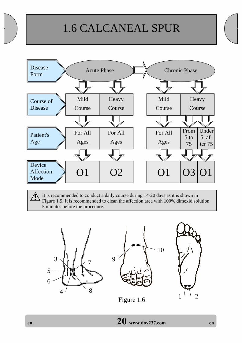
It is recommended to conduct a daily course during 14-20 days as it is shown in Figure 1.5. It is recommended to clean the affection area with 100% dimexid solution 5 minutes before the procedure.
Disease Form
Course of Disease
Patient's Age
Mild Course
Mild Course
Device Affection Mode
For All Ages
O1
For All Ages
O1 O1
From 5 to 75
Under5, af-ter 75
Heavy Course
Heavy Course
Acute Phase Chronic Phase
1.6 CALCANEAL SPUR
For All Ages
O2 O3
Figure 1.6 1
109
42
3
56
7
8

en 21 www.dov237.com en
1 Affect the point until the indica-tor shows п 2. Proceed to the next point.
Number of affection point
(Fig. 1.6)
Affection point and area
Explanation
The affection point is on the plantar side of the heel bone
2 Affect the point until the indica-tor shows п 3. Proceed to the next point.
Affect the point until the indica-tor shows п 4. Proceed to the next point.
Affect the point until the indica-tor shows п 5. Proceed to the next point.
Affect the point until the indica-tor shows п 6. Proceed to the next point.
Affect the point until the indica-tor shows п 7. Proceed to the next point.
Affect the point until the indica-tor shows п 8. Proceed to the next point.
Affect the point until the indica-tor shows п 9. Proceed to the next point.
Affect the point until the indica-tor shows п п. Proceed to the next point.
Affect the point until the indica-tor the mode numbers selected in the beginning of the proce-dure.
3
4
5
9
8
7
6
The affection point is on the Achilles tendon
The affection point is 1 cm below point 3
The affection point is close to point 1
The affection point is on the left side of the Achilles tendon
The affection point is 1 cmbelow point 5
The affection point is on the right side of the Achilles tendon
The affection point is 1 cmbelow point 7
The affection point is in the foot rise
The affection point is close to point 9 1O

en 22 www.dov237.com en
Disease Form
Course of Disease
Patient's Age
Mild Course
Mild Course
Device Affection Mode
For All Ages
O1
For All Ages
O2 O1 O1 O3 O1
From5 to 75
Under5, af-ter 75
From 5 to 75
Under5, af-ter 75
Heavy Course
Heavy Course
Acute Phase Chronic Phase
1.7 OSTEOCHONDROSIS OF BACK BONE, RADICULITIS
Figure 1.7
1
6 5
4
2 3
7 8
9 10
Area of maximal pain
It is recommended to conduct a daily course during 30 days as it is shown in Figure 1.7.
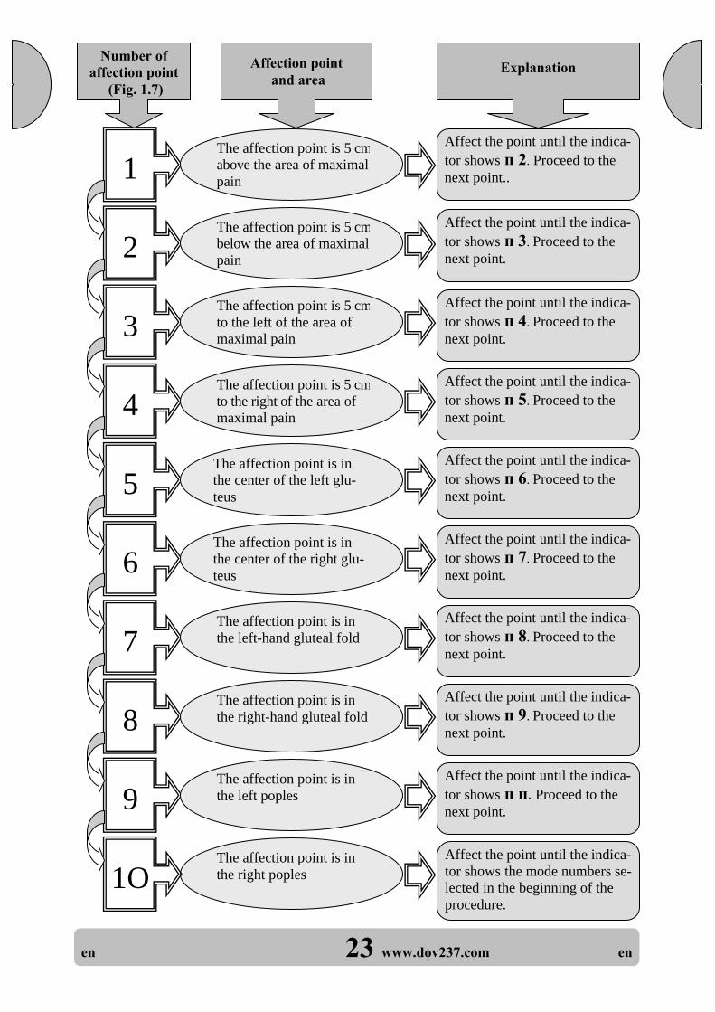
en 23 www.dov237.com en
1 Affect the point until the indica-tor shows п 2. Proceed to the next point..
Number of affection point
(Fig. 1.7)
Affection point and area
Explanation
The affection point is 5 cmabove the area of maximal pain
2 Affect the point until the indica-tor shows п 3. Proceed to the next point.
Affect the point until the indica-tor shows п 4. Proceed to the next point.
Affect the point until the indica-tor shows п 5. Proceed to the next point.
Affect the point until the indica-tor shows п 6. Proceed to the next point.
Affect the point until the indica-tor shows п 7. Proceed to the next point.
Affect the point until the indica-tor shows п 8. Proceed to the next point.
Affect the point until the indica-tor shows п 9. Proceed to the next point.
Affect the point until the indica-tor shows п п. Proceed to the next point.
Affect the point until the indica-tor shows the mode numbers se-lected in the beginning of the procedure.
3
4
5
9
8
7
6
The affection point is 5 cmto the left of the area of maximal pain
The affection point is 5 cmto the right of the area of maximal pain
The affection point is 5 cmbelow the area of maximal pain
The affection point is in the center of the left glu-teus
The affection point is in the center of the right glu-teus
The affection point is in the left-hand gluteal fold
The affection point is in the right-hand gluteal fold
The affection point is in the left poples
The affection point is in the right poples 1O

en 24 www.dov237.com en
Disease Form
Course of Disease
Patient's Age
Mild Course
Mild Course
Device Affection Mode
For All Ages
O1
For All Ages
O2 O1 O1 O3 O1
From5 to 75
Under5, af-ter 75
From 5 to 75
Under5, af-ter 75
Heavy Course
Heavy Course
Acute Phase Chronic Phase
1.8 NEURODYNIA
Prior to switching on, press the Device tight to the skin. At the moment of driving the Device away from the face, it is better to close the eyes and, if necessary, interrupt the procedure.
Any damage to the eyes by the laser of this energy is excluded, but still it is not recommended to admit any direct getting of the laser radiation into the eyes. It is recommended to conduct a daily course during 2-3 weeks as it is shown in
Figure 1.8. (Points 2 and 4 are located same as 1 and 3, but on the other side). At the paresis of the left-hand nerve, the radiation is conducted in mode 3.
Figure 1.8
7
5
4
2
39
6
8
10
1

en 25 www.dov237.com en
1 Affect the point until the indica-tor shows п 2. Proceed to the next point.
Number of affection point
(Fig. 1.8)
Affection point and area
Explanation
The affection point is in the output point of the tri-facial nerve to the left
2 Affect the point until the indica-tor shows п 3. Proceed to the next point.
Affect the point until the indica-tor shows п 4. Proceed to the next point.
Affect the point until the indica-tor shows п 5. Proceed to the next point.
Affect the point until the indica-tor shows п 6. Proceed to the next point.
Affect the point until the indica-tor shows п 7. Proceed to the next point.
Affect the point until the indica-tor shows п 8. Proceed to the next point.
Affect the point until the indica-tor shows п 9. Proceed to the next point.
Affect the point until the indica-tor shows п п. Proceed to the next point.
Affect the point until the indica-tor shows the mode numbers se-lected in the beginning of the procedure.
3
4
5
9
8
7
6
The affection point is in the output point of the fa-cial nerve to the left
The affection point is in the output point of the fa-cial nerve to the left
The affection point is in the output point of the tri-facial nerve to the right
The affection point is 2 cm above the eye
The affection point is 2 cm to the left of the eye
The affection point is 2 cm below the eye
The affection point is 2 cm above the eye
The affection point is 2 cm below the eye
The affection point is 2 cm to the right of the eye 1O

en 26 www.dov237.com en
It is recommended to conduct the procedures, as shown in Figure 1.9, till the pains dis-appear 1.9:
- twice a day at affecting through the dressing; - three times a day, when affecting through the plaster.
For modes "02", "03", it is allowed to split the procedure into two-three sessions during the day.
1.9 BONE FRACTURES Disease Form
Course of Disease
Patient's Age
After splinting (plastering)
Till the pains disappear
Device Affection Mode
For All Ages
O3 O2 O1
From 5 to 75
Under5, af-ter 75
Three weeks after remov-ing plaster
Acute Phase After Removing
Plaster
For All Ages
O2
Figure 1.9
4
2 7
61231
3
54
7
8
568

en 27 www.dov237.com en
1 Affect the point until the indica-tor shows п 2. Proceed to the next point.
Number of affection point
(Fig. 1.9)
Affection point and area
Explanation
The affection point is in the seat of fracture
2 Affect the point until the indica-tor shows п 3. Proceed to the next point.
Affect the point until the indica-tor shows п 4. Proceed to the next point.
Affect the point until the indica-tor shows п 5. Proceed to the next point.
Affect the point until the indica-tor shows п 6. Proceed to the next point.
Affect the point until the indica-tor shows п 7. Proceed to the next point.
Affect the point until the indica-tor shows п 8. Proceed to the next point.
Affect the point until the indica-tor shows п 9. Proceed to the next point.
3
4
5
8
7
6
The affection point is be-low the seat of fracture
The affection point is close to the seat of frac-ture
The affection point is close to the seat of frac-ture (close to point 1)
The affection point is above the seat of fracture
The affection point is in the seat of fracture on the back side of the extremity
The affection point is above point 6
The affection point is be-low point 6
END OF PROCEDURE Force the mode to stop by pressing STOP button
and keep it depressed for at least 3 seconds.

en 28 www.dov237.com en
Disease Form
Patient's Age
Device Affection Mode
For All Ages
O1
Without purulence With acute pain
and edema
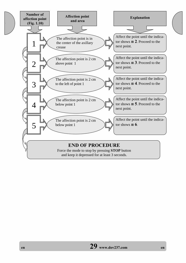
1.10 CONCUSSIONS, HYDRADENITES
It is recommended to conduct daily courses, as shown in Figure 1.10, until the inflam-mation repairs.
Treat concussions same as hydradenites.
For All Ages
O2
Figure 1.10
15
4
2
3
Hydradenitis
en 29 www.dov237.com en
1 Affect the point until the indica-tor shows п 2. Proceed to the next point.
Number of affection point
(Fig. 1.10)
Affection point and area
Explanation
The affection point is in the center of the axillary crease
2 Affect the point until the indica-tor shows п 3. Proceed to the next point.
Affect the point until the indica-tor shows п 4. Proceed to the next point.
Affect the point until the indica-tor shows п 5. Proceed to the next point.
3
4
5
The affection point is 2 cm to the left of point 1
The affection point is 2 cm below point 1
The affection point is 2 cm above point 1
Affect the point until the indica-tor shows п 6.
The affection point is 2 cm below point 1
END OF PROCEDURE Force the mode to stop by pressing STOP button
and keep it depressed for at least 3 seconds.

en 30 www.dov237.com en
1.11 OPEN WOUNDS
It is recommended to conduct daily courses till the repair of the wound by irradiating the surface of the open wound from the distance of about 1-4 cm from the dressed wound, very close to the dressing.
Prior to laser affection, treat the open wound by colourless antiseptics (3% oxygen peroxide solution, 0.05% chlorhexidine solution, 1% dioxydine solution, or others).
Make irradiation by the areas, which successively overlap the whole wound, and the ad-jacent healthy areas (0.5-2.0 cm wide). Make a transfer to the next area selected for af-fection during a pause.
If the number of areas for wound irradiation is less than 10, it is necessary to make a forced stop of the mode by pressing the STOP button and keeping it depressed for at least 3 s.
If 10 areas are insufficient to treat the whole wound surface, go on irradiating till the end of the procedure by pressing the START button.
It is allowed to split a long affection procedure into two-three sessions during the day while making breaks.
After affecting an open wound, make a triple disinfection of the radiator of the Device.
Disease Form
Course of Disease
Patient's Age
Device Affection Mode
O1 O3 O1 O2 O3 O2
From 5 to 75
Under5, af-ter 75
Deep Wound
Green Wound Old Wound
Shallow Wound
Deep Through the
Dressing
For All Ages
For All Ages
Without Dressing
Through the
Dressing
For All Ages
From 5 to 75
Under5, af-ter 75
O2

en 31 www.dov237.com en
Disease Form
Course of Disease
Patient's Age
Mild Course
Deep Ulcer
Device Affection Mode
For All Ages
O1
For All Ages
O2
Chronic Stage
1.12 TROPHIC ULCER, LONG OPEN WOUNDS,
DECUBITUS
It is recommended to conduct daily courses during 20 days or till the wound repairs, by radiating the surface of the open wound from the distance of about 1-4 cm, when the wound is dressed, close to the dressing.
Prior to laser affection, treat the wound by colourless antiseptics (3% oxygen peroxide solu-tion, 0.05% chlorhexidine solution, 1% dioxydine solution).
Make the radiation by the areas, which overlap the wound edges by 1-2 cm over the whole wound area. Move the affection points along the length of the wound.
For the "03" mode it is allowed to split the affection procedure into two-three sessions dur-ing the day.
After affecting an open wound, make a triple disinfection of the radiator of the Device.
Heavy Course
From 5 to 75
O3

en 32 www.dov237.com en
Disease Form
Course of Disease
Patient's Age
Mild Course
Mild Course
Device Affection Mode
For All Ages
O1
For All Ages
O2 O1 O1 O3 O1
From5 to 75
Under5, af-ter 75
From 5 to 75
Under5, af-ter 75
Heavy Course
Heavy Course
Acute Phase Chronic Phase
1.13 HUMEROSCAPULAR PARASYNOVITIS
It is recommended to conduct a daily course during 20 days as it is shown in Figure 1.13. It is recommended to clean the affection area with 100% dimexid solution 5 minutes before the procedure.
1
3
2
4
5
Figure 1.13

en 33 www.dov237.com en
1 Affect the point until the indica-tor shows п 2. Proceed to the next point.
Number of affection point
(Fig. 1.13)
Affection point and area
Explanation
The affection point is in the area of the acromial process
2 Affect the point until the indica-tor shows п 3. Proceed to the next point.
Affect the point until the indica-tor shows п 4. Proceed to the next point.
Affect the point until the indica-tor shows п 5. Proceed to the next point.
3
4
The affection point is in front of point 1
The affection point is be-low point 1
The affection point is above point 1
5 Affect the point until the indica-tor shows п 6.
The affection point is on the back of point 1
END OF PROCEDURE Force the mode to stop by pressing STOP button
and keep it depressed for at least 3 seconds.

en 34 www.dov237.com en
Disease Form
Course of Disease
Patient's Age
Mild Course
Mild Course
Device Affection Mode
For All Ages
O1
For All Ages
O2 O1 O1 O3 O1
From5 to 75
Under5, af-ter 75
From 5 to 75
Under5, af-ter 75
Heavy Course
Heavy Course
Acute Phase Chronic Phase
1.14 MYSITIS, PERITENDITIS, SPRAIN
It is recommended to conduct a daily course during 2-3 weeks. Place the affection points ladder-shaped (according to Fig. 1.14). It is allowed to split the affection procedure into two-three sessions during the day. To affect the hardly accessible points of the back, invite other persons (relatives).
Painful puffy muscle
2
5
1
4
Painful puffy muscle
Figure 1.14
3

en 35 www.dov237.com en
1 Affect the point until the indica-tor shows п 2. Proceed to the next point.
Number of affection point
(Fig. 1.14)
Affection point and area
Explanation
The affection point is in the upper point of the painful puffy muscle
2 Affect the point until the indica-tor shows п 3. Proceed to the next point.
Affect the point until the indica-tor shows п 4. Proceed to the next point.
Affect the point until the indica-tor shows п 5. Proceed to the next point.
Affect the point until the indica-tor shows п 6.
3
4
5
The affection point is 2 cm below point 2 along the muscle
The affection point is 2 cm below point 3 along the muscle
The affection point is 2 cmbelow point 1 along the muscle
The affection point is 2 cm below point 4 along the muscle
END OF PROCEDURE Force the mode to stop by pressing STOP button
and keep it depressed for at least 3 seconds.

en 36 www.dov237.com en
Disease Form
Course of Disease
Patient's Age
Mild Course
Mild Course
Device Affection Mode
For All Ages
O1
For All Ages
O2 O1 O1 O3 O1
From5 to 75
Under5, af-ter 75
From 5 to 75
Under5, af-ter 75
Heavy Course
Heavy Course
Acute Phase Chronic Phase
2.1 DERMATITIS, ECZEMA
The lesion foci in the extremities should be irradiated by circles while moving length-wise (those in the body should be irradiated crosswise) by areas (with overlapping edges) along the whole area at the distance of 1-5 cm from the surface.
It is recommended to conduct a daily course during 2 weeks, then to repeat the course 2 weeks later, and to take maximum 10 areas (points) per one session.
After affecting the lesion foci, make a triple disinfection of the Device irradiator.
2 SKIN DISEASES

en 37 www.dov237.com en
Disease Form
Course of Disease
Patient's Age
Device Affection Mode
2.2 FURUNCULOSIS, VERRUCAS
It is recommended to conduct a daily course during 20 days. The affection points in the extremities should be located lengthwise, and on the body – crosswise, right against the surface, or at a distance of about 1 cm.
The exposure dose for one furunculus is defined as a dose for one point, total – maximum 10 points per one session.
Prior to a quantum therapy procedure, make a surgical treatment of the inflammation area, ensuring purulence drainage.
After affecting the last current point, make a forced stop of the affection mode by pressing the STOP button and keeping it depressed for at least 3 s.
After affecting the lesion foci, make a triple disinfection of the Device irradiator. High radiation energy densities are used to treat verrucas. Therefore, affect one verruca dur-ing 10 cycles in the selected mode.
Mild Course
For All Ages
O1 O3
Heavy Course
Chronic Phase
For All Ages

en 38 www.dov237.com en
Disease Form
Course of Disease
Patient's Age
Device Affection Mode
Mild Course
For All Ages
O2 O3 O2
From5 to 75
U d
Under5, af-ter 75
Heavy Course
Acute Phase
2.3 HERPES OF SKIN AND GENITAL ORGANS
The lesion foci in the extremities should be irradiated by circles while moving length-wise, those in the body should be irradiated crosswise, flash to the surface or at the distance of about 1 cm; maximum 10 areas (points) per session.
Prior to start the treatment, consult a dermatologist, and take Zavirax or Acyclovir. It is recommended to conduct 1 session per day during 2 weeks.
For the "03" mode, it is allowed to split the affection procedure into two-three stages dur-ing the day.
After affecting the lesion foci, make a triple disinfection of the Device irradiator.

en 39 www.dov237.com en
2.4 WRINKLES
Irradiate wrinkles in the morning or in the evening in the "03" mode by scanning mo-tions.
Press the irradiator to the skin and move is slowly, as if smoothing the skin by the Device. One session – on the right-hand part of the face, one session – on the left-hand part thereof, and one session – on the forehead skin. Additionally, you may irradiate the chin and neck.
Avoid the direct laser light from getting into the eyes. It is recommended to conduct 1 session per day during 4 weeks.
Repeat the course in 1-2 months.

en 40 www.dov237.com en
3.1 ACUTE AND CHRONIC TONSILLITIS
Expose points 1, 3, 5 and 2, 4, 6 in the form of scanning, as shown in Figure 3.1. Affec-tion points 9 and 10 are irradiated through the mouth with the help of the waveguide at-tachment (by medical personnel only).
It is recommended to conduct sessions every day in the morning and in the evening during 2 weeks. The course should be intermixed with gargarism and antibacterial and deallergizing preparations. Make a triple disinfection of the attachment.
3 ENT (EAR, NOSE, THROAT) DISEASES
Disease Form
Course of Disease
Patient's Age
Mild Course
Mild Course
Device Affection Mode
For All Ages
O1
For All Ages
O1
Heavy Course
Acute Phase Chronic Phase
Heavy Course
For All Ages
For All Ages
O1 O2
Figure 3.1
1 3
2
4
5 6
7 8
Light guide, points 9, 10
1

en 41 www.dov237.com en
1 Affect the point until the indica-tor shows п 2. Proceed to the next point.
Number of affection point
(Fig. 3.1)
Affection point and area
Explanation
The affection point is on the tonsil projection to the left
2 Affect the point until the indica-tor shows п 3. Proceed to the next point.
Affect the point until the indica-tor shows п 4. Proceed to the next point.
Affect the point until the indica-tor shows п 5. Proceed to the next point.
Affect the point until the indica-tor shows п 6. Proceed to the next point.
Affect the point until the indica-tor shows п 7. Proceed to the next point.
Affect the point until the indica-tor shows п 8. Proceed to the next point.
Affect the point until the indica-tor shows п 9. Proceed to the next point.
Affect the point until the indica-tor shows п п. Proceed to the next point.
Affect the point until the indica-tor shows the mode numbers se-lected in the beginning of the procedure.
3
4
5
9
8
7
6
The affection point is on the neck to the left of the spine bone
The affection point is on the neck, to the right of the spine bone
The affection point is on the tonsil projection to the right
The affection point is below point 3, between the neck and the shoulder blade
The affection point is below point 4, between the neck and the shoulder blade
The affection point is be-low point 5, in the area of the shoulder blade
The affection point is be-low point 6, in the area of the shoulder blade
The affection point is on the left tonsil
The affection point is on the right tonsil 1O

en 42 www.dov237.com en
Disease Form
Course of Disease
Patient's Age
Mild Course
Mild Course
Device Affection Mode
For All Ages
O1
For All Ages
O2 O1 O1
From5 to 75
Under5
Heavy Course
Heavy Course
Acute Phase Chronic Phase
3.2 RHINITIS
From 5 to 75
Under5
O3 O1
Affection points 5 and 6 are exposed through the nostrils by means of the light guide attachment (by medical personnel only). Prior to use, oil the attachment with Vase-line. Refrain from any vasomotor drops on the eve of the treatment.
It is recommended to conduct a daily course during 10 days, as it is shown in Figure 3.2. At a chronic rhinitis, make a repeated 2-week course 1-2 months later. Make a triple disinfection of the attachment.
Figure 3.2
1
4
2
3Light guide, points 5, 6

en 43 www.dov237.com en
1 Affect the point until the indica-tor shows п 2. Proceed to the next point.
Number of affection point
(Fig. 3.2)
Affection point and area
Explanation
The affection point is in the tonsils projection in the left
2 Affect the point until the indica-tor shows п 3. Proceed to the next point.
Affect the point until the indica-tor shows п 4. Proceed to the next point.
Affect the point until the indica-tor shows п 5. Proceed to the next point.
Affect the point until the indica-tor shows п 6. Proceed to the next point.
3
4
5
The affection point is in the right nose wing
The affection point is in the left nose wing
The affection point is in the tonsils projection in the right
The affection point is in the left nostril
Affect the point until the indica-tor shows п 7. 6
The affection point is in the right nostril
END OF PROCEDURE Force the mode to stop by pressing STOP button
and keep it depressed for at least 3 seconds.

en 44 www.dov237.com en
Affection points 5 and 6 are irradiated with the help of the light guide attachment inserted into the nasal passages by 1-2 cm (by medical personnel only). Prior to use, apply some Vaseline oil onto the attachment.
It is recommended to run a daily course in the morning and in the evening during 14 days, as shown in Fig. 3.3. Do not take any vasomotor drops on the eve of the treatment. Disinfect the attachment.
Figure 3.3
1
3
2
4
Light guide, points 5, 6
Disease Form
Course of Disease
Patient's Age
Mild Course
Mild Course
Device Affection Mode
For All Ages
O1
For All Ages
O1 O3
From 5 to 75
Heavy Course
Acute Phase Chronic Phase
3.3 SINUITIS
For All Ages
Heavy Course
O2

en 45 www.dov237.com en
1 Affect the point until the indica-tor shows п 2. Proceed to the next point.
Number of affection point
(Fig. 3.3)
Affection point and area
Explanation
The affection point is in the projection of the ge-nyantrum to the left
2 Affect the point until the indica-tor shows п 3. Proceed to the next point.
Affect the point until the indica-tor shows п 4. Proceed to the next point.
Affect the point until the indica-tor shows п 5. Proceed to the next point.
Affect the point until the indica-tor shows п 6. Proceed to the next point.
3
4
5
The affection point is in the projection of the fron-tal sini to the left
The affection point is in the projection of the fron-tal sini to the right
The affection point is in the projection of the ge-nyantrum to the right
The affection point is in the left nostril
Affect the point until the indica-tor shows п 7. 6
The affection point is in the right nostril
END OF PROCEDURE Force the mode to stop by pressing STOP button
and keep it depressed for at least 3 seconds.

en 46 www.dov237.com en
3.4 OTITIS
Disease Form
Course of Disease
Patient's Age
Mild Course
Mild Course
Device Affection Mode
For All Ages
O1
For All Ages
O2 O1 O1 O3 O1
From5 to 75
Under5, af-ter 75
From 5 to 75
Under5, af-ter 75
Heavy Course
Heavy Course
Acute Phase Chronic Phase
Affection points 3 and 4 are exposed by means of the light guide attachment, which carefully inserted into the auditory passage (by medical personnel only). It is recommended to have daily treatment sessions in the morning and in the evening during 10-14 дней (Figure 3.4). Disinfect the attachment.
Figure 3.4
1
2
Light guide,points 3, 4

en 47 www.dov237.com en
1 Affect the point until the indica-tor shows п 2. Proceed to the next point.
Number of affection point
(Fig. 3.4)
Affection point and area
Explanation
The affection point is in the area of the tragus
2 Affect the point until the indica-tor shows п 3. Proceed to the next point.
Affect the point until the indica-tor shows п 4. Proceed to the next point. 3
The affection point is in the auditory passage
The affection point is in the area of the mastoid bone
Affect the point until the indica-tor shows п 5. 4
The affection point is in the auditory passage near the drum membrane
END OF PROCEDURE Force the mode to stop by pressing STOP button
and keep it depressed for at least 3 seconds.

en 48 www.dov237.com en
4.1 ACUTE ENDODONITIS 4 DENTISTRY
Disease Form
Course of Disease
Patient's Age
Device Affection Mode
Mild Course
For All Ages
O2
Heavy Course
Acute Phase
For All Ages
O3
Figure 4.1
Light guide, point 2
1
Irradiate point 1 by tightly pressing the Device flat to the skin. Make irradiation of point 2 through the mouth to the inflamed sections with the help of the light guide attachment (by medical personnel only).
It is recommended to conduct daily courses in the morning and in the evening during 10 days, as shown in Fig. 4.1.
In case of a heavy course of the disease, apply laser treatment only after consulting the dentist.
After affecting the lesion foci, make a triple disinfection of the attachment.

en 49 www.dov237.com en
1 Affect the point until the indica-tor shows п 2. Proceed to the next point.
Number of affection point
(Fig. 4.1)
Affection point and area
Explanation
The affection point is on the cheek, in the projec-tion of the sick tooth
Affect the point until the indica-tor shows п 3. 2
The affection point is on the gum in the projectionof the sick tooth
END OF PROCEDURE Force the mode to stop by pressing STOP button
and keep it depressed for at least 3 seconds.

en 50 www.dov237.com en
Make irradiation in the projection of the inflammatory tissue, having pressed the irra-diator to the skin, or with the mouth open by using the light guide attachment (by medi-cal personnel only).
It is recommended to conduct a daily course during 14 days, one session per day. The du-ration of one session should not exceed 30 minutes.
Disease Form
Course of Disease
Patient's Age
Mild Course
Mild Course
Device Affection Mode
For All Ages
O1
For All Ages
O1
Heavy Course
Acute Phase Chronic Phase
4.2 ALVEOLYSIS, GINGIVITIS
From 5 to 75
O3
For All Ages
O2
Heavy Course

51 en 51 www.dov237.com en
The radiation treatment of cervix is to be carried out with the help of light guide at-tachment (by medical personnel only). Put condom on light guide attachment before using.
It is recommended to conduct a daily course during 10 days. After each treatment the condom should be put off and utilized, light guide attachment should be disinfected thrice.
Disease Form
Course of Disease
Patient's Age
Mild Course
Mild Course
Device Affection Mode
For All Ages
O1
For All Ages
O1
Heavy Course
Acute Phase Chronic Phase
5.1 CERVICAL EROSION
5 GYNECOLOGY
Heavy Course
For All Ages
For All Ages
O2 O3

52 en 52 www.dov237.com en
Figure 5.2
1 3 2
Disease Form
Course of Disease
Patient's Age
Device Affection Mode
Mild Course
For All Ages
O1
Acute Phase
5.2 DISRUPTION AND HYDROPS OF MAMILLA
Heavy Course
For All Ages
O2
It is recommended to conduct a daily course during 14 days, as it is shown in Figure 5.2.Should any purulence indications appear, stop the laser therapy till lancing of the ab-
scess.

53 en 53 www.dov237.com en
1 Affect the point until the indica-tor shows п 2. Proceed to the next point.
Number of affection point
(Fig. 5.2)
Affection point and area
Explanation
Scanning of the nipple area with circular motions
2 Affect the point until the indica-tor shows п 3. Proceed to the next point.
Scanning of the neck and breast spine sections from the left
Affect the point until the indica-tor shows п 4. 3
Scanning of the neck and breast spine sections from the right
END OF PROCEDURE Force the mode to stop by pressing STOP button
and keep it depressed for at least 3 seconds.

en 54 www.dov237.com en
It is recommended to conduct a daily course during 14 days, as it is shown in Figure 6.1.During the course of treatment be sure to take antimicrobial, expectorant, antihistaminic
and tonic medication.
6 GENERAL MEDICINE
Disease Form
Course of Disease
Patient's Age
Mild Course
Mild Course
Device Affection Mode
For All Ages
O1
For All Ages
O2 O1 O1
From5 to 75
Heavy Course
Heavy Course
Acute Phase Chronic Phase
6.1 BRONCHITIS, PNEUMONIA
From 5 to 75
O3 O1
Under5, af-ter 75
Under5, af-ter 75
910
Figure 6.1
6 5
2 3
7
1
8
4

en 55 www.dov237.com en
1 Affect the point until the indica-tor shows п 2. Proceed to the next point.
Number of affection point
(Fig. 6.1)
Affection point and area
Explanation
The affection point is in the upper edge of the left shoulder blade
2 Affect the point until the indica-tor shows п 3. Proceed to the next point.
Affect the point until the indica-tor shows п 4. Proceed to the next point.
Affect the point until the indica-tor shows п 5. Proceed to the next point.
Affect the point until the indica-tor shows п 6. Proceed to the next point.
Affect the point until the indica-tor shows п 7. Proceed to the next point.
Affect the point until the indica-tor shows п 8. Proceed to the next point.
Affect the point until the indica-tor shows п 9. Proceed to the next point.
Affect the point until the indica-tor shows п п. Proceed to the next point.
Affect the point until the indica-tor shows the mode number se-lected at the beginning of the procedure.
3
4
5
9
8
7
6
The affection point is in the middle edge of the left shoulder blade
The affection point is in the middle edge of the right shoulder blade
The affection point is in the upper edge of the right shoulder blade
The affection point is in the lower edge of the left shoulder blade
The affection point is in the lower edge of the right shoulder blade
The affection point is un-der the left shoulder blade
The affection point is un-der the right shoulder blade
The affection point is below the jugular notch along the right edge of the breastbone
The affection point is be-low point 9 1O

en 56 www.dov237.com en
Disease Form
Course of Disease
Patient's Age
Mild Course
Mild Course
Device Affection Mode
For All Ages
O1
For All Ages
O2 O1 O1
From5 to 75
Heavy Course
Heavy Course
Acute Phase Chronic Phase
6.2 BRONCHIAL ALLERGY
From 5 to 75
O3 O1
Under5, af-ter 75
Under5, af-ter 75
It is recommended to conduct a daily course during 14 days as it is shown in Figure 6.2. Take 4 courses during the year. During the course of treatment be sure to take antimicrobial, expectorant, antihista-
minic and tonic medication.
12
Figure 6.2
10 9
6 7
4
5
3
8

en 57 www.dov237.com en
1 Affect the point until the indica-tor shows п 2. Proceed to the next point.
Number of affection point
(Fig. 6.2)
Affection point and area
Explanation
The affection point is in the area of the jugular notch
2 Affect the point until the indica-tor shows п 3. Proceed to the next point.
Affect the point until the indica-tor shows п 4. Proceed to the next point.
Affect the point until the indica-tor shows п 5. Proceed to the next point.
Affect the point until the indica-tor shows п 6. Proceed to the next point.
Affect the point until the indica-tor shows п 7. Proceed to the next point.
Affect the point until the indica-tor shows п 8. Proceed to the next point.
Affect the point until the indica-tor shows п 9. Proceed to the next point.
Affect the point until the indica-tor shows п п. Proceed to the next point.
Affect the point until the indica-tor shows the mode number se-lected in the beginning of the procedure.
3
4
5
9
8
7
6
The affection point is be-low point 1
The affection point is in the area of the epiga-strium
The affection point is be-low point 1
The affection point is in the upper edge of the left shoulder blade
The affection point is in the upper edge of the rightshoulder blade
The affection point is in the middle edge of the left shoulder blade
The affection point is in the middle edge of the right shoulder blade
The affection point is on the lower edge of the left shoulder blade
The affection point is on the lower edge of the right shoulder blade 1O

en 58 www.dov237.com en
6.3 PEPTIC ULCER Disease Form
Course of Disease
Patient's Age
Mild Course
Mild Course
Device Affection Mode
For All Ages
O1
For All Ages
O2 O1 O1
From5 to 75
Heavy Course
Heavy Course
Aggravation Incomplete Remission
From 5 to 75
O3 O1
Under5, af-ter 75
Under5, af-ter 75
It is recommended to conduct a daily course during 14 days, as it is shown in Figure 6.3. To avoid serious complications, make treatment of the acute phase of the
peptic ulcer after making the esophagogastroduodenoscopy and therapist (gastroenterologist) consulting. Administration of antisecretory agents (Ranisan, Fomacid) is mandatory. In case the histology examination (mandatory at gastric ulcer) detects microbs-campilobacters, they should be eliminated by special medication. The sense of laser therapy is to radically decrease the chances of surgical complications of the peptic ulcer and of the number of acute condi-tions, and to achieve faster ulcer healing. The effect of external laser therapy may be essential-ly strengthened by two sessions of local ulcer irradiation through the endoscope in combina-tion with other endoscope therapies (by medical personnel only).
12
Figure 6.3
63 5
4

en 59 www.dov237.com en
1 Affect the point until the indica-tor shows п 2. Proceed to the next point.
Number of affection point
(Fig. 6.3)
Affection point and area
Explanation
The affection point is in the area of the epiga-strium
2 Affect the point until the indica-tor shows п 3. Proceed to the next point.
Affect the point until the indica-tor shows п 4. Proceed to the next point.
Affect the point until the indica-tor shows п 5. Proceed to the next point.
Affect the point until the indica-tor shows п 6. Proceed to the next point.
3
4
5
The affection point is in the middle of the left hy-pochondrium
The affection point is in the left hypochondrium
The affection point is 1 cm below point 1
Scanning from the left shoulder blade 10 cm down the spine
Affect the point until the indica-tor shows п 7. 6
Scanning from the right shoulder blade 10 cm down the spine
END OF PROCEDURE Force the mode to stop by pressing STOP button
and keep it depressed for at least 3 seconds.

en 60 www.dov237.com en
6.4 PEPTIC ULCER DISEASES
Disease Form
Course of Disease
Patient's Age
Mild Course
Mild Course
Device Affection Mode
For All Ages
O1
For All Ages
O2 O1 O1 O3 O1
From5 to 75
Under5, af-ter 75
From 5 to 75
Under5, af-ter 75
Heavy Course
Heavy Course
Acute Phase Remission, incom-plete remission
In is recommended to conduct daily courses in the morning and in the evening during 14 days, according to Fig. 6.4.
ATTENTION!!! To avoid heavy surgical complications, be sure to follow the rules given in point 6.3.
1
2
63
54
Figure 6.4

en 61 www.dov237.com en
1 Affect the point until the indica-tor shows п 2. Proceed to the next point.
Number of affection point
(Fig. 6.4)
Affection point and area
Explanation
The affection point is in the area of the epigastrium and the ensiform process
2 Affect the point until the indica-tor shows п 3. Proceed to the next point.
To affect the point until the indi-cator shows п 4. Proceed to the next point.
Affect the point until the indica-tor shows п 5. Proceed to the next point.
Affect the point until the indica-tor shows п 6. Proceed to the next point.
3
4
5
The affection point is 5 cm to the right of the median line
The affection point is close to point 3
The affection point is 1 cm above point 1
Scanning from the left shoulder blade 10 cm down the spine
Affect the point until the indica-tor shows п 7. 6
Scanning from the right shoulder blade 10 cm down the spine
END OF PROCEDURE Force the mode to stop by pressing STOP button
and keep it depressed for at least 3 seconds.

en 62 www.dov237.com en
6.5 DYSKINESIA OF BILE PASSAGES, CHOLECYSTITIS
Disease Form
Course of Disease
Patient's Age
Mild Course
Mild Course
Device Affection Mode
For All Ages
O1
For All Ages
O2 O1 O1 O3 O1
From5 to 75
Under5, af-ter 75
From 5 to 75
Under5, af-ter 75
Heavy Course
Heavy Course
Acute Phase Chronic Phase
It is recommended to conduct a daily course in the morning and in the evening during 3 weeks, according to Fig. 6.5. Remember that treatment of the cholelithiasis and acute cholecystitis usually require surgical interference. Therefore, start your laser therapy af-ter consulting the surgeon. Application of laser in such cases removes the inflammation, which is positive even if a surgery is required. Laser therapy is indicative at chronic non-calculous cholecystitis. It is conducted by repeated courses 2-4 times per year and helps to avoid surgery.
Figure 6.5
12
6 35
4 8 7

en 63 www.dov237.com en
1 Affect the point until the indica-tor shows п 2. Proceed to the next point.
Number of affection point
(Fig. 6.5)
Affection point and area
Explanation
The affection point is in the projection of the gall bladder
2 Affect the point until the indica-tor shows п 3. Proceed to the next point.
Affect the point until the indica-tor shows п 4. Proceed to the next point.
Affect the point until the indica-tor shows п 5. Proceed to the next point.
Affect the point until the indica-tor shows п 6. Proceed to the next point.
3
4
5
The affection point is in the costal arch 2 cm above point 1
The affection point is 1 cm above point 3
The affection point is in the middle of the right hypochondrium
The affection point is in the area of the epigastrium
Affect the point until the indica-tor shows п 9. 8
The affection point is be-tween the legs of the ster-nomastoid muscle
END OF PROCEDURE Force the mode to stop by pressing STOP button
and keep it depressed for at least 3 seconds.
Affect the point until the indica-tor shows п 7. Proceed to the next point. 6
The affection point is 1 cm above point 5
Affect the point until the indica-tor shows п 8. Proceed to the next point. 7
Scanning from the right shoulder blade 10 cm downward

en 64 www.dov237.com en
6.6 CHRONIC PANCREATITIS
Disease Form
Course of Disease
Patient's Age
Mild Course
Mild Course
Device Affection Mode
For All Ages
O1
For All Ages
O1
Acute Phase Chronic Phase
Heavy Course
Heavy Course
For All Ages
For All Ages
O2 O3
1 2
6
3
5
4
Figure 6.6
7
The acute pancreatitis is a surgical disease. However, application of laser therapy, in parallel with the administration of the surgeon, may quickly stop the development of the dis-ease. At frequent acute conditions of the pancreatitis, consult the surgeon and make the re-quired analyses. The treatment course: at acute pancreatitis – 2 weeks, at the chronic one – one month, once per day (according to Fig. 6.6). Start the treatment immediately after the acute condition, without bringing the process to surgery.

en 65 www.dov237.com en
1 Affect the point until the indica-tor shows п 2. Proceed to the next point.
Number of affection point
(Fig. 6.6)
Affection point and area
Explanation
The affection point is in the area of the epiga-strium
2 Affect the point until the indica-tor shows п 3. Proceed to the next point.
Affect the point until the indica-tor shows п 4. Proceed to the next point.
Affect the point until the indica-tor shows п 5. Proceed to the next point.
Affect the point until the indica-tor shows п 6. Proceed to the next point.
3
4
5
The affection point is 1 cm to the left of point 1
The affection point is 1 cm to the left of point 2
The affection point is 1 cm above point 1
The affection point is be-tween the legs of the ster-nomastoid muscle
Affect the point until the indica-tor shows. 7
Scanning from the middle of the right shoulder blade to the waist
END OF PROCEDURE Force the mode to stop by pressing STOP button
and keep it depressed for at least 3 seconds.
Affect the point until the indica-tor shows п 7. Proceed to the next point. 6
Scanning from the middle of the left shoulder blade to the waist along the spine

en 66 www.dov237.com en
It is recommended to conduct a daily course during 3 weeks, as it is shown in Figure 6.7. Repeat the course 2 weeks later.
6.7 OBLITERATING ENDARTERITIS (ATHEROSCLEROSIS),
PHLEBEURYSM
Disease Form
Course of Disease
Patient's Age
Device Affection Mode
Mild Course
For All Ages
O1
Chronic Phase
Heavy Course
For All Ages
O2
2
5
1 6
43
10 9 8
Figure 6.7
7

en 67 www.dov237.com en
1 Affect the point until the indica-tor shows п 2. Proceed to the next point.
Number of affection point
(Fig. 6.7)
Affection point and area
Explanation
The affection point is in the middle of the front hip surface
2 Affect the point until the indica-tor shows п 3. Proceed to the next point.
Affect the point until the indica-tor shows п 4. Proceed to the next point.
Affect the point until the indica-tor shows п 5. Proceed to the next point.
Affect the point until the indica-tor shows п 6. Proceed to the next point.
Affect the point until the indica-tor shows п 7. Proceed to the next point.
Affect the point until the indica-tor shows п 8. Proceed to the next point.
Affect the point until the indica-tor shows п 9. Proceed to the next point.
Affect the point until the indica-tor shows п п. Proceed to the next point.
Affect the point until the indica-tor shows the mode number se-lected in the beginning of the procedure.
3
4
5
9
8
7
6
The affection point is on the knee-cap
The affection point is be-tween point 3 and the foot rise
The affection point is be-tween point 1 and the knee-cap
The affection point is in the foot rise
The affection point is in the gluteal fold
The affection point is in the poples
The affection point is in the middle of the calf muscle
The affection point is on the left side of the Achilles tendon
The affection point is on the right side of the Achilles tendon 1O

en 68 www.dov237.com en
Basic Affection Principles of Laser Radiation and Magnetic Fields
on Live Cell and Organism
Low-intensity laser radiation (power density from 1 to 100 mW/cm2) has now securely entered the inventory of the modern medicine as a very efficient asset to treat a broad range of diseases: old nonhealing wounds and skin ulcers; gastric and duodenal ulcers; diseases of oral cavity mucosa and paradontium; post-surgery wounds; injuries; rheumatoid arthritis; intervertebral and strain osteochondrosis; gynecopathies; dermatoses; acute and chronic diseases of ears, throat and nose; burns; prostatites; pancreatites, cholecystites and many other diseases.
Studies have shown that affection by low-intensity laser radiation provokes changes in the activity of major metabolism enzymes, cell membrane permeability, protein synthesis rates, DNA, RNA, cell fission, tissue regeneration, genetic sys-tem damage reparations, and activity of the immune system [1].
The researches [1], carried out on biological systems of various organization level (enzyme molecules in solution, human and animal cell cultures), have re-vealed that the biological activity (and hence, the therapeutic effect) are based on the changes of the spatial organization of cell components (with the liquid-crystal character of ordering), which is responsible for regulation of metabolic processes (of enzyme macromolecules, and membranes), caused by laser radiation.
The photo-physical mechanism of these changes [1] comprises reorientation of separate highly-ordered anisotropic sites (domains) of specified components of a live organism, as result of interaction of the light wave with an integrated electric dipole of the domain of the molecules and cells, which is induced by this wave.
The mechanism of such reorientation is rather deeply investigated in physics in the example of classical liquid crystals of nematic type, and is known as the light-induced analogue of the Frederix effect.
The Frederix effect consists in reorientation of a charge of molecules of liq-uid crystals in direct (constant) electric and magnetic fields.
An important peculiarity of the mechanism of the light-induced Frederix transition is its field (wave) character, that is, the reorientation of the domain charge of liquid crystals is made not due to absorption of light quanta, but as a re-sult of interaction of the light wave with the integrated electric (magnetic) dipole, induced by this wave. Experiments show that while for small isolated molecules the orienting effect of weak light fields is negligibly small, for similarly oriented, densely packed molecules, which are able to reorient their electric field only simul-taneously (by forming a domain), the aggregate electric torque will increase by several times, that is, for such domain, the ratio between the energy obtained in the field, and heat fluctuation will be several times higher than for an individual mole-cule.

en 69 www.dov237.com en
It has been established that the value of photo-biological effect depends only on the dose. Therefore, the preset dose may be absorbed by a biological object both at high radiation power during a short interval, and at a lower power of radiation, but during a longer period. [1]
Apart from primary photo-physical processes, defining the biological and therapeutic effects of low-intensity laser radiation, which is based on the light-induced analogue of the Frederix effect, the change of cell functionalities is caused also by a permanent magnetic field. A weak magnetic field, like low-intensity laser radiation, is an efficient biostimulator.
The magnetic field is a special kind of matter, by means of which communi-cation and interaction are effected between the mobile electric charges. Every-where, where there is a moving stream of charged particles, we have a magnetic field. It can exist in any physical environment. The magnetic field itself affects on-ly mobile charges. It is caused by the fact that only moving charges have their own magnetic field, and only through it the external magnetic fields can influence the particles. [2]
Therefore, the superposition of the magnetic field in addition to the laser radiation results in an essential increase of cell sensitivity to the light affection of laser radiation.
In the mechanism of initial action of magnetic fields, a great role is played by reorganization of liquid crystals, which make the basis of the cellular membrane and many endocellular structures. The observed orientation and deformation of liquid crystal structures (membrane and mitochondria, etc.) under the influence of the magnetic field are telling on their permeability, which is playing an important role in regulating the biochemical processes and performance by them of biological functions.
The biophysical and biochemical action of constant and variable magnetic fields on biological objects is charging the macromolecules (enzymes, nucleic ac-ids, proteins, etc.) and changes their magnetic susceptibility. In this connection, the magnetic energy of macromolecules may exceed the energy of thermal motion, and therefore the magnetic fields in therapeutic dozes cause orientation and concentra-tion changes of biologically active macromolecules, which tells on the kinetics of biochemical reactions and the rates of biophysical processes [2].
The effect of magnetic fields on elementary currents in atoms and molecules of extra- and endocellular water results in changes of its quasi-crystalline structure. Water properties are changing: surface tension, viscosity, conductivity, dielectric permeability, etc., due to a certain spatial orientation of elementary currents in its atoms and molecules. It promotes the performance of the specific functions by the molecules of proteins, nucleic acids, polysaccharides and other macromolecules forming a uniform system with water, the transport and metabolism of which de-pend on the condition connected with water. Physiological and therapeutic actions of the magnetic field on the organism are based on the reaction of the organism and its systems on the influence of the magnetic field.
The organs and systems of the organism are differently reacting to the effect of the magnetic field. The selectivity of responses depends on the electric and

en 70 www.dov237.com en
magnetic properties of the tissues, their distinctions in microcirculation, the inten-sity of metabolism, and the condition of neurohumoral circulation. By the degree of sensitivity of various systems of the organism to the magnetic field, the first place is taken by the nervous system, and then goes the endocrine system, followed by the sense organs, the cardiovascular system, blood, muscular, digestive, secreto-ry, and respiratory and bone systems. [2]
Thus, the therapeutic effect of laser affection on the tissues of a live organ-ism is considerably amplified in the magnetic field due to metabolism acceleration. The result of the influence of laser radiation in combination with the magnetic field on biological objects is more than a simple sum of these two factors, and is charac-terized by the synergetic-resonant action. [3]
The publication of the Methodical Guidelines on Application of the "Vi-
tyas" Quantum Therapy Device (under the general editing by Professor Ulash-chik, V. S., Chief Specialist on Physiotherapy of the Ministry of Public Health of Belarus) is intended for beginning users, laser-therapists and skilled doctors of oth-er medical specialities. It does not cover the completeness of diseases, which can be treated by the method of laser therapy. Improvement of professional skills of physicians may be achieved by training at corresponding courses and by studying the special literature.
The treatment methodologies have been designed by Dr. Ozeran, V. A., a surgeon and Candidate of Medical Sciences, on the basis of his personal expe-rience, the experience of other doctors at treatment of various diseases, and on the extensive materials in the area of quantum medicine.
The Device has successfully passed clinical tests at the Scientific-Research Institute of Traumatology and Orthopedics, the Dermatovenerologic Dispensary of Minsk, the Division of Physiotherapy of the Belarusian Medical Academy of Post-Diploma Training of the Ministry of Public Health of Belarus.

en 71 www.dov237.com en
LITERATURE
1. Edited by Doctor of Medical Sciences Lyandres, I. G. "Biostimulation Me-chanisms of Low-Intensity Laser Radiation". Minsk. 1998.
2. "New Generation Devices for Local Magnetic Therapy and Heat Treatment".
Collection of Methodical Manuals for Medical Students and Physicians in the System of Additional Training. Moscow. 2001.
3. Edited by Professor Skobelkin, O. K. "Application of Gallium Arsenide
Magnetic-Laser Therapeutic Device in Medicine". Methodology Guidelines. Moscow. 1991.

en 72 www.dov237.com en
Appendix А
Laser is a generator of coherent electromagnetic radiation in the optical range, based on use of induced transitions. Power of laser radiation (Р) is the energy, transferred by laser radiation in a unit of time.
The unit of measure is watt (1 W =1 J/s, milliwatt (1 W = 1000 mW). Power density of laser radiation represents the power of laser radiation falling on an area unit of the receiver.
The unit of measure is W/m2, mW/cm2 (1 W/m2 = 0.1 мW/cm2). Average power of pulse laser radiation (Рср) is the energy, transferred by the pulse or pulse-modulated laser radiation per unit of time.
The unit of measure is watt (1 W =1 J/s, milliwatt (1 W = 1000 mW). It is defined by the expression Рs = Рi • τ • F, Where
Рi is the pulse power of laser radiation in watts (W) or milliwatts (mW); τ is the duration of a pulse of laser radiation in seconds (s); F is the frequency of pulses in hertz (Hz). Energy of laser radiation (W) is the energy, transferred by the laser radiation. The unit of measure is joule (J), millijoule (1 J = 1000 mJ). Energy of irradiation (affection) (Е) is the energy transmitted to a body during the effect of the laser radiator at a preset radiation power.
The unit of measure is joule (J), millijoule (1 J = 1000 mJ). It is defined by the expression Е = Р • t, Where Р is the power of laser radiation in watts (W) or milliwatts (mW); t is the time of affection in seconds. Density of energy of irradiation (affection) (D) is the energy of irradiation (af-fection) transmitted through an area unit of the surface of a body.
The unit of measure is J/m2 (J/cm2), or mJ/cm2. (1 J/m2 = 0.0001 J/cm2 = 0.1 mJ/cm2).
It is defined by the expression D = E / S, Where Е is the energy of laser radiation in joules (J) or millijoules (mJ); S is the area of laser beam on the body surface in square meters (m2) or square cen-timeters (cm2).
BRIEF VOCABULARY OF PHYSICAL TERMS