Embed Size (px)
Citation preview
Lipid-derived nanoparticles forimmunostimulatory RNA adjuvant delivery
The MIT Faculty has made this article openly available. Please share how this access benefits you. Your story matters.
Citation Nguyen, D. N., K. P. Mahon, G. Chikh, P. Kim, H. Chung, A.P. Vicari, K. T. Love, et al. “Lipid-Derived Nanoparticles forImmunostimulatory RNA Adjuvant Delivery.” Proceedings of theNational Academy of Sciences 109, no. 14 (March 15, 2012): E797–E803.
As Published http://dx.doi.org/10.1073/pnas.1121423109
Publisher National Academy of Sciences (U.S.)
Version Final published version
Citable link http://hdl.handle.net/1721.1/91044
Terms of Use Article is made available in accordance with the publisher'spolicy and may be subject to US copyright law. Please refer to thepublisher's site for terms of use.

Lipid-derived nanoparticles for immunostimulatoryRNA adjuvant deliveryDavid N. Nguyena,1, Kerry P. Mahonb, Ghania Chikhc, Phillip Kimd, Hattie Chungb, Alain P. Vicaric, Kevin T. Loveb,Michael Goldbergb, Steve Chenb, Arthur M. Kriegc, Jianzhu Chenb, Robert Langera,b,d, and Daniel G. Andersona,b,d,2
aDivision of Health Sciences and Technology, Massachusetts Institute of Technology, Cambridge, MA 02142; bDavid H. Koch Institute for IntegrativeCancer Research, Massachusetts Institute of Technology, Cambridge, MA 02142; cPfizer Vaccines Research Ottawa, Pfizer Canada Inc., Ottawa, ON,Canada; and dDepartment of Chemical Engineering, Massachusetts Institute of Technology, Cambridge, MA 02142
Contributed by Robert Langer, January 4, 2012 (sent for review October 11, 2011)
The specific activation of Toll-like receptors (TLRs) has potentialutility for a variety of therapeutic indications including antiviralimmunotherapy and as vaccine adjuvants. TLR7 and TLR 8 may beactivated by their native ligands, single-stranded RNA, or by smallmolecules of the imidazoquinoline family. However the use ofTLR7/8 agonists for in vivo therapy is limited by instability, in thecase of RNA, or systemic biodistribution and toxicity in the case ofsmall molecule agonists. We hypothesized that unique lipid-likematerials, termed “lipidoids,” could be designed to efficiently de-liver immunostimulatory RNA (isRNA) to TLR-expressing cells todrive innate and adaptive immune responses. A library of lipidoidswas synthesized and screened for the ability to induce type I IFNactivation in human peripheral blood mononuclear cells whencombinedwith isRNA oligonucleotides. Effective lipidoid-isRNA na-noparticles, when tested in mice, stimulated strong IFN-α responsesfollowing subcutaneous injection, had robust antiviral activity thatsuppressed influenza virus replication, and enhanced antiovalbuminhumoral and cell-mediated responses when used as a vaccine adju-vant. Further, we demonstrate thatwhereas all immunological activ-ity was MyD88-dependent, certain materials were found to engageboth TLR7-dependent and TLR7-independent activity in the mousesuggestive of cell-specific delivery. These lipidoid formulations,which are materials designed specifically for delivery of isRNA toToll-like receptors, were superior to the commonly used N-[1-(2,3-dioleoyloxy)propyl]-N,N,N-trimethylammonium methylsulfate–RNAdelivery system and may provide new tools for the manipulationof TLR responses in vitro and in vivo.
innate immunity ∣ dendritic cell ∣ drug delivery ∣ high-throughputscreening
The development of vaccine adjuvants has focused on innateimmune activation, which is an important early aspect of the
protective immune response (1–5). A first step in triggering in-nate immunity is the activation of pattern recognition receptors(PRRs) that allow for identification of pathogens without theneed for prior education of an adaptive response. The Toll-likereceptors (TLR) recognize conserved structures among a diversegroup of pathogens (2). Nucleic acids can be recognized by TLRs7, 8, and 9, which comprise a closely related subfamily whoseexpression differs by species and cell type, and whose functionis compartmentalized to the endosome (2, 6, 7). RNA moleculesthat directly stimulate innate immune responses through mechan-isms such as the TLR pathways have been functionally termed asimmunostimulatory RNA (isRNA) (8). In humans, TLR7 ishighly expressed and functional in plasmacytoid dendritic cells(PDCs) and B cells, whereas TLR8 expression is localized mostlyto monocytes, myeloid dendritic cells (MDCs), and monocyte-derived dendritic cellss (moDCs) (9). The engagement of TLRs 7and 8 results in a characteristic type I interferon response (e.g.,IFN-α), promotion of an antiviral state with induction of IFN-sti-mulated genes, and suppression of viral replication (5, 10–13).TLR activation also leads to a coordinated Th1-biasing cytokineprofile (1) that increases immune surveillance of cancer (7) and
stimulates adaptive immunity by providing the necessary “dangersignals” for efficient production of T-cell-mediated responses andclass switching to high-affinity antibodies (1, 14).
Recent advances in characterizing and producing defined anti-gens has led to a plethora of vaccine targets, but protein vaccina-tion alone lacks the immunostimulatory danger signals presentin live attenuated or inactivated pathogen preparations (5, 15).By increasing coupling of innate and adaptive responses (4), in-troducing TLR stimulation as a vaccine adjuvant may be usefulfor improving immune responses to vaccines. However, clinicaluse of synthetic small molecule TLR7 and TLR8 agonists, suchas imiquimod, in cancer and infectious disease (3, 5) has beenrestricted to topical application to avoid systemic distribution,exuberant activation of innate immune responses (3, 16), andoff-target activity (17). Targeted delivery of nucleic acid agonistsmay allow for localized activation of specific cells without the sideeffects of systemic small molecule agonists. Utilization of unpro-tected small single-stranded RNAs (ssRNAs), the natural ligandsfor TLR7/8 (6, 18), is impractical due to low stability, nucleasedegradation (19–21), and the requirement of endosomal uptakefor induction of TLR7/8 responses (11, 18, 22). Drug deliverysystems have been shown to enhance both the efficiency and im-munostimulatory side effects of siRNAs (23–26) yet to our knowl-edge no systems designed specifically for delivery of isRNAs toTLRs have been reported.
We hypothesized that controlled delivery of isRNA to TLR7or TLR8 with synthetic nanoparticles could mimic the robustimmune responses triggered by viral infection through efficientand localized activation of innate immune responses. Here wedevelop lipid-like materials, termed “lipidoids” (27), specificallycapable of inducing robust isRNA-mediated TLR stimulation.We detail how lipidoid-isRNA nanoparticles can be synthesized,screened, and formulated to enhance humoral and cell-mediatedimmunity. Further, we discuss evidence supporting a mechanismof cell-specific targeting and MyD88-dependent activation ofTLRs by lipioid-isRNA nanopartcles in vitro and in vivo.
ResultsIn Vitro Screening of Lipidoids Capable of isRNA Delivery.A prelimin-ary library of lipidoid molecules was generated by combinatorialaddition of alkyl-acrylate or alkyl-acrylamide tails to primary or
Author contributions: D.N.N., K.P.M., G.C., P.K., H.C., A.P.V., S.C., A.M.K., J.C., R.L., andD.G.A. designed research; D.N.N., K.P.M., G.C., P.K., H.C., A.P.V., K.T.L., and S.C. performedresearch; K.P.M., K.T.L., and M.G. contributed new reagents/analytic tools; D.N.N., G.C.,P.K., H.C., A.P.V., and D.G.A. analyzed data; and D.N.N., K.P.M., G.C., R.L., and D.G.A. wrotethe paper.
The authors declare no conflict of interest.1To whom correspondence may be addressed at: Massachusetts Institute of Technology,Building 76, Room 653, 500 Main Street, Cambridge, Massachusetts 02142. E-mail:[email protected].
2Present address: Stanford University School of Medicine, Stanford, CA 94305
See Author Summary on page 5150 (volume 109, number 14).
This article contains supporting information online at www.pnas.org/lookup/suppl/doi:10.1073/pnas.1121423109/-/DCSupplemental.
www.pnas.org/cgi/doi/10.1073/pnas.1121423109 PNAS ∣ Published online March 15, 2012 ∣ E797–E803
APP
LIED
BIOLO
GICAL
SCIENCE
SEN
GINEE
RING
PNASPL
US

secondary amine-containing cores (Fig. S1A) following a simplesolvent-free synthesis (Fig. S1 B and C). Lipidoid compounds arenamed according to the tail composition (designated by two let-ters) followed by number of tails if purified (in parentheses) andthen by the amine core (designated by number) as described inFig. S1. The 96 different lipidoid synthesis reactions (Table S1)included a variety of carbon alkyl tail lengths and amine-corestructures. Lipidoid–RNA complexes were formed at four differ-ent mass ratios of lipidoid to RNA (15, 10, 5, 2.5 to 1) andscreened in vitro for delivery in human peripheral blood mono-nuclear cells (PBMCs) using a high-throughput cell-based assaythat detects type I IFN (28). The short single-stranded guanine-uracil (GU)-rich R-006 isRNA oligonucleotide has been shown tobe an active TLR7 and TLR8 agonist inducing production ofIFN-α and Th1-type cytokines across multiple species includinghuman and mouse (29). To control for direct lipidoid-mediatedimmunostimulatory activity or toxicity, we also performed nega-tive screening against the control R-1263 sequence ssRNA oligo-nucleotide, which has similar structure to R-006 but exhibits lowTLR7 and TLR8 activity (29).
Many of the lipidoid–RNA complexes tested exhibited somelevel of immunostimulatory activity (Fig. 1 and Fig. S2). Almost900 different conditions were tested that included different lipi-doids with isRNA or control RNA at various lipidoid/RNA ratios.Of these, 106 conditions generated activity greater than half thatof lipofectamine (L2K), which was used to normalize activityacross donor PBMC batches. Although too toxic for in vivo ad-ministration, L2K has proven to be an effective transfection agentin vitro and a useful delivery screening benchmark (27). Exclud-ing lipidoid formulations that were immunostimulatory with boththe R-006 and R-1263 sequences, 14 distinct lipidoid materialswere found to have activity equal to or greater than that of L2Kcombined with R-006. The 100-core diamine (Fig. S1A) washighly represented in this active subset. Of the seven 100-corelipidoid products tested, five enabled efficient delivery of R-006including the top three lipidoid compounds at any weight ratio(Fig. 1, Fig. S2, and Fig. S3A).
Second-Generation Lipidoids Based on the 100-core. The 100-corehas two primary amines that each can be substituted with up totwo alkyl tails each for a total of four possible substitutions. Asecond-generation set of 100-core lipidoids incorporating shortchain lengths and mixed alkyl chains was synthesized and evalu-ated for potential to deliver isRNA. We focused on three- andfour-tail versions of the 100-core based on prior work indicatingthat fully (n) and partially (n-1)-substituted lipidoids exhibitthe most efficient siRNA delivery (27). These 100-core lipidoidswere synthesized by stepwise substitution of each primary amine(Fig. S1D) with a mixture of ND and NA tails, further identifiedas lipidoid “I” (I-3, three tails, and I-4, four-tails), or a mixture ofND and LD tails, further identified as lipidoid “II” (Fig. 2). Pur-
ified lipidoids I-3 and II-3 were soluble in sodium acetate whereaslipidoids I-4 and II-4 were insoluble in aqueous solutions. Uponrepeat screening in vitro, lipidoids I and II exhibited high levelsof isRNA delivery that were RNA specific and greater than thatof ND(5)-98-1 (Fig. S3B), a lipidoid material previously shownto have high capacity for siRNA delivery in vitro and in vivo(27). Thus, the 100-core and second-generation derivatives werefurther investigated for in vivo isRNA delivery.
In Vivo Optimization of Lipidoid–RNA Nanoparticles. Previous workhas demonstrated that formulation parameters and lipidoid∶RNA (L∕R) ratio can affect in vivo performance of lipidoidnanoparticles (23, 26, 30). To increase solubility and stability,lipidoids at 10∶1, 11.5∶1, or 15∶1 L/R ratio were formulated withpoly(ethylene-glycol) (PEG) and cholesterol (Ch) and extrudedthrough 80 nm pores to generate nanoparticles. The particleswere then either lyophilized or dialyzed to remove ethanol. Lyo-philized nanoparticles under the conditions tested were foundto have a heterogeneous size distribution into the micron rangeand aggregated after resuspension in HBSS (Table S2). Dialyzednanoparticles remained stable in size below 200 nm for up to 1 mo(Table S3).
Nanoparticles of multiple different lipidoids encapsulatingR-006 RNA were screened by s.c. injection in BALB/c mice,and the time course of IFN-α, interferon gamma-induced protein10 (IP-10), and IL-6 induction was monitored over 24 h (Fig. S4 Aand B). These cytokines were selected as markers of PDC (IFN-αand IP-10) and MDC (IL-6) activation (31, 32) and have beenpreviously observed in response to R-006 RNA (29). N-[1-(2,3-dioleoyloxy)propyl]-N,N,N-trimethylammonium methylsulfate(DOTAP) has previously been used to facilitate cellular uptakeand to investigate isRNA effects in vivo (18, 21). Lipidoids I-4and II-4 consistently outperformed DOTAP and ND(5)-98-1.In direct comparisons, the dialyzed formulations of second-gen-eration 100-core lipidoids I-4 and II-4 at a 15∶1 L/R ratio had thehighest propensity for cytokine induction (Fig. 3). Furthermore,T, B, and NK cells were all activated in response to both I-4 andII-4 lipidoid nanoparticles (Fig. S4C). Both I-4 and II-4 nanopar-ticles exhibited dose-dependent immunostimulation followings.c. administration (Fig. S4D). Thus, dialyzed lipidoid–RNAnanoparticles (LRNP) of lipidoids I-4 and II-4 at an L/R ratioof 15∶1 were used for all subsequent in vivo investigations.
Increased Innate and Adaptive Immune Responses. We investigatedclinically relevant endpoints in two mouse models of innate im-mune activation addressing antiviral and vaccine adjuvant prop-erties. Because LRNP-II induced the greater amount of IFN-αsecretion, these nanoparticles were tested in an established invivo model of influenza infection previously shown to be sensitiveto isRNA-triggered interferon and cytokine responses (23).LRNP-II were formulated with R-006, R-1263, or without RNA,
NF
100
NG
100
ND
100
ND
95N
G87
ND
96N
G96
NG
61LF
96N
D99
QF
80N
F86
LG76
LF10
0N
D10
9LF
99L2
KN
F10
3Q
F86
NG
86LG
96N
P87
ND
98Q
F87
NG
103
ND
20N
P62
NF
95LF
94Q
D94
QG
100
NF
109
LF10
9N
F61
QF
91N
G64
NP
103
NF
87Q
G87
ND
28N
P86
NF
99N
D25
LD87
NF
96Q
D99
NP
96N
F63
NF
60N
F70
NP
63N
P10
9N
F64
ND
24N
G77
LB10
0N
G95
LB10
9Q
D10
9N
G99
NF
98N
D61
NH
103
NH
100
NG
109
NH
61N
G20
NH
109
NH
99N
H28
NH
98N
H95
NF
28N
P98
NG
28N
P95
NF
91N
G32
NH
20N
G98
LD31
NH
32N
H96
LF93
LF95
NP
99LF
31N
D94
NF
32N
D66
NF
20N
D36
NF
25LF
103
LG93
BLA
NK
ND
32LG
31
0
1
2
3
4
100 Cores
L2K
Fig. 1. Initial screening of lipidoid li-brary for isRNA delivery. The highestrelative type I IFN activity per uniquecompound, with either active or con-trol RNA at any weight ratio, is shownfor 96 lipidoids. All lipidoids werescreened for isRNA delivery to humanPBMCs in vitro independently with200 μg of either immunostimulatoryR-006 or control R-1263 comprisingover 900 unique transfection experi-ments. Type I interferon activity wasnormalized for each batch of PBMCsto activity of L2K complexed with R-006 (gray bar, dotted line). 100-corelipidoids highlighted by solid blackbars. Error bars represent standarddeviation, n ¼ 4.
E798 ∣ www.pnas.org/cgi/doi/10.1073/pnas.1121423109 Nguyen et al.

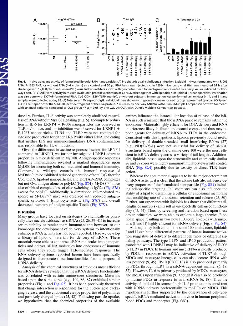
and injected s.c. into 129sv mice. After 9 h, mice were challengedintranasally with a supralethal dose (12,000 pfu) of influenzaA/PR8 virus. Lung titers were measured 24 h postchallengeand found to be reduced 10-fold by isRNA delivery (Fig. 4A) re-lative to untreated control. Further, this antiviral prophylaxis wasonly observed for the immunostimulatory R-006 RNA, whereasneither control R-1263 nanoparticles nor empty nanoparticleswithout RNA (II-4 + Blank) resulted in significant reductionof viral titer.
We further investigated the potential of both LRNP-I andLRNP-II as adjuvants of vaccination with the model protein anti-gen chicken ovalbumin (Ova). C57Bl/6 mice were vaccinatedby i.m. injections with Ova protein mixed with comparison adju-vants represented by LRNPs, DOTAP–RNA complexes, or CpGoligodeoxynucleotide (ODN) 1826, which is a mouse-specificB-class TLR9 agonist previously shown to adjuvant Ova antigenresponses (33); two different pan-species B-class CpG ODNhave shown enhancement of antibody titers against various anti-gens in clinical trials (5, 34, 35) and one is now in phase 3 testingin a hepatitis B vaccine (Heplisav, Dynavax). Immunization withLRNPs increased the magnitude of Ova-specific IgG by 3 to 4 logorders compared to immunization with protein alone (Fig. 4B). Asignificant increase in total IgG was observed for LRNP-I withboth R-006 and R-1263, but for LRNP-II only immunization withR-006 RNA resulted in statistically greater levels of IgG antibody.Vaccination with either LRNP resulted in an increase in bothIgG1 and IgG2c subclasses compared to protein alone (Fig. S5A).The increase in Th1-biased IgG2c subclass was notable comparedto immunization with Ova protein alone although not as strikingas it was for Ova mixed with CpG-ODNs, which have previouslybeen shown to induce a strong Th1 bias (36, 37). LRNP adjuvantsalso increased absolute IgG2c titers, but preserved the Th2-asso-ciated IgG1 bias of protein vaccination alone.
More strikingly, LRNP also greatly enhanced cell-mediatedimmune responses to Ova. Both LRNP-I+R-006 and LRNP-II+R-006 induced greater numbers of splenic Ova-antigen-specificCD8þ T cells than either CpG ODN 1826 or DOTAP+R-006complexes, and all LRNP formulations increased antigen-specific
CD8þ T cells to levels greater than that with Ova vaccinationalone (Fig. 4C). Using CpG ODN on its own as used here maybe suboptimal for adjuvant effect as greater immunogenicity hasbeen demonstrated in mice when CpG ODN is combined with anadditional adjuvant possessing delivery properties (34, 37, 38). Atlow dose (10 μg) of LRNP-II nanoparticles, the increase in per-centage of reactive CD8+ positive Tcells was significantly greaterwith the R-006 RNA than R-1263. However, for LRNP-I thepercentage of reactive CD8+ positive T cells was large but notsignificantly different between active R-006 and control R-1263RNA. In vitro restimulation of splenocytes from vaccinated ani-mals resulted in large increases in the Ova-specific secretion ofthe Th1-biasing cytokines IFN-γ and IL-2 (Fig. S5B). Increases ofthe Th2-associated cytokines IL-10 and IL-4 were also observed.
Characterization of Innate Immune Activation. LRNP-II exhibited10-fold greater IFN-α and IP-10 responses than LRNP-I nano-particles, which potently activated IL-6 following s.c. injection(Fig. S4D). We hypothesized that the lipidoid-specific cytokineprofiles observed were due to preferential uptake and activationof different cell types following s.c. administration. We compareddelivery of isRNA to primary monocytes or PDCs isolated fromhuman PBMCs using lipidoids I-3 and II-3, the three-tailed versionsof I and II that had greater isRNA activity in vitro (Fig. S3B).Lipidoid I-3 complexed with R-006 induced type I interferons fromisolated CD14+ cells (i.e., monocytes), but not from isolatedPDCs (Fig. S6B). Conversely, lipidoid II-3 complexed with R-006induced large amounts of type I interferon production in PDCs butconsiderably less from CD14+ cells (monocytes).
After i.v. injection in the 129sv strain of mouse, LRNPs acti-vated more robust responses than the s.c. route, and isRNAspecificity was observed in stimulation of serum IFN-α and IP-10with almost no activity of the control R-1263 RNA (Fig. S6A).However, LRNP formulated with R-1263 did induce IL-6 wheninjected i.v. raising concerns for increased nonspecific activationwith i.v. injection, and this observation was most pronounced atthe earliest time points with LRNP-I. We thus further investigateddirectly the role of TLRs in differential recognition of LRNPs.
The TLRs utilizing the common MyD88 pathway are TLR1,TLR2, TLR5, TLR6, TLR7, TLR8, and TLR9 (11). Becauseknock-out mice for all the TLRs were not readily available, weinvestigated TLR-specificity of LRNP-I in vitro in a human cellline stably expressing human TLRs. HEK293 cell lines stably ex-pressing human TLRs 2, 3, 4, 5 or 6 were incubated with LRNP+R-006 without observation of any TLR-mediated activity abovebackground (Fig. S6C). HEK293 cells stably transfected withTLR8, and TLR7 to a lesser extent, exhibited dose-dependentactivation by LRNP-I incorporating R-006 RNA (Fig. S6D).
LRNP were also investigated in knock-out mouse models ofthe MyD88, TLR4, TLR7, and TLR9 genes and compared towild-type (WT) controls on matched backgrounds (Fig. 5). Pro-duction of IFN-α, IP-10, and IL-6 were all dependent on MyD88signaling for both LRNP-I and LRNP-II. However, only IFN-αand IP-10 production were also dependent upon TLR7. In theC57BL/6 strain, we observed that IL-6 production in responseto LRNP-II was RNA specific and completely dependent uponTLR7. However, IL-6 activation in the same strain by LRNP-Iappears to be only partially TLR7-dependent at a 50 μg RNA
I-3 = ND(2)NA(1)-100
HN N N
O
NHO
NH
HN
O
O11 8
11 8
I-4 = ND(2)NA(2)-100
II-3 = ND(2)LD(1)-100
HN N N
O
NHO
O
O
O
O11 11
11 11
II-4 = ND(2)LD(2)-100
HN N
HN
O
NHO
O
O11 11
11
HN N
HN
O
NHO
HN
O11 8
11
Fig. 2. Structures of second-generation lipidoids based on 100 core. Second-generation lipidoids were designed based on the 100 core (red). An ND(2)-100 precursor was substituted with the 10-carbon alkyl-acrylamide NA (lipi-doid I) or the 12-carbon alkyl-acrylate LD (lipidoid II) and purified into singleisomer components with either three or four total tails.
Fig. 3. In vivo screening for activation of innate immuneresponses following injection of formulated lipidoid–RNA nanoparticles. Lipidoid–RNA nanoparticles formu-lated with 100 ug R-006 RNA were injected s.c. inBALB/c mice (n ¼ 3 or 4). R-006 formulated with DOTAPand a mock injection with HBSS were included as con-trols. Blood was collected at 6, 9, 12, and 24 h followinginjection and indicated cytokines were measured byELISA.
Nguyen et al. PNAS ∣ April 3, 2012 ∣ vol. 109 ∣ no. 14 ∣ E799
APP
LIED
BIOLO
GICAL
SCIENCE
SEN
GINEE
RING
PNASPL
US
dose i.v. Further, IL-6 activity was completely abolished regard-less of RNAwithout MyD88 signaling (Fig. 5). Incomplete reduc-tion in IL-6 for LRNP-I + R-006 nanoparticles was observed inTLR − ∕− mice, and no inhibition was observed for LRNP-I +R-1263 nanoparticles. TLR4 and TLR9 were not required forcytokine production for either LRNP with either RNA, indicatingthat neither LPS nor immunostimulatory DNA contaminationwas responsible for IL-6 induction.
Given the differences in vaccine responses observed for LRNP-Icompared to LRNP-II, we further investigated LRNP-I adjuvantproperties in mice deficient in MyD88. Antigen-specific responsesfollowing immunization revealed a marked dependence uponMyD88 for increasing both cell-mediated and humoral immunity.Compared to wild-type controls, the humoral response ofMyD88−∕− mice exhibited reduced generation of total IgG titer forCpG ODN, lipidoid nanoparticles, and DOTAP–RNA complexesbut not Ova antigen alone or polyI:C (Fig. S7A). MyD88−∕− micealso exhibited complete loss of class switching to IgG2c (Fig. S7B)except for polyI:C. Additionally, a diminished cell-mediated re-sponse in MyD88−∕− mice was observed with reduced antigen-specific cytotoxic T lymphocyte activity (Fig. S7C) and overalldecreased numbers of antigen-specific T cells (Fig. S7D).
DiscussionMany groups have focused on strategies to chemically or physi-cally alter nucleic acids such as siRNAs (25, 26, 39–41) to increaseserum stability or render them immuno-silent; however, to ourknowledge the development of delivery systems to intentionallyenhance isRNA activity has not been reported. Here we developa library of lipidoid materials for delivery of isRNA. Thesematerials were able to condense isRNA molecules into nanopar-ticles and deliver isRNA molecules into endosomes of immunecells where they could trigger TLR-mediated responses. TheRNA delivery systems reported herein have been specificallydesigned to incorporate these functionalities for the purpose ofisRNA delivery.
Analysis of the chemical structures of lipidoids most efficientfor isRNA delivery revealed that the isRNA delivery functionalitywas correlated with certain amine-core structures. Materialsbased upon the same cores (e.g., 100, 86, 87) exhibited similarproperties (Fig. 1 and Fig. S2). It has been previously theorizedthat charge interaction is responsible for the nucleic acid packa-ging, release, and the endosomal escape properties of polyaminesand positively charged lipids (25, 42). Following particle uptake,we hypothesize that the chemical properties of the available
amines influence the intracellular location of release of the isR-NA in such a manner that the isRNA payload remains within theendosome. Materials highly efficient for DNA delivery and RNAinterference likely facilitate endosomal escape and thus may bepoor agents for delivery of isRNA to TLRs in the endosome.Consistent with this hypothesis, lipioids previously found usefulfor delivery of double-stranded small interfering RNAs (27)(e.g., ND(5)-98-1) were not as useful for delivery of isRNA.Structures based upon the diamine core 100 were the most effi-cient in isRNA delivery across a variety of tail lengths. Addition-ally, lipidoids based upon the structurally and chemically similar86 and 87 cores were highly immunostimulatory even with controlRNA (Fig. S2A) possibly due to toxicity or direct TLR inter-action.
Whereas the core material appears to be the major determinantof isRNA activity, it is clear that the alkane tails also influence de-livery properties of the formulated nanoparticle (Fig. S3A) includ-ing cell-specific targeting. Tail chemistry can also influence theability of a lipid to destabilize a cellular or endosomal membranethus modifying rate of endosomal retention and release (25, 30).Further, our experience with lipidoids has shown that different tail-lengths or mixtures can result in unexpectedly enhanced function-ality (43, 44). Thus, by screening and iteration based upon thesedesign principles, we were able to explore a large chemical/func-tional space resulting in two novel 100-core lipidoids with mixedtails (I and II) highly efficient for isRNA delivery (Fig. 3, Fig. S4).
Although they both contain the same 100-amine core, lipidoidsI and II exhibited differential patterns of innate immune activa-tion suggestive of delivery to different cell types or different sig-naling pathways. The type I IFN and IP-10 production patternassociated with LRNP-II may be indicative of delivery of R-006to TLR7 in PDCs. In humans and mice IFN-α is mostly producedby PDCs in responses to ssRNA activation of TLR7 althoughMDCs and monocyte-lineage cells can also secrete IFN-α withless potency (9, 45). IP-10 (CXCL10) is also produced primarilyby PDCs through TLR7 in a ssRNA-dependent manner (6, 18,32). However, IL-6 is primarily produced by MDCs, monocytes,and moDCs upon stimulation (9), though it can also be producedby murine PDCs in response to viral ssRNA (6, 18). Thus theactivity of lipidoid I in terms of high IL-6 production is consistentwith isRNA delivery preferentially to moDCs or MDCs. Thishypothesis is further supported by the observation of cell-type-specific isRNA-mediated activation in vitro in human peripheralblood PDCs and monocytes (Fig. S6B).
A B C
Fig. 4. In vivo adjuvant activity of formulated lipidoid–RNA nanoparticles (A) Prophylaxis against influenza infection. Lipidoid II-4 was formulated with R-006RNA, R-1263 RNA, or without RNA (II-4 + blank) as a control and 50 μg RNA basis was injected s.c. in 129Sv mice. Lung viral titer was measured 24 h afterchallenge with 12,000 pfu of influenza (PR8) virus. Individual titers shownwith geometric mean for each group represented by a bar. p values indicated for two-way t-test. (B–C) Adjuvant activity in chicken ovalbumin protein vaccination of C57Bl/6 mice together with lipidoid I-4 or lipidoid II-4 nanoparticles. Vaccinationwas also done with DOTAP-formulated RNA, CpG ODN 1826 (TLR9 agonist), or without adjuvant. Immunization was performed i.m. on days 0, 14, and 21, andsamples were collected at day 28. (B) Total serum Ova-specific IgG. Individual titers shown with geometric mean for each group represented by a bar. (C) SpleenCD8þ Tcells specific for the SIINFEKL peptide fragment of the Ova protein. * p ¼ 0.05 by one-way ANOVAwith Dunn’s Multiple Comparison posttest for meanswith unequal variance compared to Ova group ** p ¼ 0.05 by one-way ANOVA with Dunn’s Multiple Comparison posttest.
E800 ∣ www.pnas.org/cgi/doi/10.1073/pnas.1121423109 Nguyen et al.

The LRNP may activate more than one TLR. In the mouse,cell-specific segregation of TLR7 is less distinct than it is inhumans (8, 11, 18); TLR7 is well-expressed in mouse MDCs andmoDCs. Both IFN-α and IP-10 production were clearly TLR7-and MyD88-dependent in mice (Fig. 5) confirming an isRNA-specific TLR7-mediated mechanism. Recently it has been shownthat mouse TLR8, previously thought to be nonfunctional (6, 18,19, 46), can be activated under certain conditions (29, 47, 48). Wehypothesize that TLR8 may play a partial role in LRNP-I isRNA-mediated activation of IL-6. In support of this hypothesis bothTLR7 and TLR8 showed clear activity in response to LRNP-Istimulation in a human cell culture TLR-overexpression model(Fig. S6D) whereas the remaining MyD88-dependent TLRswere not stimulated (Fig. S6C). The IL-6 production induced bylipidoid-mediated isRNA delivery in TLR7-deficienct mice but
not MyD88-deficient mice (Fig. 5) also indicates a possible rolefor isRNA stimulation of another MyD88-dependent receptoralthough no TLR8−∕− mouse model was available to directly testthe role of TLR8 responses to LRNPs in vivo. Together these dataindicate roles for TLR7 and another MyD88-dependent function,possibly TLR8, for the induction of IL-6 by LRNP-I.
Prophylactic administration of LRNP-I resulted in suppressionof influenza virus replication in the mouse lung (Fig. 4A) specificto the immunostimulatory activity of R-006 RNA. These dataindicate that s.c. injections of lipidoid-encapsulated isRNA canachieve sufficient systemic innate immune stimulation to combatviral infection. Because they activate the same receptors asviruses resulting in a variety of cytokines and interferons, lipidoidisRNA delivery may resemble the innate response to infection byRNA viruses. Imiquimod, a TLR7/8 agonist, has seen extensive
Fig. 5. Characterization of cytokine response from lipidoid–RNA nanoparticles in vivo. Lipidoids I-4 and II-4 were formulated with the strong TLR7/8 agonistR-006 RNA and the weak TLR7/8 agonist R-1263 RNA as control. Serum cytokine response of IFN-α (Top), IP-10 (Middle), and IL-6 (Bottom) at 6 h compared tostrain background-matched controls following i.v. injection of 50 μg RNA into MyD88−∕− or TLR7−∕− mice, or 30 μg RNA into TLR9−∕− or TLR4−∕− mice;LPS ¼ lipopolysaccharide; (n ¼ 3 or 4).
Nguyen et al. PNAS ∣ April 3, 2012 ∣ vol. 109 ∣ no. 14 ∣ E801
APP
LIED
BIOLO
GICAL
SCIENCE
SEN
GINEE
RING
PNASPL
US
clinical use (3, 5) as a topical chemotherapeutic and antiviral.Experimental therapies employing TLR7 agonists have also de-monstrated some clinical benefit in viral hepatitis (49), however,adverse immunologic events related to widespread biodistribu-tion were observed at therapeutic doses (50, 51) including unac-ceptable toxicity in a human trial of oral imiquimod (52). Noadverse effects were observed in mice injected with LRNPs inthis study, though long-term and dose-response effects of LRNPadministration need to be investigated. A restricted deliveryapproach such as LRNPs may limit the systemic distribution bylocalizing initiation of immune responses to the application siteand draining lymph nodes as well as targeting uptake to specificcell types. Thus the endogenous antiviral-like response activatedby LRNPs may be an attractive alternative therapy for conditionscurrently treated with recombinant IFN-α regimens.
The development of vaccine adjuvants that increase both hu-moral and cell-mediated immune responses by targeting TLRsrepresents a promising approach for the field of vaccine develop-ment (3, 5, 10). A hallmark of this cellular immunity is expansionof antigen-specific CD8þ Tcells, which was increased by immuni-zation with either LRNP-I or LRNP-II (Fig. 4C). LRNP adjuvantsincreased secretion of Th1-biasing cytokines upon restimulation(Fig. S5B), which are associated with control of intracellular patho-gens (2) and cancers (7). Efficient T-cell activation with multi-functional quality can lead to memory responses that confer pro-tective immunity for long periods of time (1, 14, 53). Further stu-dies will be necessary to evaluate the memory function and qualityof T-cell responses to protein vaccines adjuvanted by LRNPs.
Class switching and production of IgG2c in B-cells and efficientantigen-specific T-cell induction requires activation of MyD88(54). If the adjuvant mechanism of LRNP-I was completely dueto activation of TLR-dependent innate immune responses, thenthese responses would be expected to be dependent upon MyD88.However, comparison of vaccine responses in wild-type andMyD88-deficient mice showed that although loss of MyD88 mark-edly reduced both cell-mediated and humoral immune responses,some adjuvant activity was maintained particularly with respect togeneration of (non-IgG2c) IgG antibodies (Fig. S7B) indicatingan additional role of a non-MyD88-dependent adjuvant effect.Commonly used adjuvants including alum, Freund’s adjuvant,and squalene increase immune responses by both enhancing innateimmune responses (4) and sustaining antigen availability and pre-sentation in draining lymph nodes (55). Thus, in addition to pro-viding isRNA-mediated innate signals, lipidoid nanoparticles mayalso enhance the delivery of protein antigens or TLR agonists totissue-resident and lymphatic dendritic cells (55) thereby increas-ing vaccine responses.
In conclusion, combinatorial chemistry and high-throughputscreening methods have enabled development of libraries ofmaterials for isRNA delivery that provide for specific activationof immune responses. We report the development of uniquedelivery systems specifically designed to deliver isRNA in thecontext of a vaccine adjuvant or for use in immune prophylaxisagainst viral infection. Here we have directly shown LRNPs to bemore efficient for isRNA delivery than currently available agentssuch as lipofectamine, DOTAP, or even the previously reportedlipidoid ND(5)-98-1 (23, 27). Immunostimulatory RNA deliveryto PDCs using formulations such as LRNP-II may be useful forantiviral applications, whereas delivery using LRNP-I may bemost useful for activating monocytes and MDCs to enhance vac-cine responses. Our screening revealed two promising candidatematerials for future development, and further refinement of lipi-doid design and nanoparticle formulation techniques may lead toeven more specific and robust effects. Further development ofsuch vehicles for controlled TLR7 and 8 stimulation, includingadditional optimization and safety assessment in larger animals,may have future clinical utility.
Materials and MethodsA summary of experimental techniques is presented here. For full details andmethods, please see the SI Materials and Methods.
Lipidoid Synthesis and High-Throughput Screening for isRNA Delivery. Lipidoidswere synthesized in a combinatorial fashion as depicted in Fig. S1 as pre-viously described (27) by reacting primary and secondary amine-containingcores with alkyl-acrylate or alkyl-acrylamide tails. A complete list of crudelipidoids screened is found in Table S1. Second-generation lipidoids basedon the 100-core diamine were synthesized in a four-step process (Fig. S1D)and renamed lipidoids I and II for clarification. Lipidoid products were com-plexed with RNA in sodium acetate at 15, 10, 5, and 2.5 : 1mass ratios of lipidto RNA. RNAs R-006 or R-1263 were fully phosphorothioate-modified,20-base, ssRNA synthesized by Coley Pharmaceuticals with sequences aspreviously described (29). PBMCs were isolated from donor-blind buffy-coat packs obtained from the Massachusetts General Hospital blood bank.Complexes were added to PBMCs, plated at 5 × 10⁵ cells∕well, for a final RNAconcentration of 200 ng RNA per well (1 μg∕mL, approximately 140 nM).Additionally, PBMCs were incubated with R-006 and L2K, according to man-ufacturers siRNA transfection protocols, to control for donor PBMC variabilityin maximum type I IFN secretion capacity. After 16–20 h of incubation, PBMCsupernatants were stored at −80 °C for later quantification of type I interfer-on activity using a high-throughput cell-based detection assay as previouslydescribed (28).
Formulation and Characterization of Lipidoid–RNA Nanoparticles. Purified lipi-doid was codissolved in ethanol with cholesterol and C16 mPEG 2000 cera-mide at a 15∶0.8∶7 mass ratio (L∶C∶P) in a mixture of ethanol and sodiumacetate. Lipidoid/Ch/PEG were added to RNA at a 15, 11.5, or 10∶1 mass ratio(L∶R). Lipidoid–RNA nanoparticles were then extruded through a double200 nm membrane and then twice through a double 80 nm membrane(Whatman) on a Northern Lipids extrusion system at 40 °C. Prior to injection,nanoparticles were either dialyzed at 3,500 molecular weight cutoff in HBSSor lyophilized with 10 mg∕mL sucrose. Final RNA concentration was deter-mined by modified Ribogreen (Invitrogen) assay. No bacterial endotoxinwas detected by limulus amebocyte lysate assay (Lonza) in any batches ofnanoparticles.
In Vivo Characterization of Innate Immune Responses. All animal studies,except for influenza prophylaxis studies, were conducted at Coley Pharma-ceuticals (now Pfizer Canada) under the approval of the institutional carecommittees and in accordance with the guidelines set forth by the CanadianCouncil on Animal Care. Mouse studies of influenza prophylaxis were con-ducted at the Massachusetts Institute of Technology (Cambridge, MA), whereanimals were cared for according to the guidance of the Division of Compara-tive Medicine. Animals were monitored for the duration of all experimentsfor adverse behavior or decrease in normal activity that might indicate toxi-city or adverse inflammatory responses. Lipidoid–RNA nanoparticles wereformulated with either R-006 or R-1263 RNA at 15∶1 ratio (lipidoid to RNA),dialyzed to remove ethanol, and diluted in HBSS prior to injection under iso-fluorane anesthesia. Lipidoid nanoparticles were injected s.c. or by i.v. tailvein injection in 129Sv mice at indicated doses. Serum samples were analyzedfor levels of IFN-α and cytokines at indicated time points. TLR-mediated re-sponses were investigated following i.v. injection in TLR-deficient or genetic-background-matched WT controls. Prophylaxis of influenza infection modelswas conducted as previously described (23). Briefly, lipidoid–RNA nanoparti-cles were injected s.c. at 50 μg RNA per mouse. After 9 h, mice were infectedby intranasal instillation with 12,000 pfu influenza A virus A/PR/8/34 (PR8).Lungs viral titer was quantified at 24 h after infection. Adjuvant studiesof lipidoid I and II isRNA nanoparticles were performed by mixingindicated doses of lipidoid-formulated RNA with 20 μg chicken Ova immedi-ately prior to i.m. injection into C57Bl/6 mice or MyD88−∕− on the C57Bl/6background. Mice were dosed at days 0, 14, and 21, then blood and spleno-cytes were collected at day 28. Plasma anti-Ova total IgG, IgG1, and IgG2cwere determined by sandwich ELISA. Splenocytes were assessed using a chro-mium release assay for cytotoxic T cells and flow-cytometry-based tetrameranalysis for quantifying antigen-specific T cells as previously described (56).
ACKNOWLEDGMENTS. This work was supported in part by grants from theNational Institutes of Health EB00244 (to R.L. and D.G.A.) and AI56267(to J.C.) and from the National Institute of Allergy and Infectious DiseasesContract HHSSN266200400044C (to G.C. and A.M.K.).
E802 ∣ www.pnas.org/cgi/doi/10.1073/pnas.1121423109 Nguyen et al.

1. Iwasaki A, Medzhitov R (2004) Toll-like receptor control of the adaptive immune re-sponses. Nat Immunol 5:987–995.
2. Zhang SY, et al. (2007) Human Toll-like receptor-dependent induction of interferons inprotective immunity to viruses. Immunol Rev 220:225–236.
3. Meyer T, Stockfleth E (2008) Clinical investigations of Toll-like receptor agonists. ExpertOpin Investig Drugs 17:1051–1065.
4. Pashine A, Valiante NM, Ulmer JB (2005) Targeting the innate immune response withimproved vaccine adjuvants. Nat Med 11(Suppl 4):S63–68.
5. Kanzler H, Barrat FJ, Hessel EM, Coffman RL (2007) Therapeutic targeting of innateimmunity with Toll-like receptor agonists and antagonists. Nat Med 13:552–559.
6. Heil F, et al. (2004) Species-specific recognition of single-stranded RNA via toll-likereceptor 7 and 8. Science 303:1526–1529.
7. Schon MP, Schon M (2008) TLR7 and TLR8 as targets in cancer therapy. Oncogene27:190–199.
8. Hornung V, et al. (2005) Sequence-specific potent induction of IFN-alpha by shortinterfering RNA in plasmacytoid dendritic cells through TLR7. Nat Med 11:263–270.
9. Jarrossay D, Napolitani G, Colonna M, Sallusto F, Lanzavecchia A (2001) Specializationand complementarity in microbial molecule recognition by human myeloid andplasmacytoid dendritic cells. Eur J Immunol 31:3388–3393.
10. Borden EC, et al. (2007) Interferons at age 50: Past, current and future impact onbiomedicine. Nat Rev Drug Discov 6:975–990.
11. Akira S, Takeda K (2004) Functions of toll-like receptors: Lessons from KOmice. C R Biol327:581–589.
12. Cristofaro P, Opal SM (2006) Role of Toll-like receptors in infection and immunity:Clinical implications. Drugs 66:15–29.
13. Honda K, Taniguchi T (2006) IRFs: Master regulators of signalling by Toll-like receptorsand cytosolic pattern-recognition receptors. Nat Rev Immunol 6:644–658.
14. Steinman RM, Banchereau J (2007) Taking dendritic cells into medicine. Nature449:419–426.
15. Delgado MF, et al. (2009) Lack of antibody affinity maturation due to poor Toll-likereceptor stimulation leads to enhanced respiratory syncytial virus disease. Nat Med15:34–41.
16. Marshak-Rothstein A (2006) Toll-like receptors in systemic autoimmune disease. NatRev Immunol 6:823–835.
17. Romagne F (2007) Current and future drugs targeting one class of innate immunityreceptors: The Toll-like receptors. Drug Discov Today 12:80–87.
18. Diebold SS, Kaisho T, Hemmi H, Akira S, Reis e Sousa C (2004) Innate antiviralresponses by means of TLR7-mediated recognition of single-stranded RNA. Science303:1529–1531.
19. Lan T, et al. (2007) Stabilized immune modulatory RNA compounds as agonists ofToll-like receptors 7 and 8. Proc Natl Acad Sci USA 104:13750–13755.
20. Scheel B, et al. (2006) Therapeutic anti-tumor immunity triggered by injections ofimmunostimulating single-stranded RNA. Eur J Immunol 36:2807–2816.
21. Eberle F, et al. (2008) Modificationsin small interfering RNA that separate immuno-stimulation from RNA interference. J Immunol 180:3229–3237.
22. Sioud M (2005) Induction of inflammatory cytokines and interferon responses bydouble-stranded and single-stranded siRNAs is sequence-dependent and requiresendosomal localization. J Mol Biol 348:1079–1090.
23. Nguyen DN, et al. (2009) Drug delivery-mediated control of RNA immunostimulation.Mol Ther 17:1555–1562.
24. Robbins M, et al. (2008) Misinterpreting the therapeutic effects of small interferingRNA caused by immune stimulation. Hum Gene Ther 19:991–999.
25. Schroeder A, Levins CG, Cortez C, Langer R, Anderson DG (2010) Lipid-based nanother-apeutics for siRNA delivery. J Intern Med 267:9–21.
26. Whitehead KA, Langer R, Anderson DG (2009) Knocking down barriers: advances insiRNA delivery. Nat Rev Drug Discov 8:129–138.
27. Akinc A, et al. (2008) A combinatorial library of lipid-like materials for delivery of RNAitherapeutics. Nat Biotechnol 26:561–569.
28. Nguyen DN, et al. (2009) A novel high-throughput cell-based method for integratedquantification of type I interferons and in vitro screening of immunostimulatory RNAdrug delivery. Biotechnol Bioeng 103:664–675.
29. Forsbach A, et al. (2008) Identification of RNA sequence motifs stimulating sequence-specific TLR8-dependent immune responses. J Immunol 180:3729–3738.
30. Semple SC, et al. (2010) Rational design of cationic lipids for siRNA delivery. Nat Bio-technol 28:172–176.
31. Barchet W, et al. (2005) Dendritic cells respond to influenza virus through TLR7- andPKR-independent pathways. Eur J Immunol 35:236–242.
32. Megjugorac NJ, Young HA, Amrute SB, Olshalsky SL, Fitzgerald-Bocarsly P (2004)Virally stimulated plasmacytoid dendritic cells produce chemokines and induce migra-tion of T and NK cells. J Leukoc Biol 75:504–514.
33. Davila E, Celis E (2000) Repeated administration of cytosine-phosphorothiolated gua-nine-containing oligonucleotides together with peptide/protein immunization resultsin enhanced CTL responses with anti-tumor activity. J Immunol 165:539–547.
34. Halperin SA, et al. (2006) Comparison of the safety and immunogenicity of hepatitis Bvirus surface antigen co-administered with an immunostimulatory phosphorothioateoligonucleotide and a licensed hepatitis B vaccine in healthy young adults. Vaccine24:20–26.
35. Vollmer J, Krieg AM (2009) Immunotherapeutic applications of CpG oligodeoxynu-cleotide TLR9 agonists. Adv Drug Deliv Rev 61:195–204.
36. Malyala P, O’Hagan DT, Singh M (2009) Enhancing the therapeutic efficacy of CpG oli-gonucleotides using biodegradable microparticles. Adv Drug Deliv Rev 61:218–225.
37. Wilson KD, de Jong SD, Tam YK (2009) Lipid-based delivery of CpG oligonucleotidesenhances immunotherapeutic efficacy. Adv Drug Deliv Rev 61:233–242.
38. Cooper CL, Angel JB, Seguin I, Davis HL, Cameron DW (2008) CPG 7909 adjuvant plushepatitis B virus vaccination in HIV-infected adults achieves long-term seroprotectionfor up to 5 years. Clin Infect Dis 46:1310–1314.
39. Schlee M, Hornung V, Hartmann G (2006) siRNA and isRNA: Two edges of one sword.Mol Ther 14:463–470.
40. Sioud M (2007) RNA interference and innate immunity. Adv Drug Deliv Rev59:153–163.
41. Weinstein S, Peer D (2010) RNAi nanomedicines: Challenges and opportunities withinthe immune system. Nanotechnology 21:232001.
42. PutnamD (2006) Polymers for gene delivery across length scales.NatMater 5:439–451.43. Love KT, et al. (2010) Lipid-like materials for low-dose, in vivo gene silencing. Proc Natl
Acad Sci USA 107:1864–1869.44. Mahon KP, et al. (2010) Combinatorial approach to determine functional group effects
on lipidoid-mediated siRNA delivery. Bioconjug Chem 21:1448–1454.45. Asselin-Paturel C, Brizard G, Pin J-J, Briere F, Trinchieri G (2003) Mouse strain differ-
ences in plasmacytoid dendritic cell frequency and function revealed by a novelmonoclonal antibody. J Immunol 171:6466–6477.
46. Jurk M, et al. (2002) Human TLR7 or TLR8 independently confer responsiveness to theantiviral compound R-848. Nat Immunol 3:499–499.
47. Gorden KK, et al. (2006) Oligodeoxynucleotides differentially modulate activation ofTLR7 and TLR8 by imidazoquinolines. J Immunol 177:8164–8170.
48. Martinez J, Huang X, Yang Y (2010) Toll-like receptor 8-mediated activation ofmurine plasmacytoid dendritic cells by vaccinia viral DNA. Proc Natl Acad Sci USA107:6442–6447.
49. Horsmans Y, et al. (2005) Isatoribine, an agonist of TLR7, reduces plasma virus concen-tration in chronic hepatitis C infection. Hepatology 42:724–731.
50. Fidock MD, et al. (2011) The innate immune response, clinical outcomes,and ex vivoHCVantiviral efficacy of a TLR7 agonist (PF-4878691). Clin Pharmacol Ther 89:821–829.
51. Fletcher S, Steffy K, Averett D (2006) Masked oral prodrugs of toll-like receptor 7agonists: A new approach for the treatment of infectious disease. Curr Opin InvestigDrugs 7:702–708.
52. Savage P, et al. (1996) A phase I clinical trial of imiquimod, an oral interferon inducer,administered daily. Br J Cancer 74:1482–1486.
53. Seder RA, Darrah PA, Roederer M (2008) T-cell quality in memory and protection:Implications for vaccine design. Nat Rev Immunol 8:247–258.
54. Barr TA, Brown S, Mastroeni P, Gray D (2009) B cell intrinsic MyD88 signals driveIFN-gamma production from T cells and control switching to IgG2c. J Immunol183:1005–1012.
55. Tritto E, Mosca F, De Gregorio E (2009) Mechanism of action of licensed vaccineadjuvants. Vaccine 27:3331–3334.
56. Chikh G, et al. (2009) Synthetic methylated CpG ODNs are potent in vivo adjuvantswhen delivered in liposomal nanoparticles. Int Immunol 21:757–767.
Nguyen et al. PNAS ∣ April 3, 2012 ∣ vol. 109 ∣ no. 14 ∣ E803
APP
LIED
BIOLO
GICAL
SCIENCE
SEN
GINEE
RING
PNASPL
US

Supporting InformationNguyen et al. 10.1073/pnas.1121423109Material and Methods.RNA. RNAs were fully phosphorothioate-modified, 20-base,single-stranded RNA synthesized by Coley Pharmaceuticals withsequences as previously described (1): R-006 [5′-UUGUUGUU-GUUGUUGUUGUU-3′] and R-1263 [5′-GCCACCGAGCC-GAAGGCACC-3′].
Combinatorial Lipidoid Synthesis.Lipidoids (2) were synthesized, aspreviously described, in a combinatorial fashion as depicted inFig. S1B in solvent-free conditions by reacting primary and sec-ondary amine-containing cores (Fig. S1A, Right) with alkyl-acry-late or alkyl-acrylamide (Fig. S1A, Left) tails at a high tail-to-coremonomer ratio to drive synthesis of fully (n)-substituted lipidoids.Lipidoid products were purified of unreacted core and side-chainreactants resulting in crude mixtures of fully and incompletelysubstituted lipidoids by silica gel chromatography. Some alkyl-acrylate-tail lipidoids were further reacted with methyl iodide(MeI) (Fig. S1C) to form quaternized amines with a permanentpositive charge.
Second-generation lipidoids were synthesized in a four-stepprocess (Fig. S1D). The 100-core diamine was monoprotectedby reacting 10x molar excess pure diamine with di-tert-butyl di-carbonate (Boc2O). ND tails were reacted with the free primaryamine in excess prior to deprotection and regeneration of the op-posite primary amine resulting in ND(2)-100. ND(2)-100 wasfurther reacted with NA or LD tails and purified into three-tailor four-tail derivatives, which have been renamed lipidoids I andII for clarification. Lipidoid nomenclature reflects alkyl tail link-age (acrylate ¼ L, acrylamide ¼ N), alkyl tail carbon-chainlength (A ¼ 9, B ¼ 10, D ¼ 12, F ¼ 14, G ¼ 15, P ¼ 16, H ¼ 18carbons), and amine-containing core. Quaternized core-aminesare renamed with a Q designation instead of L. Purified lipidoidsinclude number of tails in parentheses following tail name. Acomplete list of crude lipidoids screened is found in Table S1.
High-Throughput Screening for Lipidoid-Mediated Immunostimula-tory RNA (isRNA) Delivery. Donor-blind buffy-coat packs were ob-tained from the Massachusetts General Hospital blood bank.Peripheral blood mononuclear cells (PBMC) were isolated byFicoll-Paque Plus (Amersham Biosciences) density centrifuga-tion. PBMCs were resuspended in supplemented RPMI medium1640 (RPMI medium 1640 with 10% FCS, 1 mM MEM sodiumpyruvate, 10 mM Hepes, and 100 U∕mL penicillin/streptomycin)and plated at 5 × 10⁵ cells∕well in 175 μL in 96-well tissue cultureplates. Lipofectamine 2000 (L2K) (Invitrogen) was used as apositive control for transfection of RNA according to manufac-turer’s protocols and to normalize interferon responses acrossdifferent donors.
Crude or purified lipidoid products were dissolved to0.5 mg∕mL in 25 mM sodium acetate (pH 5), followed by briefsonication. For lipidoids with poor solubility, up to 10% DMSOwas added to stock lipidoid solutions. RNA was dissolved to50 μg∕mL in sodium acetate. Lipidoids were arrayed in 96-wellround-bottom reaction plates and mixed at 15, 10, 5, and 2.5∶1mass ratios of lipid to RNA for 80 μL total volume. After 20 minincubation at room temperature to allow for nanoparticle com-plexes to form, complexes were diluted with 120 μL RPMI med-ium 1640. In quadruplicate, 25 μL of diluted complexes wereadded to PBMCs for a final RNA concentration of 200 ng RNAper well in 200 μLmedia (1 μg∕mL approximately 140 nM). After16–20 h of incubation, PBMC cultures were centrifuged at 400 ×
g relative centrifugal force (rcf) for 10 min, and supernatantswere stored at −80 °C for later quantification.
Type I interferon activity was quantified using a high-through-put (HT)-compatible cell-based detection assay as previouslydescribed (3). Briefly, 293T-ISRE-RFP cells were incubated with50 μL PBMC supernatant overnight prior to HT-FACS analysisof red fluorescence. Recombinant human interferon alpha (hIFN-α) (PBL Laboratories) serially diluted in supplementedRPMI medium 1640 was used as a standard, and type I interferonactivity of each screening well was normalized to L2K-mediatedtransfection with R-006 to control for donor PBMC variability inmaximum type I interferon secretion capacity.
Formulation and Characterization of Lipidoid-RNA Nanoparticles.Purified lipidoid was dissolved to 120 mg∕mL in ethanol. Choles-terol (Ch) (Sigma Aldrich) was dissolved to 25 mg∕mL in etha-nol. N-palmitoyl-sphingosine-1-[succinyl(methoxypolyethyleneglycol)2000] (C16 mPEG 2000 ceramide) (PEG) (Avanti PolarLipids) was dissolved to 100 mg∕mL in ethanol. Lipidoid, Ch,and PEG were combined at a 15∶0.8∶7 mass ratio (L∶C∶P), vor-texed briefly, and diluted in a mixture of ethanol and 200 mMsodium acetate (with 16.67 mg∕mL sucrose for lyophilization)for a final lipidoid concentration of 7.5 mg∕mL in 35% ethanol,65% sodium acetate. RNAs were resuspended in water to 10 mg∕mL and diluted with 35% ethanol. Lipidoid/Ch/PEG were addedto diluted RNA at a 15, 11.5, or 10∶1 mass ratio (L∶R) andvortexed for 20 min to allow complexes to form. Complexedlipidoid–RNA nanoparticles were extruded once through a dou-ble 200 nm membrane and then twice through a double 80 nmmembrane (Whatman) on a Northern Lipids extrusion system at40 °C. To remove ethanol prior to injection, nanoparticles weredialyzed in a Slide-A-Lyzer 3500 molecular weight cutoff dialysiscassette (Pierce Biotech) against HBSS. For lyophilization, 10 mgof sucrose was added per milliliter of extruded complexes priorto freezing at −80 °C for >2 h followed by >1 d lyophilization.
For quantification and encapsulation efficiency of RNA, a50 μL sample of nanoparticles was diluted 200-fold in Tris-EDTAbuffer (TE), mixed with either 50 μL of TE buffer or 50 μL of 2%Triton-X-100 (T-X) in TE, and incubated with 100 μL Quant-ItRibogreen reagent (Invitrogen) for 20 min at 37 °C in a 96-wellblack plate. Total fluorescence in the presence of T-X was com-pared to a standard curve of RNA diluted in TE to determinetotal RNA concentration; RNA encapsulation efficiency wasdetermined by the ratio of fluorescence signal without T-X andwith T-X. Nanoparticle size and zeta potential of formulatedlipidoid–RNA nanoparticles was assayed by dynamic light scat-tering using a Zeta-PALS instrument (Brookhaven Instruments)after 1∕50 dilution in PBS or with a Mastersizer instrument (Mal-vern Instruments) after dilution in HBSS. No bacterial endotoxinwas detected by limulus amebocyte lysate assay (Lonza) in anybatches of nanoparticles.
In Vitro Characterization of Innate Immune Responses. HumanPBMCs were collected as described above and further purifiedinto PDC (BDCA2þ) and monocyte (CD14þ) populations bypositive selection with magnetic-assisted sorting according tomanufacturer’s protocols (Miltenyi Biotech). Cells were thenincubated for 24 h with three-tail lipidoid-RNA nanoparticlesformulated for in vitro transfection. Supernatants were analyzedfor type I interferon activity by cell-based 293T-ISRE-RFP assayas described above.
Nguyen et al. www.pnas.org/cgi/doi/10.1073/pnas.1121423109 1 of 10

A reporter plasmid expressing luciferase controlled by multipleNf-κB-inducible promoter regions, pNifty-Luciferase (Invivo-gen), was transfected into HEK293T cell lines stably expressingindicated human TLRs (Invivogen). These cells were stimulatedin vitro with lipidoid I-4 + isRNA nanoparticles at 4 μg∕mLRNA for HEK293Tcells expressing TLR2/6, TLR3, TLR4, TLR5,or TLR9; or at a range of RNA concentrations for HEK293Tcellsexpressing TLR7 or TLR8. As positive controls, cells were alsostimulated with known TLR-specific ligands: synthetic diacylatedlipoprotein FSL-1 (TLR2/6), poly(I:C) (TLR3), lipopolysacchar-ide (LPS) (TLR4), flagellin (TLR5), garadiquimod (TLR7),CL097 (TLR8), CpG oligodeoxynucleotide 2395 (TLR9); all pur-chased from Invivogen and used at manufacturer’s recommendedconcentrations. Luciferase activity was determined after 24 h usingthe Bright-Glo assay (Promega) according to manufacturer’s pro-tocols.
In Vivo Characterization of Innate Immune Responses. To further in-vestigate RNA-specific immune responses, lipidoid–RNA nano-particles were formulated with either R-006 or R-1263 at 15∶1ratio (lipidoid to RNA), dialyzed to remove ethanol, and dilutedinHBSS prior to injection under isofluorane anesthesia. All animalstudies, except for influenza prophylaxis studies, were conductedat Coley Pharmaceuticals (now Pfizer, Canada; Ottawa, ON,Canada) under the approval of the institutional care committeesand in accordance with the guidelines set forth by the CanadianCouncil on Animal Care. Mouse studies of influenza prophylaxiswere conducted at the Massachusetts Institute of Technology(Cambridge, MA). Male 129Sv mice were purchased from TaconicFarms and cared for according to the standards of the Massachu-setts Institute of Technology under the guidance of the Divisionof Comparative Medicine. Animals were monitored for the dura-tion of all experiments for adverse behaviors, which might indicatetoxicity or adverse inflammatory responses, such as shivering,abnormal secretions, lethargy, decreased social interactions, ordecrease in normal activity.
Formulated lipidoid–RNA nanoparticles, or blank nanoparti-cles without RNA at an equivalent total lipidoid dose, werediluted in HBSS prior to subcutaneous injection of 25 μg RNAin each flank under isofluorane anesthesia for a total dose of50 μg RNA per mouse. After 9 h, mice were anesthetized with amixture of ketamine (1 wt%) and xylazine (0.15 wt%) in PBSby i.p. injection. Mice were immediately infected by intranasalinstillation with a supralethal dose of 12,000 plaque-forming units(pfu) of influenza A virus A/PR/8/34 (PR8) strain diluted in PBSwith 0.3 wt% BSA and 100 U∕mL penicillin/streptomycin. Micewere sacrificed 24 h after infection, whole lungs were flash frozenin 2 mL of PBS/BSA solution in liquid nitrogen, and lungs weresubjected to two freeze-thaw cycles followed by sonication. Viruswas extracted from lung homogenates after centrifugation at800 × g rcf. Viral titer in harvested lungs was determined by pla-
que-forming assay. Madine–Darby canine kidney cells wereseeded at 0.5 × 10⁶ cells∕well in six well plates in DMEM (with10 mM Hepes, 10% FBS, 100 U∕mL penicillin/streptomycin,2 mM glucose) and allowed to grow to single-layer confluenceovernight. Media was aspirated from wells, and 200 μL of virus-containing samples serially diluted 10-fold in PBS were addedonto cells in triplicate. Following a 1 h incubation period withperiodic shaking to distribute viral particles evenly, cells werecovered with 2 mL of semisolid 1% agar/media solution to limitviral particle spread to cell-to-cell contacts. Plaques were countedafter 3 d.
Innate immune responses of multiple lipidoid formulationswith R-006 RNA were compared after s.c. injections at increasingdoses in BALB/c mice or i.v. tail vein injection in 129Sv mice.TLR-mediated responses were investigated following i.v. injec-tion in C57Bl/6, C57Bl/6 TLR7−∕−, C57Bl/6 TLR9−∕−, C57Bl/6MyD88−∕−, C3H, or C3H TLR4−∕−. As a control, DOTAP [1,2-dioleoyloxy-3-(trimethylammonium)propane] (Roche) was com-plexed with RNA at a 2 : 1 weight ratio (L∶R). Blood samples forserum isolation were taken by direct cardiac puncture on heparinat indicated timepoints. Serum samples were analyzed by ELISAwith commercially available antibodies (IFN-α, PBL Labs) (IP-10, BD Pharmingen) or 10-plex Luminex technology (BioSourceInternational). Splenocytes were also harvested for surface ex-pression of activation markers by staining with anti-CD69-FITC,anti-CD86-APC, anti-CD3-PE/Cy7, anti-CD19-ECD, and anti-DX5-PE. Cytolytic activity of NK cells was measured by chro-mium release following in vitro incubation of splenocytes withYAC-1 target cells at indicated effector:target ratios.
Adjuvant studies of lipidoid–RNA nanoparticles were per-formed by mixing indicated doses of lipidoid-formulated RNAwith 20 μg chicken ovalbumin (Ova) protein antigen immediatelyprior to i.m. injection into C57Bl/6 mice or MyD88−∕− on theC57Bl/6 background. Mice were vaccinated with either Ova pro-tein antigen alone, or Ova protein mixed with the TLR9 agonistCpG ODN 1826, the TLR3 agonist poly(I:C), or DOTAP-formu-lated RNA. Mice were dosed three times, at days 0, 14, and 21.After 28 d, blood was collected by direct cardiac puncture onheparin. Plasma antibody levels of anti-Ova total IgG, IgG1, andIgG2c were determined by sandwich ELISA. Splenocytes werealso harvested at day 28 for flow cytometric analysis of surfaceexpression markers and cytokine production. Splenocytes weredirectly stained for antigen-specific T-cells with anti-CD44-FITC,SIINFEKL-tetramer-PE, anti-CD8-ECD, anti-CD62L-APC, andanti-CD3-PE/Cy7. Separate aliquots of the same splenocyte sam-ples were also independently stimulated in vitro with Ova proteinin RPMI medium 1640 + 1% L-glutamine + 1% penicillin/strep-tomycin + 10% FBS + β-mercaptoethanol for 24 h. Supernatantswere analyzed by ELISA after 24 h for IL-2, IL-4, IL-10, andTNFα, or after 72 h for IFN-γ.
1. Forsbach A, et al. (2008) Identification of RNA sequence motifs stimulating sequence-specific TLR8-dependent immune responses. J Immunol 180:3729–3738.
2. Akinc A, et al. (2008) A combinatorial library of lipid-like materials for delivery of RNAitherapeutics. Nat Biotechnol 26:561–569.
3. Nguyen DN, et al. (2009) A novel high-throughput cell-based method for integrated
quantification of type I interferons and in vitro screening of immunostimulatory RNA
drug delivery. Biotechnol Bioeng 103:664–675.
Nguyen et al. www.pnas.org/cgi/doi/10.1073/pnas.1121423109 2 of 10

A
B
C
D
Fig. S1. (A) A subset of components from the larger lipidoid library (2) used in this study. When combined with amine-containing cores (red) to form lipid-likestructures, alkyl-acrylate (L) tails form a hydrolysable ester bond, and alkyl-acrylamides (N) form nondegradable amine linkages. Tail groups (blue) are codedaccording to linkage and number of carbons in the alkyl chain. Lipidoid nomenclature reflects alkyl tail linkage (acrylate = L, acrylamide = N), alkyl tail carbon-chain length (A ¼ 9, B ¼ 10, D ¼ 12, F ¼ 14, G ¼ 15, P ¼ 16, H ¼ 18 carbons), and amine-containing core (number). (B) Schematic of solvent-free batch synthesisprocess resulting in crude mixtures of lipidoid components. All reactions were performed in excess of tail groups to drive towards full substitution of all coreamine groups. Primary amines may accept up to two tail substitutions and secondary amines can accept up to one. Crude products typically contain a mixture offully n-substituted and n-1 substituted lipidoids with rare n-2 substitutions. (C) Lipidoids can be further reacted with methyl iodide (MeI) to form quaternaryamines carrying a permanent positive charge. Quaternized lipidoids are renamed with a Q designation (Q lipidoids). (D) Synthesis of second-generation li-pidoids (lipidoids I and II) based on the 100-core. Intermediate protection and deprotection steps were used to generate the ND(2)-100 precursor, which wasfurther reacted with the NA or LD tails in the synthesis of mixed-tail lipidoids of the 100 core.
Nguyen et al. www.pnas.org/cgi/doi/10.1073/pnas.1121423109 3 of 10

Fig. S2. Activity of all formulations (different lipidid, L∶R ratio, or RNA) with relative in vitro immunostimulatory activity above 50% of the activity achievedwith L2K. All 96 lipidoid compounds were screened for isRNA delivery in vitro at four different mass ratios of lipidoid to RNA (15, 10, 5, 2.5 to 1), with animmunostimulatory RNA (R-006) and control RNA (R-1263) comprising over 900 unique transfection experiments. Complexes were added to 5 × 10⁵ humanPBMCs in 96 well plates at 200 ng RNA per well (1 μg∕mL approximately 140 nM) for 16–20 h. Type I interferon activity was determined by cell-based 293T-ISRE-RFP assay and activity was normalized for each batch of PBMCs to activity of L2K complexed with R-006 (dashed line ¼ relative activity of 1). Unless noted,formulations are with the R-006 RNA. Lipidoids with the 100 core are colored in red. Each bar is the average of four repeats with standard deviation shown.
Nguyen et al. www.pnas.org/cgi/doi/10.1073/pnas.1121423109 4 of 10

A
B
Fig. S3. Screening highlights immunostimulatory RNA delivery in vitro with 100-core lipidoids. (A) Type I interferon activity following human PBMC transfec-tion with 100 core shown for all four weight ratios of lipidoid to RNA using immunostimulatory RNA R-006. Type I interferon activity was normalized to L2Kdelivery of R-006. Activity in vitro of all 100-core materials complexed with control RNA R-1263 was near or below detection limits. Lipidoid NH100 was in-soluble in sodium acetate even after heating and sonication. Whereas NF100 had the highest activity of all lipidoids, the degradable version, LF100, was not asactive. Further, quaternization of the degradable version of NG100 severely reduced delivery potential. (B) Structures and in vitro isRNA delivery character-ization of second-generation lipidoids. Purified first- and second-generation lipidoids based on 100 core were screened in vitro for R-006 isRNA delivery with R-1263 RNA as a control. Fully substituted lipidoids (i.e., 100-core with four tails) were poorly soluble in sodium acetate. Type I interferon activity is normalized totransfection of R-006 with L2K (dotted line).
Nguyen et al. www.pnas.org/cgi/doi/10.1073/pnas.1121423109 5 of 10

A B
C D
Fig. S4. In vivo screening for activation of innate immune responses following injection of formulated lipidoid–RNA nanoparticles. Lipidoid–RNA nanopar-ticles formulated with 100 ug R-006 RNA were injected s.c. in BALB/c mice (n ¼ 3 or 4). R-006 formulated with DOTAP and a mock injection with HBSS wereincluded as controls. (A) First round screening with lyophilized nanoparticles resuspended in HBSS. (B) Second round screening with nanoparticles dialyzedagainst HBSS for 2 h. (A–B) Blood was collected at 6, 9, 12, and 24 h following injection. Serum interferon-alpha, IP-10, and IL-6 cytokine levels were measured.(c) Comparison of lyophilized and dialyzed nanoparticles at either 10∶1 or 15∶1wt∶wt ratio of lipidoid:RNA. Spleens were also collected at 24 h for staining andFACS analysis of CD3þ T-cell activation, CD19þ B-cell activation and maturation, and DX5þ NK-cell activation. Cytolytic activity of NK cells was measured bychromium release following in vitro incubation of splenocytes with YAC-1 target cells at indicated effector:target ratios. (D) Lipidoids I-4 [ND(2)NA(2)-100] and
Nguyen et al. www.pnas.org/cgi/doi/10.1073/pnas.1121423109 6 of 10

II-4 [ND(2)LD(2)-100] were formulated with the strong TLR7/8 agonist R-006 RNA as well as the weak TLR7/8 agonist R-1263 RNA as control in liquid form. Dose–response of IFN-α (Top), IP-10 (Middle), and IL-6 (Bottom) at 9 and 12 h following s.c. injection into 129sv mice (n ¼ 4) at 3, 10, 30, and 100 μg of nanoparticle-encapsulated R-006 RNA in lipidoid I-4 (Left) or II-4 (Right, note scale change for IFN-α and IP-10).
A B
Fig. S5. Adjuvant activity in protein vaccination. Chicken ovalbumin protein vaccination in C57Bl/6 mice together with lipidoid I-4 or lipidoid II-4 nanoparticlesformulated with R-006 RNA or R-1263 RNA at indicated doses. Mice were also vaccinated with Ova protein and DOTAP formulated RNA, unmethylated CpGODN 1826, or without adjuvant. Immunization was performed intramuscularly on days 0, 14, and 21, and samples were collected at day 28. (A) Serum titers ofOva-specific IgG subclasses. (B) Relative increase in cytokine secretion following restimulation in vitro with Ova protein antigen for 24 h compared to un-stimulated.
Nguyen et al. www.pnas.org/cgi/doi/10.1073/pnas.1121423109 7 of 10

A
C D
B
Fig. S6. (A) Lipidoids I-4 and II-4 were formulated with the strong TLR7/8 agonist R-006 RNA and the weak TLR7/8 agonist R-1263 RNA as control in dialyzedform. Serum cytokine responses of IFN-α (Left), IP-10 (Center), and IL-6 (Right) following i.v. injection into 129sv mice (n ¼ 4) of 75 μg of active R-006 RNA orcontrol R-1263 RNA formulated in lipidoid nanoparticles at 3, 6, and 9 h after injection. (B) Human PBMCs purified into PDC (BDCA2þ) and monocyte (CD14þ)populations were incubated for 24 h with nanoparticles of R-006 RNA and lipidoids I-3 [ND(2)NA(1)-100] or II-3 [ND(2)LD(1)-100], which are the three-tailversions of lipidoids I-4 and II-4, respectively. Supernatants were analyzed for type I interferon activity by cell-based 293T-ISRE-RFP assay, and activity is ex-pressed in U∕mL normalized to the total number of cells/well. (C–D) Nf-kB activity measured by luciferase reporter assay after stimulation of HEK293T cell linesstably expressing indicated human TLRs. Luciferase activity was determined after 24 h; data are expressed normalized to nonstimulated control wells filled withmedia only and indicated by the dashed line. (C) Stimulation of indicated TLR-expressing cell lines with lipidoid–RNA nanoparticles at 4 μg∕mL RNA. (D) A rangeof lipidoid–RNA nanoparticle concentrations (expressed as an amount per well RNA basis) were used to stimulate HEK293T cells expressing TLR7 or TLR8.
Nguyen et al. www.pnas.org/cgi/doi/10.1073/pnas.1121423109 8 of 10

A B
C D
Fig. S7. Lipidoid I-4 nanoparticle adjuvant dependence on MyD88 following i.m. injection of 20 μg Ova protein with 35 μg RNA, CpG ODN 1826, or poly(I:C)RNA analogue at days 0, 14, and 21. (A) Total serumOva-specific IgG titers in bothWTandMyD88−∕− mice, measured 28 d after first injection. (B) Serum titers inMyD88−∕− mice of specific IgG1 and IgG2c classes measured 28 d after first injection. (C) Cytotoxic T lymphocyte (CTL) activity of both WT and MyD88−∕−
splenocytes after 5 d of restimulation with Ova antigen in vitro as measured by lysis of target cells at a 50∶1 CTL:target ratio. (D) Total SIINFEKL-specificCD8þ T cells of both WT and MyD88−∕− splenocytes after 5 d of restimulation with Ova antigen in vitro as measured by tetramer staining and flow cytometry.Poly(I:C) is a TLR3 ligand that acts independently of MyD88.
Nguyen et al. www.pnas.org/cgi/doi/10.1073/pnas.1121423109 9 of 10
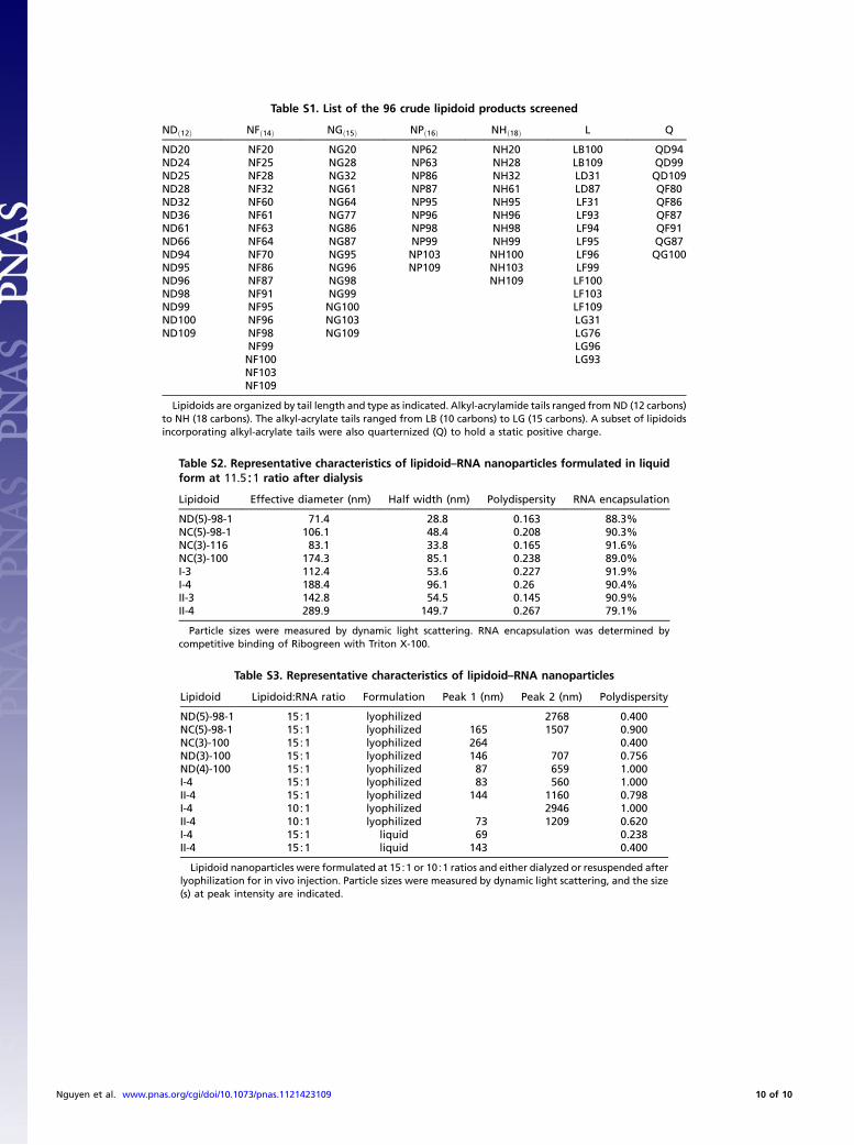
Table S1. List of the 96 crude lipidoid products screened
NDð12Þ NFð14Þ NGð15Þ NPð16Þ NHð18Þ L Q
ND20 NF20 NG20 NP62 NH20 LB100 QD94ND24 NF25 NG28 NP63 NH28 LB109 QD99ND25 NF28 NG32 NP86 NH32 LD31 QD109ND28 NF32 NG61 NP87 NH61 LD87 QF80ND32 NF60 NG64 NP95 NH95 LF31 QF86ND36 NF61 NG77 NP96 NH96 LF93 QF87ND61 NF63 NG86 NP98 NH98 LF94 QF91ND66 NF64 NG87 NP99 NH99 LF95 QG87ND94 NF70 NG95 NP103 NH100 LF96 QG100ND95 NF86 NG96 NP109 NH103 LF99ND96 NF87 NG98 NH109 LF100ND98 NF91 NG99 LF103ND99 NF95 NG100 LF109ND100 NF96 NG103 LG31ND109 NF98 NG109 LG76
NF99 LG96NF100 LG93NF103NF109
Lipidoids are organized by tail length and type as indicated. Alkyl-acrylamide tails ranged from ND (12 carbons)to NH (18 carbons). The alkyl-acrylate tails ranged from LB (10 carbons) to LG (15 carbons). A subset of lipidoidsincorporating alkyl-acrylate tails were also quarternized (Q) to hold a static positive charge.
Table S2. Representative characteristics of lipidoid–RNA nanoparticles formulated in liquidform at 11.5∶1 ratio after dialysis
Lipidoid Effective diameter (nm) Half width (nm) Polydispersity RNA encapsulation
ND(5)-98-1 71.4 28.8 0.163 88.3%NC(5)-98-1 106.1 48.4 0.208 90.3%NC(3)-116 83.1 33.8 0.165 91.6%NC(3)-100 174.3 85.1 0.238 89.0%I-3 112.4 53.6 0.227 91.9%I-4 188.4 96.1 0.26 90.4%II-3 142.8 54.5 0.145 90.9%II-4 289.9 149.7 0.267 79.1%
Particle sizes were measured by dynamic light scattering. RNA encapsulation was determined bycompetitive binding of Ribogreen with Triton X-100.
Table S3. Representative characteristics of lipidoid–RNA nanoparticles
Lipidoid Lipidoid:RNA ratio Formulation Peak 1 (nm) Peak 2 (nm) Polydispersity
ND(5)-98-1 15∶1 lyophilized 2768 0.400NC(5)-98-1 15∶1 lyophilized 165 1507 0.900NC(3)-100 15∶1 lyophilized 264 0.400ND(3)-100 15∶1 lyophilized 146 707 0.756ND(4)-100 15∶1 lyophilized 87 659 1.000I-4 15∶1 lyophilized 83 560 1.000II-4 15∶1 lyophilized 144 1160 0.798I-4 10∶1 lyophilized 2946 1.000II-4 10∶1 lyophilized 73 1209 0.620I-4 15∶1 liquid 69 0.238II-4 15∶1 liquid 143 0.400
Lipidoid nanoparticles were formulated at 15∶1 or 10∶1 ratios and either dialyzed or resuspended afterlyophilization for in vivo injection. Particle sizes were measured by dynamic light scattering, and the size(s) at peak intensity are indicated.
Nguyen et al. www.pnas.org/cgi/doi/10.1073/pnas.1121423109 10 of 10


