Embed Size (px)
Citation preview

Linköping University Medical Dissertations
No. 952
SURGERY FOR AORTIC STENOSIS with special reference to myocardial metabolism, postoperative
heart failure and long-term outcome
Farkas Vánky
Division of Cardiothoracic Surgery, Department of Medicine and Care,
Faculty of Health Sciences, SE-581 85 Linköping, Sweden
Linköping 2006

© Farkas Vánky, 2006
Printed by LIU-Tryck, Linköping, Sweden 2006 ISBN: 91-85497-89-4 ISSN: 0345-0082

“Evolution of science develops by a zigzag course of trial and error….”
Richard J. Bing
To Jane
Kristin, Charlotte and Katarina


ABSTRACT
Postoperative heart failure (PHF) remains a major determinant of the outcome after cardiac surgery.
However, characteristics of and risk factors for PHF after valve surgery have received little attention.
Post-ischaemic disturbances of myocardial metabolism that may contribute to PHF and are amenable
to metabolic treatment have been identified early after coronary surgery (CABG). Knowledge derived
from these studies may not be applicable to other patient groups. We therefore studied myocardial
energy metabolism in 20 elective patients undergoing aortic valve replacement (AVR) for isolated
aortic stenosis (AS). The metabolic studies indicated that myocardial oxidative metabolism had not
fully recovered when the procedure was completed. Free fatty acids were the only major substrates
taken up by the heart. Signs of preoperative and postoperative metabolic adaptation with substantial
uptake of glutamate, previously demonstrated in patients with coronary artery disease, were found.
Postoperative infusion of glutamate, (2 mL per kg body weight and hour of 0.125 M solution) based
on assessment of myocardial glutamate requirements in CABG patients, resulted in a two-fold increase
in myocardial glutamate uptake and a seven-fold increase in AV differences across the leg. This was
associated with a significant myocardial uptake of lactate and metabolic changes in the leg suggesting
mitigation of net amino acid loss and peripheral tissue lipolysis.
Characteristics of and risk factors for PHF were evaluated in 398 patients undergoing isolated AVR
for AS from 1 January 1995 to 31 December 2000. These were compared with 398 patients, matched
for age and sex, undergoing on-pump isolated CABG. Forty-five AVR and 47 CABG patients fulfilled
criteria for PHF and these were studied in detail. PHF usually presented at weaning from
cardiopulmonary bypass. After CABG it was closely associated with preoperative ischaemic events
and intraoperatively acquired myocardial infarction. Potential causes and eliciting events of PHF after
AVR for AS were obvious only in one-third of the patients. Risk factors for PHF after AVR for AS
indicated either pre-existing myocardial dysfunction, increased right or left ventricular after-load, or
intraoperatively acquired myocardial injury. PHF was associated with high early mortality after
CABG, whereas the consequences of PHF after AVR for AS became evident only with time, resulting
in a 42% five-year mortality. Although PHF had a different temporal impact on late mortality after
CABG and AVR for AS, it emerged as the statistically most significant risk factor for mortality
occurring within 5 years from surgery both after AVR for AS and after CABG. Potential implications
of our findings include needs for greater focus on preoperative surveillance of patients with AS for
optimal timing of surgery, mitigation of intraoperatively acquired myocardial injury and tailoring of
treatment for PHF. Furthermore, the findings have implications for long-term follow up of AS patients
after surgery.

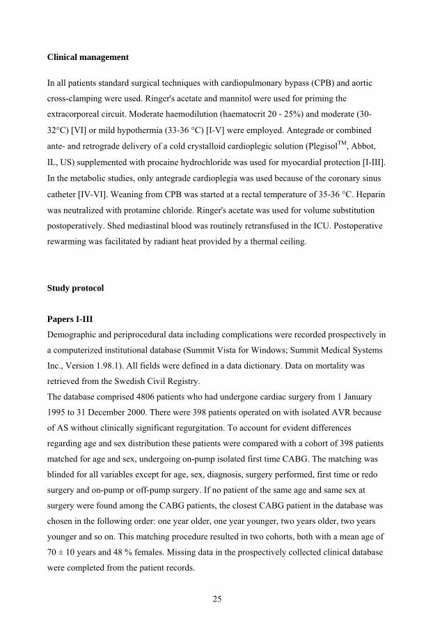
LIST OF ORIGINAL ARTICLES
This thesis is based on the following papers, which are referred by their Roman numerals:
[I]: Different characteristics of postoperative heart failure after surgery for aortic stenosis and
coronary disease. Vánky FB , Håkanson E, Maros T, Svedjeholm R. Scand Cardiovasc J.
2004;38:152-8.
[II]: Risk factors for postoperative heart failure in patients operated on for aortic stenosis.
Vánky FB , Håkanson E, Tamás E, Svedjeholm R. Ann Thorac Surg 2006;81:1297-304
[III]: Influence of early postoperative heart failure on five-year survival after surgery for
aortic stenosis compared with CABG. Vánky FB, Håkanson E, Svedjeholm R. Manuscript
[IV]: Myocardial metabolism before and after valve replacement for aortic stenosis. Vánky
FB, Håkanson E, Szabó Z, Jorfeldt L, Svedjeholm R. J Cardiovasc Surg 2006; in press
[V]: Does glutamate influence myocardial and peripheral tissue metabolism after valve
replacement for aortic stenosis? Vánky FB , Håkanson E, Jorfeldt L, Svedjeholm R. Clin Nutr
2006; in press
[VI]: Assessment of myocardial glutamate requirements early after coronary artery bypass
surgery. Vanhanen I, Svedjeholm R, Håkanson E, Joachimssom PO, Jorfeldt L, Vánky FB.
Scand Cardiovasc J. 1998;32:145-152.


TABLE OF CONTENTS
ABBREVIATIONS……………………………………………………………… 11
INTRODUCTION……………………………………………………………….. 13
AIMS OF THE STUDY…………………………………………………………. 21
MATERIAL AND METHODS…………………………………………………. 23
RESULTS……………………………………………………………………….. 35
DISCUSSION…………………………………………………………………… 57
SUMMARY AND CONCLUSIONS…………………………………………… 71
ACKNOWLEDGEMENTS…………………………………………………….. 74
REFERENCES………………………………………………………………….. 75


Abbreviations
A-Cs Arterial – coronary sinus difference
AS Aortic stenosis
AV Arterial – venous
AVR Aortic valve replacement
BCCA Branched-chain amino acids
BMI Body mass index
BSA Body surface area
CABG Coronary artery bypass grafting
CI Cardiac index
CPB Cardiopulmonary bypass
EOA Effective orifice area
FFA Free fatty acids
ICU Intensive care unit
LOS Low cardiac output syndrome
LV Left ventricle
LVSWI Left ventricular stroke work index
NYHA New York Heart Association
OR Odds ratio
PHF Postoperative heart failure
SAP Systolic arterial pressure
SD Standard deviation
SvO2 Mixed venous oxygen saturation
SVRI Systemic vascular resistance
11

12

Introduction
In the early years of its development cardiac surgery with deep hypothermia or with
cardiopulmonary bypass was a fairly traumatic procedure with high mortality and morbidity
rates. It was therefore mostly performed on children and young adults suffering from cardiac
diseases in which other treatment options had a very poor prognosis1. Improvements in
diagnostics, anaesthesia, surgical techniques, cardiopulmonary bypass and postoperative care
have resulted in an excellent outcome for cardiac surgery patients of today. It is logical that
research on patients undergoing aortic valve replacement has focused mainly on the
prosthesis. With the continuous improvements in and development of valve prostheses and the
concurrent advances in the surgical technique and perioperative care, cardiac surgery is no
longer withheld from elderly patients or patients with co-morbidities. Thus, the challenge of
today is to improve the good results in a patient population with increasing age and co-
morbidities. To succeed, it is necessary to identify and explore variables that have the most
pronounced influence on the postoperative outcome. The Ad Hoc Liaison Committee for
Standardizing Definitions of Prosthetic Heart Valve Morbidity has acknowledged that patient
variables may be more responsible for the outcome than valve-related factors2. The
Committee has also pointed out the lack of data on this issue and has encouraged investigators
to identify relevant patient factors in addition to factors related to operated valves.
Aortic stenosis
Aortic stenosis (AS) is the most common valvular disease among adults in the Western
hemisphere3, 4. It is characterized by an obstruction of the left ventricular outflow tract, which
may be either congenital or acquired. The origin of acquired AS is usually classified into
rheumatic and degenerative. The prevalence of AS increases with age and is around 2% in the
population older than 65 years and around 4% in that older than 85 years5, 6.
Aethiology
Before 1950 rheumatic disease was assumed to be the predominant cause of isolated aortic
valve stenosis, but this cause has declined since then in the western countries7-9. Rheumatic
valvulitis may produce oedema, lymphocytic infiltration and neovascularization of the
leaflets, followed later by leaflet thickening, commissural fusion, rolling of the leaflet edges
and later valvular calcification10. Today, degenerative calcification is considered to be the
13

most common cause of clinically evident aortic stenosis. It begins with collagen disruption
and small calcific deposits and ends up with heavily calcified immobile valve leaflets and
annulus10. Recent studies suggest, however, that lipoproteins may play a key role in the
development of aortic valve sclerosis11-13. Thus, sclerosis cannot be considered as a simple
degenerative process, but on the contrary it is complex and involves multiple pathogenic
mechanisms. Experimental, clinical and epidemiological data all support a link between aortic
valvulopathy and atherosclerosis: both are caused by inflammation, lipid deposition, and
accumulation of extracellular bone matrix protein11-13. The progress of calcification takes
place over several years and usually becomes symptomatic after the fifth decade of life,
earlier in bicuspid valves (present in up to 1% of the population14) and earlier in men than in
women.
Pathophysiology
The primary effect of aortic stenosis is an increased left ventricular afterload with secondary
impairment of left ventricular emptying in systole. Aortic stenosis may be quantified by
measuring the systolic pressure gradient across the aortic valve or by calculating an effective
aortic valve orifice area. Normal values for these two variables are around 5 mmHg and 3 to 4
cm2 respectively, while an aortic orifice valve area below 0.7 to 1.0 cm2 and a mean aortic
valve gradient above 50 mmHg correspond to severe aortic stenosis9, 10. An increase in the
pressure gradient across the aortic valve results in an elevated left ventricular pressure, in
order to maintain a normal pressure in the aorta. In turn, increased left ventricular wall stress
is thought to be the stimulus for left ventricular hypertrophy15. Severe left ventricular
hypertrophy may lead histologically to sarcomere disruption, disarray of myocardial filaments
and disappearance of organelles16. If left ventricular hypertrophy is insufficient to normalize
left ventricular wall stress, a chronic increase in wall stress may result in left ventricular
failure with decreased left ventricular contractility and progressive left ventricular dilatation.
Decreased left ventricular diastolic compliance appears as a consequence of both the left
ventricular hypertrophy and dilatation10.
The compensated phase of aortic stenosis with progressive left ventricular hypertrophy may
leave the patient asymptomatic for some decades10. The rate at which mild aortic stenosis
progresses to severe stenosis is variable, but different studies have shown a mean decrease in
the aortic valve orifice area of about 0.1 cm2/year9, 17, 18. However, most patients with
moderate to severe aortic stenosis ultimately develop symptoms of congestive heart failure,
angina or syncope9.
14

Congestive heart failure generally reflects elevated pulmonary venous pressure as a direct
effect of an increase in afterload or resulting from left ventricular diastolic dysfunction19.
Exertional angina pectoris in patients with AS is related to an impairment of subendocardial
blood flow due to increased left ventricular pressure20. Syncope also occurs, as a result either
of arrhythmia or of exercise-induced vasodilatation due to abnormal baroreceptor activity and
sudden changes in left ventricular pressure21.
Surgical correction of aortic stenosis immediately improves the left ventricular ejection
fraction, left ventricular end-diastolic volume and pulmonary capillary wedge pressure, as a
consequence of a reduced left ventricular afterload22, 23. Left ventricular hypertrophy from
aortic stenosis tends to decrease over a period of 6 to 12 months after aortic valve
replacement, but may not become totally normalized24.
Prognosis
Asymptomatic aortic stenosis has a good prognosis and the life expectancy is close to that in
the normal population25-29. Most patients eventually develop symptoms and the favourable
prognosis changes markedly when this occurs25-29. The survival rates 1, 2 and 3 years have
been found to be roughly 50%, 30% and 20% respectively in patients with symptomatic aortic
stenosis managed medically10. It is important to remember that the transition from an
asymptomatic to a symptomatic stage may be difficult to detect, particularly in elderly
patients, because of a gradual decrease in activity and a sedentary life style3, 30 .
Treatment
At present, medical therapy has a limited role in the treatment of aortic stenosis. Diuretics to
minimize symptoms of congestive heart failure and antiarrhythmics to control atrial
fibrillation may provide some symptomatic relief, but without altering the unfavourable
natural history of symptomatic aortic stenosis10. Afterload reduction may excessively reduce
the coronary perfusion pressure and is relatively contraindicated10.
The suggestion that high serum lipids may be contributory to aortic valve sclerosis has
resulted in recently completed and other, still ongoing studies. However, in the SATIRE trial,
lipid-lowering treatment with atorvastatin did not delay the progression of calcific aortic
stenosis31. Thus, aortic valve replacement is the treatment of choice for the majority of
patients with symptomatic aortic stenosis.
15

History of aortic valve surgery
In 1914 Tuffier described the first successful attempt to correct human aortic stenosis by
inserting a finger through the aortic wall to dilate the aortic valve32. Smithy and Parker
reported on an experimental study of aortic valvulotomy in 194710. Aortic valve dilatation
was subsequently performed with a mechanical dilator inserted through the left ventricle or
with a finger through a sleeve sewn onto the aorta (Ellis and Kirklin in 1955)10, 33. The first
time prosthesis was used to treat aortic valve disease was when Hufnagel and Harvey inserted
a ball valve in the descending aorta in 1953 in a patient with aortic valve regurgitation10.
However, this method left the patient with aortic insufficiency of the upper body. Treatment
of AS by replacement of the aortic valve did not become possible until after the development
of cardiopulmonary bypass by Gibbon in 195410. The first successful replacements of aortic
valves in 1960 by Bahnson and Harken, were followed by development of numerous different
prosthesis models10. There is still an ongoing search for the optimal prosthesis that needs no
anticoagulation, has lifelong durability even in young patients, does not reduce the aortic
orifice area and is easy to implant.
Prognosis after surgery for AS
The age- and gender-corrected survival after surgery for aortic stenosis has been reported to
be almost normalized from the first or second postoperative year34, 35. However, the surgical
procedure still carries an operative mortality of approximately 2 - 5% in spite of the fact that
results have improved dramatically over the last decades36-41. Most studies on the long-term
outcome are difficult to interpret because of mixed diagnoses and concomitant procedures.
Available data from patients operated on for isolated AS suggest that preoperative factors
such as high age, advanced functional class, reduced systolic and diastolic LV function,
hypertension, impaired renal function and a high left ventricular mass index are associated
with shortened long-term survival42-46. The role of patient-prosthesis mismatch, i.e. a small
effective orifice area in relation to body size, for the long-term outcome remains equivocal45-
49. The impact of periprocedural events on the long-term outcome has not been elucidated.
Postoperative heart failure
As early as in 1967, John Kirklin noted that when death occurs early after cardiac surgery it
was often related to low cardiac output50. In spite of the progress in cardiac surgery and
perioperative management, postoperative heart failure (PHF) remains one of the most
important causes of an adverse outcome of cardiac surgery41, 51, 52. It is therefore surprising
16

that although numerous articles have addressed different treatments of PHF, only a few
studies have addressed the issue of PHF per se. In 1973 Jarvinen et al reported a high
mortality and an increased incidence of extra-cardiac complications in association with low
cardiac output syndrome (LOS) after cardiac surgery involving both valve procedures and
coronary artery bypass grafting (CABG)53. In 1996 Rao reported a 9.1% incidence of LOS
after CABG54. In these patients the operative mortality was 16.9%, compared to 0.9% in those
without LOS (Table 1). A recent survey by the Northern New England study group of
outcomes in 8,641 patients who underwent CABG revealed that almost two-thirds of all in-
hospital deaths could be directly attributed to PHF. Furthermore, fatal heart failure accounted
for 80% of the difference between the surgeons with the highest and lowest mortalities.
Recently Maganti et al reported a high operative mortality in association with LOS after aortic
valve surgery in a heterogenous cohort41.
In spite of the grave consequences of PHF, there is still no generally accepted definition of
this condition and this might partly explain the limited number of studies on PHF. Available
data regarding PHF from the last two decades mainly derive from studies on CABG patients.
Studies addressing causes of and risk factors for PHF are sparse and the treatment of it, in
general, appears uniform regardless of the procedure and underlying causes55. Efforts to
prevent PHF and to tailor causal treatment require greater knowledge and insight into these
issues. Interpretation of available studies on different treatments of PHF is obscured by the
varying criteria used. This is demonstrated by the wide range (21-73%) in mortality reported
after use of intra-aortic balloon pump treatment56.
Haemodynamic criteria based on cardiac output measurements have been used by some
investigators. Such criteria are distinct and easy to comprehend, but it may be questioned
whether they actually always represent insufficient supply, as they do not give an account of
how well the systemic requirements are met. Heart failure, in physiological terms, reflects a
cardiac output insufficient to meet the systemic requirements. Evidences of such mismatch
between supply and demand are low mixed venous saturation and inadequate organ function.
In fact, the oxygen demand of peripheral tissues is the main determinant of cardiac output in
humans57, 58. This was illustrated by metabolic studies performed at our institution on elective
low risk CABG patients with well-preserved left ventricular function and uneventful
postoperative outcome59, 60. When anaesthetized, several of these patients, in spite of excellent
recovery of the myocardial metabolism and high mixed venous oxygen saturation, had a
cardiac output that would have been classified as heart failure with the use of haemodynamic
criteria alone.
17
Reference Surgicalprocedure
Numberof patients
Definition Prevalence(mortality)*
Kirklin 196750 Open heartsurgery
49 LCO: CI < 2.0 L/min/m2 BSA NA
Järvinen 197553 AVRMVRCABGCombination
1888110616
LOS: intraoperative death or requiring postoperative inotropic support to maintain ABPs > 90 mm Hg and diuresis > 30 mL/h
12% (70%) 33% (67%) 16% (77%) 81% (47%)
Hammermeister199061
CABGNon CABG
85691912
LCO: definition not given 3.8% (48%) 8.2% (52%)
Creswell 199262 Cardiacsurgery
7884 LCO treated by IABP: definition notgiven
4.0% (28.7%)
Rao 199654
Rao 200163CABG (all)CABG (low risk patients)
5113623
LOS: requiring postoperative IABP or inotropic support for > 30 minutes to maintain ABPs > 90 mm Hg and CO > 2.2 L/min
9.1% (16.9%)5.8% (NA)
Freeman 199764 CABG 3014 LOS: CI 2.0 L/min/m2 BSA, ABPm60 mm Hg and PCWP 8 mm Hg
NA
O’Connor 199851
Surgenor 200152CABG 8641 Fatal PHF: recognised from coding of
mode of death by a committee2.9%
Charlson 199965 CABG(elective)
248 Cardiogenic shock: urine output < 10 cc/h for > 2 h with ABPs < 90 mmHg or ABPm < 65 mm Hg and PCWP > 18 mm Hg in combination with CI < 2.2 L/min/m2 BSA
1.6% (75%)
Gorman 200055 All cardiacsurgery
NA LCO: CI < 2.2 L/min/m2 BSA NA
Hogue 200166 CABG 5113 LOS: CI < 2.0 L/min/m2 BSA for > 8 h 4.3% (18.9%)Shernan 200467
(multicenter trial, USA)
CABG+/- valve
914 LV dysfunction: use of 4 inotropic drugs, LVAD or IABP
4% (NA)
Maganti 200541 AVR 2255 LOS: requiring postoperative IABP or inotropic support for > 30 minutes to maintain ABPs > 90 mm Hg and CO > 2.2 L/min
3.9% (38%)
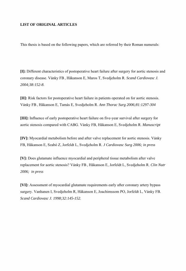
Table 1. Definitions and prevalence of low cardiac output (LCO), low cardiac output syndrome (LOS), postoperative heart failure (PHF), cardiogenic shock and left ventricular dysfunction (LV-dysfunction) after cardiac surgery in some previous scientific publications that either focus on postoperative heart failure, evaluate its treatment or use it as a study end-point. *Operative mortality rates in patients with PHF. AVR = aortic valve replacement;MVR = mitral valve replacement; CABG = coronary artery bypass grafting; CI = cardiac index; BSA = body surface area; ABPs = systolic arterial blood pressure; ABPm = meanarterial blood pressure; PCWP = pulmonary capillary wedge pressure; LVAD = left ventricular assist device; IABP = intra-aortic balloon pump; CO = cardiac output; NA = not available.
18

Myocardial metabolism
Studies of myocardial metabolism have revealed ways in which myocytes adapt to different
pathophysiological states68, 69. They have also demonstrated metabolic disturbances that may
contribute to contractile dysfunction, and hence provide a rationale and guidance for
metabolic interventions59, 70-74.
The heart is a continuously working muscle with limited energy storage and it therefore
requires a continuous supply of fuel. To cope with this, the heart is able to utilize a variety of
energy substrates. The choice of fuel, in the normal state, is primarily determined by the
availability of substrates, i.e. their arterial levels. In the fasting state, with elevated arterial
levels of free fatty acids (FFA) and low levels of glucose, FFA oxidation predominates. A
carbohydrate-rich meal shifts the myocardial metabolism towards glucose oxidation and
during vigorous exercise lactate may become the dominating fuel. In conditions such as
starvation or diabetic ketosis, ketone bodies may contribute significantly to myocardial energy
production75. Amino acids have limited importance as fuels, but certain amino acids seem to
play an important role for intermediary metabolism of the heart. Under normal conditions
cardiac function is not affected by the utilization of specific substrates75.
Myocardial metabolism in association with cardiac surgery has been studied in patients with
coronary artery disease59, 71, 73, 76-78. Various degrees of metabolic disturbances have been
described and these appear to be related to the magnitude of ischaemic insult and subside with
time after the release of the aortic cross-clamp59, 70, 73, 76, 78. Available data also suggest a
relation between degree of metabolic disturbance and myocardial function59, 70, 73, 76, 78. FFA
are the major source of energy postoperatively, whereas uptake of carbohydrates is restricted
by the systemic response to surgical stress71, 73, 79, 80. This constitutes a potentially
unfavourable metabolic state during recovery from ischaemia. Oxidation of FFA increases
myocardial oxygen expenditure and FFA cannot replenish Krebs cycle intermediates that are
depleted during ischaemia75, 81. These intermediates are essential for normal oxidative
metabolism. Studies early after CABG have demonstrated mechanisms to remedy this
situation. Certain amino acids, in particular glutamate, which serve as sources of Krebs cycle
intermediates and also contribute to clearing of metabolic waste such as lactate and ammonia,
are taken up abundantly during reperfusion71, 77, 80, 82. However, it is not known to what extent
the myocardial capacity to extract glutamate postoperatively is dependent on an adaptation to
chronic or repetitive ischaemia preoperatively, since available data derive from patients with
19

coronary artery disease68, 69. Other pathophysiological differences between patients with
coronary disease and those with valvular disease could also the influence pre- and
postoperative metabolic states.
20

Aims of the study
to determine the incidence of postoperative heart failure in patients operated on for
aortic stenosis compared with an age- and sex-matched cohort undergoing CABG
to identify characteristics of postoperative heart failure and potentially eliciting events
in patients operated on for AS and coronary artery disease, respectively
to describe the short-term outcome in patients with postoperative heart failure operated
on for AS compared with an age- and sex-matched cohort undergoing CABG
to analyse risk factors for postoperative heart failure in patients undergoing surgery for
isolated aortic stenosis
to investigate the impact of postoperative heart failure on the overall and late mortality
after AVR for AS compared with an age- and sex -matched cohort undergoing CABG
to describe the myocardial energy metabolism before and after surgery in patients
operated on for isolated AS
to investigate the impact of different rates of glutamate infusion on arterial levels of
glutamate and their relation to myocardial glutamate uptake after cardiac surgery
to evaluate the effects of glutamate infusion on myocardial metabolism after surgery
for AS
to describe peripheral tissue (leg) metabolism after surgery for aortic stenosis
to evaluate the effects of glutamate infusion on peripheral tissue (leg) metabolism after
surgery for AS
21

22

Material and methods
Patients
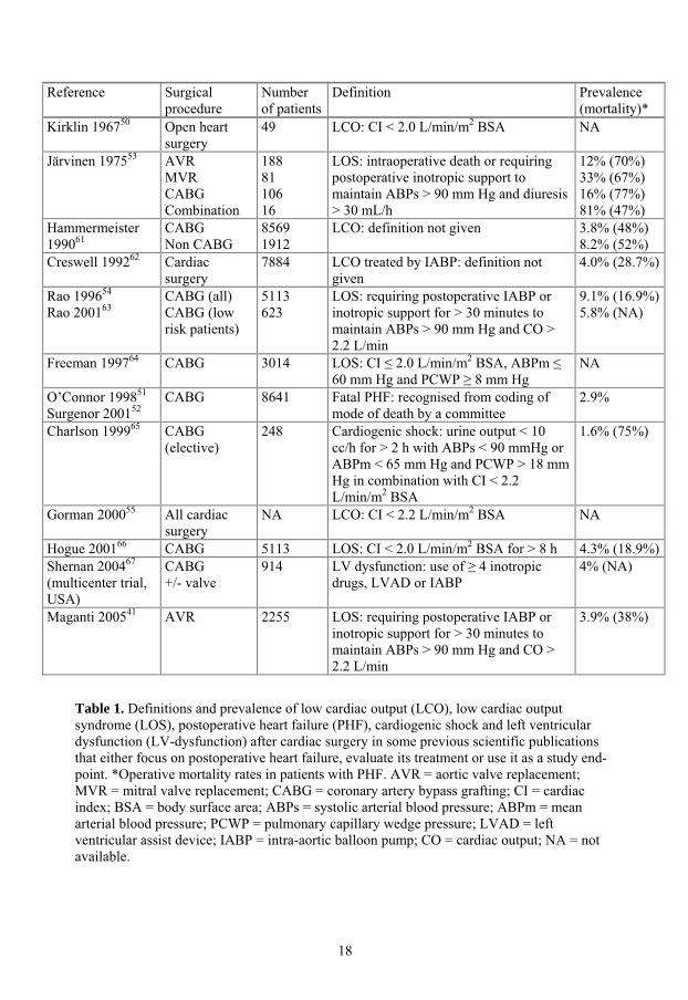
Basic preoperative demographics and intraoperative data on patients included in papers I-VI
are presented in Table 2.
Papers I-III
The selection of patients in papers I-III is depicted in Figure 1. All patients who underwent
isolated AVR because of AS without clinically significant regurgitation from 1 January 1995
to 31 December 2000 (n=398) were studied II-III . In paper III one patient was lost to
follow-up. A cohort of 398 patients matched to the AVR patients for age and sex, undergoing
on-pump isolated first time CABG was identified for comparison III . The total number of
patients included in papers I – III was thus 796. From these cohorts all AVR patients (n=45)
and CABG patients (n=47) that fulfilled our criteria for PHF were studied in detail I .
Papers IV-V
Twenty-two patients undergoing elective isolated AVR for AS were included prospectively in
papers IV - V. Patients with coronary artery disease, significant aortic valve regurgitation,
diabetes mellitus, left ventricular ejection fraction < 40%, weight > 100 kg or age > 75 years
were not included. Exclusion criteria were surgical difficulties resulting in an aortic cross-
clamp time exceeding 120 minutes and postoperative heart failure requiring inotropic or
metabolic treatment. Two patients were excluded on these grounds and 20 patients completed
the study.
Paper VI
This study comprised ten male patients with stable angina and well-preserved LV-function
undergoing elective CABG. Patients with diabetes mellitus or other major metabolic disorders
were not included.
Comments: AVR for AS represents the largest homogeneous group of valve procedures in our
practice. Our material included all patients operated on during a six-year period for AS
without clinically significant regurgitation or associated coronary artery disease, within an
23
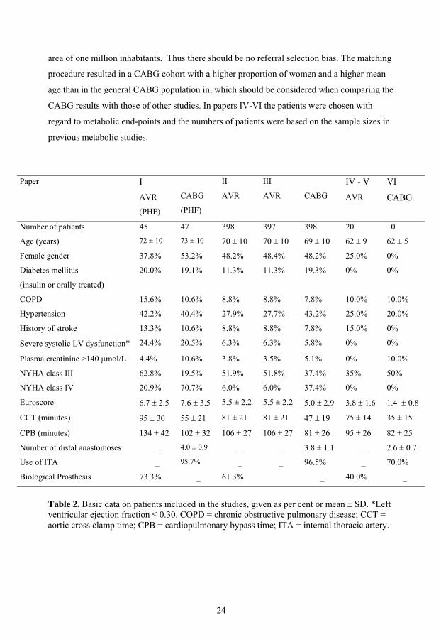
area of one million inhabitants. Thus there should be no referral selection bias. The matching
procedure resulted in a CABG cohort with a higher proportion of women and a higher mean
age than in the general CABG population in, which should be considered when comparing the
CABG results with those of other studies. In papers IV-VI the patients were chosen with
regard to metabolic end-points and the numbers of patients were based on the sample sizes in
previous metabolic studies.
Paper I
AVR
(PHF)
CABG
(PHF)
II
AVR
III
AVR CABG
IV - V
AVR
VI
CABG
Number of patients 45 47 398 397 398 20 10
Age (years) 72 ± 10 73 ± 10 70 ± 10 70 ± 10 69 ± 10 62 ± 9 62 ± 5
Female gender 37.8% 53.2% 48.2% 48.4% 48.2% 25.0% 0%
Diabetes mellitus
(insulin or orally treated)
20.0% 19.1% 11.3% 11.3% 19.3% 0% 0%
COPD 15.6% 10.6% 8.8% 8.8% 7.8% 10.0% 10.0%
Hypertension 42.2% 40.4% 27.9% 27.7% 43.2% 25.0% 20.0%
History of stroke 13.3% 10.6% 8.8% 8.8% 7.8% 15.0% 0%
Severe systolic LV dysfunction* 24.4% 20.5% 6.3% 6.3% 5.8% 0% 0%
Plasma creatinine >140 μmol/L 4.4% 10.6% 3.8% 3.5% 5.1% 0% 10.0%
NYHA class III 62.8% 19.5% 51.9% 51.8% 37.4% 35% 50%
NYHA class IV 20.9% 70.7% 6.0% 6.0% 37.4% 0% 0%
Euroscore 6.7 2.5 7.6 3.5 5.5 ± 2.2 5.5 ± 2.2 5.0 2.9 3.8 1.6 1.4 0.8
CCT (minutes) 95 30 55 21 81 ± 21 81 ± 21 47 19 75 ± 14 35 ± 15
CPB (minutes) 134 ± 42 102 ± 32 106 ± 27 106 ± 27 81 ± 26 95 ± 26 82 ± 25
Number of distal anastomoses _ 4.0 ± 0.9 _ _ 3.8 ± 1.1 _ 2.6 ± 0.7
Use of ITA _ 95.7% _ _ 96.5% _ 70.0%
Biological Prosthesis 73.3% _ 61.3% _ 40.0% _
Table 2. Basic data on patients included in the studies, given as per cent or mean SD. *Left ventricular ejection fraction 0.30. COPD = chronic obstructive pulmonary disease; CCT = aortic cross clamp time; CPB = cardiopulmonary bypass time; ITA = internal thoracic artery.
24
Clinical management
In all patients standard surgical techniques with cardiopulmonary bypass (CPB) and aortic
cross-clamping were used. Ringer's acetate and mannitol were used for priming the
extracorporeal circuit. Moderate haemodilution (haematocrit 20 - 25%) and moderate (30-
32 C) VI or mild hypothermia (33-36 C) I-V were employed. Antegrade or combined
ante- and retrograde delivery of a cold crystalloid cardioplegic solution (PlegisolTM, Abbot,
IL, US) supplemented with procaine hydrochloride was used for myocardial protection I-III .
In the metabolic studies, only antegrade cardioplegia was used because of the coronary sinus
catheter IV-VI . Weaning from CPB was started at a rectal temperature of 35-36 C. Heparin
was neutralized with protamine chloride. Ringer's acetate was used for volume substitution
postoperatively. Shed mediastinal blood was routinely retransfused in the ICU. Postoperative
rewarming was facilitated by radiant heat provided by a thermal ceiling.
Study protocol
Papers I-III
Demographic and periprocedural data including complications were recorded prospectively in
a computerized institutional database (Summit Vista for Windows; Summit Medical Systems
Inc., Version 1.98.1). All fields were defined in a data dictionary. Data on mortality was
retrieved from the Swedish Civil Registry.
The database comprised 4806 patients who had undergone cardiac surgery from 1 January
1995 to 31 December 2000. There were 398 patients operated on with isolated AVR because
of AS without clinically significant regurgitation. To account for evident differences
regarding age and sex distribution these patients were compared with a cohort of 398 patients
matched for age and sex, undergoing on-pump isolated first time CABG. The matching was
blinded for all variables except for age, sex, diagnosis, surgery performed, first time or redo
surgery and on-pump or off-pump surgery. If no patient of the same age and same sex at
surgery were found among the CABG patients, the closest CABG patient in the database was
chosen in the following order: one year older, one year younger, two years older, two years
younger and so on. This matching procedure resulted in two cohorts, both with a mean age of
70 ± 10 years and 48 % females. Missing data in the prospectively collected clinical database
were completed from the patient records.
25

Cardiac surgery patients 1995-20004806
AVR for Aorticstenosis
398CABG
398Matched for sex and age
PHF45
PHF47
Figure 1. Selection of patients in paper I – III. AVR = aortic valve replacement; CABG = coronary artery bypass surgery; PHF = postoperative heart failure.
Paper I
Two co-workers recognized PHF patients independently, on the basis of the criteria given
below in the section Calculations and Definitions. A second opinion was obtained from a third
co-worker in doubtful cases. Forty-five patients undergoing AVR (11.3%) and 47 patients
undergoing CABG (11.8%) were found to have had PHF. The records of these patients were
scrutinized for further details regarding different characteristics such as eliciting events,
presentation of heart failure, treatment and outcome.
26

Paper II
To identify risk factors for PHF in patients undergoing AVR for AS univariate and
multivariate logistic regression analysis was performed on all patients operated on for isolated
AS.
Paper III
To investigate the impact of PHF, in relation to other risk factors, on the long-term outcome,
univariate and multivariate logistic regression analysis with regard to five-year survival was
performed on the patients who had undergone surgery for isolated AS and on the matched
CABG cohort. The patients were followed up, regarding mortality over an average period of
7.2 ± 1.7 years that ranged from 5.2 to 11.2 years.
Papers IV – V
Twenty low-risk patients undergoing surgery for isolated aortic stenosis were studied pre- and
and postoperatively with respect to myocardial metabolism [IV,V], peripheral (leg) tissue
metabolism [V] and haemodynamic state [IV-V].
27

Catheterization
Surgerystarts
X-clamp
On - - - - - - - - - - - - - Off
Anaesthesia Surgeryends
Preop
T 1
Postop
Study infusion 1h
Start - - - - - - - - - - - - - - - - - Stop
T 0
Paper IV Paper V
Figure 2. Study protocol for papers IV and V. T0 = immediately before infusion of glutamate/placebo; T1 = 1 hour of infusion of glutamate/placebo.
Paper IV
To assess the myocardial metabolism and haemodynamic state in patients with isolated aortic
stenosis before and after surgery, 20 low-risk patients were studied immediately before
(preop) and after the procedure (postop) (Fig. 2). Blood samples were taken simultaneously
from the radial artery, coronary sinus and pulmonary artery. They were analysed for plasma
FFA, plasma amino acids and for the whole blood contents of glucose, lactate, glycerol and
oxygen.
The preoperative samples were taken immediately before the skin incision and the
postoperative samples were taken after the skin closure. Haemodynamic measurements were
also performed at preop and postop.
28

Paper V
To assess the impact of postoperative glutamate infusion on the myocardial metabolism,
peripheral (leg) tissue metabolism and haemodynamic state, the 20 patients from paper IV
were randomised to blinded infusion of 2 mL per kg body weight and hour of 0.125 mol/L L-
glutamate (glutamate group) or saline (control group). Blood samples were taken
simultaneously from the radial artery, the coronary sinus, the femoral vein and the pulmonary
artery immediately before (T0) the infusion was started and after 1 hour of infusion (T1).
Haemodynamic measurements were also performed at T0 and T1.
Comment: The postoperative state in paper IV and T0 in paper V correspond (Fig. 2). These
samples were taken on average 78 ± 16 minutes after release of the aortic cross-clamp.
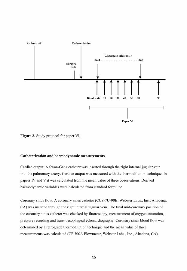
Paper VI
To assess the relationship between infusion rates, arterial levels and myocardial arterial-
coronary sinus differences of glutamate, 10 low risk CABG patients received a postoperative
infusion of 0.1 M glutamate solution over a period of one hour. To achieve a wide variation in
arterial glutamate levels, the infusion rate was varied between all patients and in addition the
infusion rate was changed for the second 30-minute period. Blood samples for whole blood
measurements of glutamate were taken simultaneously from the radial artery and the coronary
sinus before the start of glutamate infusion (basal state = 0), every 10 minutes during infusion
and 30 minutes after its termination. Arterial and coronary sinus plasma glutamate levels were
determined in the basal state, after 60 minutes of infusion and 30 minutes after termination of
the infusion.
Comment: The average time from release of the aortic cross-clamp to the basal state was 174
24 minutes. The glutamate infusion rates of glutamate varied from 10 to 87 mg per kg body
weight and hour.
29
Surgeryends
Glutamate infusion 1hStart - - - - - - - - - - - - - - - - - - - - - - Stop
Basal state 40 9050 60302010
Paper VI
X-clamp off Catheterization
Figure 3. Study protocol for paper VI.
Catheterization and haemodynamic measurements
Cardiac output: A Swan-Ganz catheter was inserted through the right internal jugular vein
into the pulmonary artery. Cardiac output was measured with the thermodilution technique. In
papers IV and V it was calculated from the mean value of three observations. Derived
haemodynamic variables were calculated from standard formulae.
Coronary sinus flow: A coronary sinus catheter (CCS-7U-90B, Webster Labs., Inc., Altadena,
CA) was inserted through the right internal jugular vein. The final mid-coronary position of
the coronary sinus catheter was checked by fluoroscopy, measurement of oxygen saturation,
pressure recording and trans-oesophageal echocardiography. Coronary sinus blood flow was
determined by a retrograde thermodilution technique and the mean value of three
measurements was calculated (CF 300A Flowmeter, Webster Labs., Inc., Altadena, CA).
30

Femoral vein: A 4 Fr (200 mm) catheter (BD Care FlowTM Becton Dickinson Critical Care
Systems, Singapore) was introduced into the right femoral vein [V]. The catheter was
positioned with the tip at the level of the inferior acetabulum with the aid of fluoroscopy and
verified by injection of 5 ml (320 mg I/ml) contrast medium (VisipaqueTM Amersham
Health).
Comment: A coronary sinus catheter provides information about the flow at the tip of the
catheter and this is dependent on the anatomy of the coronary sinus and the exact position of
the catheter tip. Owing to the anatomy of the coronary sinus, differences in position of the
catheter will influence the measurements. Thus, direct comparison of flux values between
individual patients should be avoided. However, the method is appropriate for assessment of
within-group changes and to some extent for inter-group comparisons.
In view of the larger myocardial mass in aortic stenosis patients, a higher coronary sinus
blood flow might have been expected in paper IV and V than in paper VI. Contrary to
expectation, we found a slightly lower flow in aortic stenosis patients. However, since the
catheter was inserted preoperatively in paper IV and V, a stable mid-coronary position was
aimed for to avoid intraoperative catheter displacement. This might have resulted in a
somewhat deeper tip location compared to that obtained in paper VI, which in turn could
explain the lower flow values.
Biochemical analyses IV - VI
Whole blood samples were immediately deproteinized with ice-cold perchloric acid as
described by Jorfeldt and Juhlin-Dannfelt83. After centrifugation, the protein-free extracts
were deep-frozen to -70ºC. The glutamate concentration in whole blood was determined
fluorometrically by an adapted glutamate dehydrogenase method84 [VI]. Alanine, D-glucose,
lactate and glycerol were also determined fluorometrically83, 85, 86 [IV-V]. Amino acids in
plasma were measured with a conventional amino acid analyser, AminoTac JLC-500, JEOL
Inc., Tokyo, Japan [IV-V] or Beckman system 6300, Beckman Instruments, Inc., Palo Alto,
CA [VI]. FFA were assayed in plasma as described by Ho87.
31

Comment: Because of the documented storage effect on plasma glutamate, all analyses were
done batchwise to minimize the effect on arterial-venous differences, care being taken that
corresponding arterial and venous samples were analysed simultaneously88.
Calculations and Definitions
PHF was defined as a haemodynamic state secondary to pump failure that is unable to meet
systemic demands without supportive measures other than correction of volume or vascular
resistance. A low cardiac output can be sufficient to supply the body demands in an
anaesthetized or sedated patient, and hence reliance on markers for adequate circulation, in
particular mixed venous oxygen saturation (SvO2), and echocardiographic evaluation rather
than fixed haemodynamic criteria, were employed to diagnose PHF57, 58. The following
relationship between SvO2 and systolic arterial pressure (SAP) provide our guidelines to
recognize inadequate circulation: SvO2<50% and SAP < 130 mmHg; SvO2<55% and
SAP<110 mmHg; SvO2<60% and SAP<90 mmHg; SvO2<65% and SAP<70 mmHg, after
correction of shivering and hypovolaemia. In the majority of patients PHF was evident at
weaning from cardiopulmonary bypass as an inability to wean from CPB or deteriorating
circulation and increasing filling pressures after weaning from CPB. In the remaining patients
echocardiographic evidence of left ventricular and /or right ventricular dysfunction associated
with above mentioned signs of inadequate circulation was used to diagnose PHF. The atrial
filling pressure, systemic pressure and Swan-Ganz data were used to aid the interpretations.
PHF in this thesis refers to heart failure occurring during the hospitalisation in association
with surgery. Supportive measures or treatment consisted of intraaortic balloon pump,
inotropic treatment and metabolic support with glucose-insulin-potassium and / or intravenous
glutamate 89.
Inotropic treatment [I] was defined as continuous infusion for more than 30 minutes of
catecholamines (adrenaline, dobutamine) or phosphodiesterase inhibitor (milrinone).
Effective orifice areas II-III for the different prostheses used are based on in vivo Doppler
echocardiographic measurements reported in the literature90-98.
32
Early or late mortality III was defined as mortality occurring within or later than 30 days
from surgery.
Myocardial flux of substrates [IV-VI] was calculated as the product of arterial-coronary sinus
concentration difference of blood or plasma and coronary sinus blood or plasma flow.
Coronary sinus plasma flow [IV-VI] was calculated as the product of coronary sinus blood
flow and 1-haematocrit.
Uptake or release of a substrate [IV-VI] was defined as flux or arterial-venous difference
significantly different from zero (p<0.05).
Oxygen consumption of the heart [IV-V] was estimated as the product of the arterial-coronary
sinus blood oxygen content difference and the coronary sinus blood flow. In calculations of
the O2 consumption of the whole body, the product of cardiac output and arterial-pulmonary
artery blood oxygen content difference was used.
Oxygen content of the blood [IV-V] was calculated according to the following formula:
Oxygen content (mmol/L) = (B-Hb g/L x sO2 % x 0.00062) + (pO2 kPa x 0.01).
Extraction ratio [IV-V] refers to the quotient of the arterial-venous difference and the arterial
level of a substrate.
Substrate oxygen equivalent [IV-V] was defined as the potential contribution of substrates to
myocardial oxygen consumption, assuming that the substrates taken up were fully oxidized.
According to stoichiometric analysis 3 mol of oxygen per mol of lactate, 6 mol of oxygen per
mol of glucose, and 23 mol of oxygen per mol of FFA (palmitate) are consumed73.
Statistical analysis
Data are presented as mean ± standard deviation (SD) [I-V] or mean ± standard error of the
mean (SEM) [VI]. Statistical significance was defined as p < 0.05. Statistical analyses were
performed with computerized statistical packages (Statistica 5.1 - 7, StatSoft Inc.,Tulsa,
MINITAB 13, Minitab Inc., Pennsylvania and SPSS 14.0 SPSS Inc., Chicago, IL.
33

Non-parametric tests, the Mann-Whitney U test and the Wilcoxon matched pairs test for
continuous variables with independent and dependent samples respectively and Fisher´s exact
test for categorical variables were used I - V . Correlations were computed with Spearman
rank IV .
In order to evaluate the independent risk factors II - III , univariate logistic regression was
first employed. Variables were then tested in a stepwise forward multivariate logistic
regression model if the univariate p value was less than 0.25. Hosmer-Lemeshow goodness-
of-fit statistics was calculated for the final models99. Cumulative long-term survival III was
assessed by Kaplan-Meier analysis.
In paper [VI] analysis of variance and post hoc comparisons with the LSD test were carried
out for evaluation of repeated measurements. For comparison of two sets of observations the
t-test was used if appropriate, and for samples lacking a normal distribution the Wilcoxon test
for matched pairs was used. Linear regression was used for analysis of correlation.
Comment: Conventionally, relative hazards for the risk of death during long-term follow up
are calculated with Cox proportional hazard model. However, the proportionality assumption
was not met in paper III and therefore univariate and multivariate logistic regression was
chosen to evaluate the impact of risk factors on defined time periods.
Ethical aspects
The studies were performed according to the principles of the Helsinki Declaration of Human
Rights and were approved by the Ethics Committee for Medical Research at the University
Hospital of Linköping. Written informed consent from the patients was obtained in the
prospective studies [IV - VI .
34

Results
Characteristics of PHF after surgery for AS and after CABG [I]
AVR (n=45) CABG (n=47) p value
Age (years) 72 ± 10 73 ± 10 n.s.
Female gender 37.8% (17/45) 53.2% (25/47) n.s.
Body mass index (kg/m2) 26.5 ± 4.1 27.2 ± 5.0 n.s.
Blood haemoglobin (g/L) 135 ± 15 123 ± 18 <0.001
Plasma creatinine (μmol/L) 107 ± 20 110 ± 33 n.s.
Plasma creatinine >167 μmol/L 0.0% (0/45) 10.6% (5/47) 0.06
DM (insulin or orally treated) 20.0% (9/45) 19.1% (9/47) n.s.
COPD 15.6% (7/45) 10.6% (5/47) n.s.
Cerebrovascular disease 13.3% (6/45) 10.6% (6/47) n.s.
Peripheral artery disease 4.7% (2/43) 8.7% (4/46) n.s.
Hypertension 42.2% (19/45) 40.4% (19/47) n.s.
Atrial fibrillation or flutter 22.2% (10/45) 2.1% (1/47) <0.01
NYHA III 62.8% (27/43) 19.5% (8/41) <0.001
NYHA IV 20.9% (5/43) 70.7% (20/41) <0.001
Severe systolic LV dysfunction* 24.4% (11/45) 20.5% (9/44) n.s.
Cardiogenic shock 2.2% (1/45) 4.3% (2/47) n.s.
Elective operation 73.3% (33) 12.8% (6) <0.001
Urgent operation 22.2% (10) 61.7% (29) <0.001
Emergency operation 4.4% (2) 25.5% (12) <0.01
Euroscore 6.7 ± 2.5 7.6 ± 3.5 n.s
Cross-clamp time (minutes) 95 ± 30 55 ± 21 <0.001
CPB time (minutes) 134 ± 42 102 ± 32 <0.001
Table 3. Demographic and clinical data for AVR and CABG patients with postoperative heart failure. Figures within brackets are the number of patients with the characteristics in question, obtained from those with available data. Results are given as percentage or as mean ± SD. *Left ventricular ejection fraction 0.30. DM = diabetes mellitus; COPD = chronic obstructive pulmonary disease; NYHA = New York Heart Association class; CPB = cardiopulmonary bypass.
35
Preoperative data
Both the AVR group and the CABG group had a high prevalence of severe systolic left
ventricular dysfunction preoperatively. In the CABG group 83.0% had unstable coronary
artery disease, 25.5% had significant left main stenosis and 91.3% had 3-vessel disease. The
AVR group compared favourably with the CABG group regarding NYHA class IV and
preoperative the haemoglobin level. However, preoperative atrial fibrillation was more
common in the AVR group (Table 3).
Intraoperative data
Compared with the AVR group, a higher proportion of the patients in the CABG group
underwent urgent or emergency procedures and myocardial ischaemia on induction occurred
more frequently. The aortic cross-clamp time and cardiopulmonary bypass (CPB) time were
significantly longer in the AVR group than in the CABG group (Table 3).
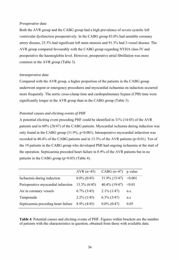
Potential causes and eliciting events of PHF
A potential eliciting event preceding PHF could be identified in 31% (14/45) of the AVR
patients and in 60% (28/47) of the CABG patients. Myocardial ischemia during induction was
only found in the CABG group (31.9%; p<0.001). Intraoperative myocardial infarction was
recorded in 40.4% of the CABG patients and in 13.3% of the AVR patients (p<0.01). Ten of
the 19 patients in the CABG group who developed PMI had ongoing ischaemia at the start of
the operation. Septicaemia preceded heart failure in 8.9% of the AVR patients but in no
patients in the CABG group (p=0.05) (Table 4).
AVR (n=45) CABG (n=47) p value
Ischaemia during induction 0.0% (0/45) 31.9% (15/47) <0.001
Perioperative myocardial infarction 13.3% (6/45) 40.4% (19/47) <0.01
Air in coronary vessels 6.7% (3/45) 2.1% (1/47) n.s.
Tamponade 2.2% (1/45) 6.3% (3/47) n.s
Septicaemia preceding heart failure 8.9% (4/45) 0.0% (0/47) 0.05
Table 4. Potential causes and eliciting events of PHF. Figures within brackets are the numberof patients with the characteristics in question, obtained from those with available data.
36

Presentation of PHF
Postoperative heart failure was evident at weaning from CPB in the majority of the patients in
both groups. PHF that presented later in the postoperative period was usually preceded by
tamponade or septicaemia. Seven of the AVR patients (15.6%) were considered to have
isolated right ventricular failure, compared to none in the CABG group (p<0.01) (Table 5).
Comment:
In patients that could sustain circulation that was sufficient to permit haemodynamic
measurements before treatment, available data showed SvO2 values of 53.7 ± 11.0 % (n=23)
and 48.0 ± 9.3 % (n=23) in the AVR and CABG groups respectively. At this stage the systolic
blood pressures were 84 ± 16 mmHg (n=30) and 82 ± 16 mmHg (n=31) while the pulmonary
arterial diastolic pressures were 22 ± 5 mmHg (n=19) and 18 ± 4 mmHg (n=26) in the
respective groups. Left ventricular stroke work indexes were 23.4 ± 10.2 6 gram-metres*m-2
(n=8) and 17.1 ± 7.3 6 gram-metres*m-2 (n=3) and cardiac indexes were 1.9 ± 0.5 (n=8) and
1.5 ± 0.2 L*min-1*m-2 (n=3) in patients with available Swan-Ganz data in the AVR group and
CABG groups respectively.
AVR (n=45) CABG (n=47) p value
Isolated left ventricular failure 75.0% (33/44) 79.5% (35/44) n.s.
Isolated right ventricular failure 15.6% (7/44) 0.0% (0/44) <0.01
Left + right ventricular failure 9.1% (4/44) 20.9% (9/44) n.s.
PHF at weaning off CPB 73.3% (33/45) 57.4% (27/47) n.s.
Early PHF in the OR 2.2% (1/45) 23.4% (11/47) <0.01
PHF in ICU 20.0% (9/45) 14.9% (7/47) n.s.
Table 5. Presentation of postoperative heart failure (PHF). Figures within brackets are the number of patients with the characteristics in question, obtained from those with available data. CPB = cardiopulmonary bypass; OR = operating room; ICU = intensive care unit.
37
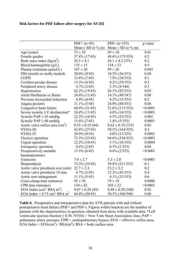
Risk factors for PHF failure after surgery for AS [II]
PHF+ (n=45)Mean ± SD or % (n)
PHF- (n=353) Mean ± SD or % (n)
p value
Age (years) 72 ± 10 69 ± 10 0.01 Female gender 37.8% (17/45) 49.6% (175/353) 0.2 Body mass index (kg/m2) 26.5 ± 4.1 26.1 ± 4.2 (351) 0.2 Blood haemoglobin (g/L) 135 ± 15 134 ± 13 0.5 Plasma creatinine (μmol/L) 107 ± 20 99 ± 26 0.003 DM (insulin or orally treated) 20.0% (9/45) 10.2% (36/353) 0.08 COPD 15.6% (7/45) 7.9% (28/353) 0.1 Cerebrovascular disease 13.3% (6/45) 8.2% (29/353) 0.3 Peripheral artery disease 4.7% (2/43) 2.3% (8/348) 0.3 Hypertension 42.2% (19/45) 26.1% (92/353) 0.03Atrial fibrillation or flutter 24.4% (11/45) 14.1% (49/347) 0.08 Previous myocardial infarction 8.9% (4/45) 4.2% (15/353) 0.2Angina pectoris 11.1% (5/45) 24.9% (88/353) 0.04 Congestive heart failure 68.9% (31/45) 32.6% (115/353) <0.0001Severe systolic LV dysfunction* 24.4% (11/45) 4.0% (14/353) <0.0001Systolic PAP 45 mmHg 22.2% (10/45) 6.5% (23/353) 0.002 Systolic PAP 60 mmHg 15.6% (7/45) 1.4% (5/353) 0.0001 Aortic valve orifice area (cm2) 0.55 ± 0.19 (44) 0.62 ± 0.19 (335) 0.03 NYHA III 62.8% (27/43) 50.5% (164/325) 0.2 NYHA IV 20.9% (9/43) 4.0% (13/325) 0.0003 Elective operation 73.3% (33/45) 94.6% (334/353) <0.0001Urgent operation 22.2% (10/45) 5.1% (18/353) 0.0004 Emergency operation 4.4% (2/45) 0.3% (1/353) 0.04 Preoperatively unstable haemodynamics
13.3% (6/45) 0.6% (2/353) <0.0001
Euroscore 7.0 ± 2.7 5.3 ± 2.0 <0.0001Bioprosthesis 73.3% (33/45) 59.8% (211/353) 0.1 Aortic valve prosthesis size (mm) 22.7 ± 2.3 23.2 ± 2.2 0.2 Aortic valve prosthesis 19 mm 6.7% (3/45) 12.2% (43/353) 0.4 Aortic root enlargement 11.1% (5/45) 9.1% (32/353) 0.6 Cross-clamp time (minutes) 95 ± 30 79 ± 18 0.0006 CPB time (minutes) 134 ± 42 103 ± 22 <0.0001EOA Index (cm2/ BSA m2) 0.87 ± 0.20 (45) 0.88 ± 0.20 (348) 0.92 EOA Index < 0.75 cm2/ BSA m2 44.4% (20/45) 30.5% (106/348) 0.06
Table 6. Preoperative and intraoperative data for AVR patients with and without postoperative heart failure (PHF+ and PHF-). Figures within brackets are the number of patients with the characteristics in question, obtained from those with available data. *Left ventricular ejection fraction 0.30. NYHA = New York Heart Association class; PAP = pulmonary artery pressure; CPB = cardiopulmonary bypass; EOA = effective orifice area; EOA Index = EOA(cm2) / BSA(m2); BSA = body surface area.
38
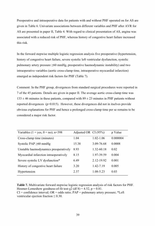
Preoperative and intraoperative data for patients with and without PHF operated on for AS are
given in Table 6. Univariate associations between different variables and PHF after AVR for
AS are presented in paper II, Table 4. With regard to clinical presentation of AS, angina was
associated with a reduced risk of PHF, whereas history of congestive heart failure increased
this risk.
In the forward stepwise multiple logistic regression analysis five preoperative (hypertension,
history of congestive heart failure, severe systolic left ventricular dysfunction, systolic
pulmonary artery pressure 60 mmHg, preoperative haemodynamic instability) and two
intraoperative variables (aortic cross-clamp time, intraoperative myocardial infarction)
emerged as independent risk factors for PHF (Table 7).
Comment: In the PHF group, divergences from standard surgical procedures were reported in
7 of the 45 patients. Details are given in paper II. The average aortic cross-clamp time was
133 ± 46 minutes in these patients, compared with 89 ± 23 minutes in PHF patients without
reported divergences (p=0.015). However, these divergences did not in itselves provide
obvious explanations for PHF and hence a prolonged cross-clamp time per se remains to be
considered a major risk factor.
Variables (1 = yes, 0 = no); n=398 Adjusted OR CI (95%) p Value
Cross-clamp time (minutes) 1.04 1.02-1.06 0.000004
Systolic PAP 60 mmHg 15.38 3.09-76.68 0.0008
Unstable haemodynamics preoperatively 8.93 1.32-60.18 0.02
Myocardial infarction intraoperatively 8.15 1.97-39.59 0.004
Severe systolic LV dysfunction* 6.49 2.12-19.92 0.001
History of congestive heart failure 3.20 1.42-7.19 0.005
Hypertension 2.37 1.08-5.23 0.03
Table 7. Multivariate forward stepwise logistic regression analysis of risk factors for PHF. Hosmer-Lemeshow goodness-of-fit test- 2 (df 8) = 4.52, p = 0.81. CI = confidence interval; OR = odds ratio; PAP = pulmonary artery pressure; *Left ventricular ejection fraction 0.30.
39
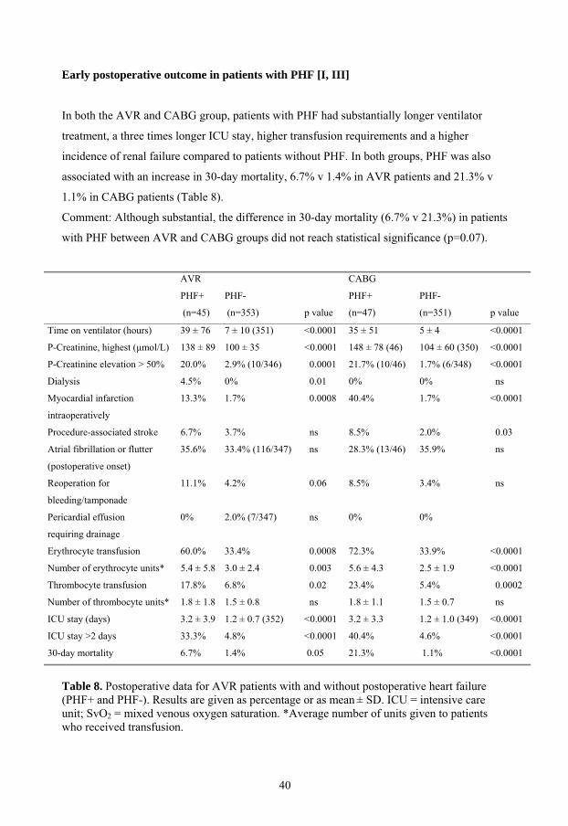
Early postoperative outcome in patients with PHF [I, III]
In both the AVR and CABG group, patients with PHF had substantially longer ventilator
treatment, a three times longer ICU stay, higher transfusion requirements and a higher
incidence of renal failure compared to patients without PHF. In both groups, PHF was also
associated with an increase in 30-day mortality, 6.7% v 1.4% in AVR patients and 21.3% v
1.1% in CABG patients (Table 8).
Comment: Although substantial, the difference in 30-day mortality (6.7% v 21.3%) in patients
with PHF between AVR and CABG groups did not reach statistical significance (p=0.07).
AVR
PHF+
(n=45)
PHF-
(n=353) p value
CABG
PHF+
(n=47)
PHF-
(n=351) p value
Time on ventilator (hours) 39 ± 76 7 ± 10 (351) <0.0001 35 ± 51 5 ± 4 <0.0001
P-Creatinine, highest (μmol/L) 138 ± 89 100 ± 35 <0.0001 148 ± 78 (46) 104 ± 60 (350) <0.0001
P-Creatinine elevation > 50% 20.0% 2.9% (10/346) 0.0001 21.7% (10/46) 1.7% (6/348) <0.0001
Dialysis 4.5% 0% 0.01 0% 0% ns
Myocardial infarction
intraoperatively
13.3% 1.7% 0.0008 40.4% 1.7% <0.0001
Procedure-associated stroke 6.7% 3.7% ns 8.5% 2.0% 0.03
Atrial fibrillation or flutter
(postoperative onset)
35.6% 33.4% (116/347) ns 28.3% (13/46) 35.9% ns
Reoperation for
bleeding/tamponade
11.1% 4.2% 0.06 8.5% 3.4% ns
Pericardial effusion
requiring drainage
0% 2.0% (7/347) ns 0% 0%
Erythrocyte transfusion 60.0% 33.4% 0.0008 72.3% 33.9% <0.0001
Number of erythrocyte units* 5.4 ± 5.8 3.0 ± 2.4 0.003 5.6 ± 4.3 2.5 ± 1.9 <0.0001
Thrombocyte transfusion 17.8% 6.8% 0.02 23.4% 5.4% 0.0002
Number of thrombocyte units* 1.8 ± 1.8 1.5 ± 0.8 ns 1.8 ± 1.1 1.5 ± 0.7 ns
ICU stay (days) 3.2 ± 3.9 1.2 ± 0.7 (352) <0.0001 3.2 ± 3.3 1.2 ± 1.0 (349) <0.0001
ICU stay >2 days 33.3% 4.8% <0.0001 40.4% 4.6% <0.0001
30-day mortality 6.7% 1.4% 0.05 21.3% 1.1% <0.0001
Table 8. Postoperative data for AVR patients with and without postoperative heart failure(PHF+ and PHF-). Results are given as percentage or as mean ± SD. ICU = intensive care unit; SvO2 = mixed venous oxygen saturation. *Average number of units given to patients who received transfusion.
40

Risk factors for early mortality [III]
The number of events was too few to permit meaningful analysis in the AVR group. In the
CABG group PHF (OR 15.93; CI(95%) 4.53-56.0; p=0.00002) and preoperative haemoglobin
(OR 0.96; CI(95%) 0.92-0.99; p=0.03) emerged as independent risk factors for 30-day
mortality. The Hosmer-Lemeshow goodness of fit p-value was 0.49.
Late postoperative outcome in patients with PHF [III]
Mortality
The crude mortality rates at 1 and 5 years in patients with and without PHF after AVR were
8.9% v 4.0% (p=0.13) and 42.2% v 14.2% (p<0.0001) respectively. Corresponding figures for
patients with and without PHF in the CABG group were 25.5% v 3.1% (p<0.0001) and 36.2%
v 11.1% (p=0.0015). The average time to death within the 5-year follow-up period for PHF
patients in the AVR and CABG group was 2.5 ±1.5 and 0.9 ± 1.5 years respectively
(p=0.007). Further details are presented in Table 9 and cumulative survival is illustrated in
Figure 4.
Mortality AVR
PHF
(n=45)
No PHF
(n=353)
p value CABG
PHF
(n=47)
No PHF
(n=351)
p value p value
AVR PHF v
CABG PHF
30-day (n) 6.7% (3) 1.4% (5) 0.05 21.3% (10) 1.1% (4) <0.0001 0.07
1-year (n) 8.9% (4) 4.0% (14) 0.13 25.5% (12) 3.1% (11) <0.0001 0.05
3-year (n) 22.2% (10) 8.0% (28) 0.005 29.8% (14) 6.0% (21) <0.0001 ns
4-year (n) 35.6% (16) 10.8% (38) 0.0001 36.2% (17) 8.8% (31) <0.0001 ns
5-year (n) 42.2% (19) 14.2% (50) <0.0001 36.2% (17) 11.1% (39) 0.0015 ns
Table 9. Crude mortality in patients undergoing AVR for AS and in the matched CABG patients.
41

Cumulative survival
0 (n=796)1 (n=795)
2 (n=795)3 (n=795)
4 (n=795)5 (n=795)
6 (n=497)7 (n=355)
Years (patients at risk)
0,0
0,1
0,2
0,3
0,4
0,5
0,6
0,7
0,8
0,9
1,0
Cum
ulat
ive
Prop
ortio
n Su
rviv
ing
AVR PHF- (n=353)
AVR PHF+ (n=45)
CABG PHF- (n=351)
CABG PHF+ (n=47)
Figure 4. Kaplan-Meier survival curves for AVR patients operated on for AS and for the matched CABG patients. PHF+ = patients with postoperative heart failure; PHF- = patients without postoperative heart failure. The p values are given in Table 9.
The role of PHF in relation to other risk factors for late mortality after AVR for AS
Postoperative heart failure emerged as the independent risk factor with the lowest p value for
both overall five-year mortality and for late mortality (between 30 days and 5 years).
Preoperative renal dysfunction, procedure-associated stroke, BMI<19 kg/m2, increasing age,
preoperative atrial fibrillation and preoperative anaemia also turned out as independent risk
factors for overall five-year mortality and late mortality occurring between 30 days and 5
years. Details regarding risk factors for mortality are presented in Table 10.
42
Variables (1 = yes, 0 = no) Overall mort (389)
OR (95% CI) p value
Late mort (381)
OR (95% CI) p value
PHF 5.14 (2.36-11.20) 0.00004 5.08 (2.28-11.31) 0.00006
Plasma creatinine >140 μmol/L
preoperatively
15.97 (3.69-69.11) 0.0002 16.88 (3.95-72.19) 0.0001
Stroke (procedure-associated) 4.36 (1.76-20.63) 0.004 4.80 (1.27-18.12) 0.02
Body mass index <19 kg/m2 7.71 (1.86-32.03) 0.005 9.03 (2.15-37.93) 0.003
Age (per year) 1.06 (1.02-1.11) 0.006 1.05 (1.00-1.09) 0.03
Preoperative atrial fibrillation 2.75 (1.31-5.74) 0.007 2.60 (1.20-5.61) 0.01
Preoperative anaemia* 2.27 (1.15-4.46) 0.02 2.33 (1.16-4.69) 0.02
Table 10. Multiple forward stepwise logistic regression analysis of risk factors for overall and late mortality after AVR for AS. Number of events: overall mortality = 69; late mortality = 61. Hosmer-Lemeshow goodness-of-fit test- 2 (df 8) = 7.3, p=0.50 and (df 8) = 7.0, p=0.53 for the overall mortality and late mortality model, respectively. *Blood haemoglobin < 134 g/L for men and < 117 g/L for women. OR = odds ratio. CI = confidence interval.
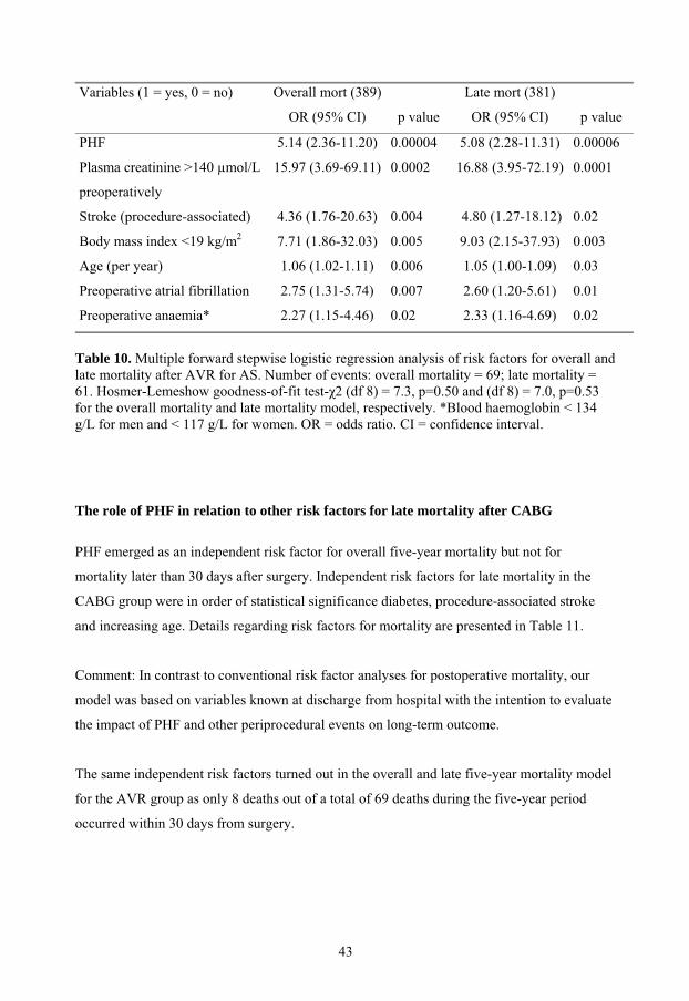
The role of PHF in relation to other risk factors for late mortality after CABG
PHF emerged as an independent risk factor for overall five-year mortality but not for
mortality later than 30 days after surgery. Independent risk factors for late mortality in the
CABG group were in order of statistical significance diabetes, procedure-associated stroke
and increasing age. Details regarding risk factors for mortality are presented in Table 11.
Comment: In contrast to conventional risk factor analyses for postoperative mortality, our
model was based on variables known at discharge from hospital with the intention to evaluate
the impact of PHF and other periprocedural events on long-term outcome.
The same independent risk factors turned out in the overall and late five-year mortality model
for the AVR group as only 8 deaths out of a total of 69 deaths during the five-year period
occurred within 30 days from surgery.
43

It may be argued that the event PHF is only a substitute for other risk factors and underlying
causes of PHF. To take these risk factors in account, they were also tested in the multivariate
model. None of these turned out as an independent risk factor for five-year mortality.
Variables (1 = yes, 0 = no) Overall mort (398)
OR (95% CI)
p value Late mort (383)
OR (95% CI)
p value
PHF 3.86 (1.87-7.95) 0.0003
Diabetes (insulin or orally treated) 2.65 (1.36-5.19) 0.004 2.76 (1.34-5.69) 0.006
Age (per year) 1.05 (1.01-1.09) 0.01 1.06 (1.01-1.11) 0.009
Stroke (procedure-associated) 4.69 (1.26-17.53) 0.02 6.45 (1.69-24.71) 0.006
Table 11. Multiple forward stepwise logistic regression analysis of risk factors for overall and late mortality in the matched CABG patients. Number of events: overall mortality = 56; late mortality = 42. Hosmer-Lemeshow goodness-of-fit test- 2 (df 8)= 11.5, p=0.18 and (df 8)= 11.3, p=0.18 for the overall mortality and late mortality model, respectively. OR = odds ratio. CI = confidence interval.
Haemodynamics and oxygen delivery before and after surgery for AS [IV]
Postoperatively a relative decrease in the myocardial oxygen extraction ratio (p=0.0001) and
oxygen consumption (p=0.14) by approximately 20% was observed. Coronary sinus oxygen
saturation consequently increased from 37 ± 5% to 49 ± 7% (p=0.0001). In contrast, a
significant increase in systemic oxygen consumption by 12% (p=0.0004) was observed
postoperatively.
Cardiac index increased by 22% from 1.8 ± 0.2 L/min preoperatively to 2.2 ± 0.4 L/min
postoperatively (p=0.002). A decrease in the systemic vascular resistance index of a similar
magnitude (23%) was found (p=0.009). Further details are given in Table II in paper IV.
44
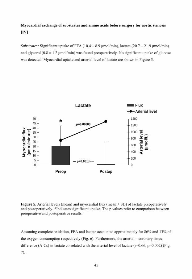
Myocardial exchange of substrates and amino acids before surgery for aortic stenosis
[IV]
Substrates: Significant uptake of FFA (10.4 ± 8.9 μmol/min), lactate (20.7 ± 21.9 μmol/min)
and glycerol (0.8 ± 1.2 μmol/min) was found preoperatively. No significant uptake of glucose
was detected. Myocardial uptake and arterial level of lactate are shown in Figure 5.
Lactate
05
101520253035404550
Preop Postop
Myo
card
ial f
lux
(µm
ol/m
inut
e)
0
200
400
600
800
1000
1200
1400
Arte
rial l
evel
(µm
ol/L
)
FluxArterial level
p=0.00009
--- p=0.0013 ---
*
Figure 5. Arterial levels (mean) and myocardial flux (mean ± SD) of lactate preoperatively and postoperatively. *Indicates significant uptake. The p values refer to comparison between preoperative and postoperative results.
Assuming complete oxidation, FFA and lactate accounted approximately for 86% and 13% of
the oxygen consumption respectively (Fig. 6). Furthermore, the arterial – coronary sinus
difference (A-Cs) in lactate correlated with the arterial level of lactate (r=0.66; p=0.002) (Fig.
7).
45

.
0
20
40
60
80
100
120
Preoperative Postoperative
(VO
2equ
ival
ents
/VO
2a-c
s)x1
00%
LactateFFA
86%
13%
88%
0
20
40
60
80
100
120
Preoperative Postoperative
(VO
2equ
ival
ents
/VO
2a-c
s)x1
00%
LactateFFA
86%
13%
88%
Figure 6. Substrate oxygen equivalents for FFA and lactate preoperatively and postoperatively. Substrate oxygen equivalents (VO2equivalents) are presented as percentage of the measured myocardial oxygen uptake (VO2a-cs), assuming complete oxidation of substrates taken up by the heart.
-0,8
-0,6
-0,4
-0,2
0,0
0,2
0,4
0,6
0,8
1,0
0,0 0,5 1,0 1,5 2,0 2,5 3,0Arterial level of lactate (mmol/L)
A-C
s di
ffere
nce
of la
ctat
e (m
mol
/L)
PreopPostop
r = 0.66p = 0.002
r = 0.45p = 0.047
Figure 7. Arterial level of lactate versus arterial-coronary sinus (A-Cs) difference in lactate preoperatively and postoperatively. r = Spearman rank correlation coefficient.
46
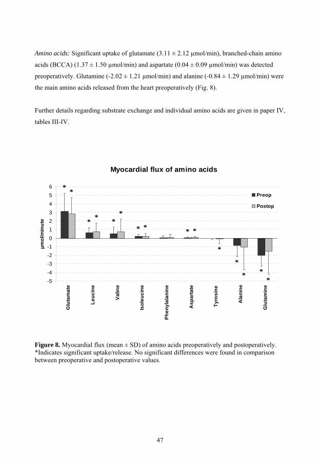
Amino acids: Significant uptake of glutamate (3.11 ± 2.12 μmol/min), branched-chain amino
acids (BCCA) (1.37 ± 1.50 μmol/min) and aspartate (0.04 ± 0.09 μmol/min) was detected
preoperatively. Glutamine (-2.02 ± 1.21 μmol/min) and alanine (-0.84 ± 1.29 μmol/min) were
the main amino acids released from the heart preoperatively (Fig. 8).
Further details regarding substrate exchange and individual amino acids are given in paper IV,
tables III-IV.
Myocardial flux of amino acids
-5-4-3-2-10123456
Glu
tam
ate
Leuc
ine
Valin
e
Isol
euci
ne
Phen
ylal
anin
e
Asp
arta
te
Tyro
sine
Ala
nine
Glu
tam
ine
µmol
/min
ute
Preop
Postop
* *
* * **
* * * *
*
*
* **
Figure 8. Myocardial flux (mean ± SD) of amino acids preoperatively and postoperatively. *Indicates significant uptake/release. No significant differences were found in comparisonbetween preoperative and postoperative values.
47

Myocardial exchange of substrates and amino acids after surgery for aortic stenosis [IV]
Substrates: The arterial level of lactate was significantly higher postoperatively compared to
the preoperative level (1.32 ± 0.46 v 0.75 ± 0.31 μmol/L, p<0.001). In spite of this, there was
a significant decrease and cessation of myocardial lactate uptake (Fig. 1). Furthermore, the
arterial level threshold for lactate uptake increased from approximately 0.25 mmol/L
preoperatively to 1.4 mmol/L postoperatively (Fig. 3). No glucose uptake was detected.
The arterial level of FFA decreased from the preoperative value (780 ± 119 μmol/L) to 646 ±
242 μmol/L (p = 0.03), but the myocardial uptake of FFA remained unchanged.
Amino acids: Like preoperatively, significant uptake of glutamate (2.80 ± 1.93 μmol/min) and
BCAA (1.67 ± 2.87 μmol/min), and a low uptake of aspartate (0.07 ± 0.11 μmol/min) were
found postoperatively. A significant correlation between arterial level of glutamate and A-Cs
difference was only found postoperatively (r=0.58; p=0.008) (Fig. 9).
0
10
20
30
40
50
60
70
80
0 50 100 150 200 250 300 350 400
Arterial level of glutamate (µmol/L)
A-C
sdi
ffere
nce
of g
utam
ate
(µm
ol/L
)
r = 0.58p = 0.008
Figure 9. Arterial plasma glutamate versus arterial-coronary sinus (A-Cs) difference in plasma glutamate postoperatively. r = Spearman rank correlation coefficient.
48

Glutamine (-1.52 ± 2.66 μmol/min) and alanine (-1.05 ± 2.62 μmol/min) remained the main
amino acids released from the heart postoperatively. In addition, a slight net release of
tyrosine (-0.07 ± 0.56 μmol/min; p=0.007) was observed in spite of a net uptake of total
amino acids (4.29 ± 17.19 μmol/min; p=0.02) (Fig. 4).
Further details regarding substrate exchange and individual amino acids are given in paper IV,
Tables III-IV.
Comment: The postoperative blood samples were taken on average 78 ± 16 minutes after
release of the aortic-cross clamp.
Effects of glutamate infusion on arterial levels of substrates and amino acids after
surgery for aortic stenosis [V]
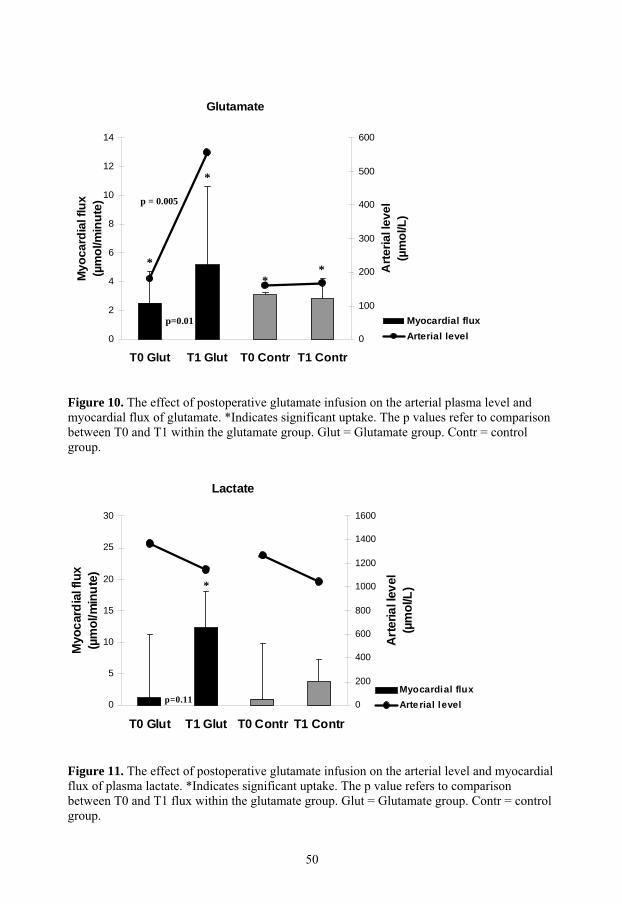
Glutamate infusion resulted in an approximately by three-fold increase in the arterial plasma
level of glutamate (from 166 ± 49 to 555 ± 177 μmol/L; p=0.005). No significant changes in
arterial levels of other substrates were observed from the basal state to one hour after the start
of the study infusion (from T0 to T1). However, the change in the plasma level of FFA from
T0 to T1 differed between the groups, with a decrease in the glutamate group and an increase
in the placebo group (p=0.049) (Figs 10-12).
49
Glutamate
0
2
4
6
8
10
12
14
T0 Glut T1 Glut T0 Contr T1 Contr
Myo
card
ial f
lux
(µm
ol/m
inut
e)
0
100
200
300
400
500
600
Arte
rial l
evel
(µm
ol/L
)
Myocardial fluxArterial level
p = 0.005
p=0.01
* **
*
Figure 10. The effect of postoperative glutamate infusion on the arterial plasma level and myocardial flux of glutamate. *Indicates significant uptake. The p values refer to comparisonbetween T0 and T1 within the glutamate group. Glut = Glutamate group. Contr = control group.
Lactate
0
5
10
15
20
25
30
T0 Glut T1 Glut T0 Contr T1 Contr
Myo
card
ial f
lux
(µm
ol/m
inut
e)
0
200
400
600
800
1000
1200
1400
1600
Art
eria
l lev
el(µ
mol
/L)
Myocardial fluxArte rial levelp=0.11
*
Figure 11. The effect of postoperative glutamate infusion on the arterial level and myocardial flux of plasma lactate. *Indicates significant uptake. The p value refers to comparisonbetween T0 and T1 flux within the glutamate group. Glut = Glutamate group. Contr = control group.
50

FFA
0
5
10
15
20
25
30
T0 Glut T1 Glut T0 Contr T1 Contr
Myo
card
ial f
lux
(µm
ol/m
inut
e)
0
100
200
300
400
500
600
700
800
Arte
rial l
evel
(µm
ol/L
)
Myocardial fluxArterial level
*
*
*
*
Figure 12. The effect of postoperative glutamate infusion on the arterial level and myocardial flux of plasma free fatty acids (FFA). *Indicates significant uptake. No significant differences were found between groups and within groups. The change in the arterial plasmalevel of FFA from T0 to T1 was significantly different between the glutamate and the control group (p = 0.049). Glut = Glutamate group. Contr = control group.
Effects of glutamate infusion on myocardial exchange of substrates and amino acids
after surgery for aortic stenosis [V]
Infusion of glutamate induced an approximate by two-fold increase in myocardial glutamate
uptake (from 2.5 ± 2.2 to 5.2 ± 5.4 μmol/min; p=0.013), and was associated with a close
significant increase in myocardial lactate uptake from T0 to T1 (p=0.11), resulting in a
significant uptake of lactate (12.3 ± 30.3 μmol/min) at T1 (Figs. 6-7). FFA (10.9 ± 3.5
μmol/min), glutamate (2.8 ± 1.4 μmol/min) and branched-chain amino acids (1.5 ± 0.7
μmol/min) were the only substrates or amino acids taken up by the heart in the placebo group
at T1 (Figs. 10-12).
The potential contributions of lactate and FFA to myocardial oxygen consumption are
illustrated in Figure 13. Further details regarding substrate exchange and individual amino
acids postoperatively in the glutamate and placebo groups are given in paper V, Tables 2-3.
51

T1
0
20
40
60
80
100
120
Glutamate group Control group
(VO
2equ
ival
ents
/VO
2a-c
s)x1
00%
Lactate
FFA
15%
89% 99%
Figure 13. Substrate oxygen equivalents (VO2equivalents) for FFA and lactate, as percentage of the measured myocardial oxygen uptake (VO2a-cs), assuming complete oxidation of substrates taken up by the heart. FFA = free fatty acids; T1 = 1 hour after start of infusion.
Effects of glutamate infusion on arterial-venous differences in substrates and amino
acids across the leg after surgery for aortic stenosis [V]
There was a net release of FFA, glycerol, lactate and amino acids from the leg in the
glutamate group at the start of the study infusion (T0). Glutamate was the only amino acid
taken up by the leg. Glutamate infusion was associated with an approximately seven-fold
increase in arterial – venous (AV) differences in glutamate (p=0.005). In the control group the
negative AV differences in FFA (p=0.007) and amino acids (p=0.047) increased from T0 to
T1. In contrast, AV-differences of FFA and amino acids tended to change in the opposite
direction in association with glutamate infusion, leading to significantly smaller negative AV
differences in FFA (p=0.04) and amino acids (p=0.01) at T1, compared with the control group
(Figs. 14-17). Further details regarding the substrate exchanges and individual amino acids
across the leg postoperatively, in the glutamate and placebo groups are given in paper V,
Table 4.
52

Comment: Leg blood flow measurements were not available. The haemodynamic variables
were stable in both groups. No significant differences were seen between the glutamate and
the control group and no significant changes occurred from T0 to T1. At T0 cardiac index was
2.3 ± 0.5 and 2.1 ± 0.3 L*min-1*m-2body surface area (BSA) in the glutamate and control
group respectively. At T1 it was 2.3 ± 0.2 and 2.3 ± 0.3 L*min-1*m-2BSA in the glutamate
and control group respectively. At T0 systemic vascular resistance index was 1873 ± 263 and
2341 ± 683 dyne*s*cm-5*m-2BSA in the glutamate group and control group respectively. At
T1 it was 2046 ± 288 and 2184 ± 357 dyne*s*cm-5*m-2BSA in the glutamate group and
control group respectively.
FFA
-200
-150
-100
-50
0
50
100
150
200
T0 Glut T1 Glut T0 Contr T1 Contr
AV
diffe
renc
e(µ
mol
/L)
-800
-700
-600
-500
-400
-300
-200
-100
0
100
200
300
400
500
600
700
800
Arte
rial l
evel
(µm
ol/L
)
AV differenceArterial level
*
*
*
p=0.04
p=0.007
Figure 14. The effect of postoperative glutamate infusion on arterial-venous (AV) differences in FFA over the leg. *Indicates significant release. Glut = Glutamate group. Contr = control group.
53

Glutamate
0
100
200
300
400
500
T0 Glut T1 Glut T0 Contr T1 Contr
AV
diffe
renc
e(µ
mol
/L)
0
100
200
300
400
500
600
Arte
rial l
evel
(µm
ol/L
)
AV differenceArterial level
*
*
* *
p = 0.005
p = 0.005
Figure 15. The effect of postoperative glutamate infusion on arterial-venous (AV) differences in glutamate over the leg. *Indicates significant uptake. The p values refer to comparisonbetween T0 and T1 within the glutamate group. Glut = Glutamate group. Contr = control group.
AV differences in total amino acids
-700
-600
-500
-400
-300
-200
-100
0
100
Glutamate Control
AV
diff
eren
ce (µ
mol
/L)
T0T1
*
**
*
p=0.047p=0.01
Figure 16. The effect of postoperative glutamate infusion on arterial-venous (AV) differences in total amino acids over the leg. *Indicates significant release.
54

AV differences in AA over the leg at T1
-700
-600
-500
-400
-300
-200
-100
0
100
200
300
400
Glu
tam
ate
Leuc
ine
Valin
e
Isol
euci
ne
Aspa
rtate
Phen
ylal
anin
e
Tyro
sine
Ala
nine
Glu
tam
ine
Tota
l AA
µmol
/L
GlutamateControl
* * * * ****
*
** * * *
**
*
*
*
p<0.001
p=0.01
Figure 17. Arterial-venous (AV) differences in amino acids (AA) over the leg after 1 hour of infusion of glutamate/placebo (T1). *Indicates significant uptake/release. The p values refer to comparison between the glutamate and the control groups.
55

Effects of different infusion rates of glutamate on arterial levels and myocardial uptake
of glutamate after CABG [VI]
The rates of glutamate infusion varied from 10 to 87 mg per kg body weight and hour during
the 60-minute period of glutamate infusion (Figure 1, paper VI). A dose-dependent linear
increase in arterial whole blood glutamate levels was found (r= 0.95; p<0.001; Figure 2, paper
VI).
Myocardial glutamate uptake correlated with arterial whole blood levels of glutamate during
the first 30-minute period (r = 0.60 p<0.001; Figure 4, paper VI) and uptake of glutamate
increased from 0.7 ± 0.2 μmol/min (basal state) to an early peak of 5.7 ± 1.2 μmol/min at 20
minutes. The arterial whole blood glutamate level had by then increased from 101 ± 10 to 186
± 18 μmol/L.
A further increase in the arterial glutamate level was not associated with an increase in
myocardial glutamate uptake. The significant correlation between arterial whole blood levels
and A-Cs differences in glutamate disappeared during the second 30-minute period when
higher infusion rates were used. Fractional extraction of glutamate by the heart from the
plasma decreased from 43.5% in the basal state to 11.0% at 60 minutes (p<0.001).
However, the individual peak uptake occurred during the first 30-minute period in 6 patients
and during the second 30-minute period in 4 patients. The mean of the individual peak
uptakes was 6.6 ± 1.1 μmol/min and occurred at an increase in arterial whole blood glutamate
by 172 ± 34 μmol/L.
Comment: A strong correlation was found between change in arterial plasma level and
change in whole blood level of glutamate from the basal state to 60 minutes after the start of
glutamate infusion (r= 0.90; p < 0.001; Figure, 3 paper VI). An increase in arterial whole
blood glutamate by 84 μmol/L (after 20 minutes when the mean peak uptake was recorded)
and 172 μmol/L (individual peak uptakes) corresponded to increases in arterial plasma levels
of glutamate by approximately 230 and 480 μmol/L respectively. Notably, the infusion rate
36.8 mg per kg body weight and hour, in paper V, resulted in an increase in plasma glutamate
by almost 400 μmol/L.
56

Discussion
Although postoperative heart failure (PHF) is a major determinant of the outcome after
cardiac surgery, the literature on this issue is surprisingly sparse. With the exception of a few
early studies, paper I is to our knowledge the first to address PHF per se in non-CABG
patients50, 53. The results corroborate previous findings suggesting that PHF is a common and
serious complication with high early mortality in CABG patients54, 61, 65, 66. Aortic valve
replacement (AVR) for aortic stenosis (AS) is a surgical procedure that provides immediate
relief of left ventricular outflow obstruction. In spite of this we found that the incidence of
PHF in patients operated on for AS was similar to that in an age- and sex-matched cohort of
CABG patients. However, different characteristics of PHF after AVR for aortic stenosis and
after CABG were identified. PHF after surgery for AS was not as closely associated with
preoperative ischaemic events and intraoperative myocardial infarction as PHF after CABG.
Although PHF after surgery for AS was associated with a marked increase in postoperative
morbidity and in the length of postoperative ICU stay, it appeared to be a comparatively
benign condition compared with PHF after CABG. However, our further studies showed that
the serious consequences of PHF after surgery for AS became evident only with time,
resulting in a 5-year mortality not less than that in patients with PHF after CABG.
Potentially eliciting causes of PHF in AVR patients were identified in only one-third of the
patients. However, risk factor analysis revealed a strong influence of extended aortic cross-
clamp times on development of PHF. This is in agreement with studies that have
demonstrated some degree of left and right ventricular dysfunction early after release of the
aortic cross-clamp in virtually all patients100. The term myocardial stunning has been coined
to describe post ischaemic myocardial dysfunction that persists after reperfusion despite the
absence of irreversible damage and despite restoration of normal flow. The pathogenesis of
myocardial stunning has not been definitively established. Generation of oxygen radicals and
calcium overload during reperfusion are among the proposed mechanisms that have received
the greatest attention101-103. Unfortunately, strategies to address these mechanisms specifically
have so far not been clinically successful in preventing or treating postoperative myocardial
dysfunction. Impaired energy production by mitochondria has also been suggested as an
underlying mechanism of myocardial stunning101. In CABG patients an association between
metabolic disturbances of the heart and transient contractile dysfunction has been
57
demonstrated72, 103-106. There are also reports of encouraging clinical results of metabolic
interventions to enhance myocardial recovery in patients with severe heart failure after
cardiac surgery70, 107-109. Our study is the first to address myocardial exchange of substrates in
patients undergoing surgery for AS.
METABOLIC CONSIDERATIONS IN SURGERY FOR AORTIC STENOSIS
Myocardial metabolism before surgery for AS
The myocardial metabolism in patients with coronary artery disease is characterized by an
increased uptake of glutamate and a release of alanine68, 110. It has been suggested that this
may represent metabolic adaptation to repetitive ischaemic episodes, as glutamate plays a key
role in the intermediary metabolism of the cardiomyocytes, particularly in association with
ischaemia. When the glycolytic activity exceeds the oxidative capacity, excess pyruvate is
mainly converted to lactate. However, the amino transferase reaction involving glutamate and
pyruvate results in production of alanine (instead of lactate) and alpha-ketoglutarate, of which
the latter enters the Krebs cycle. Alanine formed during this process is released from the
myocytes75. In our study the arterio-venous differences in both glutamate and alanine in
patients with AS were close to those originally reported by Mudge et al in patients with
coronary artery disease68. None of our patients had coronary artery disease and, hence, it is
conceivable that other conditions such as an increased myocardial workload and left
ventricular hypertrophy lead to similar adaptation possibly due to mismatch between oxygen
supply and demand.
FFA and lactate were taken up by the heart immediately before surgery in patients with aortic
stenosis. Assuming that all substrates taken up by the heart were completely oxidized, FFA
would account for approximately 85% of the myocardial oxygen consumption before surgery.
A predominant reliance on FFA uptake has previously been reported in the fasting state both
in normal post-absorptive man and in patients with coronary artery disease75, 78. The
remaining oxygen consumption can be explained by myocardial uptake of lactate. The high
extraction ratio of lactate and the clear correlation between arterial levels and uptake suggest
an undisturbed oxidative metabolism.
58

Myocardial metabolism after surgery for AS
FFA was the only major substrate taken up postoperatively. Lactate is a preferred substrate
under normal conditions and its uptake correlates with the arterial level78, 111. The fact that
myocardial lactate uptake had decreased and disappeared despite increasing arterial lactate
levels postoperatively suggests that normal Krebs cycle activity had not been resumed. In
view of the oxygen wasting properties of FFA and their inability to replenish Krebs cycle
intermediates, the predominant reliance on FFA as an energy source represents a potentially
unfavourable metabolic state under these circumstances and could further delay postoperative
recovery81, 112, 113.
Glutamate, BCAA and aspartate were the only amino acids taken up by the heart. The
magnitude of glutamate uptake, in particular, contributed to a total net uptake of amino acids.
Nevertheless, mild myocardial protein degradation was indicated by a small but significant
net release of tyrosine. The main amino acids released from the heart were alanine and
glutamine. These findings imply a metabolic state similar to that described early after
CABG71, 80. It is suggested that amino acids, particularly glutamate, play an important role. It
is conceivable that mitochondria were fed by amino acids both by external uptake and by
degradation of cardiac protein, whereas excess amino groups that formed during this process
were exported mainly as alanine and glutamine. Evidence for such a role of amino acids after
cardiac surgery has been provided by tracer studies in CABG patients77, 82, 114.
The significant correlation between arterial levels of plasma glutamate and arterial-coronary
sinus differences (r=0.58; p=0.008) found postoperatively could have therapeutic
implications, as it also suggests that glutamate uptake can be increased postoperatively by
exogenous supply in analogy with the observations reported after CABG.
Dosage of glutamate after cardiac surgery
It is not known to what extent myocardial glutamate uptake is determined by myocardial
needs or to what extent it may be limited by the amino acid carrier capacity. In an
investigation of L-glutamate transport in sarcolemmal vesicles from rat heart, Dinkelborg
found that acidosis enhanced glutamate transport, and such a mechanism could explain the
increased uptake of glutamate observed in ischaemic and post-ischaemic hearts115. Patients
59

with PHF can be expected to have greater requirements of glutamate than the low-risk
patients in our studies.
Pisarenko was the first to investigate the effect of intravenous glutamate infusion after cardiac
surgery70. He studied patients with postoperative heart failure treated with inotropic support.
Glutamate infusion resulted in functional recovery that was associated with a marked
improvement of the previously deranged myocardial metabolism, including conversion of
release of lactate and ammonia to uptake. The total amount of L-glutamate administrated was
approximately the same as in paper VI, but the infusions were given over a 15-minute period.
The arterial-coronary sinus plasma differences in glutamate achieved were almost twice as
large as those attained in our paper VI. Arterial plasma glutamate rose approximately 15-fold
and the fractional uptake decreased to less than 15%.
In CABG patients a strong correlation between arterial levels and myocardial uptake of
glutamate with fractional extraction rates of up to 50% was later reported71, 80. In view of the
high capacity to extract glutamate we concluded, even though patients with postoperative
heart failure appeared to have greater requirements of glutamate, that lower infusion rates
would have sufficed in Pisarenkos study. This could be of importance, as glutamate is not
considered a completely innocuous substance116, 117. Hence, our aim in paper VI was to
explore the relationship between infusion rates, arterial levels and myocardial uptake of
glutamate in an attempt to roughly estimate the lowest infusion rate sufficient to supply the
postoperative needs of glutamate. We found that glutamate infusion early after CABG
induced a dose-dependent linear increase in the arterial level of glutamate and that the
correlation between arterial glutamate level and glutamate uptake was lost during the second
half hour when the infusion rates were increased. Elevation of the arterial whole blood level
by more than two- to three-fold did not substantially increase the myocardial glutamate
uptake. This implied that glutamate infusion rates of 30-40 mg per kg body weight and hour
could suffice to achieve near maximal glutamate uptake in the heart, at least in routine
patients.
In clinical practice the lower range has subsequently been used with encouraging results in
patients with postoperative heart failure after CABG107. In patients with aortic stenosis in
paper V, we assumed that a slightly higher dose might be appropriate in view of their
hypertrophied hearts. Therefore, in paper V a dose of 2 ml per kg body weight and hour of a
60

0.125 M glutamate solution (36.8 mg of glutamate per kg body weight and hour) was infused
and this resulted in an increase in arterial plasma glutamate to the desired level. The
myocardial glutamate uptake was doubled and the significant correlation between arterial
level of glutamate and myocardial uptake, that was seen before the infusion, disappeared
suggesting that the dosage was appropriate to match the myocardial uptake capacity after
surgery for AS. This is true at least for the group in general, but owing to the large variations
between patients it cannot be ruled out that higher doses might be required in occasional
patients to optimize myocardial glutamate uptake.
Impact of glutamate infusion on myocardial metabolism after surgery for AS
Our study demonstrates that amplified glutamate uptake by the myocardium during glutamate
infusion is not restricted to patients with coronary artery disease. Glutamate infusion has
previously been reported to improve recovery of the myocardial metabolism and
haemodynamic state after CABG59. We found similar metabolic effects of postoperative
glutamate infusion on the heart after AVR for aortic stenosis, albeit somewhat more discrete.
The uptake of glutamate was doubled and associated with a significant uptake of lactate.
Since these findings associated with glutamate infusion are consistent with results from
previous animal and human studies using glutamate or its metabolite -ketoglutarate, it is
conceivable that they reflect a true effect59, 70, 78, 118-122. On reviewing the literature it seems
that the magnitude of the metabolic effect of glutamate on the heart has been related to the
magnitude of metabolic disturbance present when the glutamate infusion was started72. In our
study the change in lactate uptake within and between groups did not quite reach statistical
significance. Our sample size was chosen with regard to previous studies but in retrospect we
appreciate that a larger sample could have provided more conclusive results.
Assessment of myocardial metabolism with the coronary sinus catheter technique is
reproducible and well documented, which makes comparisons with previous studies
appropriate. However, the methodology does not provide direct information on the
intracellular metabolic pathways of substrates. Available tracer studies from humans in
association with coronary surgery have shown that glutamate taken up by the heart is fed into
the Krebs cycle and preferentially oxidized via the alanine aminotransferase reaction77, 114.
Thus, and also in agreement with animal experimental data, glutamate can be expected to
stimulate Krebs cycle activity in humans after ischaemia, which would be reflected by
enhanced lactate uptake.
61

Exchange of substrates in the leg (peripheral tissue) after surgery for AS
AV differences in substrates and amino acids across the leg revealed signs of net protein
catabolism, peripheral tissue lipolysis and lactate release postoperatively. The only amino acid
taken up by the leg was glutamate. This is in accordance with the post-traumatic neuro-
endocrine stress response and has been demonstrated previously after coronary surgery80, 123.
Impact of glutamate infusion on exchange of substrates in the leg (peripheral tissue)
after surgery for AS
The impact of glutamate on peripheral tissue metabolism has not been studied previously.
Glutamate infusion resulted in an approximately seven-fold increase in the AV difference in
glutamate and this was associated with significantly smaller negative AV differences in FFA
and amino acids compared with the control group.
The peripheral tissue results need to be interpreted with caution, owing to the lack of flow
measurements. Assessment of cardiac output, systemic vascular resistance and oxygen
extraction ratios across the leg argue against significant changes in leg blood flow in either
group. Furthermore, venous occlusion plethysmography measurements early after coronary
surgery have revealed stable flow in the leg during this time frame after cardiac surgery80, 123.
Thus it is conceivable that glutamate infusion counteracted an increase in peripheral tissue
release of FFA, which is consistent with the observed changes in plasma FFA. Decreased
plasma levels of FFA have been reported previously in association with glutamate
administration59, 124. This does not necessarily imply a direct effect of glutamate on lipolysis,
as glutamate has been shown to increase insulin secretion124. In normal subjects a maximal
reduction of plasma FFA may be seen at plasma insulin levels as low as 30 mU/L, which
correspond to the reported increment in insulin during glutamate infusion124, 125.
Our data also indicate that the marked increase in glutamate uptake in the leg was associated
with a mitigation of net amino acid loss. This appears to be explained by net uptake of
glutamate per se and is probably not directly connected to protein turnover, as no evident
change was observed in the exchange of non-metabolizable amino acids.
62

Taken together, the findings suggest that glutamate has a potentially favourable effect not
only on myocardial metabolism but also on peripheral tissue metabolism in the postoperative
phase.
POSTOPERATIVE HEART FAILURE AFTER SURGERY FOR AORTIC STENOSIS
Different characteristics of PHF after CABG and after surgery for AS
The incidence of PHF after surgery for isolated, first time AS was 11.3%, which was similar
to the incidence found in an age- and sex-matched cohort of patients undergoing CABG
(11.8%). However, the eliciting events and presentation of heart failure differed between the
groups. PHF presented at weaning from cardiopulmonary bypass or within the first hours after
surgery in the majority of patients both after AVR for AS and after CABG. PHF that had its
onset later in the postoperative period was usually preceded by tamponade or septicaemia.
PHF in the CABG group was more clearly related to preoperative ischaemia and perioperative
myocardial infarction. Forty per cent of the patients with PHF after CABG had perioperative
myocardial infarction. Half of these showed signs of ischaemia at the induction of
anaesthesia. This is in agreement with previous studies in which preoperative ischaemia was
identified as a major cause of perioperative myocardial infarction in association with
CABG126, 127.
The AVR group had longer aortic cross-clamp time and a higher rate of isolated right
ventricular heart failure postoperatively. PHF after surgery for AS was not associated with
preoperative ischaemic events and perioperative myocardial infarction was only found in 13%
of the patients. This could explain the more favourable short-term outcome in patients with
PHF after surgery for AS. The few patients in the AVR group with perioperative myocardial
infarction had a high postoperative mortality rate. Mechanical circulatory assist was less
frequently used in the AVR group, which also suggests less severe heart failure in the AVR
patients. However, it could also imply a lower threshold for using mechanical circulatory
assistance in association with myocardial ischaemia.
There are data suggesting that right ventricular dysfunction may contribute to low output
syndrome in almost half of the cases after CABG128. Isolated right ventricular failure, on the
63
other hand, was found in only 2 out of 31 patients with low output syndrome after CABG. In
contrast it was noted in 2 out of 7 patients with low output syndrome after AVR. In agreement
with these findings, isolated right ventricular failure appeared to be a problem mainly
associated with heart failure after AVR in our study. Several factors may have contributed to
right ventricular dysfunction after AVR for AS. The occurrence of an increased after-load for
the right ventricle due to left ventricular hypertrophy and diastolic dysfunction, insufficient
protection of the right ventricle during retrograde delivery of cardioplegia, and air embolism
into the right coronary artery due to inadequate de-airing procedures can all lead to right
ventricular dysfunction128-131.
Events eliciting PHF could be identified in approximately 2/3 of the CABG patients but only
1/3 of the AVR patients. In addition, the surgical procedure per se, especially cardioplegic
arrest, contributes to postoperative depression of myocardial function100. In particular, patients
with preoperatively compromised left ventricular function are at increased risk of developing
clinical evidence of heart failure after surgery54. Hence, it is conceivable that preoperative left
ventricular dysfunction, which was common in both groups in our study, was an important
contributor to PHF54, 132, 133. To shed further light on the mechanisms behind PHF in these
patients, we decided to analyse risk factors for PHF in the total cohort of patients undergoing
surgery for isolated AS.
Risk factors for PHF after surgery for AS
With regard to the clinical presentation of AS, angina according to univariate analysis was
associated with a reduced risk of PHF, whereas history of congestive heart failure increased
this risk. Multivariate logistic regression analysis identified seven variables as independent
risk factors for PHF after surgery for isolated AS: five preoperative (hypertension, history of
congestive heart failure, severe systolic left ventricular function, pulmonary hypertension,
haemodynamic instability preoperatively) and two intraoperative (aortic cross-clamp time,
intraoperative myocardial infarction).
After submission of paper II we found a recent paper by Maganti et al, on risk factors for low
cardiac output syndrome (LOS) after AVR41. Their paper focused on preoperative variables in
order to predict operative mortality and LOS in a heterogeneous cohort including all
indications for AVR. They identified renal failure, earlier year of operation, left ventricular
64

dysfunction, shock, female gender and increasing age as risk factors for LOS. Out of these,
left ventricular dysfunction and shock agree with our results.
In order to define preventive measures and to improve the therapeutic approach, it can be
more useful to establish pathophysiological relationships between risk factors and
complications. Thus, our aim was to identify not only predictive variables, but also factors
with a mechanistic relationship to PHF. Variables tested in our models were chosen to meet
these aims. Those identified in the final model seem to indicate either preoperatively existing
or intraoperatively acquired myocardial dysfunction. Furthermore, arterial and pulmonary
hypertension indicating increased left and right ventricular after-load, respectively, emerged
as independent risk factors.
Pulmonary hypertension, haemodynamic instability before anaesthesia, intraoperative
myocardial infarction and severe systolic left ventricular dysfunction preoperatively had the
highest odds ratios for PHF. However, the hazards associated with them were not matched by
their explanatory role in the final model. In contrast, the statistically most significant risk
factor was aortic cross-clamp time, which was also found to have the highest explanatory role.
Extensions of aortic-cross clamp times were partly explained by intraoperative surgical
problems. However, these surgical problems did not in themselves provide obvious
explanations for PHF and hence a prolonged cross-clamp time per se remains to be considered
a major risk factor.
The influence of PHF on the early postoperative outcome after AVR for AS
Although PHF after AVR for AS appeared to be a less serious condition than PHF after
CABG, it was associated with an almost five-fold increase in 30-day mortality (6.7% v 1.4%),
a substantial increase in postoperative morbidity and consequently increased utilization of
ICU resources compared with AVR patients without PHF. The average ICU stay was more
than doubled and the time on ventilator was increased more than five-fold. Transfusion
requirements were approximately doubled.
Different temporal impact of PHF on mortality after AVR for AS compared with CABG
Our study demonstrates differences in the temporal patterns of the mortality after PHF in
CABG and AVR patients, which to our knowledge has not previously been described. PHF is
a complication that carries a high early mortality after CABG, whereas its influence on late
65

mortality after this operation is limited. In contrast, to fully recognize the serious
consequences of PHF on survival after AVR for AS an extended follow-up is required. After
five years, the mortality exceeded that found in the CABG patients with PHF.
Risk factors for late mortality after AVR for AS
To clarify the relative importance of PHF for late mortality, multivariate risk factor analyses
were performed. In order of statistical significance, PHF, preoperative renal dysfunction, low
body mass index, preoperative atrial fibrillation, procedure-associated stroke, preoperative
anaemia and increasing age emerged as independent risk factors for mortality occurring
between 30 days and five years after AVR for AS.
It may be argued that PHF as an event is only a substitute for other risk factors and underlying
causes of PHF. However, the previously identified risk factors for PHF (hypertension, history
of congestive heart failure, severe systolic left ventricular dysfunction, pulmonary
hypertension, preoperative haemodynamic instability, aortic cross-clamp time, intraoperative
myocardial infarction) were also tested in the multivariate model. None of these emerged as
an independent risk factor for late mortality. This emphasizes the importance of PHF per se as
a prognostic factor for late mortality after surgery for AS.
The reasons for the extended unfavourable impact of PHF after surgery for AS remain to be
clarified. A similar sustained negative influence of preoperative left ventricular diastolic
dysfunction on long-term survival has recently been demonstrated45. The authors speculated
that diastolic dysfunction represented irreversible structural myocardial abnormalities,
although they acknowledged the lack of direct histopathological evidence. We had no specific
data on left ventricular diastolic dysfunction in our database, but pulmonary hypertension, a
related variable, emerged as a strong risk factor for PHF and was associated with a high
mortality rate.
Risk factors for early and late mortality after CABG
In CABG patients, PHF has previously been reported to be the major cause of early
postoperative mortality51, 52. Our results are consistent with these findings and this is well
illustrated by the survival curves (Fig. 4). PHF had the highest significance of the risk factors
for early mortality after CABG in our study. Its impact on early mortality was so pronounced
that PHF also emerged as the risk factor with the highest significance for mortality occurring
66

within five years from surgery, although it did not turn out as an independent risk factor for
late mortality. The finding in paper I, which indicates that PHF after CABG was closely
related to ischaemic events during the early stages of surgery and to intraoperative myocardial
infarction, could explain the unfavourable short-term outcome.
Procedure-associated stroke, diabetes and increasing age were the only significant
independent risk factors for mortality occurring between 30 days and 5 years after surgery.
These results may not be entirely representative for CABG patients, as the cohort was
selected, but nevertheless the identified risk factors are in general agreement with previous
experience61, 66, 134, 135.
The patient, the procedure, the prosthesis and PHF
Research in valve surgery has mainly focused on the prosthesis. In addition, preoperative risk
factors and postoperative echocardiographic characteristics, such as regression of left
ventricular mass, early improvement of the left ventricular ejection fraction and diastolic
function, have been evaluated as prognostic factors for the long-term outcome after valve
surgery.
Several studies have dealt with preoperative risk factors for operative and in-hospital
mortality after AVR37-40, 46, 135-137 and a few have also addressed risk factors for long-term
mortality42-46. In contrast to most previous studies, our study is characterized by its
homogeneous cohort of AS patients, and that the analysis is based on variables known at
discharge from hospital, with the intention of evaluating the impact of PHF and other
periprocedural events in relation to other risk factors on long-term survival. Interestingly PHF
emerged as the risk factor with the highest statistical significance for mortality within 5-years
after AVR for AS although virtually all previously documented preoperative and
periprocedural risk factors were included in the analysis. However, evaluation of follow-up
variables such as echocardiographic data, medication or biochemical markers such as
natriuretic peptides were beyond the scope of this thesis.
The other independent risk factors for mortality within 5-years after AVR for AS besides PHF
that emerged in our model were procedure-associated stroke, preoperative renal dysfunction,
increasing age, preoperative atrial fibrillation, low BMI and anaemia, which all to some extent
have been reported being risk factors for mortality after cardiac surgery37-40, 42, 46, 135-141.
67
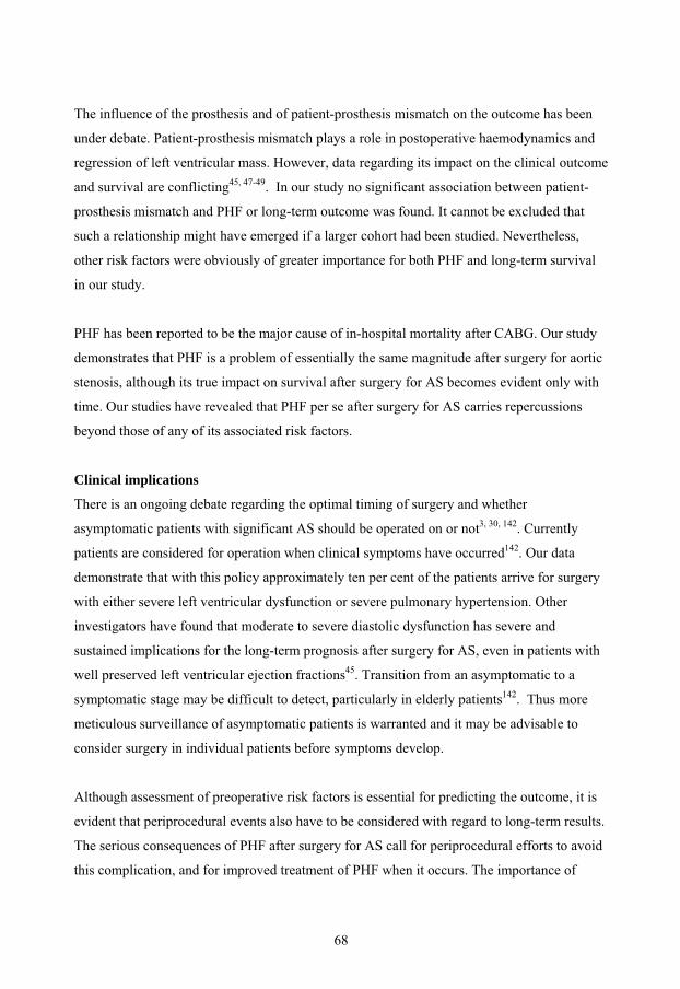
The influence of the prosthesis and of patient-prosthesis mismatch on the outcome has been
under debate. Patient-prosthesis mismatch plays a role in postoperative haemodynamics and
regression of left ventricular mass. However, data regarding its impact on the clinical outcome
and survival are conflicting45, 47-49. In our study no significant association between patient-
prosthesis mismatch and PHF or long-term outcome was found. It cannot be excluded that
such a relationship might have emerged if a larger cohort had been studied. Nevertheless,
other risk factors were obviously of greater importance for both PHF and long-term survival
in our study.
PHF has been reported to be the major cause of in-hospital mortality after CABG. Our study
demonstrates that PHF is a problem of essentially the same magnitude after surgery for aortic
stenosis, although its true impact on survival after surgery for AS becomes evident only with
time. Our studies have revealed that PHF per se after surgery for AS carries repercussions
beyond those of any of its associated risk factors.
Clinical implications
There is an ongoing debate regarding the optimal timing of surgery and whether
asymptomatic patients with significant AS should be operated on or not3, 30, 142. Currently
patients are considered for operation when clinical symptoms have occurred142. Our data
demonstrate that with this policy approximately ten per cent of the patients arrive for surgery
with either severe left ventricular dysfunction or severe pulmonary hypertension. Other
investigators have found that moderate to severe diastolic dysfunction has severe and
sustained implications for the long-term prognosis after surgery for AS, even in patients with
well preserved left ventricular ejection fractions45. Transition from an asymptomatic to a
symptomatic stage may be difficult to detect, particularly in elderly patients142. Thus more
meticulous surveillance of asymptomatic patients is warranted and it may be advisable to
consider surgery in individual patients before symptoms develop.
Although assessment of preoperative risk factors is essential for predicting the outcome, it is
evident that periprocedural events also have to be considered with regard to long-term results.
The serious consequences of PHF after surgery for AS call for periprocedural efforts to avoid
this complication, and for improved treatment of PHF when it occurs. The importance of
68

addressing myocardial protection and minimizing the cross-clamp time, particularly in
patients with preoperatively compromised myocardial function, is highlighted by our results.
It is apparent from our study and from the literature that treatment of PHF seems to be quite
standardized, with use of inotropic agents regardless of the cause of PHF or the type of
procedure64. On the basis of our findings in and available data in the literature we suggest that
greater focus on causal aspects of heart failure, tailoring of treatment and prophylactic
measures is warranted. In patients with a severe ischaemic insult and a high probability of
myocardial infarction, the use of adrenergic agents that increase the myocardial oxygen
demand should be avoided or minimized143, 144. Instead, early insertion of an intra-aortic
balloon pump and after-load reduction that could enhance myocardial recovery and improve
cardiac output without an unnecessary increase of the myocardial workload should be
favoured. Adrenergic drugs may not be ideal for treatment of PHF after surgery for AS, as
they also have a negative influence on diastolic relaxation in hypertrophic myocardium and
may contribute to left ventricular outflow obstruction145. Predominant right ventricular failure
calls for use of drugs that reduce the right ventricular after-load and stimulate right ventricular
contractility. Given the potential adverse effects of inotropic drugs used for treatment of PHF,
alternative approaches to enhance myocardial recovery deserve to be explored.
Restrictive use of inotropic agents has been found feasible in surgery for ischaemic heart
disease by employing a metabolic strategy, which included metabolic treatment with
glutamate and GIK (glucose-insulin-potassium) in selected cases58, 107, 146. Glutamate has also
been shown to improve myocardial performance and enhance metabolic recovery of the heart
after ischaemia59, 70. Our data on the effects of postoperative glutamate infusion in low-risk
patients undergoing surgery for AS suggest that glutamate deserves evaluation in PHF, also
after surgery for AS. Given the lack of conclusive evidence for benefits of any type of
treatment, it appears that current treatment of PHF is based mainly on opinion and tradition.
Thus, it is obvious that evaluations of treatment strategies of PHF are needed and in view of
the long-term consequences after AVR for AS, these evaluations require more than
haemodynamic assessment and short-term follow-up. One of the reasons for this scientific
deficiency is the absence of generally accepted and uniform diagnostic criteria for PHF. Thus,
efforts to reach a consensus about diagnostic criteria for PHF that are easily applicable in the
routine clinical setting are desirable to facilitate future research on this important issue.
69

In chronic congestive heart failure active surveillance and treatment has been shown to be
associated with improved prognosis147. The recognition of PHF as an important long-term
prognostic factor after surgery for AS suggests that PHF as an event deserves to be
emphasized in the discharge records, as it cannot be ruled out that these patients could benefit
from a more active follow-up.
70
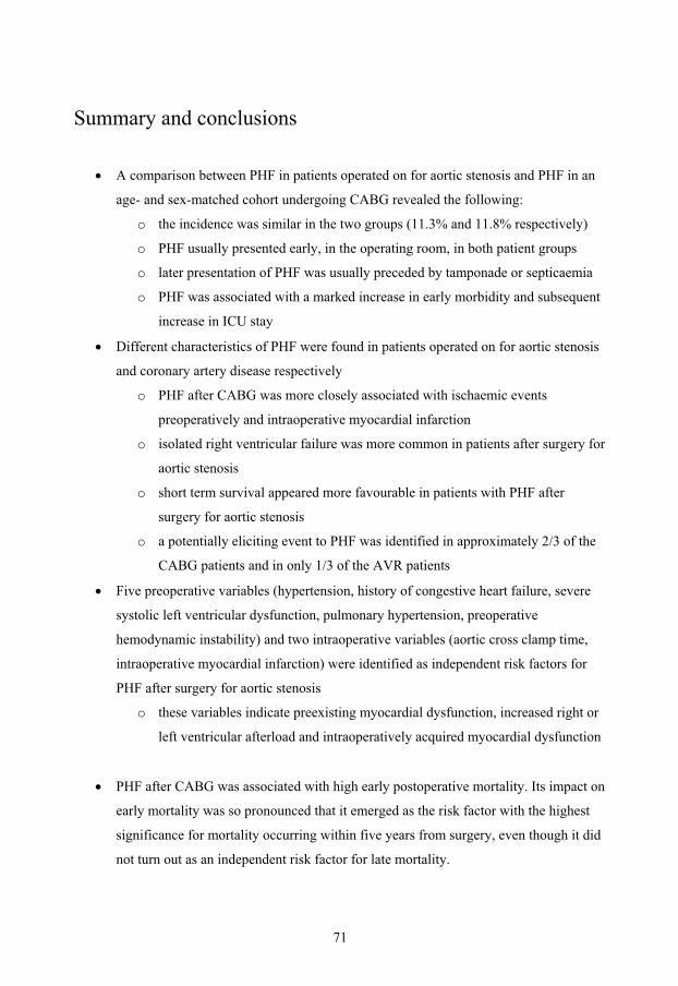
Summary and conclusions
A comparison between PHF in patients operated on for aortic stenosis and PHF in an
age- and sex-matched cohort undergoing CABG revealed the following:
o the incidence was similar in the two groups (11.3% and 11.8% respectively)
o PHF usually presented early, in the operating room, in both patient groups
o later presentation of PHF was usually preceded by tamponade or septicaemia
o PHF was associated with a marked increase in early morbidity and subsequent
increase in ICU stay
Different characteristics of PHF were found in patients operated on for aortic stenosis
and coronary artery disease respectively
o PHF after CABG was more closely associated with ischaemic events
preoperatively and intraoperative myocardial infarction
o isolated right ventricular failure was more common in patients after surgery for
aortic stenosis
o short term survival appeared more favourable in patients with PHF after
surgery for aortic stenosis
o a potentially eliciting event to PHF was identified in approximately 2/3 of the
CABG patients and in only 1/3 of the AVR patients
Five preoperative variables (hypertension, history of congestive heart failure, severe
systolic left ventricular dysfunction, pulmonary hypertension, preoperative
hemodynamic instability) and two intraoperative variables (aortic cross clamp time,
intraoperative myocardial infarction) were identified as independent risk factors for
PHF after surgery for aortic stenosis
o these variables indicate preexisting myocardial dysfunction, increased right or
left ventricular afterload and intraoperatively acquired myocardial dysfunction
PHF after CABG was associated with high early postoperative mortality. Its impact on
early mortality was so pronounced that it emerged as the risk factor with the highest
significance for mortality occurring within five years from surgery, even though it did
not turn out as an independent risk factor for late mortality.
71

PHF after AVR for AS appeared comparatively benign looking at early mortality but
was associated with a sustained negative influence on survival resulting in a five-year
survival that was at least as poor as after PHF in the CABG group
PHF emerged as the risk factor with highest statistical significance for mortality
within the five years after AVR for AS
o other independent risk factors for five-year mortality were preoperative renal
dysfunction, procedure-associated stroke, BMI < 19 kg/m2, increasing age,
preoperative atrial fibrillation and preoperative anaemia
o risk factors for PHF did not emerge as independent risk factors for late
mortality emphasizing the significance of the event of PHF per se
FFA and lactate were the major substrates taken up by the heart in patients with aortic
stenosis preoperatively
Metabolic adaptation with a substantial myocardial uptake of glutamate of a
magnitude previously described in patients with coronary artery disease was observed
preoperatively
Postoperatively a potentially unfavorable metabolic state was observed as indicated by
the following findings:
o FFA were the only major substrates taken up by the heart
o no significant uptake of lactate uptake indicating not fully recovered oxidative
metabolism
Postoperative infusion of glutamate after CABG indicated:
o a correlation between arterial levels of glutamate and myocardial uptake
o that elevation of arterial whole blood levels of glutamate by more than 2-3 fold
did not lead to a further substantial increase of myocardial uptake in CABG
patients
o such levels were achieved with a glutamate infusion of 30-40 mg per kg body
weight and hour
72

Postoperative infusion of glutamate 36.8 mg per kg body weight and hour after
surgery for AS was associated with:
o an increase of arterial whole blood glutamate by almost three fold
o a two fold increase of myocardial uptake of glutamate
o a net uptake of lactate by the heart
Studies of peripheral tissue metabolism as indicated by AV differences across the leg
after surgery for aortic stenosis revealed:
o no significant uptake of major substrates across the leg
o a net loss of amino acids
o that glutamate was the only amino acid taken up by the leg
o that an infusion of glutamate 36.8 mg per kg body weight and hour after
surgery for aortic stenosis resulted in a seven fold increase of AV differences
of glutamate
o that glutamate infusion appeared to counteract peripheral tissue release of FFA
suggesting an anti-lipolytic effect
o that glutamate infusion appeared to mitigate net amino acid loss
A beneficial effect of glutamate on myocardial metabolism is suggested by the results
and, hence, glutamate deserves further evaluation in patients operated on for aortic
stenosis
The potentially beneficial effect of glutamate on peripheral tissue metabolism
suggested by this study warrants further exploration in other conditions associated
with activated neuroendocrine stress response
73
Acknowledgements
The work presented in this thesis was carried out at the Department of Cardiothoracic
Surgery, University Hospital in Linköping, Sweden.
Financial support was received from The Swedish Heart Lung Foundation, The Swedish
Medical Research Council, The Lions Research Foundation, The Östergötlands Läns
Landsting and The Linköping Heart Center.
I would like to express my gratitude to all of those who have supported me and helped me to
complete the studies that have finally resulted in this thesis. I especially want to thank:
First and foremost, my principal supervisor Rolf Svedjeholm, for his never-failing support,
enthusiasm and optimism. You showed me that scientific work can be joyful. Furthermore,
you proved to me that a rewriting of manuscript can make it better than any of its 119
previous versions.
Erik Håkanson, my second supervisor, who has scrutinized all my work and contributed with
valuable input during these years.
My other co-authors, PO Joachimsson, Lennart Jorfeldt, Tamás Maros, Lennart Nilsson,
Zoltán Szabó, Éva Tamás and Ingemar Vanhanen for their important contributions to this
work.
Eva Nylander, who has always been willing to help me and explain any difficulties I have
come across regarding echocardiography and haemodynamics.
Mats Broqvist, for his friendly advice and support in the field of cardiology.
Olle Eriksson, for helping me with the statistics. I have learnt a lot, but most of all I have
realized how much more I have to learn.
Inger Huljebrant, Birgitta Lönnkvist, Marie Hill, Lena Hassel and Ragna Lindström all of
whom have helped me in different ways in the practical work that makes research possible.
Finally, my collegues for their support and understanding during my recent physical and
mental absence from clinical work.
74
References
1. Miller GW. King of hearts. New York: Crown Publishers; 2000. 2. Edmunds LH, Jr., Clark RE, Cohn LH, Grunkemeier GL, Miller DC, Weisel RD.
Guidelines for reporting morbidity and mortality after cardiac valvular operations. TheAmerican Association for Thoracic Surgery, Ad Hoc Liaison Committee for Standardizing Definitions of Prosthetic Heart Valve Morbidity. Ann Thorac Surg1996;62:932-5.
3. Freeman RV, Otto CM. Spectrum of calcific aortic valve disease: pathogenesis,disease progression, and treatment strategies. Circulation 2005;111:3316-26.
4. Thom T, Haase N, Rosamond W, Howard VJ, Rumsfeld J, Manolio T, Zheng ZJ, Flegal K, et al. Heart disease and stroke statistics--2006 update: a report from the American Heart Association Statistics Committee and Stroke Statistics Subcommittee.Circulation 2006;113:85-151.
5. Otto CM, Lind BK, Kitzman DW, Gersh BJ, Siscovick DS. Association of aortic-valve sclerosis with cardiovascular mortality and morbidity in the elderly. N Engl J Med 1999;341:142-7.
6. Stewart BF, Siscovick D, Lind BK, Gardin JM, Gottdiener JS, Smith VE, KitzmanDW, Otto CM. Clinical factors associated with calcific aortic valve disease.Cardiovascular Health Study. J Am Coll Cardiol 1997;29:630-4.
7. Davies MJ, Treasure T, Parker DJ. Demographic characteristics of patients undergoing aortic valve replacement for stenosis: relation to valve morphology. Heart1996;75:174-8.
8. Roberts WC. The structure of the aortic valve in clinically isolated aortic stenosis: anautopsy study of 162 patients over 15 years of age. Circulation 1970;42:91-7.
9. Bonow RO, Carabello B, de Leon ACJ, Edmunds LHJ, Fedderly BJ, Freed MD, Gaasch WH, McKay CR, et al. Guidelines for the management of patients with valvular heart disease: executive summary. A report of the American College of Cardiology/American Heart Association Task Force on Practice Guidelines (Committee on Management of Patients with Valvular Heart Disease). Circulation1998;98:1949-84.
10. Glower DD. Aquired aortic valve disease. In: Sabastion DC, Spencer FC, editors. Surgery of the chest. 6 ed. Philadelphia: W.B. Saunders Company; 1995.
11. Prasad Y, Bhalodkar NC. Aortic sclerosis--a marker of coronary atherosclerosis. ClinCardiol 2004;27:671-3.
12. Rossebo AB, Pedersen TR. Hyperlipidaemia and aortic valve disease. Curr Opin Lipidol 2004;15:447-51.
13. Yilmaz MB, Guray U, Guray Y, Cihan G, Caldir V, Cay S, Kisacik HL, Korkmaz S. Lipid profile of patients with aortic stenosis might be predictive of rate of progression. Am Heart J 2004;147:915-8.
14. Fenoglio JJJ, McAllister HAJ, DeCastro CM, Davia JE, Cheitlin MD. Congenital bicuspid aortic valve after age 20. Am J Cardiol 1977;39:164-9.
15. Grossman W, Jones D, McLaurin LP. Wall stress and patterns of hypertrophy in the human left ventricle. J Clin Invest 1975;56:56-64.
75
16. Maron BJ, Ferrans VJ, Roberts WC. Myocardial ultrastructure in patients with chronic aortic valve disease. Am J Cardiol 1975;35:725-39.
17. Bellamy MF, Pellikka PA, Klarich KW, Tajik AJ, Enriquez-Sarano M. Association of cholesterol levels, hydroxymethylglutaryl coenzyme-A reductase inhibitor treatment,and progression of aortic stenosis in the community. J Am Coll Cardiol 2002;40:1723-30.
18. Otto CM, Pearlman AS, Gardner CL. Hemodynamic progression of aortic stenosis in adults assessed by Doppler echocardiography. J Am Coll Cardiol 1989;13:545-50.
19. Sabiston DC, Spencer FC. Surgery of the chest. 6 ed. Philadelphia: W.B. Saunders Company; 1995.
20. Bache RJ. Effects of hypertrophy on the coronary circulation. Prog Cardiovasc Dis1988;30:403-40.
21. Johnson AM. Aortic stenosis, sudden death, and the left ventricular baroceptors. BrHeart J 1971;33:1-5.
22. Harpole DH, Jones RH. Serial assessment of ventricular performance after valve replacement for aortic stenosis. J Thorac Cardiovasc Surg 1990;99:645-50.
23. Hwang MH, Hammermeister KE, Oprian C, Henderson W, Bousvaros G, Wong M, Miller DC, Folland E, et al. Preoperative identification of patients likely to have left ventricular dysfunction after aortic valve replacement. Circulation 1989;80:I65-76.
24. Monrad ES, Hess OM, Murakami T, Nonogi H, Corin WJ, Krayenbuehl HP. Timecourse of regression of left ventricular hypertrophy after aortic valve replacement.Circulation 1988;77:1345-55.
25. Horstkotte D, Loogen F. The natural history of aortic valve stenosis. Eur Heart J1988;9:57-64.
26. Kelly TA, Rothbart RM, Cooper CM, Kaiser DL, Smucker ML, Gibson RS. Comparison of outcome of asymptomatic to symptomatic patients older than 20 yearsof age with valvular aortic stenosis. Am J Cardiol 1988;61:123-30.
27. Pellikka PA, Nishimura RA, Bailey KR, Tajik AJ. The natural history of adults with asymptomatic, hemodynamically significant aortic stenosis. J Am Coll Cardiol1990;15:1012-7.
28. Rosenhek R, Klaar U, Schemper M, Scholten C, Heger M, Gabriel H, Binder T, Maurer G, et al. Mild and moderate aortic stenosis. Natural history and risk stratification by echocardiography. Eur Heart J 2004;25:199-205.
29. Turina J, Hess O, Sepulcri F, Krayenbuehl HP. Spontaneous course of aortic valve disease. Eur Heart J 1987;8:471-83.
30. Otto CM. Aortic stenosis--listen to the patient, look at the valve. N Engl J Med2000;343:652-4.
31. Cowell SJ, Newby DE, Prescott RJ, Bloomfield P, Reid J, Northridge DB, Boon NA. A randomized trial of intensive lipid-lowering therapy in calcific aortic stenosis. NEngl J Med 2005;352:2389-97.
32. Tuffier T. État actuel de la chirurgie intrathoracicue. In: Transact XVII:th Internat Congr Medicine; 1913; London; 1913. p. Section VII:316-7.
33. Bailey CP, Glover RP, O'Neill TJ. Surgery of stenotic valvular disease of the heart. JAm Med Assoc 1950;144:1049-57.
34. Lindblom D, Lindblom U, Qvist J, Lundström H. Long-term relative survival rates after heart valve replacement. J Am Coll Cardiol 1990;15:566-73.
35. Dellgren G, Eriksson MJ, Brodin LA, Rådegran K. Eleven years' experience with the Biocor stentless aortic bioprosthesis: clinical and hemodynamic follow-up with long-term relative survival rate. Eur J Cardiothorac Surg 2002;22:912-21.
76

36. Lund O, Pilegaard H, Nielsen TT, Knudsen MA, Magnussen K. Thirty-day mortalityafter valve replacement for aortic stenosis over the last 22 years. A multivariate risk stratification. Eur Heart J 1991;12:322.
37. Astor BC, Kaczmarek RG, Hefflin B, Daley WR. Mortality after aortic valve replacement: results from a nationally representative database. Ann Thorac Surg2000;70:1939-45.
38. Edwards FH, Peterson ED, Coombs LP, DeLong ER, Jamieson WR, Shroyer ALW,Grover FL. Prediction of operative mortality after valve replacement surgery. J Am Coll Cardiol 2001;37:885-92.
39. Hannan EL, Racz MJ, Jones RH, Gold JP, Ryan TJ, Hafner JP, Isom OW. Predictors of mortality for patients undergoing cardiac valve replacements in New York State. Ann Thorac Surg 2000;70:1212-8.
40. Jamieson WR, Edwards FH, Schwartz M, Bero JW, Clark RE, Grover FL. Risk stratification for cardiac valve replacement. National Cardiac Surgery Database. Database Committee of The Society of Thoracic Surgeons. Ann Thorac Surg1999;67:943-51.
41. Maganti MD, Rao V, Borger MA, Ivanov J, David TE. Predictors of low cardiac output syndrome after isolated aortic valve surgery. Circulation 2005;112:I448-52.
42. Cohen G, David TE, Ivanov J, Armstrong S, Feindel CM. The impact of age, coronary artery disease, and cardiac comorbidity on late survival after bioprosthetic aortic valvereplacement. J Thorac Cardiovasc Surg 1999;117:273-84.
43. Connolly HM, Oh JK, Orszulak TA, Osborn SL, Roger VL, Hodge DO, Bailey KR, Seward JB, et al. Aortic valve replacement for aortic stenosis with severe leftventricular dysfunction. Prognostic indicators. Circulation 1997;95:2395.
44. Gaudino M, Alessandrini F, Glieca F, Luciani N, Cellini C, Pragliola C, Morelli M, Canosa C, et al. Survival after aortic valve replacement for aortic stenosis: does leftventricular mass regression have a clinical correlate? Eur Heart J 2005;26:51-7.
45. Gjertsson P, Caidahl K, Farasati M, Oden A, Bech-Hanssen O. Preoperative moderateto severe diastolic dysfunction: a novel Doppler echocardiographic long-termprognostic factor in patients with severe aortic stenosis. J Thorac Cardiovasc Surg2005;129:890-6.
46. Lytle BW, Cosgrove DM, Taylor PC, Goormastic M, Stewart RW, Golding LA, Gill CC, Loop FD. Primary isolated aortic valve replacement. Early and late results. JThorac Cardiovasc Surg 1989;97:675-94.
47. Blackstone EH, Cosgrove DM, Jamieson WR, Birkmeyer NJ, Lemmer JH, Jr., Miller DC, Butchart EG, Rizzoli G, et al. Prosthesis size and long-term survival after aortic valve replacement. J Thorac Cardiovasc Surg 2003;126:783-96.
48. Medalion B, Blackstone EH, Lytle BW, White J, Arnold JH, Cosgrove DM. Aortic valve replacement: is valve size important? J Thorac Cardiovasc Surg 2000;119:963-74.
49. Pibarot P, Dumesnil JG. Hemodynamic and clinical impact of prosthesis-patient mismatch in the aortic valve position and its prevention. J Am Coll Cardiol2000;36:1131-41.
50. Kirklin JW, Rastelli GC. Low cardiac output after open intracardiac operations. ProgCardiovasc Dis 1967;10:117-22.
51. O'Connor GT, Birkmeyer JD, Dacey LJ, Quinton HB, Marrin CA, Birkmeyer NJ, Morton JR, Leavitt BJ, et al. Results of a regional study of modes of death associated with coronary artery bypass grafting. Northern New England Cardiovascular Disease Study Group. Ann Thorac Surg 1998;66:1323-8.
77

52. Surgenor SD, O'Connor GT, Lahey SJ, Quinn R, Charlesworth DC, Dacey LJ, Clough RA, Leavitt BJ, et al. Predicting the risk of death from heart failure after coronaryartery bypass graft surgery. Anesth Analg 2001;92:596-601.
53. Jarvinen A. Low-output syndrome as a complication of open-heart surgery in 85 patients. Ann Clin Res 1975;7:379-93.
54. Rao V, Ivanov J, Weisel RD, Ikonomidis JS, Christakis GT, David TE. Predictors of low cardiac output syndrome after coronary artery bypass. J Thorac Cardiovasc Surg1996;112:38-51.
55. Gorman JH, III, Gorman RC, Milas BL, Acker MA. Circulatory management of the unstable cardiac patient. Semin Thorac Cardiovasc Surg 2000;12:316.
56. Baskett RJ, Ghali WA, Maitland A, Hirsch GM. The intraaortic balloon pump in cardiac surgery. Annals of Thoracic Surgery 2002;74:1276.
57. Håkanson E, Svedjeholm R, Vanhanen I. Physiologic aspects in postoperative cardiac patients. Ann Thorac Surg 1995;59:S12-4.
58. Svedjeholm R, Håkanson E, Szabo Z. Routine SvO2 measurement after CABG surgery with a surgically introduced pulmonary artery catheter. Eur J Cardiothorac Surg 1999;16:450-7.
59. Svedjeholm R, Vanhanen I, Håkanson E, Joachimsson PO, Jorfeldt L, Nilsson L. Metabolic and hemodynamic effects of intravenous glutamate infusion early after coronary surgery. J Thorac Cardiovasc Surg 1996;112:1468-1477.
60. Vanhanen I, Håkanson E, Jorfeldt L, Svedjeholm R. Intravenous aspartate infusion after a coronary operation: effects on myocardial metabolism and hemodynamic state. Ann Thorac Surg 1998;65:1296-1302.
61. Hammermeister KE, Burchfiel C, Johnson R, Grover FL. Identification of patients at greatest risk for developing major complications at cardiac surgery. Circulation1990;82:IV380-9.
62. Creswell LL, Rosenbloom M, Cox JL, Ferguson TB, Kouchoukos NT, Spray TL, Pasque MK, Ferguson Jr TB, et al. Intraaortic balloon counterpulsation: patterns of usage and outcome in cardiac surgery patients. Ann Thorac Surg 1992;54:11-20.
63. Rao V, Ivanov J, Weisel RD, Cohen G, Borger MA, Mickle DA. Lactate release during reperfusion predicts low cardiac output syndrome after coronary bypass surgery. Ann Thorac Surg 2001;71:1925.
64. Freeman RK, Daily PO, Dembitsky WP, Adamson RM, Moreno-Cabral RJ. The treatment of low cardiac output syndrome following cardiopulmonary bypass using delayed sternal closure. Am Surg 1997;63:882.
65. Charlson M, Krieger KH, Peterson JC, Hayes J, Isom OW. Predictors and outcomes of cardiac complications following elective coronary bypass grafting. Proc Ass Am Physicians 1999;111:622.
66. Hogue CW, Jr., Sundt T, III, Barzilai B, Schecthman KB, Davila-Roman VG. Cardiac and neurologic complications identify risks for mortality for both men and womenundergoing coronary artery bypass graft surgery. Anesthesiology 2001;95:1074.
67. Shernan SK, Fitch JC, Nussmeier NA, Chen JC, Rollins SA, Mojcik CF, Malloy KJ, Todaro TG, et al. Impact of pexelizumab, an anti-C5 complement antibody, on total mortality and adverse cardiovascular outcomes in cardiac surgical patients undergoing cardiopulmonary bypass. Ann Thorac Surg 2004;77:942-9; discussion 949-50.
68. Mudge GH, Mills RM, Taegtmeyer H, Gorlin R, Lesch M. Alterations of myocardial amino acid metabolism in chronic ischemic heart disease. J Clin Invest 1976;58:1185-1192.
78
69. Thomassen A, Nielsen TT, Bagger JP, Thuesen L. Myocardial glutamate and alanine exchanges related to carbohydrate metabolism in patients with normal and stenotic coronary arteries. Clin Physiol 1984;4:425-434.
70. Pisarenko OI, Lepilin MG, Ivanov VE. Cardiac metabolism and performance during L-glutamic acid infusion in postoperative cardiac failure. Clin Sci 1986;70:7-12.
71. Svedjeholm R, Ekroth R, Joachimsson PO, Ronquist G, Svensson SE, Tydén H. Myocardial uptake of amino acids and other substrates in relation to myocardialoxygen consumption four hours after cardiac surgery. J Thorac Cardiovasc Surg1991;101:688-694.
72. Svedjeholm R, Håkanson E, Vanhanen I. Rationale for metabolic support with aminoacids and glucose-insulin-potassium (GIK) in cardiac surgery. Ann Thorac Surg1995;59:S15-S22.
73. Svensson S, Svedjeholm R, Ekroth R, Milocco I, Nilsson, Sabel KG, William-OlssonG. Trauma metabolism and the heart. Uptake of substrates and effects of insulin early after cardiac operations. J Thorac Cardiovasc Surg 1990;99:1063-1073.
74. Teoh KH, Mickle DAG, Weisel RD, Fremes SE, Christakis GD, Romaschin AD, Harding RS, Madonik MM, et al. Decreased postoperative myocardial free fatty acid oxidation. J Surg Res 1988;44:36-44.
75. Opie LH, Opie LH. Fuels: Carbohydrates and lipids. In: The heart: physiology and metabolism. New York: Raven Press; 1991. p. 208-246.
76. Pietersen HG, Langenberg CJ, Geskes G, Kester A, de Lange S, Van der Vusse GJ, Wagenmakers AJ, Soeters PB. Myocardial substrate uptake and oxidation during and after routine cardiac surgery. J Thorac Cardiovasc Surg 1999;118:71-80.
77. Pietersen HG, Langenberg CJ, Geskes G, Soeters PB, Wagenmakers AJ. Glutamatemetabolism of the heart during coronary artery bypass grafting. Clin Nutr 1998;17:73-75.
78. Vanhanen I, Håkanson E, Jorfeldt L, Svedjeholm R. Myocardial uptake and release of substrates in patients operated for unstable angina: impact of glutamate infusion. Scand Cardiovasc J 2003;37:113-120.
79. Nilsson F, Ekroth R, Milocco I, Nilsson NJ, Svensson S, Berglin E, William-OlssonG. Splanchnic glucose balance and insulin resistance in the early postoperative phase of cardiac surgery. JPEN J Parenter Enteral Nutr 1988;12:574-578.
80. Svedjeholm R, Svensson S, Ekroth R, Milocco I, Nilsson F, Vinnars E, Wernerman J. Trauma metabolism and the heart: studies of heart and leg amino acid flux after cardiac surgery. Thorac Cardiovasc Surg 1990;38:1-5.
81. Peuhkurinen KJ. Regulation of the tricarboxylic acid pool size in heart muscle. J Mol Cell Cardiol 1984;16:487-495.
82. Pietersen HG, Langenberg CJ, Geskes G, Soeters PB, Wagenmakers AJM. Protein synthesis and protein degradation of the human heart after coronary artery bypass graft surgery. Thesis: Myocardial metabolism in the coronary artery bypass graft patient. Maastrich: University of Maastrich; 2003.
83. Jorfeldt L, Juhlin-Dannfelt A. The influence of ethanol on splanchnic and skeletal muscle metabolism in man. Metabolism 1978;27:97-106.
84. Bernt E, Bergmayer HU, Bergmayer HU. L-Glutamat: UV-test mit Glutamat-Dehydrogenase und NAD. In: Metoden der enzymatischen analyse. Weinheim: Verlag Chemie GMBH; 1974. p. 1749-1753.
85. Barthelmai W, Czok R. Enzymatische bestimmungen der glukose in blut, liquor und hirn. Klin Wochenschr 1962;40:585-589.
86. Schmidt FH. Die enzymatische bestimmung von glukose und fructose. KlinWochenschr 1961;39:1244-1247.
79

87. Ho RJ. Radiochemical assay of long-chain fatty acids using 63 Ni as tracer. AnalBiochem 1970;36:105-113.
88. Kornhuber ME, Balabanova S, Heiligensetzer GV, Kornhuber C, Kornhuber AW.Stability of human blood serum aminoacids after storage at different pH and temperature conditions. Clin Chim Acta 1991;197:189-200.
89. Svedjeholm R, Huljebrant I, Hakanson E, Vanhanen I. Glutamate and high-dose glucose-insulin-potassium (GIK) in the treatment of severe cardiac failure after cardiac operations. Ann Thorac Surg 1995;59:S23.
90. Aupart MR, Sirinelli AL, Diemont FF, Meurisse YA, Dreyfus XB, Marchand MA. The last generation of pericardial valves in the aortic position: ten-year follow-up in 589 patients. Ann Thorac Surg 1996;61:615-20.
91. Bernal JM, Martin-Duran R, Rabasa JM, Revuelta JM. The CarboMedics "Top-Hat" supraannular prosthesis. Ann Thorac Surg 1999;67:1299-303.
92. Chafizadeh ER, Zoghbi WA. Doppler echocardiographic assessment of the St. Jude Medical prosthetic valve in the aortic position using the continuity equation. Circulation 1991;83:213-23.
93. Chambers J, Cross J, Deverall P, Sowton E. Echocardiographic description of the CarboMedics bileaflet prosthetic heart valve. J Am Coll Cardiol 1993;21:398-405.
94. Chambers J, Ely JL. Early postoperative echocardiographic hemodynamicperformance of the On-X prosthetic heart valve: a multicenter study. J Heart Valve Dis 1998;7:569-73.
95. Chambers JB. Studies on the use of Doppler ultrasound in the assessment of prosthetic cardiac valve function. Thesis. Cambridge: Cambridge University; 2000.
96. De Paulis R, Sommariva L, De Matteis GM, Polisca P, Tomai F, Bassano C, Penta de Peppo A, Chiariello L. Hemodynamic performances of small diameter carbomedicsand St. Jude valves. J Heart Valve Dis 1996;5 Suppl 3:S339-43.
97. Dumesnil JG, LeBlanc MH, Cartier PC, Metras J, Desaulniers D, Doyle DP, LemieuxMD, Raymond G. Hemodynamic features of the freestyle aortic bioprosthesis compared with stented bioprosthesis. Ann Thorac Surg 1998;66:S130-3.
98. Zingg U, Aeschbacher B, Seiler C, Althaus U, Carrel T. Early experience with the new Masters series of St. Jude Medical heart valve: in vivo hemodynamic and clinical results in patients with narrowed aortic annulus. J Heart Valve Dis 1997;6:535-41.
99. Hosmer DW, Lemeshow S. Applied logistic regression. New York: Wiley; 1989. 100. Breisblatt WM, Stein KL, Wolfe CJ, Follansbee WP, Capozzi J, Armitage, Jm,
Hardesty RL. Acute myocardial dysfunction and recovery: a common occurrence after coronary bypass surgery. J Am Coll Cardiol 1990;15:1261.
101. Bolli R. Mechanism of myocardial stunning. Circulation 1990;82:723-738. 102. Bolli R, Marban E. Molecular and cellular mechanisms of myocardial stunning.
Physiol Rev 1999;79:609-34. 103. Wesslén Ö. Beta1-blockade early after coronary surgery. A hemodynamic, metabolic
and electrophysiologic study with special reference to effects of metoprolol. Thesis. Uppsala: Acta Univ Ups; 1990.
104. Svedjeholm R. Myocardial uptake of substrates early after cardiac surgery. A clinical study of metabolic derangement and recovery with special reference to effects of dopamine and insulin. Thesis. Uppsala: Acta Univ Ups; 1990.
105. Svensson S. Effects of insulin early after cardiac surgery. A clinical study of myocardial metabolism and central hemodynamic parameters. Thesis. Gothenburg:University of Gothenburg; 1988.
80

106. Vanhanen I. Metabolic intervention with amino acids in coronary surgery. A clinical study with special reference to effects of glutamate and aspartate on myocardialmetabolism. Thesis. Linköping: University of Linköping; 2000.
107. Svedjeholm R, Huljebrant I, Håkanson E, Vanhanen I. Glutamate and high-dose glucose-insulin-potassium (GIK) in the treatment of severe cardiac failure after cardiac operations. Ann Thorac Surg 1995;59:S23-S30.
108. Beyersdorf F, Kirsh M, Buckberg GD, Allen BS. Warm glutamate/aspartate-enrichedblood cardioplegic solution for perioperative sudden death. J Thorac Cardiovasc Surg1992;104:1141-1147.
109. Gradinac S, Coleman GM, Taegtmeyer H, Sweeney MS, Frazier OH. Improved cardiac function with Glucose-Insulin-Potassium after aortocoronary bypass grafting. Ann Thorac Surg 1989;48:484-489.
110. Thomassen A, Bagger JP, Nielsen TT, Henningsen P. Altered global myocardialsubstrate preference at rest and during acing in coronary artery disease with stable angina pectoris. Am J Cardiol 1988;62:686-693.
111. Drake AJ, Haines JR, Noble MM. Preferential uptake of lactate by the normalmyocardium in dog. Cardiovasc Res 1980;14:65-72.
112. Liedtke AJ, Nellis S, Neely JR. Effects of excess free fatty acids on mechanical and metabolic function in normal and ischemic myocardium in swine. Circ Res1978;43:652-661.
113. Simonsen S, Kjekshus JK. The effect of free fatty acids on myocardial oxygen consumption during atrial pacing and catecholamine infusion in man. Circulation1978;58:484-491.
114. Pietersen HG, Langenberg CJ, Geskes G, Wagenmakers AJM, Soeters PB. Metabolic fate of glutamate in the human heart during and after routine cardiac surgery. Thesis. Maastricht: University of Maastricht; 2003.
115. Dinkelborg LM, Kinne RK, Grieshaber MK. Characterization and pH dependence of L-glutamate transport in sarcolemmal vesicles from rat hearts. Am J Physiol1995;268:H194-201.
116. Daabees TT, Finkelstein MW, Stegink LD, Applebaum AE. Correlation of glutamateplus aspartate dose, plasma amino acid concentration and neuronal necrosis in infant mice. Food Chem Toxicol 1985;23:887-893.
117. Thomassen AR. Myocardial uptake and effects of glutamate during non-ischaemic and ischaemic conditions. A clinical study with special reference to possibleinterrelationships between glutamate and myocardial utilization of carbohydrate substrates. Dan Med Bull 1992;39:471-488.
118. Kimose HH, Helligso P, Randsbaek F, Kim Y, Botker HE, Hansen SB, ThomassenAR, Nielsen TT. Improved recovery after cold crystalloid cardioplegia using low- dose glutamate enrichment during reperfusion after aortic unclamping: a study in isolated blood-perfused pig hearts. Thorac Cardiovasc Surg 1996;44:118-125.
119. Kjellman UW, Björk K, Ekroth R, Karlsson H, Jagenburg R, Nilsson FN, Svensson G, Wernerman J. Addition of alpha-ketoglutarate to blood cardioplegia improvescardioprotection. Ann Thorac Surg 1997;63:1625-1633.
120. Thomassen A, Nielsen TT, Bagger JP, Pedersen AK, Henningsen P. Antiischemic and metabolic effects of glutamate during pacing in patients with stable angina pectoris secondary to either coronary artery disease or syndrome X. Am J Cardiol1991;68:291-295.
121. Lazar HL, Buckberg GD, Manganaro AJ, Becker H. Myocardial energy replenishmentand reversal of ischemic damage by substrate enhancement of secondary blood
81
cardioplegia with amino acids during reperfusion. J Thorac Cardiovasc Surg1980;80:350-359.
122. Lazar HL, Buckberg GD, Manganaro AJ, Becker H, Maloney JV, Jr. Reversal of ischemic damage with amino acid substrate enhancement during reperfusion. Surgery1980;80:702-709.
123. Nilsson F, Ekroth R, Milocco I, Nilsson NJ, Sabel KG, Svensson S, Berglin E, William-Olsson G. The effect of insulin on the leg and splanchnic balance of glucose and gluconeogenic substrates after cardiac surgery. Thorac Cardiovasc Surg1988;36:343-347.
124. Thomassen A, Nielsen TT, Bagger JP, Henningsen P. Effects of intravenous glutamateon substrate availability and utilization across the human heart and leg. Metabolism1991;40:378-384.
125. Nilsson FN, Berglin EE, Ekroth R, Holm G, Milocco I, Mjös OD, Waldenström A, William-Olsson GE. Effects of graded insulin infusions on plasma levels of free fattyacids, adrenaline and noradrenaline directly after open heart surgery :published erratum appears in Thorac Cardiovasc Surg 1987 Aug;35(4):262. Thorac Cardiovasc Surg 1987;35:96-100.
126. Dahlin LG, Olin C, Svedjeholm R. Perioperative myocardial infarction in cardiac surgery--risk factors and consequences. A case control study. Scand Cardiovasc J2000;34:522-527.
127. Slogoff S, Keats AS. Does perioperative myocardial ischemia lead to postoperative myocardial infarction? Anesthesiology 1985;62:107.
128. Davila-Roman VG, Waggoner AD, Hopkins WE, Barzilai B. Right ventricular dysfunction in low output syndrome after cardiac operations: assessment by transesophageal echocardiography. Ann Thorac Surg 1995;60:1081.
129. Allen BS, Winkelmann JW, Hanafy H, Hartz RS, Bolling KS, Ham J, Feinstein S. Retrograde cardioplegia does not adequately perfuse the right ventricle. J Thorac Cardiovasc Surg 1995;109:1116.
130. Gonzalez AC, Brandon TA, Fortune RL, Casano SF, Martin M, Benneson DL, Guilbeau EJ, Fisk RL. Acute right ventricular failure is caused by inadequate right ventricular hypothermia. J Thorac Cardiovasc Surg 1985;89:386.
131. Greeley WJ, Kern FH, Ungerleider RM, Kisslo JA. Intramyocardial air causes right ventricular dysfunction after repair of a congenital heart defect. Anesthesiology1990;73:1042.
132. Kawachi K, Kitamura S, Hasegawa J, Kawata T, Kobayashi S, Mizuguchi K, Nishioka H, Taniguchi S, et al. Increased risk of coronary artery bypass grafting for left ventricular dysfunction with dilated left ventricle. J Cardiovasc Surg 1997;38:501.
133. Yau TM, Fedak PW, Weisel RD, Teng C, Ivanov J. Predictors of operative risk for coronary bypass operations in patients with left ventricular dysfunction. J Thorac Cardiovasc Surg 1999;118:1006.
134. Dacey LJ, Likosky DS, Leavitt BJ, Lahey SJ, Quinn RD, Hernandez FJ, Quinton HB, Desimone JP, et al. Perioperative stroke and long-term survival after coronary bypass graft surgery. Ann Thorac Surg 2005;79:532-6.
135. Gardner SC, Grunwald GK, Rumsfeld JS, Cleveland JC, Jr., Schooley LM, Gao D, Grover FL, McDonald GO, et al. Comparison of short-term mortality risk factors for valve replacement versus coronary artery bypass graft surgery. Ann Thorac Surg2004;77:549-56.
136. Christakis GT, Weisel RD, David TE, Salerno TA, Ivanov J. Predictors of operative survival after valve replacement. Circulation 1988;78:I25-34.
82

137. Florath I, Rosendahl UP, Mortasawi A, Bauer SF, Dalladaku F, Ennker IC, Ennker JC. Current determinants of operative mortality in 1400 patients requiring aortic valve replacement. Ann Thorac Surg 2003;76:75-83.
138. Engelman DT, Adams DH, Byrne JG, Aranki SF, Collins JJJ, Couper GS, Allred EN, Cohn LH, et al. Impact of body mass index and albumin on morbidity and mortality after cardiac surgery. J Thorac Cardiovasc Surg 1999;118:866-73.
139. Maurer MS, Luchsinger JA, Wellner R, Kukuy E, Edwards NM. The effect of body mass index on complications from cardiac surgery in the oldest old. J Am Geriatr Soc2002;50:988-94.
140. Rapp-Kesek D, Ståhle E, Karlsson TT. Body mass index and albumin in the preoperative evaluation of cardiac surgery patients. Clin Nutr 2004;23:1398-404.
141. DeFoe GR, Ross CS, Olmstead EM, Surgenor SD, Fillinger MP, Groom RC, Forest RJ, Pieroni JW, et al. Lowest hematocrit on bypass and adverse outcomes associated with coronary artery bypass grafting. Northern New England Cardiovascular Disease Study Group. Ann Thorac Surg 2001;71:769-76.
142. Lung B, Gohlke-Barwolf C, Tornos P, Tribouilloy C, Hall R, Butchart E, Vahanian A. Recommendations on the management of the asymptomatic patient with valvular heart disease. Eur Heart J 2002;23:1252-66.
143. Maroko PR, Kjekshus JK, Sobel BE, Watanabe T, Covell JW, Ross Jr J, Braunwald E. Factors influencing infarct size following experimental coronary artery occlusions. Circulation 1971;43:67.
144. Lazar HL, Buckberg GD, Foglia RP, Manganaro AJ, Maloney JV. Detrimental effectsof premature use of inotropic drugs to discontinue cardiopulmonary bypass. J Thorac Cardiovasc Surg 1981;82:18-25.
145. Dawn B, Paliwal VS, Raza ST, Mastali K, Longaker RA, Stoddard MF. Left ventricular outflow tract obstruction provoked during dobutamine stress echocardiography predicts future chest pain, syncope, and near syncope. Am Heart J2005;149:908-16.
146. Svedjeholm R. Intravenous metabolic support with GIK (glucose-insulin-potassium)and amino acids in cardiac surgery. In: Beyersdorf F, editor. Ischemia-reperfusioninjury in cardiac surgery. Georgetown: Eurecah.com / Landes Bioscience; 2001. p. 135-156.
147. Strömberg A, Dahlström U. Heart failure clinics have decreased mortality and hospitalisation rates in Sweden. Eur Heart J 2004;25:1368-9.
83